HAL Id: tel-01174416
https://tel.archives-ouvertes.fr/tel-01174416
Submitted on 9 Jul 2015
HAL is a multi-disciplinary open access archive for the deposit and dissemination of sci-entific research documents, whether they are pub-lished or not. The documents may come from teaching and research institutions in France or
L’archive ouverte pluridisciplinaire HAL, est destinée au dépôt et à la diffusion de documents scientifiques de niveau recherche, publiés ou non, émanant des établissements d’enseignement et de recherche français ou étrangers, des laboratoires
lung imaging
Andrea Bianchi
To cite this version:
Andrea Bianchi. Magnetic resonance imaging techniques for pre-clinical lung imaging. Imaging. Université de Bordeaux, 2014. English. �NNT : 2014BORD0060�. �tel-01174416�
THÈSE PRÉSENTÉE
POUR OBTENIR LE GRADE DE
DOCTEUR DE
L’UNIVERSITÉ DE BORDEAUX
ÉCOLE DOCTORALE DES SCIENCES DE LA VIE ET DE LA SANTÉ
SPECIALITÉ BIOIMAGERIE
Par Andrea, BIANCHI
MAGNETIC RESONANCE IMAGING TECHNIQUES FOR
PRE-CLINICAL LUNG IMAGING
Sous la direction de : Yannick, CRÉMILLIEUX
Soutenue le 28 mars 2014
Membres du jury :
M. QUESSON, Bruno Université de Bordeaux Président M. BARBIER, Emmanuel Université Joseph Fourier, Grenoble Rapporteur M. BERTHAULT, Patrick CEA – Saclay, Gif sur Yvette Rapporteur M. BECKMANN, Nicolau Novartis Pharma AG, Bâle Examinateur
radiales à temps d’écho ultra-court (UTE) sont analysées pour évaluer leur potentiel dans l’étude non-invasive de différents modèles expérimentaux de maladies pulmonaires chez la souris. Chez le petit animal, les séquences radiales UTE peuvent efficacement limiter l’impact négatif sur la qualité de l’image dû au déphasage rapide des spins causé par les nombreuses interfaces air/tissu. En plus, les séquences radiales UTE sont moins sensibles aux artefacts de mouvement par rapport aux séquences Cartésiennes classiques. En conséquence, chez le petit animal, les séquences radiales UTE peuvent permettre d’obtenir des images du poumon avec une résolution bien inférieure au millimètre avec des rapports signal/bruit importants dans le parenchyme pulmonaire, tout en travaillant en conditions physiologiques (animaux en respiration spontanée).
Dans cette thèse, il sera démontré que les séquences d’IRM protonique UTE sont outils efficaces dans l’étude quantitative et non-invasive de différents marqueurs distinctifs de certaines pathologies pulmonaires d’intérêt général. Les protocoles développés seront simples, rapides et non-invasifs, faciles à implémenter, avec une interférence minimale sur la pathologie pulmonaire étudiée et, en définitive, potentiellement applicables chez l’homme. Il sera ainsi démontré que l’emploi des agents de contraste, administrés via les voies aériennes, permet d’augmenter la sensibilité des protocoles développés. Parallèlement, dans cette thèse des protocoles suffisamment flexibles seront implémentés afin de permettre l’étude d’un agent de contraste paramagnétique générique pour des applications aux poumons.
Mots clés :
Poumon, Imagerie par Résonance Magnétique, Temps d’écho ultra-court, IRM UTE, Asthme, Cancer du poumon, Agents de contraste, Nanoparticules à base de gadolinium, Imagerie optique, Modèles animauxTitle :
Magnetic resonance imaging techniques for pre-clinical lung imaging
Abstract :
In this work, ultra-short echo time (UTE) Magnetic Resonance Imaging (MRI) sequences are investigated as flexible tools for the noninvasive study of experimental models of lung diseases in mice. In small animals radial UTE sequences can indeed efficiently limit the negative impact on lung image quality due to the fast spin dephasing caused by the multiple air/tissue interfaces. In addition, radial UTE sequences are less sensitive to motion artifacts compared to standard Cartesian acquisitions. As a result, radial UTE acquisitions can provide lung images in small animals at sub-millimetric resolution with significant signal to noise ratio in the lung parenchyma, while working with physiological conditions (freely-breathing animals).In this thesis, UTE proton MRI sequences were shown to be efficient instruments to quantitatively investigate a number of hallmarks in longitudinal models of relevant lung diseases with minimal interference with the lung pathophysiology, employing easily-implementable fast protocols. The synergic use of positive contrast agents, along with an advantageous administration modality, was shown to be a valuable help in the increase of sensitivity of UTE MRI. At the same time, UTE MRI was shown to be an extremely useful and efficacious sequence for studying positive contrast agents in lungs.
Keywords :
Lung, Magnetic Resonance Imaging, Ultra-short echo time, UTE MRI, Asthma, Lung cancer, Contrast agents, Gadolinium nanoparticles, Optical imaging, Mice modelsmais de le rendre possible” Antoine de Saint-Exup´ery
Contents i
Abstract v
Preface vii
R´esum´e xiii
List of Figures xix
List of Acronyms and Abbreviations xxi
1 Introduction 1
1.1 Anatomopathology of lung . . . 1
1.1.1 Lung anatomophysiology and morphology . . . 1
1.1.1.1 Lung structure and physiology . . . 1
1.1.1.2 Lung morphology . . . 4
1.1.1.3 Defense system . . . 6
1.1.1.4 Rodents and human’s lungs comparative anatomy 7 1.1.2 Lung pathologies . . . 8 1.1.2.1 Asthma . . . 10 Pathophysiology . . . 10 Treatments . . . 12 1.1.2.2 Lung Cancer . . . 12 Pathophysiology . . . 13
Diagnosis and treatments . . . 14
1.2 MRI of the lung . . . 15
1.2.1 Main challenges in MR lung imaging . . . 15
1.2.1.1 Lung proton density . . . 15
1.2.1.2 Lung air-tissue interfaces . . . 16
1.2.1.3 Lung cardiorespiratory motion . . . 18
Non-forced-breathing gating methods . . . 19
Free-breathing methods . . . 20
1.2.2 MR techniques for lung parenchyma imaging . . . 20
1.2.2.1 The k-space concept . . . 20
1.2.2.2 Cartesian vs. non-Cartesian trajectories . . . . 21
Cartesian k-space trajectories . . . 22
Non-Cartesian k-space trajectories . . . 23
1.2.2.3 UTE radial sequences . . . 24
Short TE . . . 24
Reduced motion sensitivity . . . 25
Drawbacks . . . 25
Applications to lung imaging . . . 26
1.2.2.4 Other techniques for lung parenchyma imaging 27 Spoiled gradient-echo sequences . . . 27
Balanced steady-state free precession sequences. 28 Single-shot fast spin echo sequences . . . 29
1.3 MRI contrast agents for lung imaging . . . 30
1.3.1 MR contrast agents applied to lung imaging . . . 30
Paramagnetic contrast agents . . . 30
Superparamagnetic contrast agents . . . 31
Hyperpolarized gases . . . 32
Fluorinated gases . . . 32
Oxygen-enhanced proton MRI . . . 33
1.3.2 Contrast agent requirements . . . 34
Factors affecting clinical safety . . . 34
1.3.3 Paramagnetic contrast agents relaxivity theory . . . 35
Inner sphere relaxation . . . 36
Outer sphere relaxation . . . 36
1.3.3.1 General considerations . . . 37
Contrast agent design . . . 37
T2∗effect . . . 38
2 Material and methods 41 2.1 UTE sequence and image reconstruction . . . 41
2.1.1 UTE sequence acquisition scheme. . . 41
2.1.2 UTE sequence reconstruction . . . 43
2.1.2.1 Regridding algorithm . . . 43
Density compensation . . . 43
Convolution with a gridding kernel. . . 44
2.2 Ultra-small rigid platforms . . . 46
2.2.1 Synthesis . . . 47
2.2.2 Properties . . . 48
3 UTE MRI applications on lung diseases 53 3.1 A chronic model of asthma in mice . . . 53
3.2 An orthotopic model of lung cancer in mice . . . 64
4 UTE MRI and intratracheally administered contrast agents 75 4.1 Proof-of-concept . . . 75
4.2 Biodistribution and pharmacokinetics . . . 85
5 A new protocol for lung tumor detection 101 6 Discussion and perspectives 111 6.1 UTE MRI applications on lung diseases . . . 111
6.1.1 Asthma . . . 112
Perspectives . . . 113
6.1.2 Lung cancer . . . 115
6.2 UTE MRI and intratracheally administered contrast agents . . 115
Perspectives . . . 116
6.3 A new protocol for lung tumor detection . . . 117
6.4 General perspectives . . . 118
6.4.1 Oral administration and active targeting USRPs. . . 118
Integrin targeting and oral administration . . . . 118
On-going studies . . . 119
Future studies . . . 120
6.4.2 Application of USRPs to other lung models . . . 121
Lung fibrosis . . . 121
6.4.3 Application of USRPs to other tumors: brain cancer study123 Material and methods. . . 123
Results . . . 124
Discussion . . . 125
6.4.4 Translational considerations . . . 126
6.5 Other applications of the described protocols . . . 127
6.5.1 Application to upconverting nanoparticles . . . 127
6.5.2 Application to MEMRI . . . 128
7 Conclusion 131
C A review about USRPs 159
D USRPs synthesis and characterization: Supplementary Material 195
Bibliography 199
List of Publications, Patent, and Communications 217
Magnetic Resonance Imaging for pre-clinical lung
imaging
In this work, ultra-short echo time (UTE) Magnetic Resonance Imaging (MRI) sequences are investigated as flexible tools for the noninvasive study of ex-perimental models of lung diseases in mice. In small animals radial UTE
sequences can indeed efficiently limit the negative impact on lung image qual-ity due to the fast spin dephasing caused by the multiple air/tissue interfaces. In addition, radialUTEsequences are less sensitive to motion artifacts com-pared to standard Cartesian acquisitions. As a result, radialUTEacquisitions can provide lung images in small animals at submillimetric resolution with significant signal to noise ratio in the lung parenchyma, while working with physiological conditions (freely-breathing animals).
In this thesis,UTEprotonMRIsequences were shown to be efficient
in-struments to quantitatively investigate a number of hallmarks in longitudinal models of relevant lung diseases with minimal interference with the lung pathophysiology, employing easily-implementable fast protocols. The syner-gic use of positive contrast agents, along with an advantageous administration modality, was shown to be a valuable help in the increase of sensitivity of
UTE MRI. At the same time,UTE MRIwas shown to be an extremely useful and efficacious sequence for studying positive contrast agents in lungs. Keywords:Lung, Magnetic Resonance Imaging, Ultra-short echo time, UTE MRI, Asthma, Lung cancer, Contrast agents, Gadolinium nanoparticles, Opti-cal imaging, Mice models
Techniques d’IRM pour l’imagerie pr´eclinique du
poumon
Dans ce travail, les s´equences Imagerie par R´esonance Magn´etique (IRM) radiales `a temps d’´echo ultra-court (UTE) sont analys´ees pour ´evaluer leur potentiel dans l’´etude non-invasive de diff´erents mod`eles exp´erimentaux de maladies pulmonaires chez la souris. Chez le petit animal, les s´equences ra-diales UTEpeuvent efficacement limiter l’impact n´egatif sur la qualit´e de
l’image d ˆu au d´ephasage rapide des spins caus´e par les nombreuses interfaces air/tissu. En plus, les s´equences radialesUTEsont moins sensibles aux arte-facts de mouvement par rapport aux s´equences Cart´esiennes classiques. En cons´equence, chez le petit animal, les s´equences radialesUTEpeuvent per-mettre d’obtenir des images du poumon avec une r´esolution bien inf´erieure au millim`etre avec des rapports signal/bruit importants dans le parenchyme pulmonaire, tout en travaillant en conditions physiologiques (animaux en respiration spontan´ee).
Dans cette th`ese, il sera d´emontr´e que les s´equences d’IRMprotonique
UTEsont outils efficaces dans l’´etude quantitative et non-invasive de diff´erents
marqueurs distinctifs de certaines pathologies pulmonaires d’int´erˆet g´en´eral. Les protocoles d´evelopp´es seront simples, rapides et non-invasifs, faciles `a impl´ementer, avec une interf´erence minimale sur la pathologie pulmonaire ´etudi´ee et, en d´efinitive, potentiellement applicables chez l’homme. Il sera ainsi d´emontr´e que l’emploi des agents de contraste, administr´es via les voies a´eriennes, permet d’augmenter la sensibilit´e des protocoles d´evelopp´es. Par-all`element, dans cette th`ese des protocoles suffisamment flexibles seront im-plement´es afin de permettre l’´etude d’un agent de contraste paramagn´etique g´en´erique pour des applications aux poumons.
Mots cl´es:Poumon, Imagerie par R´esonance Magn´etique, Temps d’´echo ultra-court, IRM UTE, Asthme, Cancer du poumon, Agents de contraste, Nanopar-ticules `a base de gadolinium, Imagerie optique, Mod`eles animaux
Motivation The spectrum of pulmonary pathologies is extremely broad and includes, among others, lower respiratory tract infections, chronic obstructive pulmonary diseases, asthma, and respiratory apparatus tumors. According to the World Health Organization surveys these diseases, altogether, represent the first cause of death worldwide [1]. The burden of lung pathologies could be greatly reduced with an effective policy of prevention and early diagnostics, which often results in a positive prognosis.
In the race to improve global health through an early detection of diseases, imaging techniques play a major role since they can provide early diagnostics of pathologies, noninvasive longitudinal follow-ups of the patients, and can help preparing and guiding surgery.
Chest radiography and computed tomography currently represent the gold standard for lung diseases diagnostics. Nuclear medicine imaging techniques (e.g., gamma scintigraphy, positron-emission tomography, single photon-emission computer tomography) have shown to be of significant aid in the validation of X-ray or computed tomography observations, and can provide further information about several lung pathologies (e.g., stage of lung can-cer) [2]. Nonetheless, all these in vivo imaging techniques imply the exposure of the patient to ionizing radiation. As a consequence, repeated acquisitions are discouraged to reduce the cumulative radiation to which patients are exposed, which has been shown to be associated with an increased risk of developing cancer [3].
In this context, magnetic resonance imaging (MRI) has shown to be espe-cially promising because of its high soft tissue contrast, good spatial resolution, and absence of ionizing radiation [2,4–6].MRIwas proven to be the most adequate imaging technique for the screening and diagnostics of a number of pathologies in brain, heart or liver. Nevertheless, this consideration cannot yet be applied to the lung, which remains one of the most difficult organs to
image withMRIbecause of its intrinsic properties [5,7–9].
In this work, ultra-short echo time (UTE)MRIsequences are investigated as flexible tools for the noninvasive study of experimental models of lung diseases in mice. In small animals, radialUTEsequences can indeed efficiently
limit the negative impact on image quality due to the fast spin dephasing caused by the multiple air/tissue interfaces [10,11]. In addition, radialUTE
sequences are less sensitive to motion artifacts compared to standard Cartesian acquisitions [12]. As a result, radialUTEacquisitions can provide lung images in small animals at submillimetric resolution with significant signal to noise ratio in the lung parenchyma, while working with physiological conditions (freely-breathing animals) [13,14].
Aim The aim of this thesis is to implement, apply and validateMRI tech-niques dedicated to the investigation and diagnostic of lung diseases, and to the monitoring of drug effects in animal models of pulmonary patholo-gies.UTEprotonMRIsequences are investigated to evaluate their potential in the quantitative and noninvasive study of a number of hallmarks in lon-gitudinal models of relevant lung diseases with minimal interference with the lung pathophysiology. The objective of this work is to develop easily-implementable noninvasive protocols that are fast, simple and potentially translatable to human studies. The possibility of using positive contrast agents to increase the sensitivity of the developed protocols is a main objective of this work. On the other hand, this thesis aims also at the development of imaging protocols flexible enough to guarantee their applicability to the study of a generic paramagnetic contrast agent applied to lungs. Multimodal imaging techniques and conventional ex vivo procedures are used to validateMRI
results.
The main contributions of this thesis are the following:
• Implementation of a robust method to longitudinally characterize with
MRIa murine model of chronic asthma in a completely noninvasive way. The characterization includes both the standard detection of inflamma-tory fluids and the quantification of the subtle changes that take place in the bronchial walls after bronchial remodeling.
• Implementation of a protocol able to detect lung cancer in a repro-ducible and precise way. The protocol, validated with complementary techniques (bioluminescence imaging and histology), shows the
advan-compared to standard gradient-echo sequences.
• Implementation of anMRIprotocol to study the Nuclear Magnetic Res-onance (NMR) properties of T1-shortening contrast agents. The protocol
enables to study the gross biodistribution and elimination pathways of the contrast agent and to test its potential interest in lungs. The imaging protocol can be applied to study any positive contrast agent with special interest for theMRIof the lung.
• Implementation of anMRIpharmacokinetic modelization of the biodis-tribution of a positive contrast agent in lungs. TheMRIprotocol is based on the negligibility of the T2∗effect generated by the contrast medium when an ultra-short echo time is used and it allows to compute the concentration of the contrast agent in the organ of interest. In addi-tion, the protocol permits to obtain quantitative information about the pharmacokinetics of the contrast agent.
• Application of the previously implemented protocols to improve the detection of lung cancer in a mouse model. A detailed comparison be-tween two different administration routes (orotracheal and intravenous) is carried out and the advantages and limitations of both are critically discussed.
Organization The research work of this thesis is organized as a collection of articles, either published or submitted for revision in peer-reviewed journals. Each of them will be introduced in the context of the aforementioned aims and an overall discussion of the results will be provided in the last chapter. Supplementary material related to the thesis research outputs is provided in the appendices to complement the data presented and analyzed in the body of this work.
In chapter1, the basic concepts and terminology about the functions and anatomy of lungs are introduced. A special attention is given to the descrip-tion of the main pulmonary pathologies, with a major focus on asthma and lung cancer, the diseases which will be object of investigation throughout the entire work. An overview of the main challenges to image lung withMRIis provided along with a short summary of the main techniques used to encom-pass these limiting factors. A synopsis of the contrast agents principles and of the state-of-the-art of contrast media applied to lungs is presented as well.
In chapter2, two tools used throughout the whole thesis are described in detail: theUTEreconstruction program and the ultra-small multimodal rigid gadolinium-based platforms (USRPs). The former was optimized in order to obtain the most accurate reconstruction for the subsequent analysis ofUTE
MR images with standard medical imaging softwares in chapters3,4and5. TheUSRPs have been used to improve the sensitivity ofUTE MRI and are largely employed in the work described in chapters4and5.
In chapter3, theUTEMR imaging technique was applied to longitudi-nally investigate a chronic murine model of asthma. This study showed for the first time that high-resolutionUTEprotonMRIof the lungs may allow non-invasive quantification of the main hallmarks of asthma, namely the peri-bronchial inflammation with airway occlusion by mucus and the peri-bronchial remodeling. The data obtained with MRIsuccessfully correlated with the functional information obtained from plethysmography, used to assess the bronchial hyperresponsiveness.
The same imaging technique was then applied to an orthotopic model of lung tumor in immunodeficient mice. The study showed thatUTE MRIis an effective sequence to detect carcinogenic formations in the lung parenchyma. Nevertheless, the need to visualize small metastatic lesions suggested the potential interest in using positive contrast agents to improveUTE MRI sensi-tivity.
In chapter4, ultra-small multimodal rigid gadolinium-based platforms were therefore chosen for their interesting characteristics, including their mul-timodal capacity and their high longitudinal relaxivity. In order to test their MR properties in the lung in conjunction with an optimized T1-weightedUTE
sequence, an intra-tracheal instillation was selected as route of administration. The study of the signal enhancements evolution in time after the administra-tion of the contrast agent allowed detecting the eliminaadministra-tion pathway of the administered nanoparticles. TheMRImeasurements confirmed that the size of the nanoparticle is small enough to ensure a negligible hepatic clearance, making the contrast agent and the instillation protocol good candidates for applications in imaging and treatment of selected lung diseases.
To further exploit the wealth of information whichMRIcan provide, a pro-tocol for the quantification of the contrast agent concentration in the lung was proposed. Quantitative pharmacokinetics models of tracheally-administered ultra-small rigid platforms were implemented and validated against optical imaging of fluorescentUSRPs.
tested on the lung tumor model in mice mentioned above. The results showed that a significant increase in the contrast-to-noise and signal-to-noise ratios can be observed in the tumor after administration of the nanoparticles. The orotracheal administration was proven to be more effective in the detection of lung tumors, compared to the standard intravenous injection. The localization of the tumor observed withUTEprotonMRIwas confirmed with complemen-tary imaging modalities such as bioluminescence, fluorescence imaging, and histology.
In chapter6, the results obtained in this thesis are discussed in the light of the data previously published in scientific literature. The perspectives and limitations of the present work are critically analyzed and some close- and long-term possible applications of the innovative protocols proposed in this thesis are discussed. Finally, in chapter7, the conclusions of this thesis are presented considering the aforementioned aims.
In appendixA, the patent publication relative to the variety of applica-tions which may potentially arise from the use of theUSRPs administered in animals or humans through the airways is introduced and referenced. The patent discusses the interest of these nanoassemblies administered via the oral route for the diagnostics and therapeutics of lung diseases. The patent request was submitted in April 2012, before the submission of the articles presented in chapter4. The work presented in this thesis was the basis for and the natural development of the ideas presented in the patent.
In appendix B, the theranostic potential of the USRPs applied to lung cancer was investigated in mice which underwent xenotransplantation of lung tumor. The pre-clinical study showed that the direct administration of the nanoparticles into the airways before conventional X-ray radiation significantly increased the life-span of mice compared to control mice that did not receive any administration before the radiotherapy. The encouraging result, most probably due to the radiosensitizing properties of the USRPs, can be seen as a first step towards theranostic and image-guided therapy. The work presented in chapter5was an important contribution to this work for the investigation about the diagnostic potential of USRPs. In addition, the fluorescence imaging technique presented in this section further validates the results presented in chapter5.
properties of theUSRPs is presented. The work includes part of the results obtained in this thesis along with additional applications to other heterotopic and orthotopic tumor models. The review represents a summary of the results obtained with such a contrast agent up to now and a starting point for future developments.
In appendixD, the synthesis and characterization of theUSRPs used in chapters4and5is presented for sake of completeness.
Motivation Le spectre des pathologies pulmonaires est extrˆemement vaste et inclut, entre autres, les infections des voies respiratoires inf´erieures, broncho-pneumopathies chroniques obstructives, asthme et tumeurs du syst`eme respi-ratoire. Selon les rapports de l’Organisation Mondiale de la Sant´e, l’ensemble de ces maladies repr´esente la premi`ere cause de mortalit´e dans le monde [1]. Par ailleurs, la mise en place d’une politique de pr´evention avec l’aide d’un diagnostic pr´eventif plus efficace pourrait sensiblement am´eliorer la prise en charge des pathologies li´ees au poumon.
Dans la course pour am´eliorer la sant´e globale `a travers la d´etection pr´ecoce des maladies, les techniques d’imagerie jouent un rˆole fondamental. En effet ces techniques permettent un diagnostic rapide et pr´ecis des patholo-gies ainsi qu’un suivi non-invasif des patients. Enfin, elles peuvent aider `a la pr´eparation et au guidage des interventions.
La radiographie et la tomodensitom´etrie du thorax sont actuellement les techniques de r´ef´erence pour le diagnostic des maladies pulmonaires. Les techniques d’imagerie en m´edecine nucl´eaire (scintigraphie, tomoscintigra-phie par ´emission de positons, tomogratomoscintigra-phie d’´emission monophotonique) se sont r´ev´el´ees ˆetre des instruments pr´ecieux pour valider les observations faites en radiographie ou tomodensitom´etrie. En outre, elles peuvent fournir des informations importantes sur plusieurs pathologies (par exemple, le stade d’une tumeur) [2]. Cependant, toutes ces techniques d’imagerie in vivo im-pliquent l’exposition du patient aux radiations ionisantes. Par cons´equent, les acquisitions r´ep´et´ees sont d´econseill´ees afin de r´eduire la dose totale de radiations `a laquelle les patients sont expos´es. Il a, en effet, ´et´e d´emontr´e que cela augmente le risque de d´eveloppement d’un cancer [3].
Dans ce contexte, l’imagerie par r´esonance magn´etique (IRM) est parti-culi`erement prometteuse puisque elle permet d’obtenir un contraste ´elev´e dans les tissus mous et une bonne r´esolution spatiale sans utiliser des
ra-diations ionisantes [2,4–6]. Plusieurs ´etudes ont d´ej`a d´emontr´e que l’IRM
est la technique d’imagerie la plus appropri´ee pour le d´epistage et le diag-nostic de plusieurs pathologies du cerveau, du cœur et du foie. Cependant, cette consid´eration n’est pas encore valable pour le poumon, qui reste un des organes les plus difficiles `a imager avec l’IRM `a cause de ses propri´et´es intrins`eques [5,7–9].
Dans ce travail, les s´equencesIRMradiales `a temps d’´echo ultra-court (UTE) sont analys´ees pour ´evaluer leur potentiel dans l’´etude non-invasive de diff´erents mod`eles exp´erimentaux de maladies pulmonaires chez la souris. Chez le petit animal, les s´equences radialesUTEpeuvent efficacement limiter
l’impact n´egatif sur la qualit´e de l’image d ˆu au d´ephasage rapide des spins caus´e par les nombreuses interfaces air/tissu [10,11]. En plus, les s´equences radialesUTEsont moins sensibles aux artefacts de mouvement par rapport aux s´equences Cart´esiennes classiques [12]. En cons´equence, chez le petit animal, les s´equences radialesUTEpeuvent permettre d’obtenir des images du poumon avec une r´esolution bien inf´erieure au millim`etre avec des rapports signal/bruit importants dans le parenchyme pulmonaire, tout en travaillant en conditions physiologiques (animaux en respiration spontan´ee) [13,14].
Aim L’objectif de ce travail de th`ese est d’impl´ementer, d’appliquer et de valider des techniquesIRMd´edi´ees `a l’investigation et au diagnostic de mal-adies pulmonaires ainsi qu’au suivi des effets de mol´ecules pharmaceutiques dans des mod`eles de pathologies du poumon. Les s´equences d’IRMprotonique `a temps d’´echo ultra-court seront analys´ees pour ´evaluer leur potentiel dans l’´etude quantitative et non-invasive de diff´erents marqueurs distinctifs de cer-taines pathologies pulmonaires d’int´erˆet g´en´eral. Ces ´etudes seront effectu´ees sur des mod`eles animaux avec un suivi dans le temps. L’objectif de cette th`ese est ainsi de d´evelopper des protocoles simples, rapides et non-invasifs, faciles `a impl´ementer, avec une interf´erence minimale sur la pathologie pulmonaire ´etudi´ee et, en d´efinitive, potentiellement applicables chez l’homme. En outre, la possibilit´e d’utiliser des agents de contraste pour augmenter la sensibilit´e des protocoles d´evelopp´es est un des objectifs principaux de ce travail. Par-all`element, cette th`ese a pour objectif suppl´ementaire l’impl´ementation de protocoles suffisamment flexibles pour permettre l’´etude d’un agent de con-traste paramagn´etique g´en´erique pour des applications aux poumons. Les techniques d’imagerie multimodales et les proc´edures conventionnelles ex vivoseront utilis´ees dans ce travail de th`ese pour valider les r´esultats obtenus parIRM.
• Impl´ementation d’une m´ethode robuste et compl`etement non-invasive pour la caract´erisation longitudinale parIRMd’un mod`ele murin d’asthme chronique. La caract´erisation inclut la d´etection standard des zones d’inflammation ainsi que la quantification de subtiles modifications qui ont lieu dans la paroi bronchique suite au remodelage bronchique. • Impl´ementation d’un protocole capable de d´etecter la tumeur du poumon
de fac¸on pr´ecise et reproductible. Ce protocole, valid´e au moyen de tech-niques compl´ementaires (bioluminescence et histologie), montre les avantages et les limites des s´equences radialesUTEchez le petit animal par rapport aux s´equences classiques `a ´echo de gradient.
• Impl´ementation d’un protocole IRM pour ´etudier les propri´et´es de la r´esonance magn´etique nucl´eaire des agents de contraste qui sont responsables du raccourcissement du temps de relaxation longitudinal T1. Le protocole permet d’´etudier la biodistribution g´en´erale et les voies
d’´elimination de l’agent de contraste ainsi que de tester son int´erˆet potentiel dans les poumons.
• Impl´ementation d’un protocole d’IRMpour la mod´elisation pharma-cocin´etique de la biodistribution d’un agent de contraste positif dans les poumons. Le protocoleIRMse base sur le fait que l’effet T2∗g´en´er´e par l’agent de contraste est n´egligeable quand un temps d’´echo tr`es court est utilis´e. Ce protocole permet de calculer la concentration de l’agent de contraste dans l’organe d’int´erˆet et d’obtenir des informations quantitatives sur la pharmacocin´etique de l’agent.
• Application des protocoles pr´ec´edemment d´evelopp´es afin d’am´eliorer la d´etection de la tumeur du poumon dans un mod`ele murin. Une comparaison entre deux diff´erentes modalit´es d’administration (intra-trach´eale et intraveineuse) a ´et´e effectu´ee et les limites de chacune sont discut´ees en portant un regard critique.
Organization Dans cette th`ese, le travail de recherche est organis´e `a la mani`ere d’une collection d’articles scientifiques. Certains sont d´ej`a publi´es tandis que d’autres ont ´et´e soumis dans des journaux de peer-review et sont maintenant sous r´evision. Chacun de ces articles sera introduit dans le con-texte des objectifs mentionn´es pr´ec´edemment et une discussion g´en´erale des r´esultats sera propos´ee dans le dernier chapitre. Afin de compl´eter les donn´ees pr´esent´ees et analys´ees, des documents suppl´ementaires relatifs aux r´esultats
scientifiques de cette th`ese seront propos´es en annexe.
Dans le chapitre 1, les concepts principaux et la terminologie sur les fonctions et l’anatomie des poumons seront introduits. Les pathologies pul-monaires principales y seront d´ecrites avec une attention toute particuli`ere pour l’asthme et le cancer du poumon. Ces deux maladies feront l’objet du travail d’investigation pr´esent´e dans cette th`ese. Enfin, une vue d’ensemble des principaux d´efis `a surmonter pour obtenir une image de poumon de bonne qualit´e parIRMsera aussi expos´ee, ainsi qu’un bref r´esum´e des techniques couramment utilis´ees pour surmonter les facteurs limitant les applications de l’IRMpulmonaire.
Dans le chapitre2, deux outils utilis´es dans tout le travail de th`ese seront pr´esent´es en d´etail : le programme de reconstruction UTEet des nanopar-ticules multimodales ultrafines `a base de gadolinium (USRPs, ultra-small rigid platforms). Le premier outil a ´et´e optimis´e dans le but d’obtenir une reconstruction pr´ecise des images IRM UTEet pour l’analyse par des logi-ciels standards d’analyse d’imagerie m´edicale dans les chapitres3,4et5. Le deuxi`eme outil sera utilis´e pour am´eliorer la sensibilit´e de l’IRM UTEet sera largement employ´e dans les travaux d´ecrits dans les chapitres4et5.
Dans le chapitre3, les techniques d’IRMradiales `a temps d’´echo ultra-court seront appliqu´ees `a l’´etude longitudinale d’un mod`ele d’asthme chronique chez la souris. Cette ´etude montrera, pour la premi`ere fois, que l’IRM pro-toniqueUTE`a haute r´esolution du poumon permet la quantification des fac-teurs distinctifs de l’asthme de mani`eres non-invasive, c’est-`a-dire l’inflammation peribronchique avec occlusion li´ee `a la pr´esence de mucus ainsi que le remod-elage bronchique. Les donn´ees obtenues parIRMseront corr´el´ees avec succ`es aux informations fonctionnelles obtenues par la pl´ethysmographie, utilis´ee pour ´evaluer l’hyperr´eactivit´e bronchique.
Une technique semblable sera ensuite appliqu´ee dans un mod`ele ortho-topique de tumeur pulmonaire chez la souris immunod´eficiente. L’´etude montrera que l’IRM UTEest une s´equence efficace pour la d´etection des
forma-tions carcinog`enes dans le parenchyme pulmonaire. Cependant, la n´ecessit´e de visualiser les petites l´esions m´etastatiques sugg´erera l’int´erˆet potentiel des agents de contraste positifs pour am´eliorer la sensibilit´e de d´etection de l’IRM UTE.
Dans le chapitre4, des nanoparticules ultrafines `a base de gadolinium seront alors utilis´ees pour leurs caract´eristiques particuli`eres telles que leur
re-leurs propri´et´es RM dans le poumon conjointement avec une s´equenceUTE
optimis´ee et fortement pond´er´ee en T1, une instillation intratrach´eale sera
employ´ee comme voie d’administration. L’´etude de l’´evolution du rehausse-ment du signal en fonction du temps apr`es administration de l’agent de contraste montrera la possibilit´e de d´etecter les voies d’´elimination des nanoparticules. Les observationsIRMconfirmeront, en outre, que la taille des nanoparticules est suffisamment r´eduite pour assurer une clairance h´epatique n´egligeable. Ces r´esultats d´emontreront donc que cet agent de contraste administr´e selon le protocole d’instillation est un bon candidat pour le d´eveloppement d’applications d’imagerie et le traitement th´erapeutique de certaines maladies pulmonaires.
Pour davantage exploiter la richesse des informations fournies par l’IRM, un protocole de quantification de la concentration de l’agent de contraste sera propos´e. De plus, des mod`eles pharmacocin´etiques quantitatifs des nanoparticules administr´ees intratrach´ealement seront impl´ement´es et valid´es `a travers l’utilisation de l’imagerie par fluorescenceeffectu´ee grˆace aux pro-pri´et´es multimodales des USRPs.
Dans le chapitre 5, les nanoparticules et le protocole d´evelopp´e seront enfin appliqu´es au mod`ele de tumeur pulmonaire d´ecrit ci-avant. Les r´esultats montreront qu’une augmentation significative des rapports signal/bruit et contraste/bruit peut ˆetre atteinte suite `a l’administration des nanoparticules. Il sera par ailleurs montr´e que l’administration intratrach´eale est plus ef-ficace qu’une administration intraveineuse classique pour la d´etection des tumeurs. La localisation de la tumeur parIRM UTEsera confirm´ee avec l’aide de diff´erentes techniques d’imagerie (bioluminescence, fluorescence) et de l’histologie.
Dans le chapitre6, les r´esultats obtenus pendant ce travail de th`ese seront discut´es `a la lumi`ere des connaissances disponibles dans la litt´erature scien-tifique. Les perspectives tout comme les limites de ce travail seront analys´ees avec un regard critique. De plus, ces protocoles innovants ainsi pr´esent´es feront alors l’objet d’une discussion dans le cadre de plusieurs applications `a court et long terme. Enfin, dans le chapitre7, les conclusions de cette th`ese seront propos´ees en consid´erant les objectifs susmentionn´es.
Dans l’annexe A, un brevet relatif aux nombreuses applications poten-tielles pouvant ˆetre d´evelopp´ees sur l’administration par voie respiratoire des USRPs chez la souris ou l’homme est introduit et r´ef´erenc´e. Le brevet
expose l’int´erˆet de ces nanoparticules administr´ees par voie intratrach´eale pour le diagnostic et la th´erapie des maladies pulmonaires. La demande de brevet a ´et´e soumise au mois d’avril 2012, avant la soumission des articles pr´esent´es dans le chapitre4. Les travaux de cette th`ese sont bas´es sur les id´ees pr´esent´ees dans ce brevet et en sont le d´eveloppement naturel.
Dans l’annexeB, le potentiel th´eranostique des USRPs appliqu´ees `a la tumeur du poumon sera investigu´e sur le mod`ele de cancer pulmonaire dis-cut´e dans les chapitres4et5. L’´etude pr´eclinique montrera que l’administration directe des nanoparticules dans les voies ariennes avant radioth´erapie con-ventionnelle augmente significativement la survie des souris par rapport `a celles qui rec¸oivent la radioth´erapie sans administration pr´eliminaire des nanoparticules. Le r´esultat encourageant, probablement li´e aux propri´et´es radiosensibilisantes des USRPs, peut ˆetre vu comme un premier pas vers la th´eranostic et la th´erapie guid´ee par imagerie. Le travail pr´esent´e dans le chapitre5constitue une contribution importante au manuscrit pr´esent´e dans cette annexe en ce qui concerne l’investigation du potentiel diagnostique des USRPs. En outre, l’imagerie par fluorescence pr´esent´ee dans cette annexe valide d’autant plus les r´esultats pr´esent´es dans le chapitre5.
Dans l’annexeC, une revue invit´ee sur les propri´et´es diagnostiques et th´erapeutiques des USRPs sera pr´esent´ee. Le manuscrit contient une partie des r´esultats pr´esent´es dans cette th`ese ainsi que des applications suppl´ementaires des USRPs dans d’autres mod`eles h´et´erotopiques et orthotopiques de tumeur. Cette revue repr´esente une synth`ese des r´esultats obtenus avec cet agent de contraste jusqu’`a pr´esent et un point de d´epart pour de futurs d´eveloppements.
Dans l’annexeD, la synth`ese et la caract´erisation des USRPs utilis´ees dans les chapitres4et5sont pr´esent´ees par souci d’exhaustivit´e.
1.1 Schematic representation of the tracheobronchial tree . . . 2
1.2 Terminal alveoli connected to the network of the capillary system 3
1.3 Microscopic view of lung alveoli. . . 4
1.4 Schematic diagram and real image of a medium-sized bronchus . 5
1.5 Tissue layers of the tracheobronchial tree. . . 5
1.6 Schematic representation of mouse and human’s lung structure . . 8
1.7 Asthma pathophysiology . . . 11
1.8 Typical images of the lung with standard gradient-echo sequences 16
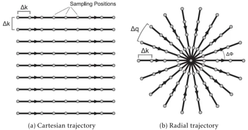
1.9 Cartesian vs. non-Cartesian trajectories. . . 22
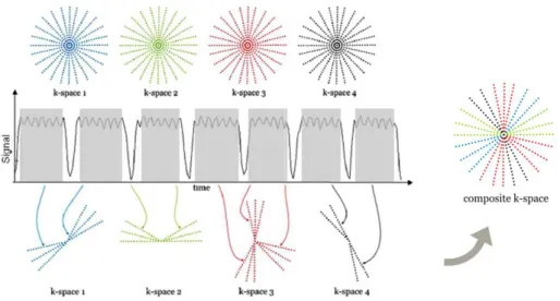
1.10 UTE self-navigating property . . . 26
1.11 FLASH sequence. . . 28
2.1 UTE pulse sequence . . . 42
2.2 Gridding problem . . . 44
2.3 Gridding kernel convolution and oversampling correction . . . 45
2.4 USRPs synthesis and size . . . 47
2.5 USRP schematic representation and relaxivity properties . . . 49
2.6 Multimodal USRPs biodistribution after intravenous injection. . . 50
2.7 SPECT biodistribution of USRPs after intravenous administration 51
6.1 Ovalbumin model histology for different time points . . . 113
6.2 Extension of the OVA model to a third time point . . . 114
6.3 USRPs active targeting potential. . . 120
6.4 Application of USRPs to lung fibrosis . . . 122
6.5 Application of USRPs to brain cancer study . . . 124
6.6 Application of USRPs to brain cancer study: SNR and SE. . . 125
6.7 Upconverting nanophosphors for lung imaging . . . 128
ADC Analog to Digital Converter BHR Bronchial Hyperresponsiveness BLI Bioluminescence Imaging
BW Bandwidth
CNR Contrast-to-Noise Ratio
COPD Chronic Obstructive Pulmonary Disease cRAD Cyclic arginine-alanine-aspartic acid cRGD Cyclic arginine-glycine-aspartic acid
CT Computed Tomography
Cy Cyanine dye
DOTA 1,4,7,10-tetraazacyclododecane-1,4,7,10-tetraacetic acid ECG Electrocardiogram
EPR Enhanced Permeability and Retention FA Flip Angle
FFT Fast Fourier Transform FID Free Induction Decay FLASH Fast Low-Angle Shot FOV Field Of View
FRI Fluorescence Imaging FT Fourier Transform
HES Haematoxylin-Eosin-Safran HP Hyperpolarized
ICC Interclass Correlation Coefficient ID Injected Dose
IRM Imagerie par R´esonance Magn´etique IS Inner Sphere
LPS Lipopolysaccharide
MEMRI Manganese-enhanced Magnetic Resonance Imaging
MR Magnetic Resonance
MRI Magnetic Resonance Imaging
NMR Nuclear Magnetic Resonance
NMRD Nuclear Magnetic Resonance Dispersion
NSCLC Non-Small-Cell Lung Cancer
OE Oxygen-enhanced
OS Outer Sphere
OVA Ovalbumin
PBSI Peribronchial Signal Index Penh Enhanced Pause
PET Positron Emission Tomography PK Pharmacokinetic
PR Projection Reconstruction PS Peribronchial Signal
RES Reticuloendothelial System RF Radiofrequency
RGD Arginine-glycine-aspartic acid ROI Region Of Interest
SCLC Small-Cell Lung Cancer SD Standard Deviation
SE Signal Enhancement
SEM Standard Error of the Mean SNR Signal-to-Noise Ratio
SPECT Single-Photon Emission Computed Tomography
SPIO Super Paramagnetic Iron Oxide
TE Echo Time
TR Repetition Time
TRE Typical Respiratory Epithelium
USPIO Ultra-small Super Paramagnetic Iron Oxide USRP Ultra-Small Rigid Platform
UTE Ultra-short Echo Time
1
Introduction
This chapter will provide the readers with the basic concepts and terminology about the functions and anatomy of lungs. In view of the in vivo pre-clinical studies presented in this work, a special attention will be given to the dif-ferences between rodents and human’s respiratory systems. In addition, the main pulmonary pathologies will be reviewed, with special focus on asthma and lung cancer, the diseases which will be object of investigation in the next chapters.
An overview of the main challenges to image lung withMRIwill be pro-vided along with a short summary of the main techniques used to encompass these limiting factors.
Finally, a synopsis of the contrast agents principles and of the state-of-the-art of contrast media applied to lungs will be presented.
1.1
Anatomopathology of lung
1.1.1 Lung anatomophysiology and morphology
The lung is the essential respiration organ in air-breathing animals. Its prin-cipal function is to fulfill gas exchanges, transporting oxygen from the at-mosphere to the bloodstream and removing carbon dioxide from the blood circulation. In addition, the lungs carry out other functions like the metabo-lization of some compounds and the filtration of undesired materials from the circulation; moreover, it acts as a reservoir for blood and as a blood pH balance [15].
1.1.1.1 Lung structure and physiology
In order to perform these physiologic functions, a specific architecture of the lung is needed. Even though this differs between species, many struc-tural components remain consistent among animals [16–18]. In reptiles, birds and mammals, respiration takes place through a sequence of steps where air is brought into the lungs through the airways. Since the diffusion of a gas
through a sheet of tissue is proportional to the area of the sheet and inversely proportional to its thickness (Fick’s law of diffusion), commonly the blood-gas barrier is exceedingly thin and has an internal total area which is much larger than the outer surface of the lung (between 50 and 100 m2in humans) [15]. Such enormous surface is achieved through a complex branching pattern of the tracheobronchial tree.
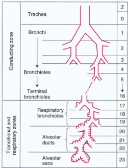
The airways consist in a series of branching conducting tubes, which be-come narrower, shorter and more numerous as they penetrate deeper into the lung. As shown in Figure1.1they can be divided into two zones: the conduct-ing airways and the respiratory ones. The conductconduct-ing zone, whose function
Figure 1.1: Schematic representation of the tracheobronchial tree. The major bronchi, bronchi-oles, and terminal bronchioles make up the conducting zone, while the respiratory bronchibronchi-oles, alveolar ducts, and alveolar sacs make up the transitional and respiratory zones. Z denotes the airway generation. Reprinted from [15].
is to transport the inspired air to the gas-exchanging regions of the lungs, is formed by the trachea which divides into the bronchi, bronchioles and terminal bronchioles. The respiratory zone, where the gas-exchange occurs, begins when the terminal bronchioles divide into the respiratory bronchioles and eventually to the alveolar ducts, which are lined with alveolar sacs (alve-oli). Alveoli associated with the respiratory airways and with the more distal alveolar sacs constitute the lung parenchyma [19].
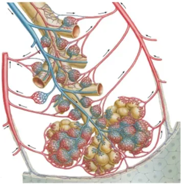
The portion of the cardiovascular system which carries deoxygenated blood away from the heart, to the lungs (via the pulmonary artery), and returns oxygenated blood back to the heart (via the pulmonary veins) consti-tutes the pulmonary circulation. The pulmonary blood vessels branch from
Figure 1.2: Terminal alveoli connected to the network of the capillary system. Alveoli are in contact with capillaries. The thin barrier between the alveolar and the capillary walls allows a rapid exchange of oxygen and carbon dioxide. Adapted from [20].
the pulmonary artery accompanying the tracheobronchial tree deep into the respiratory zone. The smallest branches of the cardiovascular system, the cap-illaries, form a dense network of short interconnecting segments which cover and embed the alveoli, providing an efficient arrangement for gas exchange, as shown in Figure1.2[15].
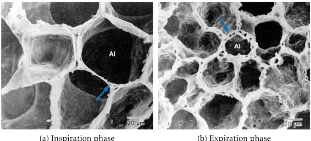
From a mechanical point of view, the respiration in mammals is made pos-sible thanks to several muscles: the diaphragm, the intercostal and accessory muscles, and the abdominal muscles. During inhalation, the diaphragm is the most important muscle. Briefly, when it contracts, it moves in the inferior direction, thus enlarging the volume of the thoracic cavity. The decrease of the internal pressure generates a gradient of pressure which allows air to enter the lungs. During the inspiration phase, alveoli expand in volume, resulting in an overall lower parenchyma density (Figure1.3a).
(a) Inspiration phase (b) Expiration phase
Figure 1.3: Scanning electron microscope view of lung alveoli, magnified × 750, showing the way in which their shape is retained as their size alters with changes in lung volume. The blue arrows underline the changes in capillaries shape, which varies proportionally to the degree of stretching undergone by the alveolar walls. Adapted from [21]
return to their equilibrium position because of elastic recoil. The decrease in lung volume and the increase in the alveoli pressure generate the flow of air from the lungs to the atmosphere, increasing the parenchyma density (Figure
1.3b) [15,19].
1.1.1.2 Lung morphology
The airway wall can be considered as being composed of inner and outer layers, the former extending from the lumen to the outermost layer of smooth muscle and the latter from the smooth muscle to the airway parenchymal boundary, as shown in Figure1.4.
The basic morphology of lung varies while moving from the conducting to the respiratory zone, as schematically shown in Figure1.5. The conducting airways are characterized by a surface epithelium largely composed of cili-ated and secretory cells (e.g., goblet cells), overlying subepithelial tissue that consists predominantly of connective tissues and glands. The proportion and type of these elements vary at different levels of the conducting system [19]. The tracheal and proximal bronchial epithelia are composed predomi-nantly of columnar ciliated and goblet cells, attached to the underlying basal lamina. These cells are attached to one another by tight junctions, forming a barrier physically impermeable to most substances [9,19].
The subepithelial tissue can be subdivided into a lamina propria and a submucosa, consisting of all the remaining airway tissue.
Figure 1.4: Schematic diagram and real image of a medium-sized bronchus. The cartilage reinforcement is clearly visible in dark blue. In the wall of the bronchus it is possible to recognize the smooth muscle. The lining of all bronchi is the typical respiratory epithelium (TRE). In the image on the right it is possible to observe one of the large arteries that run alongside the bronchi. Numerous smaller blood vessels are present among the profiles of the gas exchange areas. Bronchioles have a similar structure but do not present cartilage plates since, when the airways diameter is smaller, smooth muscle in the wall has sufficient tone to resist collapse [22].
The lamina propria consists principally of a network of capillaries, of reticular fibers, and bundles of elastic and nerve fibers. These fibers are believed to help transmit to the more rigid and stronger cartilaginous fibrous tissue the tension that arises in the airway epithelium and lung parenchyma during breathing [9,19].
The submucosa contains cartilage, muscle, and other supportive connec-tive tissue elements, including the loose connecconnec-tive tissue known as adventi-tia (whom mainly serves as a support for trachea and bronchi). The majority of the tracheobronchial glands are located in the submucosa and they are almost absent in the smallest bronchi and bronchioles. These glands are mainly responsible to secrete mucus. A variety of substances that are poten-tially important in local airway defense are also secreted by serous cells [9,19].
Moving towards the respiratory zone, the surface of respiratory bronchi-oles is lined by cuboidal epithelium that gradually decreases in extent as the number of alveoli increases. Bronchiolar-type epithelium and lamina propria are absent in alveolar ducts [9,19].
Alveoli are demarcated by septa composed of a continuous layer of epithe-lial cells overlying a thin interstitium. The interstitium contains capillaries involved in gas exchange (Figure 1.3), as well as connective tissue and a variety of cells responsible for maintaining alveolar shape and defense. Of particular interest is their secretion of surfactant, a phospholipid which forms an extremely thin (4 nm) layer of material that covers the alveolar epithelial surface and is responsible to reduce the alveoli surface tension, preventing them from collapsing [15,19].
1.1.1.3 Defense system
Letting aside the first-pass metabolism constituted by the nasopharyngeal anatomy (able to stop particles with size ≥ 2-3 µm), lungs have several defense mechanisms to clean and protect themselves from external potentially harm-ful pathogens. Some of the most relevant of such mechanisms include the mucociliary clearance , the epithelial barrier , and the phagocytic system [24].
The mucociliary clearance is the removal of impacted particles from the terminal bronchioles to the trachea by the ciliary beats of epithelial cells in the mucus of bronchi. The mucus acts as a barrier for bacteria as well.
Epithelial cells provide a mucosal barrier (with antimicrobial properties) and contribute to the mucociliary clearance function just mentioned. At the same time, lining the luminal surface of the airways and being tightly attached one to the other, they form a physical barrier between the luminal space and
the pulmonary parenchyma. Similarly, the alveolar epithelial cells strongly contribute to pulmonary host defense through the surfactant.
Many cells concerned with airways defense are found in the lungs, in-cluding lymphocytes, dendritic cells, and mast cells [19,24]. Nevertheless, macrophages are the resident mononuclear phagocytes of the lung which pro-vide the first line of defense against organisms or particles reaching the lower airways. In a normal lung, they account for about the 95% of all the nonep-ithelial cells in lung parenchyma [19]. Even though still matter of debate, it is acknowledged the possibility that pulmonary macrophages have different functional capabilities. For this reason they are often divided in several groups on the basis of their anatomic location [19,24,25]. Commonly the following groups are distinguished: (a) the airway macrophages, situated on the epithe-lial lining of conducting airways; (b) the alveolar interstitial macrophages; (c) the alveolar surface macrophages; (d) the intravascular macrophages, located adjacent to the capillary endothelial cells; and (e) the pleural macrophages.
Finally, depending on the load of pathogens, various non-resident effector cells can be recruited. For example, eosinophils are abundant in the infiltrates of late phase reactions and contribute to many of the pathological processes in allergic diseases whereas the recruitment of neutrophils is a major component of the protective host response to bacterial infections [24].
1.1.1.4 Rodents and human’s lungs comparative anatomy
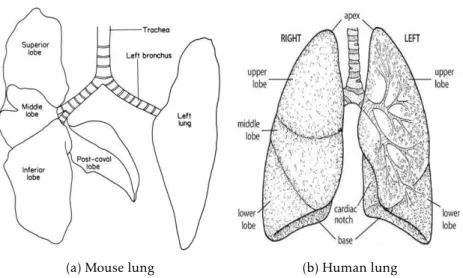
In both rodents and humans, the right and the left lungs are enveloped by a thin layer of connective tissue (the visceral pleura), which extends into the parenchyma, dividing it into several lobes. Nevertheless, at this level, substan-tial structural differences can be found between the two orders (Rodentia and Primates). In mice (Figure1.6a) and rats the left lung consists of one single lobe whereas the right one consists of four lobes (superior, middle, inferior and post-caval lobes) [26]. Conversely, as shown in Figure1.6b, human’s left lung is composed of two lobes (upper and lower) while the right side is di-vided into three lobes (upper, middle, and lower) [15].
The total capacity of mouse lungs has been reported to be about 1 ml, com-pared to 10 ml of the rat and 6,000 ml of a human. Mouse lungs have fewer respiratory bronchioles and airways generations (13-17 generations) than human lungs (17-21 generations) [27,28]. It has also been found that, while the branching pattern in humans is dichotomous (that is, the parent branch divides into two parts), in rodents monopody (a small segment branching off from the main stem) is commonly observed [16,27,29]. The parenchyma in
(a) Mouse lung (b) Human lung
Figure 1.6: Schematic representation of mouse and human’s lung structure.
mice occupies roughly the 18% of the total lung (24% in rats), with an average alveolus mean linear intercept of 80 µm (100 µm for rats) and a blood-gas barrier thickness of 0.32 µm (0.38 µm for rats) [27,29,30]. The parenchyma of the human occupies a smaller fraction of the total lung (12%), with larger alveoli mean linear intercept (210 µm) and gas-blood barrier (0.62 µm) [15,27]. An important functional difference observed between rodents and humans is the paucity of submucosal glands and the presence of a high number of nonciliated cells (Clara cells) in rodents [27].
Even though the significance of all these anatomical features of the mouse lung for lung function is matter of debate, it is generally believed that they might explain the different inflammatory effects of specific allergens on ro-dents and humans [27].
1.1.2 Lung pathologies
Respiratory diseases comprise a vast number of pathological conditions affect-ing the pulmonary tract, includaffect-ing conditions of the upper respiratory tract, trachea, bronchi, bronchioles, alveoli, pleura, pleural cavity, and the nerves and muscles of breathing. They range from mild (e.g., common cold) or self-limiting to life-threatening (e.g., bacterial pneumonia, lung cancer, etc.) [1,31].
While a comprehensive classification of these pathologies can be found in specific reports of the World Health Organization (WHO) [32], for practical
reasons the major part of the most common lung diseases can be categorized as follows [31]:
• Obstructive diseases : All these diseases are characterized by airways obstruction, which can originate for different reasons. Firstly, the lumen can be partially or totally obstructed by excessive secretions (e.g. in chronic bronchitis); secondly, the obstruction can take place in the wall of the airway because of an excessive contraction of the bronchial smooth muscle (e.g. in asthma), hypertrophy of mucous glands (e.g. chronic bronchitis) or inflammation and edema of the wall (like both in asthma and chronic bronchitis). Finally, the obstruction can be due to changes that take place in the peribronchial region. This is for example the case of the lung parenchyma destruction typical of emphysema (with its loss of radial traction and consequent narrowing of the airway) or peribronchial edema [31].
The most frequent pathologies which belong to this category are asthma [33] (see Section 1.1.2.1) and the so-called Chronic Obstructive Pul-monary Diseases (COPDs) [31], which include emphysema (enlarge-ment of the air spaces distal to the terminal bronchioles, with destruc-tion of their walls [9]), chronic bronchitis (excessive mucus producdestruc-tion in the bronchial tree, sufficient to cause excessive expectoration of spu-tum) or a mixture of the two [31].
• Restrictive diseases : All these diseases are characterized by a restric-tion of lung expansion, resulting in a decreased lung volume due to alterations of the lung parenchyma or to pathological conditions of the pleura (e.g., pneumothorax), chest wall (e.g., scoliosis) or neuromus-cular apparatus (e.g., musneuromus-cular dystrophies) [31]. The most frequent pathology which belong to this category is the diffuse interstitial pul-monary fibrosis, whose main feature is the excessive thickening of the interstitium of the alveolar wall [9].
• Vascular diseases : All these diseases are characterized by pathological conditions of the pulmonary vasculature. The most frequent pathologies which belong to this category are the pulmonary embolism (blockage of the main artery of the lung or one of its branches by a substance that has travelled from elsewhere in the body through the bloodstream) and the pulmonary hypertension (increase of pulmonary blood pressure which may lead to heart failure). The abnormal accumulation of fluids in the extravascular spaces and tissues, known as pulmonary edema, belongs
to this category as well, even though it’s a complication of a variety of heart and lung diseases rather than a disease on its own [31].
• Environmental, infectious, suppurative, and diseases : This category comprises a large range of diseases which do not fit in the previous three classes of pathologies, including many occupational lung diseases caused by inhaled dusts, abnormal functioning of exocrine glands (e.g., cystic fibrosis) or viruses and bacteria (e.g., pneumonia and tubercolo-sis). In addition, neoplastic diseases of the lung are included in this category (see Section1.1.2.2).
1.1.2.1 Asthma
Asthma is a complex chronic inflammatory disorder of the airways which involves several inflammatory cells and mediators and which results in patho-physiological changes of the lungs. As the pathogenesis of asthma is not clear, its description is generally based on the functional consequences of airway in-flammation, including the airways excessive responsiveness to various stimuli that leads to recurrent episodes of wheezing, breathlessness, chest tightness, and coughing. These episodes are often associated with diffuse airflow ob-struction within the lungs. For these reasons, for didactic purposes, asthma is generally categorized as an ‘obstructive disease’ [31].
The burden of asthma is consistent worldwide, with an estimate of about 300 million individuals affected by this airways disorder and 250 000 annual deaths [34]. The economic impact of asthma is substantial all over the world, both for direct medical costs (cost of medications and hospitalizations) and indirect non-medical costs (asthma is a major cause of absence from work in many countries) [34].
Pathophysiology The three main hallmarks of asthma are airway inflam-mation , bronchial remodeling and airway hyperresponsiveness [33–37], as schematically shown in Figure1.7. The relation intercurring among them is not clear; it is supposed that the inflammatory process results in the charac-teristic pathological changes of the airways in ways that are not well under-stood. Even though it is still matter of debate, it seems that inflammation and bronchial remodeling may be strongly associated to airways hyperresponsive-ness and the other asthma symptoms [34,38].
• Airways inflammation : the presence of inflammation is a constant feature in all asthmatic patients [34]. Typically mucosal mast cells and
macrophages are activated by allergens and release bronchoconstrictor and inflammatory mediators. Increased number of mast cells in airway smooth muscle may be linked to airway hyperresponsiveness while macrophages mediators amplify the inflammatory response [34,36]. A significant increase in the number of airways eosinophils, neutrophils, and T-lymphocytes is commonly observed. While the pathophysiological role of neutrophils and T-lymphocytes is still unclear, eosinophils are suspected to be at the basis of the damaging process of epithelial cells and airway remodeling [34,36].
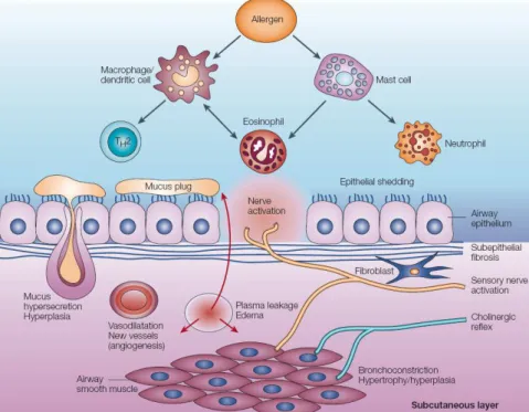
Figure 1.7: Schematic representation of asthma pathophysiology. Several inflammatory cells and mediators are recruited or activated, producing acute effects on the airway (bronchocon-striction, plasma leakage, mucus secretion, sensory nerve activation, . . . ), together with remod-eling structural changes (subepithelial fibrosis, angiogenesis, increased thickness of airway smooth muscle, hyperplasia of mucus-secreting cells, . . . ). Reprinted from [36].
• Bronchial remodeling : with this term it is generally indicated all the characteristic structural changes in the airways of asthma patients, which often result in an irreversible thickening of bronchial walls and, consequently, narrowing of the airways [33,34,36,38]. These changes include subepithelial fibrosis with deposition of collagen fibers (base-ment membrane thickening), airway smooth muscle hypertrophy
(in-creased size of individual cells) and hyperplasia (in(in-creased cell divi-sion), blood vessels proliferation (angiogenesis) and increased mucous secretory cells (hypertrophy of mucosal glands, hyperplasia of goblet cells,. . . ) [33–35,39–42].
• Airway Hyperresponsiveness : with this term it is generally indicated the excessive airways narrowing in response to an external stimulus which would be innocuous in a person not affected by asthma. This airway narrowing, as a consequence, leads to airflow limitation. Its mechanism is probably connected to the excessive contraction of airway smooth muscle, thickening of the airway wall, and sensitized sensory nerves [34–36,43]. It is therefore hypothesized that both inflammation and bronchial remodeling play an important role in the hyperesponsive-ness development [34].
Treatments Medications to treat asthma are classified as controllers or relievers [34]. The formers are medications which are based on a long-term approach and are taken daily to keep asthma under clinical control mainly thanks to their anti-inflammatory effects; they include inhaled and systemic glucocorticosteroids (e.g., budesonide) and long-acting inhaled β2
-agonists [36]. The latters are medications used on a as-needed basis in order to quickly reverse acute asthma symptoms [34], mainly acting on the bron-choconstriction; short-acting β2-agonists belong to this category [36].
While it has been shown that controller medications can strongly improve patients’ quality of life (controlling airways inflammation, decreasing airways hyperresonsiveness and severity of exacerbations), it is widely known that they do not cure asthma and they have to be taken continuously [34]. All these therapies are intended to treat inflammation and bronchoconstriction whereas bronchial remodeling still remains insensitive to current asthma treatments [44]. Whether and when to begin treating patients with asthma to prevent or reverse the negative effects of remodeling, which components of remodeling to target, and how to monitor remodeling are still matters of debate.
1.1.2.2 Lung Cancer
Lung cancer is a disease characterized by uncontrolled cell growth in the lung tissues. Its development is the result of multiple alterations which include the classical genetic abnormalities that cause the overactivity of growth pro-moting oncogenes and the inactivation of the tumor suppressor gene [45]. The most common cause of lung cancer is long-term exposure to tobacco
smoke, which accounts for 80-90% of all lung cancers [46,47]. The other cases are generally attributed to a combination of genetic factors and exposure to a number of carcinogenic substances like radon gas, asbestos, some metals compounds (cadmium, chromium, beryllium, nickel, arsenic, . . . ), ionizing radiations and air pollution (including second-hand smoke) [31,46,47].
Lung cancer is the leading cause of cancer deaths worldwide and the third cancer for occurrence in both sexes (after breast and prostate cancers) [48]. The burden of this disease is impressive since it is estimated that lung cancer is responsible for more than 1.3 million deaths per year [46,48].
Pathophysiology Lung cancers are mainly classified as either Small-Cell Lung Cancers (SCLCs) or Non-Small-Cell Lung Cancers (NSCLCs) [47].SCLCs account for approximately 15% of all lung cancers, they develop mainly in the main bronchi (bronchial submucosa), and are highly malignant, spreading early in the course of the disease. The name comes from the fact that the oval-shaped cells which compose this type of tumor are smaller than normal cells with scant cytoplasm. Virtually all patients withSCLChave a smoking his-tory [45–47]. On the other hand,NSCLCs account for approximately 85% of all lung cancers and are commonly further divided into three sub-categories: adenocarcinoma, squamous cell carcinoma, and large cell carcinoma [49].
• Adenocarcinoma : it is a malignant epithelial tumor with glandular differentiation and mucus production which generally develops periph-erally in the lungs. Adenocarcinomas account for approximately 40-45% of lung cancers. The majority of adenocarcinoma cases are associated with smoking; nonetheless, this type of lung cancer is the most common in non-smokers [47,49].
• Squamous cell carcinoma : it is a malignant epithelial tumor showing keratinization and/or intercellular bridges that arises from bronchial epithelium. It generally arises centrally in the main bronchi and it accounts for approximately 30% of lung cancers. Like forSCLCs, over 90% of squamous cell lung carcinomas occur in cigarette smokers [49]. • Large cell carcinoma : it is, by definition, a poorly differentiated tumor. It is a diagnosis of exclusion made after ruling out the presence of a component of squamous cell carcinoma, adenocarcinoma or small-cell carcinoma. Large cell carcinoma typically appears as a large, peripheral mass, but may also involve large bronchi. It accounts for approximately 10% of lung cancers [49].
Diagnosis and treatments Prognosis of lung cancer is generally very poor, with an overall five-year survival rate of 15% [46,47,50]. The main reason of this is that the disease is often diagnosed at its latest stages [45,47]. Surgery, when applicable, is the treatment of preference. Radiotherapy and chemother-apy are generally used as adjuvant therapies or as primary/palliative therapies when the tumors are inoperable [46,47].
The diagnosis mainly relies on chest radiograph whereas Computed To-mography (CT) imaging is generally employed to better understand the type and extent of the disease [47,50]. The final diagnosis of lung cancer is based on histological examination of the suspected tissues after bronchoscopy in order to avoid the typical false positives ofCTimaging [51]. Even though it has been shown that the benefit in monitoring subjects at risk (e.g., smok-ers) with conventional imaging techniques (e.g.,CT) brings great advantages in terms of early diagnosis and hence prognosis [50,52–54], concerns have been raised about the radiation risk [3,47]. Preliminary modeling studies suggest that potential risks may largely outweigh benefits in nonsmokers or in young patients [51,55]. No current guidelines recommend mass screening to diagnose this pathology, not even for at-risk individuals [56,57].
![Figure 1.5: Tissue layers of the tracheobronchial tree. Reprinted from [23].](https://thumb-eu.123doks.com/thumbv2/123doknet/14740619.755018/34.892.202.734.666.1017/figure-tissue-layers-tracheobronchial-tree-reprinted.webp)
![Figure 2.1: Pulse sequence for a radial UTE trajectory with FID acquisition. Adapted from [129].](https://thumb-eu.123doks.com/thumbv2/123doknet/14740619.755018/71.892.206.640.259.580/figure-pulse-sequence-radial-ute-trajectory-acquisition-adapted.webp)