HAL Id: tel-01359195
https://tel.archives-ouvertes.fr/tel-01359195
Submitted on 2 Sep 2016HAL is a multi-disciplinary open access archive for the deposit and dissemination of sci-entific research documents, whether they are pub-lished or not. The documents may come from teaching and research institutions in France or abroad, or from public or private research centers.
L’archive ouverte pluridisciplinaire HAL, est destinée au dépôt et à la diffusion de documents scientifiques de niveau recherche, publiés ou non, émanant des établissements d’enseignement et de recherche français ou étrangers, des laboratoires publics ou privés.
Nuria Socoro Yuste
To cite this version:
Nuria Socoro Yuste. Proteomic and functional deregulations in Philadelphia negative Myeloprolifer-ative Neoplasms. Hematology. Université Grenoble Alpes, 2015. English. �NNT : 2015GREAS018�. �tel-01359195�
THÈSE
Pour obtenir le grade de
DOCTEUR DE L’UNIVERSITÉ GRENOBLE ALPES
Spécialité : BIS - Biotechnologie, instrumentation, signal et imagerie pour la biologie, la médecine et l'environnementArrêté ministériel : 7 août 2006
Présentée par
« Nuria SOCORO YUSTE »
Thèse dirigée par « Pascal MOSSUZ »
préparée au sein du Laboratoire TIMC-IMAG - Techniques de l'Ingénierie Médicale et de la Complexité - Informatique, Mathématiques et Applications, Grenoble – UMR 5525 CNRS dans l'École Doctorale Ingénierie Pour La Santé, La Cognition et L’environnement
« Dérégulations protéomiques et
fonctionnelles des Syndromes
Myéloprolifératifs Philadelphia
négatifs »
Thèse soutenue publiquement le « 17 décembre 2015 », devant le jury composé de:
Mme Isabelle PLO
DR-INSERM, Université Paris Diderot-Paris 7 (Rapporteuse)
Mr Wassim EL NEMER
CR1-INSERM, Université Paris Diderot-Paris 7 (Rapporteur)
Mr Jean-Yves CAHN
PU-PH, Université Grenoble Alpes, CHU Grenoble (Examinateur)
Mme Valérie UGO
PU-PH, Université d’Angers, CHU Angers (Examinatrice)
Mme Marie Claire DAGHER
CR-CNRS, Université Grenoble Alpes (Examinatrice)
Mr Pascal MOSSUZ
INDEX
BACKGROUND ... 1 MYELOPROLIFERATIVE NEOPLASMS ... 3 POLYCYTHEMIA VERA ... 4 ESSENTIAL THROMBOCYTHEMIA ... 5 PRIMARY MYELOFIBROSIS ... 6 GENETIC COMPLEXITY OF MPNs ... 8 VASCULAR COMPLICATIONS ... 19 LEUKEMIC PROGRESSION ... 23 MYELOFIBROTIC EVOLUTION ... 25 TREATMENT ... 26PREVIOUS REPORTED DYSFUNCTIONS IN MPNs ... 34
PROTEOMICS, the proteome study ... 39
PROTEOMIC METHODS ... 39
SELDI TOF MS (115) ... 41
LC MS/MS (116) ... 42
ERYTHROCYTE PROTEOME ... 45
OBJECTIVE ... 49
CHAPTER I - Ph-negative MPN red blood cells display deregulation of IQGAP1-Rho GTPase signaling depending on CALR/JAK2 status ... 53
BACKGROUND ... 55
MATERIALS AND METHODS ... 56
Patients ... 56
Erythrocyte and Protein Preparation ... 56
Magnetic Nanoparticle Hemoglobin Depletion ... 57
Proteomic Analysis ... 58
Western-Blotting ... 60
Immunoprecipitation ... 60
Antibodies ... 61
Rac1Q61L-GST Protein Production ... 61
KG1 and K562 Lysates ... 62
RESULTS ... 63
1-RBC proteome of MPN patients display specific alterations of protein expression ... 63
3-IQGAP1 interacts selectively with Rho GTPase proteins in MPN RBCs ... 68
4-Calreticulin but not JAK2 protein is expressed by MPN RBCs ... 70
5-CALR mutated patients displayed distinct IQGAP1/Rho GTPase protein interactions compared with JAK2V617F ones ... 72
DISCUSSION ... 76
RAS subfamilies... 78
CONCLUSION AND PERSPECTIVES ... 87
1-Does a link between JAK2 and IQGAP1 exist? ... 87
2-Is there any link between JAK2 and IQGAP1/Rho GTPase signaling? ... 87
RESUME DU CHAPITRE I ... 89
CHAPTER II - Proteomic study of granulocytes in Ph-negative MPNs ... 91
BACKGROUND ... 93
MATERIAL AND METHODS... 95
Patients ... 95 Sample Preparation ... 95 iTRAQ Labeling ... 96 Protein Identification ... 96 Protein Quantification ... 97 Statistical Analysis ... 97 Functional Analysis ... 98 RESULTS ... 99
1-Granulocyte proteome of Ph- MPN patients displays alterations in protein expression ... 99
2-Impact of JAK2V617F allele burden in granulocyte protein expression ... 105
3-Pathway alterations in Ph-MPN granulocytes ... 107
4-Rho GTPase deregulations in Ph-MPN granulocytes ... 107
5-Oxidative stress in granulocytes ... 109
6-Transcriptomic analysis ... 110
7-Comparison between transcriptomic and proteomic analysis ... 111
DISCUSSION ... 112
CONCLUSION AND PERSPECTIVES ... 119
RESUME DU CHAPITRE II ... 121
CHAPTER III - Ba/F3 and HEL cells as models on the study of MPN deregulations. ... 123
BACKGROUND ... 125
Cell Lines ... 127
Western-Blotting ... 127
Immunoprecipitation ... 127
Antibodies ... 127
Inhibitors ... 127
Cell cycle analysis by Flow Cytometry ... 128
RESULTS ... 129
HEL CELLS ... 129
1-Effects of JAK2 inhibitors and hydroxyurea on cell concentration and cell viability ... 129
2-Cell cycle analysis... 130
3-IQGAP1 and Rho GTPases expression in HEL cells ... 136
4-Both Rac1 and Cdc42 are activated in HEL cells ... 140
5-JAK2 and Calreticulin expression in HEL cells ... 141
Ba/F3 CELLS ... 144
1-Effects of inhibitors on cell concentration and cell viability ... 144
2-Rho GTPase family expression in Ba/F3 cells ... 146
3-Rac1 and Cdc42 are activated in Ba/F3 EpoR JAK2 V617F cells ... 149
4-JAK2 and Calreticulin expression in Ba/F3 cells ... 149
5-IQGAP1 effectors in Ba/F3 cells ... 151
DISCUSSION ... 154
1-Effects of inhibitors on cell concentration and cell viability ... 154
2-Cell cycle analysis... 155
3-Protein expression in Ba/F3 EpoR, Ba/F3 JAK2 WT, Ba/F3 EpoR JAK2V617F and HEL cells ... 155
CONCLUSION AND PERSPECTIVES ... 158
1-Does JAK2V617F have any influence on the IQGAP1/Rho GTPase complexes? ... 158
2-Is there any link between STAT5/3 and IQGAP1/Rho GTPase signaling? ... 158
3-Does CALR mutations induce deregulation of IQGAP1/Rho GTPase signaling? ... 159
4-Does up-regulation of CALR protein participate to autonomous growth of cells? ... 159
RESUME DU CHAPITRE III ... 161
GENERAL SYNTHESIS ... 163
BIBLIOGRAPHY ... 169
SUPPLEMENTAL DATA ... 181
SUPPLEMENTAL DATA I ... 183
Supplemental Data I-1 ... 183
List of most downregulated proteins in JAK2V617F PV vs Controls ... 184
List of most upregulated proteins in JAK2V617F ET vs Controls ... 184
List of most downregulated proteins in JAK2V617F ET vs Controls ... 186
List of most deregulated proteins in JAK2V617F PV vs JAK2V617F ET ... 187
List of most deregulated proteins in JAK2V617F PV vs JAK2(-) ET ... 188
List of most deregulated proteins in JAK2V617F ET vs JAK2(-) ET ... 188
Supplemental Data I-2 ... 189
Deregulated pathways in JAK2V617F PV vs Controls ... 189
Deregulated pathways in JAK2V617F ET vs Controls ... 190
Deregulated pathways in JAK2V617F PV vs JAK2V617F ET ... 191
Deregulated pathways in JAK2V617F PV vs JAK2(-) ET ... 193
Deregulated pathways in JAK2V617F ET vs JAK2(-) ET ... 194
Supplemental Data I-3 ... 196
Supplemental Data I-4 ... 197
Supplemental Data I-5 ... 198
Supplemental Data I-6 ... 198
SUPPLEMENTAL DATA II... 199
Supplemental Data II-1 ... 199
PV vs Mut0 (JAK2(-) ET and PMF) comparison ... 199
ET vs Mut0 (JAK2(-) ET and PMF) comparison ... 200
PMF vs Mut0 (JAK2(-) ET and PMF) comparison ... 203
PV vs PMF comparison ... 206
ET vs PMFcomparison ... 207
PV vs ET comparison... 210
Supplemental Data II-2 ... 211
PV vs Mut0 (JAK2(-) ET and PMF) comparison ... 211
ET vs Mut0 (JAK2(-) ET and PMF) comparison ... 212
PMF vs Mut0 (JAK2(-) ET and PMF) comparison ... 213
PV vs PMF comparison ... 214
ET vs PMF comparison ... 214
PV vs ET comparison... 215
Supplemental Data II-3 ... 216
ABBREVIATIONS
AML: Acute Myeloid Leukemia CALR: CalreticulinCML: Chronic Myeloid Leukemia DMSO: Dimethyl Sulfoxide EPO: Erythropoietin
EpoR: Erythropoietin receptor ET: Essential Thrombocythemia GDP: Guanosine diphosphate GTP: Guanosine Triphosphate Hb: Hemoglobin
HSC: Hematopoietic Stem Cell HU: Hydroxyurea
IFN: Interferon IL: Interleukin
IPA: Ingenuity Pathway Analysis
iTRAQ: Isobaric tags for relative and absolute quantitation JAK2: Janus Kinase 2
LC: Liquid Chromatography
Lu/BCAM: Lutheran and basal cell adhesion molecule MDS: Myelodysplastic Syndromes
MF: Myelofibrosis
MPN: Myeloproliferative Neoplasm MS: Mass Spectrometry
Mut0: Non mutation m/z: mass / charge Ph: Philadelphia
PMF: Primary Myelofibrosis PV: Polycythemia Vera RBC: Red Blood Cell
SELDI: Surface-enhanced laser desorption/ionization TOF: Time of Flight
TPO: Thrombopoietin WB: Western Blot WBC: White Blood Cells
WHO: World Health Organization WT: wild type
1
BACKGROUND
3
MYELOPROLIFERATIVE NEOPLASMS
The myeloproliferative neoplasms (MPNs), are a group of hemopathies characterized by a clonal proliferation of hematopoietic stem cells that results in an augmentation of mature blood cells.
According to the World Health Organization (WHO), among the myeloid neoplasms we can distinguish, (1, 2):
1. Myeloproliferative Neoplasms
Chronic myelogenous leukemia, (CML);
Polycythemia Vera, (PV);
Primary myelofibrosis, (PMF);
Essential thrombocythemia, (ET);
Chronic neutrophilic leukemia;
Chronic eosinophilic leukemia, not otherwise specified;
Mastocytosis,
Unclassifiable MPNs,
2. Myeloid and lymphoid neoplasms associated with eosinophilia and abnormalities of PDGFRA, PDGFRB, or FGFR1.
3.
Myelodysplastic/myeloproliferative neoplasms (MDS/MPN) Chronic myelomonocytic leukemia
Atypical chronic myeloid leukemia, BCR-ABL1-negative
Juvenile myelomonocytic leukemia
Myelodysplastic/Myeloproliferative neoplasm unclassifiable 4. Myelodisplastic syndrome (MDS)
The only MPN with a specific gene abnormality is CML, which is characterized by the presence of the Philadelphia (Ph) chromosome which is a chromosomal translocation, t(9;22)(q34;q11) that fuses the abl gene from chromosome 9 with the bcr gene from chromosome 12 leading to the BCR-ABL1 fusion gene (3, 4). The other subtypes are known as Philadelphia negative (Ph-) MPNs. The most frequent are PV (35%) and ET (40% of total MPN patients). CML represents 16% of MPN cases and PMF just 5%. The rest are rare
4
diseases. The global incidence of Ph- MPNs is about 3 per 100,000 people newly diagnosed with any form of MPN.
POLYCYTHEMIA VERA
Polycythemia Vera is also known as the Osler-Vaquez disease in honor of the first ones to characterize it. Louis Henri Vaquez was a French physician who described for the first time a PV patient in 1892. This patient presented chronic cyanosis (congestion or ruddiness), vertigo, dyspnea, palpitations, hepatosplenomegaly and marked erythrocytosis. He thought that raised red blood cell count had to be due to an increased activation of hematopoiesis. The Osler term came out thanks to the first PV review by William Osler in 1903: “Chronic cyanosis, with polycythemia and enlarged spleen: a new clinical entity”. In Osler’s review, he included his own patients and other cases that had been previously reported (5, 6).
In those days, PV was defined by chronic cyanosis, polycythemia, moderate splenomegaly, weakness, constipation, headache and vertigo. Nowadays it is characterized by clonal overproduction of mature erythrocytes and, moreover variable overproduction of leukocytes and platelets. The erythroid stem cells present erythropoietin (EPO) independence and hypersensitivity. EPO independence means that these cells are capable of going on with their cell cycles without the EPO stimulation. EPO hypersensitivity is referred to as the increased reaction of myeloid stem cells to very low concentrations of this cytokine (7). Clinically, it is characterized by splenomegaly, thrombohemorrhagic complications and pruritus. The average age in patients is 60 years. Usually, survival rate is about 15 years for older patients, excepting for cases with grave thrombohemorrhagic complications such as aggressive phlebotomy where the survival average is shortened. Life-expectancy rises to 24 years in patients under 60 years old (8, 9). In addition, 5 to 15 % of patients evolve to Acute Myeloid Leukemia or to myelofibrosis (10).
Diagnosis
According to the 2008 World Health Organization (1, 2), the diagnosis of PV needs the presence of both major criteria and one minor criterion or the presence of one major criterion and two minor criteria:
5 Major criteria
1. Hemoglobin > 18.5 g/dL in men, > 16.5 g/dL in women or other evidence of increased red cell volume. (Hemoglobin or hematocrit > 99th percentile of method-specific reference range for age, sex, altitude of residence or hemoglobin > 17 g/dL in men, 15 g/dL in women if associated with a documented and sustained increase of at least 2 g/dL from baseline that cannot be attributed to correction of iron deficiency or elevated red cell mass >25% above mean normal predicted value).
2. Presence of JAK2V617F or other functionally similar mutation such as JAK2 exon 12 mutation.
Minor criteria
1. Bone marrow biopsy showing hypercellularity for age with trilineage growth (panmyelosis) with prominent erythroid, granulocytic, and megakaryocytic proliferation.
2. Serum erythropoietin level below the reference range for normal. 3. Endogenous erythroid colony formation in vitro.
ESSENTIAL THROMBOCYTHEMIA
This disease was described in 1934 by Emil Epstein and Alfred Goedel, two Australian pathologists; but it wasn’t until 1960 that two reviews regrouped the first diagnostic criteria for what they named “primary hemorrhagic thrombocythemia”: a history of thrombohemorrhagic events. These criteria included: splenomegaly or normal-size spleen; thrombocytosis without either erythrocytosis or leukocytosis; bone marrow panmyelosis with megakaryocytic hyperplasia without leukemic infiltration (6).
Nowadays, it is named “Essential Thrombocythemia” and it is defined as a myeloproliferative neoplasm characterized by high count of platelets in peripheral blood with tendency to thrombosis and hemorrhage. The typical clinical signs are predisposition to vascular occlusive events such as cerebrovascular, coronary or in peripheral circulation; and hemorrhages. Some patients are asymptomatic whereas others may present headaches, visual disturbances, distal paresthesia, lightheadedness, erythromelalgia, chest pain, and thrombotic or hemorrhagic problems (like hematomas, bruising, ecchymosis, epistaxis, melena or hematemesis). It is a disease that may occur at any age although the median age at diagnosis is 65-70 years. The incidence is twice higher in women (ratio 2:1 females:males)
6
and the prevalence is about 30/100,000. The median survival is about 20 years for older patients and 33 for younger ones (under 60). The only risks that can cause mortality in these patients are hematologic complications like arterial and venous thrombosis, platelet mediated occlusions of the microcirculation and hemorrhages. Sometimes it can evolve to myelofibrosis, acute myeloid leukemia or PV reducing patients’ life expectancy (9, 11, 12).
Diagnosis
According to the WHO (1, 2), the diagnosis of ET must meet these four criteria: 1. Sustained platelet count ≥ 450 x 109/L
2. Bone marrow biopsy specimen showing proliferation mainly of the megakaryocytic lineage with increased number of enlarged, mature megakaryocytes. No significant increase or left-shift of neutrophil granulopoiesis or erythropoiesis.
3. Not meeting WHO criteria for polycythemia vera, primary myelofibrosis, BCR-ABL1–positive CML, or myelodysplastic syndrome, or other myeloid neoplasm.
4.
Demonstration of JAK2 V617F or other clonal marker, or in the absence of JAK2 V617F, or no evidence of reactive thrombocytosis.PRIMARY MYELOFIBROSIS
Primary myelofibrosis (PMF) is the last one of the three most important Ph- MPNs. Older terms used to name PMF were “agnogenic myeloid metaplasia”; “chronic idiopathic myelofibrosis” (used in the 2001 WHO on the classification of tumors of hematopoietic and lymphoid tissues); and “myelofibrosis with myeloid metaplasia”. It was in 2006 when the International Working Group for Myelofibrosis Research and Treatment reached a consensus to exclusively use the term of Primary Myelofibrosis.
It was Gustav Heuck (a German surgeon) who gave the first description of PMF in 1879, under the title of “Two cases of leukemia with peculiar blood and bone marrow findings”. His young patients presented massive splenomegaly, circulating nucleated red blood cells, and increased number of morphologically abnormal leukocytes. He realized that these patients differed from those described for CML because of the presence of marrow fibrosis and extensive extramedullary hematopoiesis. From that date, many other cases of PMF have been described in literature (6).
7 PMF is predominantly present in males and the age at diagnosis is usually over 60. Patients are characterized by the presence of anemia, high splenomegaly, leukoerythroblastosis, dacryocytosis, increased levels of serum lactate dehydrogenase (LDH), excess circulating blasts and stromal changes in the bone marrow such as collagen and reticulin fibrosis, osteosclerosis and angiogenesis. According to the Dynamic International Prognostic Scoring System for primary myelofibrosis (DIPSS-plus), risk factors measured at diagnosis include age over 65 years, hemoglobin levels under 10 g/dL, leukocyte count more than 25x109/L, number of circulating blasts ≥1%, presence of
constitutional symptoms, red cell transfusion need, platelet count lower than 100x109/L and
unfavorable karyotype (including complex karyotype or sole or two abnormalities that comprised +8, -7/7q-, i(17q), inv(3), -5/5q-, 12p-, or 11q23 rearrangement; all other cytogenetic abnormalities were considered favorable). These risk factors are used to define low (no risk factors), intermediate-1 (1 risk factor), intermediate-2 (2 or 3 risk factors), and high (4 risk factors) risk groups. Leukemic transformation risk was higher in the presence of unfavorable karyotype or platelet count under 100x109/L and in intermediate and high
risk groups. Overall survival in low risk patients was around 17.5 years, 7.8 for the intermediate-1 risk group, 3.6 for the intermediate-2 and 1.8 for high risk patients. In young patients (aged under 60), the overall survival rises to 20 years in low risk patients and 14 in the intermediate-1 risk group (13, 14).
Generally PMF patients have poorer prognosis than PV or ET patients. PMF is clinically indistinguishable from the fibrotic transformation of polycythemia vera or essential thrombocythemia. Some patients who are diagnosed of PMF probably are in the accelerated phase of previously unrecognized PV or ET (15).
Diagnosis
According to the WHO (1, 2), the diagnosis of PMF must meet the three major criteria and at least two minor criteria:
Major criteria
1. Presence of megakaryocyte proliferation and atypia, (small to large megakaryocytes with an aberrant nuclear/cytoplasmic ratio and hyperchromatic, bulbous, or irregularly folded nuclei and dense clustering), usually accompanied by either reticulin or collagen fibrosis, or, in the absence of significant reticulin fibrosis, the megakaryocyte changes must
8
be accompanied by an increased bone marrow cellularity characterized by granulocytic proliferation and often decreased erythropoiesis.
2. Not meeting WHO criteria for polycythemia vera, BCR-ABL1–positive chronic myelogenous leukemia, myelodysplastic syndrome, or other myeloid disorders.
3. Demonstration of JAK2V617F or other clonal marker (e.g. MPLW515K/L), or, in the absence of the above clonal markers, no evidence that bone marrow fibrosis is secondary to infection, autoimmune disorder or other chronic inflammatory condition, hairy cell leukemia or other lymphoid neoplasm, metastatic malignancy, or toxic (chronic) myelopathies.
Minor criteria
1. Leukoerythroblastosis
2. Increase in serum lactate dehydrogenase level. 3. Anemia.
4. Palpable splenomegaly
GENETIC COMPLEXITY OF MPNs
Myeloproliferative neoplasms are clinical entities genetically complex. In fact, more than one mutation is usually present in these patients. Furthermore, there are not exclusive mutations for one type of MPN, but rather mutations that exclude the presence of other ones. Patients could be classified according to their genetic profile (number and description of mutations and order of appearance) to determine their diagnosis, prognostic and treatment.
The most important mutations in MPNs are grouped in four types depending on the action affected: signaling pathways, epigenetic regulation, splicing regulation and checkpoints, Figure 1.
9 Figure 1. Somatic mutations in MPNs affect four major processes: Signaling, epigenetics, splicing and checkpoints.
Mutations implicated in signaling pathways
JAK2 (16-20)
Mutation in JAK2 exon 14 is the most frequent in MPNs. It is present in more than 95% of PV patients and 50 to 60% of ET and PMF patients. It consists on the nucleotide change from Guanine to Thymine at position 1849. This causes the substitution of valine by phenylalanine at codon 617 in the Janus Kinase 2 (JAK2) protein.
The JAK family is formed by 4 kinases, JAK1, 2, 3 and TYK2. They are attached to cytokine receptors. JAK2 is formed by up to seven JAK homology domains (JH1-JH7). The most important ones are the active kinase domain, JH1 and JH2, the catalytically inactive pseudokinase domain that negatively regulates JH1 (21).
In a normal cell, erythropoietin binds its receptor which activates JAK2. It phosphorylates itself and also the erythropoietin receptor what initiates a cascade of erythroid-specific signaling promoting erythroid proliferation and differentiation by activation of STATs. This cascade is controlled thanks to a negative feedback by JAK2 itself. The key role of JAK2 in erythropoiesis is shown in JAK2 deficient mice that died at embryonic day 12.5 with complete absence of erythropoiesis (22).
10
The JAK2V617F mutation affects to the JH2 domain. This conversion implicates an autoinhibition of JH2 domain and thus, an inhibition of the downregulation of the erythropoietin signal. Due to this mutation; JAK2V617F progenitor cells become erythropoietin independent and can form erythropoietin-independent erythroid colonies. It also causes cytokine-independent activation of different pathways such as JAK-STAT, PI3K, AKT, MAPK and ERK which affect to gene transcription, apoptosis, cell cycle and differentiation (Figure 2) as it was showed in Ba/F3 EpoR and Ba/F3 EpoR JAK2V617F murine cells (17, 19, 23) and in gamma-2A human fibrosarcoma cells (17).
Among the STATs, the STAT5 activation is compulsory for MPN establishment. Recent approaches suggest that STAT3 activation opposes thrombocytosis and promotes inflammation whereas STAT1 is associated with thrombocytosis. Conformingly, STAT1 activation has been shown to be specifically activated in ET erythroid colonies but not in PV ones (21). Chen E et al. (24) showed that STAT1 inhibition in stem cells promoted erythropoiesis and reduced megakaryopoiesis. Thus, STAT1 activation via JAK2 promotes ET acquisition but not PV. The mechanisms why sometimes STAT1 is phosphorylated and sometimes it is not are not yet elucidated.
In conclusion, JAK2V617F is directly related to MPN pathogenesis by promoting spontaneous proliferation of stem cells (15). However, the main issue remains incompletely solved: why the same mutation can lead to three different phenotypes.
On the one hand, in PV patients, its presence is associated with higher hemoglobin levels, increased granulocytes, leukocytosis, endogenous erythroid colony formation, higher spleen size, higher bone marrow cellularity and inversely to lower platelet counts. On the other hand, it is also related to leukocytosis, higher spleen size and higher levels of hemoglobin in ET subjects besides older age at diagnosis and lower platelet count. In PMF, JAK2V617F patients require less red cell transfusion and present higher leukocyte counts than JAK2 wild type (25-27).
According to Alshemmari et al. (28), ET patients had lower JAK2V617F allele burden (<50% of JAK2V617F cells) whereas for PV patients the allele burden average was 40%. High allele burden levels are related to myelofibrotic evolution, leukocytosis and high hemoglobin levels. Low allele burden is associated with high platelet counts. Furthermore, 25% of PV patients are homozygous for JAK2V617F whereas ET patients are mostly heterozygous or present the wild type suggesting that higher levels of mutated JAK2 are related to an erythroid phenotype and lower ones to a megakaryocytic phenotype. Besides
11 this allele-burden-dependent mechanism, the presence of other acquired somatic mutations play very probably a role in these phenotypic variations, as well as in genetic background.
Figure 2. Left, type JAK2 is inactivated without erythropoietin stimulation. Center, wild-type JAK2 is phosphorylated and activated by erythropoietin action; it can then activate some pathways such as STAT, PI3K and RAS-MAPK. Right, JAK2 V617F is autophosphorylated in absence of EPO signal and is capable to activate the same signaling pathways as the non-mutated receptor in presence of EPO. Figure taken from Campbell et al.(15).
JAK2 exon 12 mutations were identified in JAK2V617F negative PV patients. (Its presence is rare in ET or PMF patients). These mutations are thought to modify the JH2 structure and thus to alter its negative feedback function inducing a constitutive phosphorylation of JAK2 and STAT5 (29). These patients are characterized by erythroid myelopoiesis, lower serum erythropoietin levels, younger age at diagnosis and the possibility of progression to a secondary myelofibrosis (21, 27).
MPL mutations (9, 21, 27)
MPL is the Myeloproliferative Leukemia virus oncogene. It encodes for the thrombopoietin (TPO) receptor. Gain-of-function mutations have been discovered in exon 10 with the substitution of a tryptophan at codon 515 to a leucine (MPLW515L), lysine (MPLW515K), asparagine (MPLW515N) or alanine (MPL W515A). At this position, there is
12
a group of 5 amino acids that are responsible for the cytosolic conformation of the MPL protein, (transmembrane protein). This receptor possesses an amphipathic domain R/KWQFP between the transmembrane and the cytosolic domains preventing JAK2 activation in the absence of TPO. Mutations in this group of amino acids cause the spontaneous activation of the receptor, resulting in constitutive JAK-STAT activation as well as MAP-kinase ERK1, 2. This activation leads to megakaryocytic proliferation and thus thrombocytosis.
These patients are associated with older age, female sex, higher platelet count, lower hemoglobin level, splenomegaly, myelofibrosis and an increased risk of thrombosis. They are found in about 3-5% of ET patients and in 10% of PMF ones. Their presence is rare in PV. Other mutations of this exon have been reported, (MPLW515S or MPLS505N) but at very low frequencies.
CALR (30-34)
Calreticulin is a Ca2+ binding chaperone located in the lumen of the endoplasmic
reticulum (ER) that contributes to calcium homeostasis. Together with calnexin (another chaperone), and ERp57, they form the “calreticulin/calnexin cycle” responsible for the folding control of newly-synthesized proteins. When calnexin and calreticulin recognize misfolded proteins, they bind them to prevent their export to the Golgi apparatus and consequently, guaranteeing proper folding of newly synthesized glycoproteins. Calreticulin can also be found outside the ER, at the cell surface, in the cytoplasm or in extracellular compartments where it is suggested to play a role in proliferation, antigen presentation and complement activation, apoptosis and immunogenic cell death (35).
CALR mutations are located in exon 9 and they are present in about 15 to 30% of ET and PMF subjects. Predominantly, 80% of these mutations are classified in two groups: type 1 mutations which correspond to a 5-base-pair (bp) deletion and type 2 to 5-bp insertions. All mutations result in a novel amino acid sequence at the C-terminal. This new mutated peptide contains an important number or positively charged amino acids, whereas the native C-terminal peptide is principally negatively charged. Moreover, the new C-terminal part has lost the KDEL motif (lysine, aspartic acid, glutamic acid and leucine amino acid sequence). KDEL motif allows calreticulin to anchor to the ER. Consequently, mutant calreticulin is thought to modify its cellular location and to lose its Ca2+binding function.
13 CALR mutations are suggested to activate the JAK2(-)STAT pathway in myeloid stem cells as it was suggested by Ba/F3 CALR cell mutants (33). Moreover, JAK2(-)STAT activation was shown to have the same transcriptional signature in JAK2V617F and mutated CALR peripheral blood granulocytes suggesting a common transformation (Rampal et al. (36)). Nonetheless, Lau et al. (37) argued Rampal results as theirs showed completely different signatures of STAT activation in JAK2(+) and CALR(+) ET megakaryocytes. In addition, MARIMO cell line approaches showed that JAK2(-)STAT activation was not activated by calreticulin (Kollman et al. (38)).
Generally, compared with JAK2V617F patients, CALR mutations arecorrelated with younger age, male predominance and high allele burden. They are also associated with higher platelet counts and serum erythropoietin, lower Hb and leukocyte levels and lower thrombosis risk. In addition, there is no significant difference in overall survival compared with JAK2V617F patients. Furthermore, phenotypical differences between both types of calreticulin mutations show that type 2 mutations are connected with younger patients and higher platelet counts whereas type 1 are linked to male sex. In these patients, there is no risk of polycythemic transformation (whereas the cumulative risk for JAK2V617F patients it is about 30%). In PMF, results on overall survival demonstrated differences between type 1 and type 2 mutations but are contradictory (Cabagnols et al. 2015, (39) vs Tefferi et al. 2014, (30, 40)). In addition, Cabagnols et al. study on 572 JAK2(-) MPN patients suggested that high CALR allele burdens and type 2 CALR mutations correlated with MF whereas low allele burdens with ET phenotypes.
Vannuchi et al. 2014 (41) showed, using immunohistochemistry labelling of bone marrow biopsy, that calreticulin expression (mutated and non-mutated) was higher in megakaryocytes than in myeloid or erythroid lineages suggesting an important role for calreticulin in platelet function. They suggested thatCALR mutations might use the JAK-STAT signaling to contribute to the high platelet production in MPNs. This special link between CALR and megakaryocytes was confirmed by the study of Mondet et al. (42) who showed that endogenous megakaryocytic colonies (EMC) were more frequent in mutated CALR than in JAK2V617F or “triple negative” (JAK2(-), MPL- and CALR(-)) patients and among CALR mutated patients, type 2 induced more EMC than type 1.
14
CBL (21, 27, 43)
The casitas B-cell lymphoma family (CBL) is composed by 3 proteins: CBL, CBL-b and CBL-c. The CBL gene is located at 11q23.3 and it encodes for a protein that principally is involved in negative regulation of the receptor tyrosine kinase mediated by E3 ubiquitin ligase activity. Mutations in this gene directly affect the region that is necessary for its E3 ubiquitin ligase function. It appears to be mutated in about 6% of PMF patients and in some myeloid malignancies as juvenile monomyelocytic leukemia or chronic monomyelocytic leukemia but it is rare in PV and ET patients. It’s related to myelofibrosis with poor prognosis or in AML post MPN.
LNK (21, 44)
This gene is located at chromosome 12 at 12q24 loci. The lymphocyte adaptor protein (LNK) is part of the SH2B family. It negatively regulates TPO-MPL and EPO receptor signaling, thus it inhibits the JAK/STAT signaling. Furthermore, it negatively regulates JAK2 thanks to its SH2 domain. These loss-of-function mutations cover codons 208 to 234 and can affect the C-terminal region; the SH2 domain or the PH domain. They seem to be involved in disease progression because they are not found in chronic states. In fact, some LNK mutations are found in about 13% of leukemic transformation of MPNs and in about 3 to 6% in ET and PMF subjects. A recent study by Benton et al. (45) showed that PV patients with mutations in this chromosome (Ch12) are more likely for myelofibrosis progression. In general, patients with this mutation are classified as poor prognosis.
SOCS 1, 2 and 3 (21)
Suppressor of cytokine signaling (SOCS) proteins are negative regulators of JAK signaling. Loss of their activity results in an excess of signaling by cytokines. SOCS1 gene is located at 16p13.2; SOCS2 at 12q22 and SOCS3 at 17q23.3. Mutations in SOCS genes are rare in MPNs. Nevertheless, hypermethylation of CpG sites in these genes together with a decrease in their expression was found in PV and ET mutated and non-mutated JAK2. The frequencies of these methylations in PV patients are: 11-13% for SOCS1 gene; 28% for SOCS2 and 22% for SOCS3, in ET patients: 14-25% for SOCS1 gene; 28% for SOCS2 and 10% for SOCS3, and in PMF patients: 17% for SOCS1 gene; 28% for SOCS2.
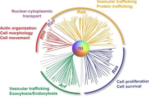
15 NRAS/KRAS (21)
The RAS family is a group of membrane proteins associated with GTPases that controls numerous signaling pathways and cellular processes. The most common mutations are found in KRAS and NRAS genes at codons 12, 13 and 61. They involve the inhibition of the GTPase activity that avoids the inhibition of these proteins and thus, the continue activation of the effector pathways. It is not clear if these mutations promote the initiation or the progression of MPNs to acute leukemia.
PTPN11 (46)
PTPN11 is the protein tyrosine phosphatase non-receptor type 11. It encodes for src homology 2 domain-containing protein-tyrosine phosphatase 2 (SHP2). It regulates proliferation, apoptosis, and differentiation.
Mutations in this gene are rare in ET and PV. Nonetheless, they have been described in PMF.
NF1 (47)
The tumor suppressor gene Neurofibromatosis-1 is located in the 17q11.2 chromosome. It is a negative regulator of the RAS pathway. Loss of NF1 may progress into a myeloproliferative neoplasm. It is related to an enhance risk of developing myeloid leukemia in some tumors, however, it is not yet clear if this is the case for MPNs and leukemic transformation. It is a rare mutation in MPNs.
Mutations implicated in epigenetics regulation
TET2 (21, 27, 48-50)
TET “ten-eleven-translocation” genes are a group of three homologous human proteins (TET1, 2 and 3). TET2 gene comprises 11 exons and is located on chromosome 4q24. It encodes for a 2-oxoglutarate and Fe(II)-dependent hydroxylase whose function is to hydroxylate methylated cytosine. Therefore, TET2 mutations result in decreased 5-hydroxymethylcytosine and thereby affect levels of DNA methylation.
TET2 appears to be mutated in 16% of PV patients, 5 to 11% of ET and 18% of PMF ones. These mutations consist on nonsense, missense, insertions and deletions which lead to loss of function. They are associated with leukocytosis, splenomegaly, extra medullary hematopoiesis, poor overall survival, leukemic transformation and older patients. The order
16
in which TET2 loss and JAK2V617F appear seems to be important. When JAK2 is acquired before TET2 mutations, it is more likely that patients develop PV; however, when TET2 goes first, it seems that it reduces the ability of JAK2 to up-regulate proliferation genes. Actually, mice studies revealed that TET2 loss increased self-renewal capacities of hematopoietic stem cells contrary to JAK2V617F which promotes proliferation and hardly reduces self-renewal of these cells. When these two mutations appeared together, TET2 competes with JAK2V617F for restoring self-renewal and quieting cell proliferation. In fact, TET2-loss-induced self-renewal is thought to be a first event on disease initiation and progression (51) that produces a pre-leukemic state with clonal expansion of HSC that, depending on the secondary molecular events, would evolve to a MPN or other myeloid malignancies.
IDH (21, 27, 49)
IDH gene encodes for isocitrate dehydrogenases (IDH1 and IDH2). They are NADP+
dependent enzymes that catalyze the oxidative decarboxylation of isocitrate to α-ketoglutarate. When mutated, they lose their affinity to isocitrate and catalyze the reduction of α-ketoglutarate (2HK) to 2-hdroxyglutatate (2HD), leading to an overproduction of 2HG instead of 2HK, which has been proposed to affect the function of some enzymes such as TET2 which are α-ketoglutarate-dependent. IDH1 an IDH2 are mutually exclusive mutations and are rare in PV and ET patients. Nonetheless they can be present in about 4% of PMF. One study has reported that the incidence of these two mutations is increased in blast transformation of MPNs (~22%) compared with its low incidence in chronic phase (1.9% in PV and 0.8% in ET). They are associated with decreased survival.
ASXL1 (21, 27, 49, 52)
ASXL1 gene is located on chromosome 20q11.1 and is similar to the Drosophila megalogaster additional sex combs gene. It is associated with the Polycomb repressive complex 2 (PRC2). The protein disrupts chromatin in localized areas, activating or suppressing the transcription activity. It is thought to have a soft role in hematopoiesis of myeloid stem cells.
This protein appears in most hematopoietic cell types and its mutated homologue is related to MPNs (19 to 40% mutated cases in PMF). Nevertheless, it is just found in less
17 than 7% of PV patients and in 5 to 8% in ET patients. However, it is significantly associated with acute leukemia post MPN (15-20%) and thus considered as a poor prognostic factor.
DNMT3A (49, 53)
This gene is implicated in DNA methylation. Even though DNMT3A mutations are more common in AML and post-MPN AML, it also appears in chronic phase MPNs. In PV patients, it is mutated in 5 to 7% of patients.
EZH2 (21, 49)
EZH1 and EZH2 proteins are the catalytic members of the PRC2, which is implicated in some cellular processes such as proliferation or differentiation. It is a methyltransferase of histone H3 at lysine 27. In MPNs, these mutations are associated with loss of activity. They are found in 3 % of PV and 13% of PMF.
SUZ12 (54)
SUZ12 gene is located on chromosome 17q and encodes for a protein of the PRC2 as EZH2. Mutations and deletions in this region cause a dysfunction on PRC2 which seems to enhance hematopoietic stem cell activity. Mutations in this gene are not frequent in PV or ET patients.
Mutations implicated in splicing regulation
Some mutations have been discovered in MPNs in genes implicated on the RNA splicing machinery: SF3B1, SRSF2, U2AF65 and ZRSR2. They are thought to alter pre-mRNA splicing although the mechanisms remain to be elucidated.
They are rare in PV and ET; however, they are more frequent in PMF.
SRSF2 is related to bad prognosis by promoting leukemic transformation of MPNs (55).
Mutations implicated in checkpoints
TP53 (21)
The TP53 gene encodes for p53, a tumor suppressor protein that controls (among others) cell cycle and apoptosis. TP53 mutations are found in a big amount of cancers. They
18
are not associated with the chronic phase of MPNs but seem to play an important role in leukemic transformation of MPNs as they are found in 20-30% of leukemic transformations.
Other gene mutations implicated in leukemic progression
Some gene mutations are thought to be involved in leukemic transformation of MPNs. To be classified in this way, these mutations must be detected in blast cells but not in most of cells from the precursory MPN clone. These mutations are found in less than 10% of MPN patients in chronic phases.
The most important genes implicated in leukemic transformation are: ASXL1, EZH2, IDH, TP53 and SRSF2, (already developed) IKZF1 and RUNX1.
IKZF1 (21, 27)
IKZF1, Ikaros family zinc finger 1 gene is located at chromosome 7p12. It encodes for the Ikaros transcription factor, which regulates lymphoid differentiation of B and T cells. IKZF1 deletions are prevalent in acute lymphoblastic leukemia (ALL), above all in BCR-ABL positive cases. In leukemic transformation of MPNs they are described as late events in the progression of MPN to AML.
RUNX1 (21, 56)
The RUNX1 gene, also known as AML-1 or CBFA2 encodes for a transcription factor with an important role in the development of normal hematopoiesis. It is believed to be one of the most common mutations in the leukemic transformation.
Generally these mutations are associated with bad prognosis (46). Nevertheless not all mutations have a prognostic value. What we can affirm is that the more mutations a patient has, the worse.
Phenotypic and clinical impact of genetic modifications
JAK2 and MPL mutations are known to be drivers of MPN disease in mice. Actually, the presence of only one of these mutations in HSC leads to PV or ET development whereas co-existence of more than one mutation predominantly produces PMF. Nevertheless, JAK2 and MPL mutations may not be the first genetic alterations in MPNs. TET2, ASXL1 and
19 EZH2 can precede JAK2V617F mutations. Even, they can appear separately from JAK2 mutations (21). When JAK2V617F is acquired before TET2 mutations, patients are more likely to develop PV (48). Moreover, CALR mutations are thought to be an early genetic event in MPNs (57). In addition, Lundberg et al. (57) showed, in a study of 197 MPN patients, that mutations in DNMT3A could also be acquired before JAK2V617F or coexisted as separate clones. They also found that IDH1 mutations occurred exclusively after JAK2V617F.
Recently, a study on induced pluripotent stem cells and primary cells from patients showed that the overexpression of ATG2B and GSKIP genes induced spontaneous formation of colony forming unit-megakaryocytes (CFU-MK) enhancing hematopoietic progenitor differentiation by increasing sensitivity to thrombopoietin. The overexpression of these genes was demonstrated to cooperate with classical mutations (JAK2, MPL and CALR) to generate MPN phenotypes, predominantly ET. Moreover, they are also associated with myelofibrosis or AML progression (58).
Mutations display a remarkable influence in clinical phenotypes of patients. In general, an increased number of somatic mutations is correlated with reduced overall survival, increased leukocyte counts and risk of leukemic transformation whereas patients that don’t present any detectable somatic mutation or only one mutation in JAK2, CALR, or MPL genes present good prognosis and low risk of leukemic transformation. The TP53 mutation is associated with AML transformation and decreased in overall survival as well as TET2. ASXL1 mutations are related to lower hemoglobin levels and EZH2 with leukocytosis (57). JAK2V617F is present in almost all PV patients whereas ET and PMF are characterized by the presence of JAK2 mutation or other genetic abnormalities such as MPL, CALR or TET2 mutations.
VASCULAR COMPLICATIONS
PV and ET are chronic diseases in which global survival is weakly decreased (PV) or not (ET) compared with healthy patients. Nevertheless, they are characterized by a significant increase of vascular events. These consist on hemorrhagic events as well as arterial and venous thrombosis that usually are first signs that lead to diagnosis. They are the principal causes of morbidity and mortality in these hemopathies. In general,
20
thrombotic events are more frequent in PV patients than in ET ones and in older patients compared with younger ones. The annual incidence of these events varies from 1 to 10%.
Some of the thrombohemorrhagic events are arterial thrombosis, (ischemic stroke, myocardial infarction, unstable angina pectoris, peripheral and visceral thromboembolism), venous thrombosis (deep venous thrombosis in legs and arms, pulmonary embolism, unusual sites venous thrombosis – mainly splanchnic vein thrombosis – superficial venous thrombosis) as well as microcirculatory disturbances (mainly in ET patients) such as erythromelalgia, ischemic attacks, visual or hearing defects, headache and peripheral paresthesia. Venous thrombosis at unusual sites are the most common in these patients. In fact, one of the most characteristic venous complications is abdominal venous thrombosis in splanchnic or portal veins, including Budd–Chiari syndrome affecting young women principally. Controversially, the prevalence of cerebral venous thrombosis is not well defined in MPNs. Some studies point that they are usual among MPN patients but recently, Dentali et al (59) concluded that these events are rarely found in MPN patients (60-62).
Risk factors
Cardiovascular factors: Principally they are hypercholesterolemia, hypertension, smoking, and diabetes. They are present in about one third of PV patients with vascular complications. More precisely, smoking has been associated with an increased risk of arterial thrombotic events such as myocardial infarction, and hyperlipidemia was also related to arterial events in one clinical study (63).
Age and sex: younger patients (under 65) have less vascular complications than >65-year-old patients, and these last ones, less than >75-year-old ones. Male patients are associated with arterial thrombotic events such as myocardial infarctions and females with thrombosis in unusual sites such as splenic or portal veins.
Number of blood cells: In PV patients, control of hematocrit under 45% is necessary so as to reduce blood hyperviscosity that is related to vascular complications. Hyperviscosity enhances erythrocyte aggregation, what apart from disturbing blood flow; it also facilitates platelet activation and aggregation by easing platelets to interact with endothelial cells or other platelets that are more activated
21 in MPN patients. Furthermore, erythrocytosis linked to leukocytosis increases leukocyte proteases that could affect the integrity of endothelial cells. In fact, some abnormalities in the endothelium have been described in MPN patients such as increased expression of adhesion molecules in these modified endothelial cells that favors platelet and leukocyte arrest causing the secretion of procoagulant particles (61). In addition, PV erythrocytes have also been reported to hyperexpress adhesive molecules that could adhere to the endothelium and act in thrombotic events. In PV patients participating in the ECLAP study (European Collaboration on Low-Dose Aspirin in Polycythemia Vera), leukocytosis higher than 15x109/L was associated
with an increased risk of major thrombosis as myocardial infarction. Similarly, in ET, arterial and venous thrombosis were associated with leukocytosis but not with platelet count or hemoglobin level. Conversely, extreme thrombocytosis (>1500x109/L) seems to reduce the risk of arterial thrombosis in ET. It may be
caused by the fact that this excessive thrombocytosis could be secondary to an acquired von Willebrand Factor (vWF) deficiency. It is suggested that increased platelet binding to vWF facilitates platelet activation and promotes vWF multimer proteolysis consuming vWF and causing erratic vWF activity (60, 61, 64, 65).
Microparticles: They are 0.1-1µm membrane fragments released by activated cells especially from platelets and endothelial cells. They are increased in thrombotic ET patients. Additionally, these microparticles are associated with thrombin generation potential that causes a thrombo-modulin-resistant phenotype in PV and ET patients (61, 66).
Neutrophil Extracellular Traps (NETs): These are a mixture of DNA fibers including histones and antimicrobial proteins such as myeloperoxidase, neutrophil elastase and cathepsin G released as defense mechanisms against pathogens by dying neutrophils. They are capable of killing both Gram-positive and Gram-negative bacteria, fungi and of sequester viruses (67). Fuchs et al. 2007 (68), proved that NET production needed NADPH oxidase activation and ROS production. NETs have been strongly associated with thrombosis risk as they are shown to promote platelet adhesion, activation, and aggregation. Their correlation with deep vein thrombosis is
22
explained as they can recruit red blood cells promoting fibrin deposition forming a red thrombus usually in deep veins (Fuchs et al. 2010 (69)). Demers et al. 2013 (70), showed that the stimulation of isolated neutrophils from mice with chronic myelogenous leukemia resulted in high levels of NET formation. They thought that the granulocyte colony-stimulating factor (G-CSF) could be a priming factor for NET generation. Furthermore, they also associated NET formation with thrombosis.
APC resistance: PC is a serine protease known to be a potent anticoagulant. It is activated by thrombin bound to the thrombomodulin receptor in the endothelium. APC complexes protein S (PS) so as to reduce clotting activation by proteolytic inactivation of coagulation factors V and VIII. APC resistance is thought to be caused by a decrease in free PS level. In fact, one study showed that ET patients presented higher thrombocytosis and PS cleavage than normal ones. Actually, a platelet protease is thought to be responsible for PS cleavage. In agreement, treatment with hydroxyurea decreased platelet and PS cleavage levels. It has been reported that ET and PV patients present high APC resistance, particularly mutated JAK2 ones and ET ones with a history of thrombosis. In fact, some studies have revealed a decrease in natural anticoagulants concentration in MPN patients.
Both, elevated levels of microparticles and APC resistance are related to an increased risk of thrombosis. These both factors were especially increased in JAK2V617F PV and ET patients (61, 66).
JAK2 V617F: The role of this mutation in vascular complications is not well defined as clinical studies differ in their conclusions. For instance, Passamonti et al. (26), did not find a connection between JAK2V617F allele burden and risk of thrombosis in PV patients. They just concluded that advanced age is the only well-known significant predictor of thrombosis. Kuriakose et al. 2012 (71), also reported that even though the thrombotic events were decreased in patients treated with pegylated interferon (IFN), JAK2V617F allele burden barely changed after treatment. Kittur et al. (72), also concluded that there is no significant bond between mutated JAK2 and arterial thrombosis (at diagnosis or post-diagnosis) or venous thrombosis at diagnosis; but there was a positive correlation in post-diagnosis venous thrombosis events. On the contrary, Vannucchi et al. 2007 (73), concluded that the
23 risk of cardiovascular events was higher in JAK2V617F PV patients. According to them, JAK2V617F ET patients are also associated with higher risk of vascular complications. Similar results were obtained by Takata et al. 2014 (74). Moreover, Borowczyk et al. 2015 (65), also associated JAK2V617F allele burden (>20%) with thrombohemorrhagic episodes as venous thromboembolisms and principally deep vein thrombosis.
CALR mutation: Rumi et al. (31), reported that thrombosis was twofold higher in JAKV617F PV and ET patients than in CALR mutated. Furthermore, the incidence of thrombosis at diagnosis in JAKV617F PV patients was also higher than in CALR mutated ET patients. In a similar study, Rotunno et al. (75) also concluded that CALR patients presented lower risk of thrombosis than those with JAK2 or MPL mutations. Therefore, CALR patients present low thrombotic risk.
LEUKEMIC PROGRESSION
Despite progresses in MPN management, 1% of ET and 4% of PV patients after 10-year diagnosis and 4 to 8% after 18-10-years diagnosis evolve to secondary acute myeloid leukemia with an awful outcome: median survival of less than 6 months.
Leukocytosis (WBC count over 15x109/L), advanced age and exposure to chemotherapy
are poor prognostic factors for AML progression. Nevertheless, the mechanisms of leukemic transformation are not well defined yet.
In general, JAK2/MPL-positive patients can evolve to JAK2-positive AML associated with the acquisition of additional genetic alterations but they can progress to JAK2 negative AML as well. This suggests that the leukemic clone could emerge from a “pre JAK2V617F cell” that has acquired secondarily the JAK2V617F mutation leading to the chronic phases, or that the leukemic cells developed from an independent clone that do not participate in the chronic phase. Passamonti et al. 2010 (26) showed that JAK2V617F allele burden is not connected to AML evolution.
Some of the potential gene mutations that are thought to play a role in AML transformation are MYC, ETV6, and RUNX1, as well as high frequent mutations in post-AML such as TET2, ASXL1, SRSF2, IDH1/2 and TP53. TP53 loss has been shown to be
24
strongly associated with leukemic transformation. Rampal et al. (76) demonstrated that the combination of JAK2V617F and loss of TP53 can cause leukemic transformation in mice. However, TP53 mutation is not frequently related to CALR mutations. Abdel-Wahab et al. 2010 (77) studied the most frequent mutations in leukemic transformation and yielded that TET2 mutations were usually acquired at AML transformation. On the opposite, ASXL1 mutations were detected in both MPN and AML states in most of the cases. The presence of more than one somatic mutation was also associated with poor prognosis and leukemic progression as well as TET2 presence (57). However, the mechanism of leukemic transformation remains poorly elucidated. Knowledge from new sequencing technologies might probably bring out the effects that other genetic alterations may have in this progression.
Another well documented factor of leukemic transformation is the impact of chemotherapy. Finazzi et al. (10) reported the largest randomized trial in PV: The European Collaboration on Low-dose Aspirin in Polycythemia Vera (ECLAP) prospective project of 1638 patients. In this study they demonstrated that treatment with the cytoreductive molecules pipobroman, busulfan or chlorambucil alone or combined with other drugs significantly increased the risk of leukemic and fibrotic transformation. Treatment with hydroxyurea alone did not increase this risk compared with patients treated with phlebotomy or interferon (the reference treatment with no influence in leukemic progression) but it did when combining hydroxyurea with cytoreductive agents. Nevertheless, this trial was criticized as the follow-up time was short (2.8 years). Furthermore, Kiladjian et al. (78) reported the results of a randomized trial initiated in 1980 from the French Polycythemia Study Group (FPSG) in 285 PV patients with a median follow-up of more than 12 years. They concluded that the use of pipobroman significantly reduced overall survival as well as it increased leukemic or dysplastic evolution. Therefore this drug should be reserved for second-line treatment. Concerning hydroxyurea, they suggested that it could have an impact in dysplastic or leukemic evolution as the number of patients evolving to AML was 5.9% against 1.5% in those ones receiving phlebotomy as treatment. They stressed the role of the time of exposure since in the second overview they showed that the frequency of AML was significantly increased in comparison with their first analysis (median follow-up of 12 versus 2.8 years). In addition, they found a higher number
25 of patients treated with hydroxyurea that evolved to myelofibrosis compared with those treated with pipobroman. These patients are more likely to develop AML (79).
MYELOFIBROTIC EVOLUTION
The risk of myelofibrotic evolution in MPN is 6% after 15 years of diagnosis. Nevertheless, patients’ survival after myelofibrotic state and prognostic factors are not well defined nowadays.
A clinical study of PV patients by Passamonti et al. 2008 (80) concluded that leukocytosis over 15x109/L at PV diagnosis was significantly correlated with myelofibrotic
evolution. They sought that 96% of patients with myelosuppressive agents developed post-PV myelofibrosis (MF). They also remarked that all post-post-PV myelofibrotic patients had high circulating CD34+ cell counts and high levels of serum lactate dehydrogenase (LDH). They developed a prognostic model to predict survival in PV patients that evolve to MF. This model is based on the fact that leukocyte count (more than 30x109/L), hemoglobin levels less
than 100g/L and platelet count less than 100x109/L at diagnosis are useful markers to
predict survival in these patients. Another clinical study of Passamonti et al. 2010 (26) highlighted that MPN patients that evolved to MF had high JAK2V617F allele burden (>50%) and that patients with homozygous JAK2V617F are more likely to develop myelofibrosis. Another study by Benton et al. (45), revealed that PV patients with chromosome 12 mutations such as those in loci 12q15 (HMGA2) and 12q24 (SH2B3 (LNK)), were more likely for myelofibrotic progression.
A recent clinical approach of PV patients that evolved to MF (81), found that patients that followed this pathway were characterized by higher leucocyte count (12.4±4.4x10⁹/L)
and splenomegaly (mean size 1.6±3.3 cm). Other facts such as Hb levels, platelet count, diabetes, hypertension, LDH values or clinical facts such as advanced age, thrombocytosis or cardiovascular risk factors weren’t correlated with myelofibrotic evolution in this study.
Summing up, leukocytosis at diagnosis is a risk factor to evolve into MF (as well as for leukemic transformation).
26
TREATMENT
Despite clinical researches with new drugs, the only curative approach that exists nowadays for MPN patients is allogenic stem cell transplantation. It is reserved for intermediate-2 or high-risk myelofibrosis or patients that already have transformed to acute leukemia or myelodysplasia. In these cases, patients present a 3-year overall survival from 39 to 67%. However, stem cell transplantation is also associated with morbidity due to GVHD (Graft versus host disease). Bad outcomes of allogenic stem cell transplantation are older age, AML diagnosed at transplant time and unrelated donor (82).
In general, MPN treatment strategy is based on reducing the risk of vascular events which are the principal actors of mortality and morbidity and on the control of cardiovascular risk factors as smoking, hypertension, dyslipidemia or diabetes.
First of all, PV and ET patients are classified as high vascular risk if they are over 60 years old or/and have a history of vascular events. Low risk patients are under 60 and with no history of vascular events.
Treatment is founded on the use of cytoreductive molecules. Hydroxyurea (HU) is the “historical” molecule commonly used. It blocks the ribonucleotide reductase inactivating DNA synthesis and thus producing cell death in S phase. It reduces myeloproliferation and displays a good efficacy and tolerability. It also reduces significantly thrombotic complications. However, it is lightly associated with leukemic progression (78).
Interferon alfa
The study of pegylated IFNα in both PV and ET is the aim of actual clinical researches in MPN treatment. It has been demonstrated that it is capable of normalizing platelet counts and leukocytosis; of reducing erythrocytosis (avoiding phlebotomies in PV patients) and splenomegaly. Furthermore, it is non leukemogenic. Interestingly, pegylated IFNα treated patients show a complete molecular response after 21 to 31 months in some patients. It is also remarkable that the pegylated form inhibits colony formation from JAK2 mutated CD34+ cells from MPN patients (83, 84). On the contrary, in a clinical study by Kuriakose et al. (71), they didn’t observe any reduction of JAK2V617F allele burden when treating with peg-IFN. They suggested that it could be due to the fact that they used higher
27 doses resulting in clinical and hematologic responses but not in molecular ones. Moreover, JAK2 mutant erythroblasts from PV patients reduced STAT1 phosphorylation after IFNα treatment which may explain thrombocytosis reduction (24).
Even it is a promising drug in MPN therapy, long-term treatments are usually required till complete remission and the adverse effects of this drug are fatigue, depression, weight loss and nausea. Lu et al. (85) proposed a combination of peg-IFNα with a MDM2 antagonist, the RG7112 as IFN effects are associated with the activation of p53 pathway. On the one hand, IFNα stimulates p53 transcription through the activation of JAK1-STAT1/2 and IRF-9 (interferon regulatory factor-9). On the other hand, in JAK2V617F cells through PI3K/Akt/mTOR pathway, it increases MDM2 expression degrading the p53 protein. Therefore, Lu et al. 2014 (85) proposed an MDM2 antagonist combined with IFNα for MPN treatment that would raise p53 expression in IFNα treatments. Their results were encouraging; however no in vivo studies have been done for the moment (86).
First approaches to determine the effects of IFN in CALR patients were studied by Cassinat et al. (87). Even though they could just treat two patients, their results showed a reduction in mutated CALR allele burden coupled to hematologic complete response (reduction of thrombocytosis) for more than 60 and 18 months (patients 1 and 2). Concluding, pegylated forms of interferon alfa represent a potential effective drug for JAK2 and CALR mutated MPNs.
JAK2 inhibitors (43)
Since its discovery in 2005, JAK2 became a potential target to treat these diseases. Controlling its activation is the main purpose of new drug design. The principal problem is that there aren’t any specific drugs that target specifically mutated JAK2 so their use will disrupt normal hematopoiesis causing anemia or thrombocytopenia. These drugs are Adenosine Tri-Phosphate (ATP) mimetic JAK inhibitors binding to the active form of JAKs. Nowadays, ruxolitinib is the only JAK2 inhibitor approved by the U.S. Food and Drug Administration (FDA) in 2011 and the European Commission in 2012 based on two clinical trials. It was approved for intermediate-2 and high risk myelofibrosis (primitive or
28
secondary) as it proved to reduce splenomegaly and to ameliorate disease-related symptoms (88). It is also proposed in PV patients with intolerance to Hydroxyurea.
Despite these advances and their potential efficacy; JAK inhibitors show little reduction of allele burden and myelofibrosis. In addition, they don’t seem to be capable of eliminating MPN-initiating cells as a mouse model showed (89). Thus they cannot be considered as effective treatment for disease eradication as peg-IFNα suggested. Moreover, they should be taken continuously to be effective as curative treatment.
Finally, it seems that their impact is mainly mediated by their action on cytokine levels via JAK regulation. Ruxolitinib showed a large and rapid downregulation of cytokine levels in PMF patients (90) and murine models (91). In addition, the interruption on Ruxolitinib administration causes a huge increased in cytokines associated with a withdrawal syndrome (92). Furthermore, splenomegaly and symptom reduction were correlated with reductions of IL-1ra, MIP-1β, IL-6 and TNF-α (90).
Another problem of these drugs is resistance. Deshpande et al. (93) showed that some JAK2 mutations presented resistance to these drugs in Ba/F3 cell models.
Other clinical approaches studied SAR302503 (TG101348, fedratinib; Sanofi) which targets preferentially JAK2. Results showed that it improved myelofibrosis related symptoms, reduced spleen size and JAK2V617F allele burden. Nevertheless, the appearance of Gayet-Wernicke encephalopathy in some cases ended with its development.
CEP701 (lestaurtinib; Cephalon) is a multikinase inhibitor. In a clinical phase II study, it displayed little reduction in spleen volume, myelofibrosis related symptoms, and leukocytosis, but it didn’t reduce either JAK2V617F allele burden or proinflammatory cytokine levels. In another clinical study of the same molecule, PV or ET patients showed a reduction of spleen size but not of thromboembolic events.
CYT387 (Gilead Sciences) another JAK1/JAK2 inhibitor also exhibited reduction of spleen size, anemia, and myelofibrosis related symptoms in almost 50% of patients. It is remarkable that CYT387 was also effective in patients in whom ruxolitinib or SAR302503 treatment had failed.
29 SB1518 (pacritinib; SBio) is a pyrimidine-based JAK2/FLT3 inhibitor. A phase II clinical trial presented reduction of spleen size and general symptoms with low frequency of myelosuppression.
Moreover, other JAK inhibitors (LY2784544, NS018, AZD1480, BMS911543, tasocitinib, NVP-BSK805, INCB16562) are being studied in preclinical or clinical approaches.
Other strategies (43)
Targeting Heat-shock protein (HSP) 90 is an alternative to classic JAK inhibitors. This protein belongs to the HSP family proteins that control correct folding, intracellular disposition and proteolytic turnover of key regulators of cell growth and survival. It presents folding and chaperone activity in molecules such as JAK2. Therefore, Hsp-90 inhibitors aim to destabilize JAK2 and promote its proteolytic degradation. Some of these candidates are 17-AGG, PU-H71, SNX5422 and NVP-AUY922.
Previous approaches presented a decrease in JAK2 protein and inhibition of cell growth of JAK2V617F or MPLW515L-positive cell lines and JAK2V617F-positive primary MPN cells by PU-H71 treatment. These results weonal similar to the ones obtained with NVP-AUY922 in primary MPN cells and cell lines. Moreover, they can be used alone or in combination with JAK2 inhibitors such as TG101348 and PU-H71 which seemed to resolve the resistance problem and presented an additive antiproliferative effect in cell lines when used together.
Hsp90 proteins are regulated by acetylation/deacetylation of their lysine residues. This process is controlled by histone acetyltransferases and HDACs. Next therapy strategies are then based on HDACs inhibitors. Several inhibitors of HDAC are already known such as givinostat (ITF235), panabinostat (LBH589), vorinostat (MK-0683), pracinostat (SB939), and abexinostat. First essays with givinostat showed a decreased in JAK2 protein level, its signaling pathways and growth inhibition in JAK2V617F-positive cell lines and primary MPN cells. The use of panabinostat gave similar results and showed more efficacy when combined with the JAK2 inhibitor TG101348.