Anatomic reconstruction of recurrent aortic arch obstruction in children
q
Alexander Kadner
a,*, Hitendu Dave
a, Dominique Bettex
b, Emanuela Valsangiacomo-Buechel
c,
Marko I. Turina
a, Rene´ Preˆtre
aaClinic for Cardiovascular Surgery, University Hospital Zurich, Raemistrasse 100, 8091 Zurich, Switzerland b
Department of Anesthesia, University Hospital Zurich, Zurich, Switzerland
c
Department of Cardiology, Children’s Hospital Zurich, Zurich, Switzerland
Received 15 October 2003; received in revised form 23 February 2004; accepted 24 March 2004; Available online 6 May 2004
Abstract
Objective: Anatomical reconstruction of the aortic arch following aortic arch surgery is challenging. The placement of an extra-anatomical aortic bypass has been proposed for these difficult cases. This approach is not ideal in children due to possible long-term complications. This study presents the results of our policy to reconstruct the aortic arch in recurrent obstruction in children, which are not amenable to balloon dilatation. Methods: Seven children with a median age of 8 years (range 1 month – 15 years) were operated for aortic arch obstruction following correction of an aortic coarctation. Six children presented another intra-cardial lesion (2 subaortic membranes; 2 VSDs, 1 ostium stenosis of the left main coronary artery, and 1 mitral valve insufficiency). The surgical approach involved a sternotomy, cardiopulmonary support using two arterial inflow cannulas (one above and one below the aortic arch), and moderate hypothermia. Enlargement of the aortic arch was performed by a sliding plasty in four patients and by a patch plasty in three patients. Associated cardiac defects were corrected as well. Results: It was technically possible to perform the planned operation in all patients. All patients survived and none presented significant postoperative complications. There were no residual gradients in six patients and a gradient of 10 mmHg in one patient postoperatively. One patient showed transient recurrent nerve palsy which recovered within 6 weeks. Follow-up echocardiographic and MRI studies revealed a normal appearing aortic arch with laminar flow. Conclusions: Although more demanding, an anatomical reconstruction of the aortic arch can be performed in infants and children with recurrent obstruction of the aortic arch with excellent initial results. This approach may prove superior to an extra-anatomic bypass in the long-term.
q2004 Elsevier B.V. All rights reserved.
Keywords: Congenital; Recurrent coarctation; Recurrent arch obstruction; Aortic arch reconstruction; Anatomic reconstruction
1. Introduction
The best surgical management of a recurrent obstructive lesion on the aortic arch is controversial in children. An anatomic repair seems attractive because it tends to restore a normal physiology, maintains the potential for harmonious growth, and should be less prone to long-term compli-cations. Reconstruction, however, is often demanding and can be associated with dire operative complications. The relatively simple implantation of an extra-anatomic bypass has resulted in good short- and mid-term results[1 – 5]and is an accepted therapy in adults. The long-term results of this
repair and the consequences to cardiac physiology are, however, unknown. In our unit, we have favoured an anatomical repair of the aortic arch, because of our conviction that this approach will lead to superior results in the long-term. The immediate results of our experience and our technique of reconstruction are presented in this study.
2. Patients and technique
Seven consecutive children, with a median age of 8 years (range 1 month – 15 years), underwent anatomic reconstruc-tion of the aortic arch after recurrent obstrucreconstruc-tion following coarctation repair between November 2001 and March 2003. The initial operation consisted in a resection of the coarctation and end-to-end anastomosis of the aorta using
www.elsevier.com/locate/ejcts
1010-7940/$ - see front matter q 2004 Elsevier B.V. All rights reserved. doi:10.1016/j.ejcts.2004.03.040
q
Presented at the joint 17th Annual Meeting of the European Association for Cardio-thoracic Surgery and the 11th Annual Meeting of the European Society of Thoracic Surgeons, Vienna, Austria, October 12 – 15, 2003.
* Corresponding author. Tel.: þ41-1-255-1111; fax: þ41-1-255-4369. E-mail address: [email protected] (A. Kadner).
a postero-lateral thoracotomy in four children, and in a resection of the coarctation with end-to-end anastomosis and an additional VSD closure through a median sternotomy in three children. The recurrent stenosis, because of its location in the aortic arch, was judged not amenable to percutaneous balloon dilatation in five patients and was attempted, but unsuccessfully, in two patients. During this time period, no child underwent the implantation of an extra-anatomic bypass. Two, with recurrent localised aortic stenosis distal to the left subclavian artery, underwent aortic enlargement with a subclavian flap and reimplantation of the subclavian artery in the left common carotid artery through a left thoracotomy. They are not included in this report.
The clinical and demographic data of the patients are summarized inTable 1. Before operation, all the patients presented an obstruction with significant gradient across the aortic arch or proximal descending aorta, documented by blood pressure analysis, echocardiography and MRI study. 2.1. Technique
2.1.1. Cardiopulmonary bypass
All operations were performed on cardiopulmonary bypass and moderate hypothermia (25 – 28 8C). Blood pressure was monitored in the right radial artery and in the left femoral artery. Cardiopulmonary bypass was established between the right atrium and two arterial sites, one above and one below the aortic arch. In young infants (age , 3 years, 2 patients in this series), the proximal
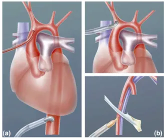
cannulation site was the ascending aorta (with subsequent advancing of the flexible cannula tip into the innominate artery) and the distal one the descending aorta underneath the heart, just above the diaphragmatic hiatus (Fig. 1a). In older children (5 patients) the proximal cannulation was the innominate artery and the distal one the iliac artery (Fig. 1b). During cooling, the ascending aorta, the aortic arch and the arch vessels were extensively dissected and mobilized. Extreme care was taken for the dissection of the distal aortic
Table 1
Patient characteristics Patient/
gender
Age Primary diagnosis Primary operation Pra¨-op echo gradient (mmHg)
Time until re-operation
Re-operation Last follow-up echo/MRI result at post-op months 1/female 3 months Interrupted AA,
VSD
AA reconstruction 20 4 months Sliding plasty: VSD closure
Echo: gradient absent at 25 months 2/male 12 years Coarctation End-to-end resection 40 12 years Sliding plasty Echo: gradient absent
at 18 months 3/female 1 month Interrupted AA,
VSD, PFO
AA reconstruction 20 1 month Sliding plasty: ASD, VSD closure
MRI: no stenosis or dilatation of AA at 6 months 4/male 1 month Coarctation,
VSD, ASD, SAS
End-to-end resection 20 1 month Patch plasty: ASD, VSD closure, SAS resection
Echo: gradient absent at 14 months 5/male 8 years Interrupted AA,
A. lusoria right, LMCA stenosis, SAS
AA reconstruction 25 8 years Patch plasty: ostium plasty, correction of A. lusoria, SAS resection
Echo: gradient absent at 13 months
6/female 13 years Coarctation, VSD, PA stenosis, SAS
End-to-end resection, VSD closure, SAS resection, Patch RVOT reconstruction
40 11 years Sliding plasty: SAS resection
Echo: gradient of 16 mmHg at 1 week
7/male 15 years Coarctation, SAS, MV insufficiency
End-to-end resection, SAS resection
25 10 years Patch plasty: MV reconstruction
Echo: gradient absent at 7 months AA, aortic arch; SAS, subaortic stenosis; MV, mitral valve; LMCA, left main coronary artery.
Fig. 1. Arterial cannulation technique. Arterial cannulation was established in young infants (age , 3 years) in the ascending aorta and in the descending aorta underneath the infant’s heart. The tip of the ascending aorta cannula was later advanced into the innominate artery (a). In older children, the innominate artery and the iliac artery were cannulated (b).
arch and the proximal descending aorta because of the possible increased friability of the arterial wall, which is commonly seen after a stenotic lesion. Once moderate hypothermia was reached, the aorta was cross-clamped and antegrade cold blood cardioplegia was infused to obtain cardiac arrest. The perfusion via the innominate artery was isolated either with a snaring tape around the aortic cannula or a clamp proximate to the insertion of the cannula in older children. The left common carotid artery and the subclavian artery were gently clipped and the proximal descending artery was cross-clamped. Perfusion flow and pressure did not change significantly with the dual arterial perfusion. Perfusion was titrated and adjusted with vasopressors to maintain a normal blood flow and an average blood pressure of 50 mmHg in the radial and femoral arteries.
2.1.2. Aortic arch repair
Two basic techniques were employed for the repair of the aortic arch.
A sliding plasty was used when the stenosis was the result of a segmental narrowing of the aortic arch and when the aortic tissue showed intra-operatively sufficient elas-ticity for adequate mobilization (Fig. 2). The aortic arch was divided in the middle of the stenotic segment. An incision was performed in the bottom of the proximal part and in the top of the distal part, and both segments were re-approximated in a bevelled fashion with a continuous resorbable suture.
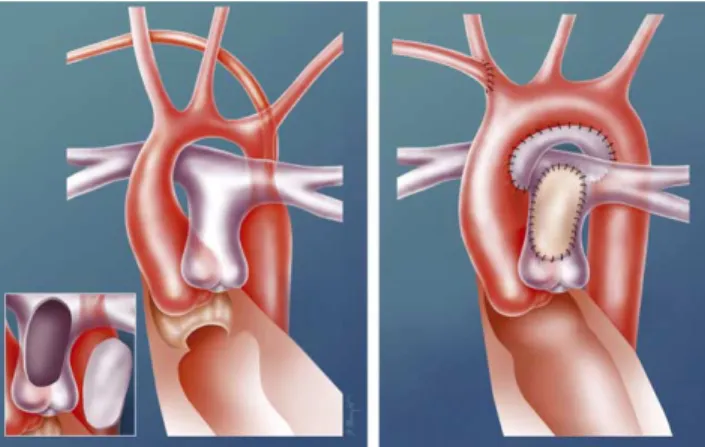
A patch plasty was used when the stenotic segment was short and found on the concavity of the aortic arch (Fig. 3). A longitudinal incision was performed in the concavity of the aortic arch (across the stenotic area). A patch of autologous arterial wall was harvested from the anterior wall of the main pulmonary artery, from the valve commissure to the pulmonary bifurcation. This patch, which was rather large and showed excellent elastic and handling characteristics, was sutured with a continuous resorbable thread to enlarge the longitudinal incision of the aortic arch. Following the defect on the pulmonary artery was covered with a patch of autologous pericardium.
Once the aortic arch reconstruction was completed, the aorta was deaired, the clamps and clips on the arch arteries
were removed and normal perfusion was resumed. If an intracardiac lesion needed repair, it was performed at that time under cardioplegic arrest. Warm blood cardioplegia was delivered shortly before release of the aortic clamp. The heart was allowed to resume work after appropriate reperfusion and rewarming.
3. Results
It was technically possible to perform the planned operation in all the patients. One patient presented a troublesome operative bleeding which required a second run on cardiopulmonary bypass to complete hemostasis. Otherwise surgery was performed without the requirement of autologous blood transfusion in all older patients (. 7 years, n ¼ 5). All the patients survived and none presented significant postoperative complications. One patient showed transient recurrent nerve palsy which completely recovered within 6 weeks. Intra-operatively the blood pressure was measured invasively above and below the repair and showed no residual gradients. Follow-up MRI and transthoracic echocardiograms demonstrated normal appearing aortic arches and a laminar flow in six patients (Table 1, andFigs. 4 and 5). One patient showed a gradient of 10 mmHg across the corrected re-stenosis in a follow-up echocardiogram at 7 months post-operatively. A patient with Shone’s syndrome required cardiac re-operation 1 year later for a sub-aortic membrane and plasty of a parachute mitral valve. In this patient, a harmonious growth of the repaired aortic arch could be further confirmed intraoperatively.
Fig. 2. Sliding plasty repair. The aortic arch is divided in the middle of the stenotic segment and an incision is performed in the bottom of the proximal part and in the top of the distal part. Following both segments are re-approximated in a bevelled fashion and end-to-end anastomosed with a continuous resorbable suture.
Fig. 3. Patch plasty repair. A longitudinal incision is performed in the concavity of the aortic arch (across the stenotic area). A patch of autologous arterial wall is harvested from the anterior wall of the main pulmonary artery (figure inset). This patch, which is rather large and showed excellent elastic and handling characteristics, is sutured with a continuous resorbable thread to enlarge the longitudinal incision of the aortic arch. The defect on the pulmonary artery is patched with autologous pericardium. This figure shows the reconstruction in patient number 5. A subaortic membrane was resected and an abnormal right subclavian artery was reimplanted on the right common carotid artery.
4. Discussion
Recurrence of obstruction after correction of an aortic coarctation is a vexing problem and its management remains problematic. The majority of residual or recurrent lesions can be handled by percutaneous balloon dilatation, sometimes with implantation of a stent[6,7]. Even though a mild residual gradient remains in many cases, this approach
has been universally accepted because of the low morbidity it entails. Balloon dilatation, however, has limits, especially in the setting of complex, tortuous lesions or those located in the aortic arch itself. The optimal surgical approach to these difficult lesions, not amenable to balloon dilatation, is controversial.
The creation of an extra-anatomic bypass between the ascending aorta and the coeliacal aorta (using sternotomy) or between the ascending and the descending thoracic aorta (using a left thoracotomy) presents the advantages of relative simplicity and efficient relief of the arterial obstruction [1 – 5]. These approaches, however, show some disadvantages, which in children, might turn to be determinant. These grafts have no potential for growth and may lead to grotesque deformities of the aorta with the patient’s growth. The development of pseudo-aneurysms at the distal suture line can be extremely troublesome and difficult to repair [8]. Aorto-enteric fistula is another potential dire complication that exists with any graft to abdominal aorta anastomosis. Furthermore, any re-oper-ation on the heart (a significant proportion of children have an additional congenital heart defect) will be much more difficult to perform with the presence of a large mediastinal graft. Finally, the loss of elastic capacitance of the aorta can lead to severe left ventricular hypertrophy with its negative long-term effects[9]. As a matter of fact, the aorta, thanks to its elastic characteristics, acts as a capacitance reservoir during heart cycle. This function reduces the upstroke and the peak pressure for any given cardiac output[10].
For these reasons, we have favoured an anatomic reconstruction of the aortic arch in children. It was also
Fig. 4. Follow-up 3D MRI reconstruction of the sliding plasty repair of patient 3.
Fig. 5. Preoperative transthoracic echocardiogram of patient 4 demonstrates the restenosis (white arrow, a) with a doppler gradient of 38 mmHg (b). Following aortic arch reconstruction with a patch plasty, no remaining stenosis (c) and no gradient (d) is studied by transthoracic echocardiography.
our impression that the restitution of an anatomic blood flow in native tissues would provide the best perfusion to the body, the best adaptation during growth and might preserve the capacitance function of the aorta.
All the aortic lesions we encountered were amenable to repair with two basic techniques. The sliding plasty results basically in the superposition of two stenotic segments with corresponding increase in diameter. This plasty was possible when the obstruction was relatively long and the tissue showed sufficient elasticity for adequate mobiliz-ation during surgery. The patch plasty was appropriate for more discrete lesions, usually in the middle of the aortic arch. As a patch, we used the anterior wall of the pulmonary artery. This patch shows excellent character-istics and produces a harmonious curve underneath the aortic arch, eliminating the risk of producing an internal fold as may occur with other synthetic patches [11]. The thickness and the tissue quality of the patch were reassuring regarding its potential to sustain systemic pressure in the long-term.
The surgical approach to the aortic arch has long been hampered by devastating neurologic complications encountered either in the brain or spinal cord [12,13]. The use of two sites for arterial perfusion—above and below the obstruction—limits the incidence of ischemic damage to the central nervous system and to other organs. We used moderate hypothermia as an additional security to prevent any ischemic injury. Our technique, in comparison to the extra-anatomic bypass, presents the disadvantage of using a heart-lung machine. In older children (over 7 years of age), we were, however, able to perform this surgery without the requirement of auto-logous blood transfusion (as occurred in five out of our patients). With the decrease of the inherent side effects of the heart-lung machine regarding inflammatory response, we believe that this type of support can be used with minimal risk and adverse effects.
In summary, an anatomic reconstruction of the aortic arch can be performed in infants and children presenting recurrent obstruction not amenable to balloon dilatation with excellent early results. This approach may provide superior long-term results compared to extra-anatomic bypasses.
5. Addendum
During the last 3 months we operated two additional children (6 and 8 months of age) for cardiac malformations involving a stenotic aortic arch. One child underwent a patch-plasty and the other one a sliding plasty repair with excellent operative results. Intra-operatively invasively measured blood pressure showed no gradient across the corrected stenosis and early follow-up echocardiograms demonstrated a harmonious reconstruction of the aortic arch and an unobstructed blood flow in both patients.
References
[1] Wukasch DC, Cooley DA, Sandiford FM, Nappi G, Reul Jr. GJ. Ascending aorta – abdominal aorta bypass: indications, technique, and report of 12 patients. Ann Thorac Surg 1977;23(5):442 – 8. [2] Pasic M, Carrel T, Tonz M, Mihaljevic T, Niederhauser U, Kariger U,
Arbenz U, Laske A, Vogt P, Jenni R, Turina M. Der extra-anatomische aszendens-suprazoliakale Aortenbypass in der Behan-dlung der komplexen oder rezidivierenden Aortenisthmusstenosen. Helv Chir Acta 1993;60:447– 50.
[3] Kanter KR, Erez E, Williams WH, Tam VK. Extra-anatomic aortic bypass via sternotomy for complex aortic arch stenosis in children. J Thorac Cardiovasc Surg 2000;120(5):885 – 90.
[4] Connolly HM, Schaff HV, Izhar U, Dearani JA, Warnes CA, Orszulak TA. Posterior pericardial ascending-to-descending aortic bypass: an alternative surgical approach for complex coarctation of the aorta. Circulation 2001;104(12 Suppl 1):I133 – 7.
[5] Berdat PA, Gober V, Carrel T. Extranatomic aortic bypass for complex (re-) coarctation and hypoplastic aortic arch in adolescents and adults. Int Cardiovasc Thorac Surg 2003;2:133 – 7.
[6] Siblini G, Rao PS, Nouri S, Ferdman B, Jureidini SB, Wilson AD. Long-term follow-up results of balloon angioplasty of postoperative aortic recoarctation. Am J Cardiol 1998;81(1):61 – 7.
[7] Suarez de Lezo J, Pan M, Romero M, Medina A, Segura J, Lafuente M, Pavlovic D, Hernandez E, Melian F, Espada J. Immediate and follow-up findings after stent treatment for severe coarctation of aorta. Am J Cardiol 1999;83(3):400 – 6.
[8] Carrel T, Berdat P. Images in cardio-thoracic surgery. Descending aortic aneurysm following ascending to descending bypass in the treatment of aortic re-coarctation. Eur J Cardiothorac Surg 2002; 21(4):760.
[9] Cooper RS, Simmons BE, Castaner A, Santhanam V, Ghali J, Mar M. Left ventricular hypertrophy is associated with worse survival independent of ventricular function and number of coronary arteries severely narrowed. Am J Cardiol 1990;65(7):441 – 5.
[10] Bonapace S, Rossi A, Cicoira M, Franceschini L, Golia G, Zanolla L, Marino P, Zardini P. Aortic distensibility independently affects exercise tolerance in patients with dilated cardiomyopathy. Circula-tion 2003;107(12):1603 – 8.
[11] Roussin R, Belli E, Lacour Gayet F, Godart F, Rey C, Bruniaux J, Planche C, Serraf A. Aortic arch reconstruction with pulmonary autograft patch aortoplasty. J Thorac Cardiovasc Surg 2002;123(3): 443 – 8.
[12] Ceriana P, Barzaghi N, Locatelli A, Veronesi R, De Amici D. Aortic arch surgery: retrospective analysis of outcome and neuroprotective strategies. J Cardiovasc Surg (Torino) 1998;39(3):337 – 42. [13] Svensson LG. Progress in ascending and aortic arch surgery:
minimally invasive surgery, blood conservation, and neurological deficit prevention. Ann Thorac Surg 2002;74(5):S1786 – 8.
Appendix. Conference discussion
Dr T. Ebels (Groningen, The Netherlands): May I start the discussion by asking you what role balloon angioplasty had in this series of patients? Dr Kadner: I think balloon angioplasty has clearly its place. But many lesions, especially more complex and tortuous lesions, are difficult to manage with balloon angioplasty and such cases should preferably undergo surgery. Furthermore, balloon angioplasty is not really suitable for lesions located in the aortic arch itself.
Dr Ebels: But this was not attempted in these 7 patients? Dr Kadner: It was attempted first, but unsuccessfully. Dr Ebels: In all 7?
Dr D. Di Carlo (Rome, Italy): I noticed that in 3 patients you used a very large patch of main pulmonary artery and presumably branches, because it’s very large.
I’d like to add a word of caution. One of the complications of the Takeuchi procedure was severe supravalvular pulmonary stenosis which tends to be recurrent. I wonder whether you have gradients in these patients, supravalvular gradients?
Dr Kadner: We are aware of these potential complications, but until now we have not observed this kind of problem. We did not have to extend the harvesting to the branch pulmonary arteries. But I would like to emphasize that the presented data shows just early results. Our longest follow-up is just 21
2 years.
Dr T. Walles (Hannover, Germany): I wonder, do you see an indication for tissue-engineered autologous material as a patch for your aortic arch instead of your pulmonary artery patch?
Dr Kadner: I can clearly see such an indication and the usage of a tissue engineered patch for reconstruction of the aortic arch, and this might be easier feasible than replacing a complete valve, especially an aortic valve. However, I think so far we are not ready for this yet.
As you probably know, we have a tissue engineering research group in Zurich, and currently long-term animal experiments are performed.
Given these results are turning out nicely, one can think further. But at this moment, I believe it’s not ready for clinical application yet.
Dr Walles: But it would be your first choice if you had a patch? Dr Kadner: If we would have a good patch, we might use it. Dr C. Knott-Craig (Oklahoma City, OK): The patient with Dysphagia lusoria interests me. Did you divide the subclavian artery and reimplant it? And did you do this from the front (via sternotomy)? Perhaps you could share the technical details of the operation with us. Dr Kadner: All patients were operated through a median sternotomy. And did you ask about the technique of the sliding plasty or about the way we managed the arteria lusoria? I’m sorry but I did not hear you properly.
Dr Knott-Craig: Arteria lusoria.
Dr Kadner: The arteria lusoria was reimplanted in the right subclavian artery. We routinely perform this reimplantation in case of interrrupted aortic arch with abnormal right subclavian artery. The reimplantation is performed during the rewarming phase of the operation.
Dr A. Sousa (Oporto, Portugal): You used two cannulas, one on the trunk and the second in the descending aorta. Are they coming from the same source, from the same pump, and divided, or two sources?