The influence of the rapamycin-derivate SDZ RAD on the healing of
airway anastomoses
q
Andre´ E. Dutly
a, Ariana Gaspert
b, Ilhan Inci
a, Didier Schneiter
a, Stephan Korom
a,
Walter Weder
a,*
aDivision of Thoracic Surgery, University of Zurich, Raemistrasse 100, 8091 Zurich, Switzerland b
Department of Pathology, University of -Zurich, Raemistrasse 100, 8091 Zurich, Switzerland Received 20 September 2002; received in revised form 26 February 2003; accepted 12 March 2003
Abstract
Objective: Among the many immunosuppressive effects of SDZ RAD (40-0(2-hydroxyethyl)-rapamycin), a rapamycin derivative, is the inhibition of fibroblast proliferation. Since the long-term success of lung transplantation is limited by the development of bronchiolitis obliterans, a fibroblast-associated progressive luminal obstruction of the terminal bronchioli, the use of SDZ RAD as immunosuppressive in pulmonary graft recipients may counteract this process. However, reduction of fibroblast activity, posttransplant, may impair the healing of the bronchial anastomoses. Materials and methods: The cervical trachea in pigs was denuded, divided and re-anastomosed with Prolene 4-0 single stitches. Control animals (group 1, n ¼ 4) were without, and study animals (group 2, n ¼ 6) were with SDZ RAD therapy (1.25 mg/kg/day, p.o., 14 days). After 14 days, the pigs were sacrificed. The anastomoses were examined histologically, and breaking strength of tracheal strips of 5-mm width was measured. Results: All animals survived without complications. Serum levels of SDZ RAD were 30.9 ^ 8.7 ng/ml (recommended level 20 – 40 ng/ml). All anastomoses healed macroscopically without difference between the two groups. Breaking strength was significantly lower in the treated animals (group 1 vs. group 2: 11.75 ^ 0.35 vs. 7.69 ^ 1.39 N, P ¼ 0:01). Histology did not show a significant change in histoarchitecture between the groups. Conclusions: Although SDZ RAD significantly reduced the breaking strength of the tracheal anastomosis, no obvious histological differences between treated and untreated animals could be detected. Since this model does not reflect the clinical situation, further investigations are necessary to reveal the effect of SDZ RAD on airway wound healing in concert with a contemporary clinically used multidrug immunosuppressive regimen in allograft recipients. q2003 Elsevier Science B.V. All rights reserved.
Keywords: Rapamycin; SDZ RAD; Lung transplantation; Wound healing
1. Introduction
Healing of the bronchial anastomoses has been the Achilles heel of clinical lung transplantation, with an associated complication rate of 0 – 12% and a mortality of 0 – 4% [1 – 3]. Although modern surgical approaches have led to a decreased complication rate [1], in many centers, insufficiency and stenosis are still frequently encountered; and despite rapid diagnosis and early intervention and intensive care, the prognosis is often poor. In spite of considerable progress in pulmonary transplantation over the last two decades, several obstacles still remain. Firstly,
cartilage is a bradytrophic tissue, with a low cellular turnover and slow-healing dynamics. Secondly, the surgical trauma itself, comprising microlesion and denudation of the anastomotic plane, impairs re-vascularization. Thirdly, continuous unspecific immunosuppression as a therapeutic cornerstone of transplantation medicine may further delay healing of the anastomoses[4,5].
SDZ RAD (40-0(2-hydroxyethyl)-rapamycin) is a new analogon of rapamycin with a high immunosuppressive activity. It inhibits cellular proliferation during mitosis (G1 to S phase). In contrast to ciclosporine, which decreases interleukin production, it reduces the interleukin-mediated cellular proliferation.
The potential synergistic effect of SDZ RAD along with the current triple immunosuppressive regimen used in lung transplantation – ciclosporine, mycophenol mofetil and
www.elsevier.com/locate/ejcts
1010-7940/03/$ - see front matter q 2003 Elsevier Science B.V. All rights reserved. doi:10.1016/S1010-7940(03)00182-9
q
Presented at the 16th Annual Meeting of the European Association for Cardio-Thoracic Surgery, Monte Carlo, Monaco, September 22 – 25, 2002. * Corresponding author. Tel.: þ41-1-255-88-01; fax: þ41-1-255-88-05.
steroids – is discussed[6]. Since rapamycin has a strong inhibitive influence on fibroblasts[7,12], which seem to be instrumental for initiating chronic rejection through the development of bronchiolitis obliterans (BO), the drug may hold a promising potential for future first line immuno-suppression in recipients of pulmonary grafts. However, since fibroblasts play a central role during the early phase of wound healing, abrogation of their proliferative cap-abilities may lead to complications with the bronchial anastomoses.
In this study, we have evaluated the influence of SDZ RAD – without rejection phenomena – on the healing process of airway anastomoses.
2. Materials and methods 2.1. Animals and groups
In ten pigs (24 – 31 kg, 3 – 4 months), the cervical trachea was transected and re-anastomosed. After surgery, the animals were randomized into two groups: group 1 ðn ¼ 4Þ animals were untreated and served as the control group; group 2 ðn ¼ 6Þ animals were treated with 1.25 mg/kg/day SDZ RAD orally (generously provided by Novartis Pharma, Switzerland) during the 14-day observation period. All animals received antibiotic prophylaxis with chlortetra-cycline, tylosin and sulfadimidine (CAS), 8 g/kg feed, 5 days prior to surgery.
2.2. Surgical procedure
The pigs were intubated and ventilated with 100% oxygen (FIO2: 1.0) during the intervention. Inhalative gas
narcosis was performed with isoflurane (Abbott, Switzer-land) at concentrations between 0.2 and 1.5%. All procedures were performed under sterile conditions with the animals fixed in supine position. After cervical exposure, the first six rings of the trachea were dissected free from the surrounding tissue. Between the third and fourth ring, the tracheal continuity was cut over 80% of its circumference to simulate local ischemia. The anastomoses were performed with Prolene 4-0 (Ethicon, Johnson & Johnson Medical, Switzerland), single stitches. The wound was closed layer by layer with Vicryl 3-0 (Ethicon, Johnson & Johnson Medical), and the skin with Dermalon 4-0 (USSDG, TYCO Healthcare, Switzerland). The duration of the procedure was 30 min.
2.3. General postoperative care
Antibiotic prophylaxis with CAS (8 g/kg feed), was continued over 5 days postoperatively. All animals received analgesics on a fixed schedule for 1 week. Blood samples including hematological parameters and SDZ RAD trough levels were taken at days 3, 7, 10 and 14. The animals were
visited twice a day. On day 14, the animals were sacrificed and the cervical trachea removed. All animals received humane care in accordance with the ‘Guide for the Care and Use of Laboratory Animals’ (National Institutes of Health publication 85-23, revised 1985). The study protocol was approved by the local animal study committee.
2.4. Macroscopic and biomechanical evaluations
The anastomoses were inspected and the trachea was cut into strips of 5 mm width. The suture material was carefully removed, and breaking strength of two strips of trachea and skin for each animal was measured. The specimen was fixed at a spring balance and tension was continuously increased until the specimen ruptured.
2.5. Histological evaluation
Tissue specimens were fixed in 4% neutral buffered aqueous formaldehyde solution. From each specimen, two longitudinal sections were taken, comprising the processed and paraffin-embedded anastomosis were taken using routine procedures. A 2 mm section was cut from each paraffin tissue block and stained with hematoxylin – eosin, elastic Van Gieson and alcian blue – periodic acid – Schiff for routine light microscopic evaluation. The site of the anastomosis was assessed morphologically for fibrosis, occurrence of granulation tissue, infiltration by lymphocy-tes/histiocytes and by neutrophils as none (0), mild (1þ ), moderate (2þ ), or severe (3þ ). All sections were examined by a pathologist (A.G.) who was blinded toward the treatment protocol (Table 1).
2.6. Statistics
Data analysis was performed with the Statview for Applew 5.0.1 (SAS Institute Inc., USA). The data are
expressed as mean values ^ standard deviation. We performed ANOVA to compare the two groups. A P value , 0.05 was considered significant.
3. Results 3.1. General data
There was no difference between the two groups in terms of postoperative course, general condition and wound healing. No local infection was encountered and none of the animals developed airway stenosis or associated pulmonary problems. SDZ RAD serum levels were 30.9 ^ 8.7 ng/ml (recommended level 20 – 40 ng/ml).
Fourteen days after surgery, the animals were sacrificed and the cervical trachea removed. At time of sacrifice, skin incision, trachea and peritracheal tissue had completely healed in both groups. The anastomotic region could not be
detected by bronchoscopy. None of the animals showed stenosis of the trachea or intraluminal hypergranulation. The Prolene stitches were easy to remove without injuring the trachea and the surrounding tissue at the site of anastomosis. The breaking strength of the trachea was measured using strips of 5-mm diameter (two for each animal;Fig. 2).
Animals in the treated group (group 2) displayed a significantly reduced anastomotic breaking strength (group 2: 6.68 ^ 1.39 N) as compared with untreated animals (group 1: 11.75 ^ 0.35 N; P ¼ 0:008). In two of the four untreated animals, the rupture occurred adjacent to, but not at the anastomosis. For the skin strips, we found a reduced breaking strength too, yet without statistical significance (group 1: 31.0 ^ 8.1 N vs. group 2: 19.38 ^ 6.2 N, P ¼ 0:116).
3.2. Histology
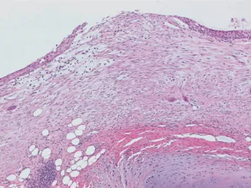
The sections from the treated animals (Fig. 1) showed mild mucosal fibrosis in four, and severe fibrosis in two cases. There was no granulation tissue in three cases, little granulation tissue in two cases and moderate granulation tissue one case. The infiltration by lymphocytes/histiocytes was mild in three and moderate in another three cases as well. There was no infiltration by neutrophils in four, and a mild and a moderate infiltration in another two cases.
The sections from the control animals (Fig. 2) revealed mild mucosal fibrosis in two, moderate fibrosis in one and severe fibrosis in one case. There was no granulation tissue in three cases and moderate granulation tissue in one animal. The infiltration by lymphocytes/histiocytes was mild in three and moderate in one case. There were no neutrophils in three cases, and there was a mild infiltration by neutrophils in one case. The cartilaginous part of the trachea was inconspicuous in both groups. Altogether, the
interindividual differences were greater than the variances between the two groups.
4. Discussion
Rapamycin (Sirolimusw) is an immunosuppressive agent
produced by fermentation of Streptomyces hygroscopicus
[9]. Its derivative, SDZ RAD, has similar immunosuppres-sive properties; however, the pharmacokinetics consist of a shorter terminal half-life and a better oral bioavailability in man[8]. The immunosuppressive properties of SDZ RAD, especially in combination with ciclosporine and FK506, are well documented[10,11].
Like rapamycin, SDZ RAD inhibits growth factor-mediated proliferation of non-immune cells: it blocks basic fibroblast growth factor (bFGF)-induced proliferation of bovine aortic and human umbilical vein endothelial cells and 3T3 fibroblasts; it also inhibits bFGF and platelet-derived growth factor (PDGF)-stimulated proliferation of smooth muscle cells[7,12].
After lung transplantation, long-term survival is limited by the development of obliterative bronchiolitis (OB). OB causes a decline of lung function that is refractory to therapy and often culminates in respiratory failure within months of its onset. Pathologically, OB is a partial or complete luminal obliteration of the terminal bronchioli by dense fibrous scarring, resulting from proliferation of fibroblasts. Since SDZ RAD inhibits fibroblast proliferative capabilities, it has been proposed as an ideal first line immunosuppressant in clinical lung transplantation. On the other hand, this unspecific impairment of fibroblast function could also affect the general quality of wound healing following engraftment. Theoretically, the region with the slowest proliferative turnover – the bronchial anastomoses – may Table 1
Morphological assessment of the anastomotic site for fibrosis, occurrence of granulation tissue, infiltration by lymphocytes/histiocytes and by neutrophils Animal
hist
Mucosal fibrosis neutrophils Occurrence of granulation tissue Infiltration by lycy/hist Infiltration by neutrophils Breaking strength (N) Trachea Skin T 1 þ 0 1 þ 0 6.75 na T 1 þ 0 1 þ 0 5.25 22.50 T 3 þ 2 þ 2 þ 2 þ 8.00 26.25 T 3 þ 1 þ 2 þ 1 þ 7.75 16.25 T 1 þ 0 1 þ 0 5.75 12.50 T 1 þ 1 þ 2 þ 0 9.00 17.00 C 1 þ 0 1 þ 0 11.5a na C 2 þ 0 1 þ 0 11.50 36.75 C 3 þ 2 þ 2 þ 1 þ 12a 25.25 C 1 þ 0 1 þ 0 13.00 21.25
Despite the fact that the breaking strength of the treated animals was significantly reduced, no histological correlation could be found. The interindividual histological differences were higher than the differences between the groups.
Classification of the pathological changes: 0, none; 1 þ , mild; 2 þ , moderate; 3 þ , severe. Abbreviations used: lycy, lymphocytes; hist, histiocytes; sqmet, squamous metaplasia.
a
be the most susceptible toward SDZ RAD-mediated fibroblast inhibition. In order to disclose the isolated influence on bronchial anastomotic healing by SDZ RAD, we devised a simplified model with tracheal anastomoses. The trachea is surgically easier to access, and since there is no structural difference between trachea and main bronchi, the impact of SDZ RAD treatment is supposedly the same. Performing an in situ repair, rather than an allogenic transplantation, we could rule out any additional alloanti-gen-triggered immune phenomenon.
Macroscopically, cartilage and mucosa in both groups had healed well, without any lesions at the time of sacrifice. The evaluation of the histomorphological anastomotic architecture did not show marked differences between the groups; as a matter of fact, greater interindividual, rather than intergroup, differences were detected. When we measured breaking strength, however, we found a statistical significant reduction in the SDZ RAD group. Although we were able to demonstrate a biological significance between
the groups – applied to the clinical setup – no differences between SDZ RAD treated and untreated animals could be demonstrated. Still, the fact that mechanical stress tests on anastomoses in the treated group showed significantly lower results implies – in spite of the unspecific morhphologic differences – an altered, yet undetected ultrastructural change. Possibly, rapamycin treatment may affect the extracellular component synthesis of chondrocytes and fibroblasts, thereby impairing mechanical properties of the surrounding matrix. In addition, the applied time frame in our setup should be taken into consideration. At 2 weeks, when the anastomoses were analyzed, structural stability was still aided by the intact suture materials. Assessment of the bronchial anastomoses at a later time point, and/or the influence of other immunosuppressive agents, may also influence the clinical course. The model itself has limitations. First of all, the tracheal anastomosis is different from the bronchial anastomosis. In lung transplantation, the blood supply of the donor main bronchi is very low, whereas in tracheal anastomoses, tension can be a limiting factor.
In summary, our data suggest a biologically significant, SDZ RAD-mediated reduction of mechanical strength in tracheal anastomoses. Since macroscopic inspection and histological evaluation did not show marked differences, the observed phenomenon does not have clinical significance in the investigated setup. In order to estimate the influence of rapamycin on anastomotic healing in a more clinical relevant model, SDZ RAD should be investigated in the context with a standard immunosuppressive regimen, possibly in recipients of lung allografts with bronchial anastomoses.
References
[1] Schmid RA, Boehler A, Speich R, Frey HR, Russi EW, Weder W. Bronchial anastomotic complications following lung transplantation: still a major cause of morbidity? Eur Respir J 1997;10:2872– 5. [2] Shennib H, Massard G. Airway complications in lung transplantation.
Ann Thorac Surg 1994;57:506– 11.
[3] Date H, Trulock EP, Arcidi JM, Sundaresan S, Cooper JD, Patterson GA. Improved airway healing after lung transplantation. An analysis of 348 bronchial anastomoses. J Thorac Cardiovasc Surg 1995;110: 1424 – 32. [discussion p. 1432 – 3].
[4] Inui K, Schafers HJ, Aoki M, Becker V, Ongsiek B, Kemnitz J, Haverich A, Borst HG. Bronchial circulation after experimental lung transplantation. The effect of long term administration of predniso-lone. . [see comments] J Thorac Cardiovasc Surg 1993;105:474 – 8. [discussion p. 478 – 9].
[5] Auteri JS, Jeevanandam V, Sanchez JA, Marboe CC, Kirby TJ, Smith CR. Normal bronchial healing without bronchial wrapping in canine lung transplantation. . [see comments] Ann Thorac Surg 1992;53: 80 – 3. [discussion p. 83 – 4].
[6] Schuurman HJ, Cottens S, Fuchs S, Joergensen J, Meerloo T, Sedrani R, Tanner M, Zenke G, Schuler W. SDZ RAD, a new rapamycin derivate: synergism with ciclosporine. . [comment] Transplantation 1997;64:32 – 5.
[7] Nair RV, Huang X, Shorthouse R, Adams B, Brazelton T, Braun Dullaeus R, Morris RE. Antiproliferative effect of rapamycin on growth factor-stimulated human adult lung fibroblasts in vitro may Fig. 1. The figure shows a section through the anastomosis of a treated
animal (magnification £ 60). No change in the histological architecture was seen when compared with the treated animals.
explain its superior efficacy for prevention and treatment of allograft obliterative airway disease in vivo. Transplant Proc 1997;29:614– 5. [8] Appel Dingemanse S, Wong R, Dou L. Multiple-dose pharmaco-kinetics of the immunosuppressant SDZ RAD in stable renal transplant patients. Transplantation 1998;65:138.
[9] Vezina C, Kudelski A, Sehgal SN. Rapamycin (AY-22,989, a new antifungal antibiotic. I. Taxonomy of the producing streptomycete and isolation of the active principle. J Antibiot 1975;28:721– 6. [10] Hausen B, Ikonen T, Briffa N, Berry GJ, Christians U, Robbins RC,
Hook L, Serkova N, Benet LZ, Schuler W, Morris RE. Combined immunosuppression with ciclosporine (Neoral) and SDZ RAD in non-human primate lung transplantation: systematic pharmacokinetic-based trials to improve efficacy and tolerability. Transplantation 2000; 69:76 – 86.
[11] Salminen US, Maasilta PK, Taskinen EI, Alho HS, Ikonen TS, Harjula ALJ. Prevention of small airway obliteration in a swine heterotopic lung allograft model. J Heart Lung Transplant 1999;19:193 – 205. [12] Sehgal SN. Rapamune (Sirolimus, rapamycin): an overview and
mechanism of action. Ther Drug Monit 1995;17:660– 5.
Appendix A. Conference discussion
Dr A. Haverich (Hannover, Germany): I liked this presentation very much because it shows a very clean experimental design, also very clear results. However, what you state in the outlook is very obvious. If you would have put in a cocktail of immunosuppressive agents in this experimental model, the results would have been quite different. This experimental design would clearly have mimicked more the clinical situation. Would you comment on that?
Dr Korom: Yes, obviously, but still, as you stated, in the first set of these experiments, we just wanted to assess the clear-cut influence of rapamycin. And, in addition, what should be taken into account, especially using an allograft model, go even one step further, adding an inflammatory reaction to that area, which adds another dimension of entropy to that level. Then it is even harder to decipher that which of the effects was rapamycin-induced. Dr W. Weder: I want to briefly add to Dr Korom’s comments that these follow-up experiments are in preparation.
Mr J. Thorpe (Leeds, UK): You mentioned there was a change in the intracellular matrix in terms of the fibroblastic activity. Was there much less activity, because this will have connotations not just for lung transplan-tation but for any tracheal resection? Can you comment on what the fibroblastic activity was like in the treatment group?
Dr Korom: Again, these investigations are under way. We are analyzing with PCR the composition of extracellular matrix in these areas, and, as a matter of fact, we have not yet isolated fibroblasts from these areas. We are planning to take them ex vivo and assess their activities and secretion dynamics. But we are in the process, and maybe a year from now we can satisfy your curiosity.
Dr S. Guth (Mainz, Germany): If I understand you right, you stated that you only cut 80% of the trachea?
Dr Korom: Right.
Dr Guth: How was the set-up of the examination of the breaking strength?
Dr Korom: We used a spring balance and the weight was gradually increased, and we measured strips of 0.5 cm diameter, including the anastomosis. We put them up, increased the weight and measured the breaking strength needed and they all broke at the anastomosis, except for one.
Dr Guth: And why didn’t you cut the whole trachea? Dr Korom: We only cut 80% of the trachea for surgical ease.