Review
Gene transfer of cytoprotective and immunomodulatory molecules
for prevention of cardiac allograft rejection
Giuseppe Vassalli
a,b,*, Sylvain Fleury
a,b, Jianping Li
a, Jean-Jacques Goy
a,
Lukas Kappenberger
a, Ludwig K. von Segesser
caDepartment of Cardiology, BH-10, CHUV, University of Lausanne Medical School, Rue du Bugnon 46, 1011 Lausanne, Switzerland b
Department of Experimental Surgery, University of Lausanne Medical School, Lausanne, Switzerland
c
Department of Cardiovascular Surgery, University of Lausanne Medical School, Lausanne, Switzerland Received 8 January 2003; received in revised form 8 April 2003; accepted 15 June 2003
Summary
Current treatments of heart transplantation are limited by incomplete effectiveness, significant toxicity, and failure to prevent chronic rejection. Genetic manipulation of the donor heart at the time of removal offers the unique opportunity to produce a therapeutic molecule within the graft itself, while minimizing systemic effects. Cytoprotective approaches including gene transfer of heme oxygenase (HO)-1, endothelial nitric oxide synthase, and antisense oligodeoxynucleotides specific for nuclear factor (NF)-kB or intercellular adhesion molecule (ICAM)-1 reduced ischaemia – reperfusion injury and delayed cardiac allograft rejection in small animals. Exogenous overexpression of immunomodulatory cytokines such as interleukin (IL)-4, IL-10 and transforming growth factor-b, as well as gene transfer of inhibitors of pro-inflammatory cytokines also delayed graft rejection. Gene transfer-based blockade of T-cell costimulatory activation with CTLA4-Ig or CD40-Ig resulted in long-lasting graft survival and donor-specific unresponsiveness, as manifested by acceptance of a second graft from the original donor strain but rejection of third-party grafts. Similar results were obtained with donor major histocompatibility complex class I gene transfer into bone marrow cells. Gene therapy approaches to chronic rejection included gene transfer of HO-1, soluble Fas, tissue plasminogen activator and antisense oligodeoxynucleotides specific for the anti-apoptotic mediator Bcl-x or the E2F transcription factor. Despite major experimental advances, however, gene therapy for heart transplantation has not entered the clinical arena yet. Fundamental questions regarding the most suitable vector, the best gene, and safety issues remain unanswered. Well-controlled studies that compare gene therapy with established treatments in non-human primates are needed before clinical trials can be started.
q2003 Elsevier B.V. All rights reserved.
Keywords: Transplantation; Heart; Gene therapy; Gene transfer
1. Introduction
Transplantation is the treatment of choice for end-stage organ failure [1]. However, maintenance of a functional allograft requires life-long immunosuppression to prevent rejection by the immune system. Current immunosuppres-sive drugs such as cyclosporine and corticoids act by indiscriminately blocking T-cell activation, the primary mechanism of graft rejection. Unfortunately, these drugs are associated with significant side effects including renal toxicity, dyslipidaemia, diabetes, and increased risk of infections and malignancies. Moreover, current treatments have failed to prevent chronic rejection, or graft arteriopathy
[2]. As a result, although 1-year survival rates for transplanted organs now exceed 90%, overall 10-year graft survival rates remain below 50% [3]. Clearly, novel approaches to organ transplantation are needed.
New immunosuppressive drugs include humanized anti-interleukin (IL)-2 receptor monoclonal antibody (daclizu-mab) [4]; tacrolimus, which blocks IL-2-dependent T-cell activation; mycophenolate mofetil, which blocks lympho-cyte purine biosynthesis; and sirolimus (rapamycin), which inhibits multiple cell cycle regulators. Initial clinical trials have shown that the new drugs improve the short-term outcome after organ transplantation [4,5]. However, they are associated with significant toxicity, and their long-term effects are unknown because the clinical follow-up is still too short.
www.elsevier.com/locate/ejcts
1010-7940/$ - see front matter q 2003 Elsevier B.V. All rights reserved. doi:10.1016/S1010-7940(03)00456-1
* Corresponding author. Tel.: þ41-21-314-0076; fax: þ41-21-314-0013. E-mail address: giuseppe.vassalli@chuv.hospvd.ch (G. Vassalli).
Gene therapy is defined as the introduction and expression of recombinant DNA in order to ameliorate or cure a disease condition. The easy access to the donor organ for genetic manipulation at the time of removal and the need for a localized biological effect make organ transplantation particularly well-suited to gene therapy approaches. Indeed, ex vivo gene transfer into the donor organ can be performed under controlled, optimized conditions. Because the foreign gene is not directly administered to the patient, its systemic dissemination is minimized. Most importantly, the protective factor can be produced for extended periods of time[6,7], potentially for a lifetime, after a single gene administration. Obviously, sustained production of the therapeutic molecule is of major relevance for organ transplantation, which requires lifelong immunosuppression.
Despite the extensive publicity devoted to gene therapy, this field is still in its infancy. Recently, cardiovascular gene therapy has entered the clinical arena, and promising results have been reported in initial trials for coronary artery disease [8]. By contrast, no clinical applications in gene therapy for heart transplantation have been reported so far. Nevertheless, increasing experimental evidence suggests that this approach may be feasible. This paper is devoted principally to a review of the theoretical basis of gene therapy for heart transplantation, as established by exper-imental studies in animal models.
2. Routes of gene administration
General requirements for a successful gene therapy strategy include: (1) a suitable route of gene administration; (2) an efficient gene transfer vector; (3) a gene product that mediates a strong biological effect; (4) a sufficient duration of gene expression; and (5) an acceptable risk profile.
Both systemic and localized approaches have been used to deliver a gene of interest to the transplanted heart. Systemic gene delivery may be suitable in the case when the delivered gene encodes a secreted factor that acts on neighbouring or remote cells via a paracrine mechanism. This approach involves systemic dissemination of the foreign gene, of course. However, targeted vectors that bind to specific surface markers or contain tissue-specific promoters have been developed[9]. After systemic administration, these vectors mediate gene transfer selec-tively to target tissues. However, targeted vectors may not be required for gene therapy for heart transplantation because transgene expression after ex vivo gene transfer into the donor heart is largely confined to the graft itself.
Various routes of administration including intracoronary infusion, intramyocardial and endomyocardial injection, and pericardial instillation have been used to deliver a gene of interest to the donor heart[7,10]. Using a Langendorff in vitro perfusion system, adenovirus-mediated gene transfer into the isolated rat heart varied as a function of vector
concentration and perfusion time [11]. Pre-treatment with hypocalcaemic solutions, serotonin or vascular endothelial growth factor (VEGF; originally termed vascular per-meability factor) increases vascular perper-meability, poten-tially enhancing myocardial gene transfer. Ex vivo gene transfer by intracoronary vector infusion into the isolated donor heart is more efficient than in vivo gene transfer by vector instillation into the coronary circulation. This difference is due to the long dwelling time of the vector within the isolated donor heart, which can be equivalent to the organ preservation time. In contrast, the transit time of vector particles through the coronary circulation in vivo is short, ranging from a few seconds to a few minutes during blood flow arrest. As a result, cardiac uptake of vector particles instilled into coronary arteries in vivo is relatively low. As an example, the number of adenovirus genomes after intracoronary infusion of an adenoviral vector was 33-fold lower than after intramyocardial injection in pigs
[12]. In the clinical setting, the isolated donor organ is routinely perfused with a tissue preserving solution. This procedure could be combined with the administration of a therapeutic gene.
3. Gene transfer systems
Several vectors including recombinant adenovirus, plasmid DNA, liposome – DNA and hemagglutinating virus of Japan (HVJ) – liposome – DNA complexes have been used to deliver a gene of interest to the donor heart
[10,13,14]. Each vector has distinct advantages and disadvantages, and hence, a perfect vector for all appli-cations does not exist. Instead, the vector used should be tailored to any given application, taking into account the cellular target, the predicted levels of transgene expression, and the duration of expression. Accordingly, gene transfer-based prevention of acute and chronic rejection may require different vectors because long-term expression of the protective gene is highly desirable in many approaches to chronic rejection. However, short-lived transgene expression can also mediate long-lasting effects, especially in the case when immunological tolerance toward donor antigens can be induced (see below). The cellular target should also be taken into account when choosing the vector. While cardiomyocytes are the primary target of most gene therapy approaches to acute rejection, endothelial cells and other vascular cells are important targets for the prevention of graft arteriopathy.
The number of cells that need to express the transgene in order to achieve a biological effect depends on the delivered gene itself. In the case when the gene encodes a secreted peptide that acts on neighbouring cells via a paracrine mechanism, limited numbers of gene-transduced cells may be sufficient to elicit a therapeutic effect. What really matters is the concentration of the protective gene product within the graft, or in the plasma, depending on
the mechanism of action. Conversely, in the case when the therapeutic gene encodes an intracellular factor, as many cardiac cells as possible should express the cytoprotective molecule.
Replication-deficient, recombinant adenoviral vectors have been used in the vast majority of gene therapy studies for heart transplantation. These vectors efficiently transduce genes into both cardiomyocytes and endothelial cells in vivo
[10 – 13]. Using a lac Z reporter gene, the b-galactosidase gene product is readily visualized by histochemical reaction with the chromogenic substrate X-gal (Fig. 1). It should be noted, however, that the efficiency of gene transfer is underestimated by X-gal staining due to lac Z expression below the detection threshold in a proportion of cells[15]. Conversely, false-positive X-gal staining due to micro-infarctions, rather than effective lac Z gene transfer, was reported after intramyocardial injection[16].
Limitations of adenoviral vectors include tissue inflam-mation and short-lived transgene expression (< 2 – 4 weeks)
[12,17,18]. The absence of vector integration into the cell genome, as well as immune responses to viral proteins are responsible for the short duration of gene expression with adenoviral vectors. Non-integrated DNA is inherently unstable due to the presence of DNA digesting enzymes within the cell. Immune responses include both cytotoxic T cells that eliminate cells that express adenoviral antigens and neutralizing antibody that preclude successful read-ministration of the adenoviral vector [17]. By analogy, pre-existing antibody as a result of previous infection with wild-type adenovirus may preclude adenoviral gene transfer in humans. In a cohort of healthy adult individuals, we found a 57%-prevalence of neutralizing antibody to adenovirus [17]. Thus, many candidate patients may be refractory to adenovirus-based gene therapy. In a clinical
trial of gene therapy for coronary artery disease, the extent of anti-adenovirus antibody formation in patients who received intramyocardial adenoviral vectors was strongly correlated with pre-existing antibodies[18].
Adenovirus-induced inflammation has been studied in donor hearts transplanted into genetically identical hosts, thus avoiding confounding alloimmune responses. Adeno-virus-mediated lac Z gene transfer into rat cardiac isografts caused significant myocardial inflammation that was associated with rapid extinction of lac Z expression [19]. In contrast, negligible inflammation and long-lasting lac Z expression were observed in adenovirally transduced mouse cardiac isografts, despite the fact that the same vector induced marked hepatic inflammation when administered to the liver [20]. Although the differences between the two organs have not been fully explained, the lower antigen-presenting cell (APC) content of cardiac tissue, as compared to the liver, may play a part. Dendritic cells as professional APCs have been shown to migrate from the donor heart and localize in the host’s spleen, where they generate productive cellular and humoral immune responses[21].
Plasmid DNA, liposome – DNA and HVJ – liposome – DNA vectors have also been used to deliver genes of interest to the donor heart[13,14,22,23]. Although these vectors are intrinsically less efficient than adenoviral vectors, they also induce less tissue inflammation. Interestingly, those rare studies that directly compared different vectors to each other showed that the most efficient vector does not always mediate the best therapeutic effect. For instance, liposome – DNA transfection of the active form of transforming growth factor (TGF)-b1 was more effective than adenovirus-mediated
Fig. 1. Adenovirus-mediated transfer of a lac Z reporter gene into a transplanted rat heart. Cryosection through the heart shows patchy yet widespread b-galactosidase expression (blue areas after X-gal staining), most abundantly in the interventricular septum. It should be considered that X-gal staining underestimates gene transfer efficiency[15]. Empty spaces within myocardium are cyrosectioning artefacts. The apparent thickening of the RV wall is an artefact due to the slightly oblique sectioning axis; LV/RV, left/right ventricular cavities.
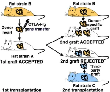
Fig. 2. Schematic of donor-specific hyporesponsiveness induced by CTLA4-Ig gene transfer. A donor heart of the strain B transduced ex vivo with the CTLA4-Ig gene and transplanted into the recipient strain A survives indefinitely (. 100 days). A second cardiac graft from the original donor strain B is accepted in the absence of new treatment. In contrast, a third-party graft from the strain C is rejected in the absence of treatment. Modified from Ref.[7].
gene transduction with respect to the prolongation of cardiac allograft survival in mice[22]. HVJ – liposome vectors are more efficient than most liposome and plasmid DNA vectors. In addition, repeated gene transfer with these vectors is feasible, and their safety profile is relatively good[23]. Thus, HVJ – liposome – DNA complexes provide a useful alterna-tive to adenoviral vectors for gene therapy applications in heart transplantation.
Recombinant adeno-associated virus (AAV) vectors can integrate into the host cell genome, thus providing a potential for permanent genetic modifications of target cells. We have shown that AAV-mediated expression of a green fluorescence protein (GFP) reporter gene lasts for extended periods of time (. 1 year) in mouse myocardium
[6]. Attractive features of AAV vectors also include negligible tissue inflammation and a good safety profile because wild-type AAV is not a human pathogen. However, delayed onset of expression (by , 1 – 2 weeks, as compared to a few hours with adenoviral vectors [6]) limits the usefulness of AAV vectors for applications in heart transplantation. Nevertheless, AAV vectors could be used in combination with immunosuppressive drugs to bridge the gap from transplantation to onset of expression.
Retroviral vectors have been used only in a few studies of heart transplantation because they do not efficiently transduce genes into non-dividing cells such as cardio-myocytes and the large majority of endothelial cells in normal vessels. On the other hand, retroviral vectors have been used to express donor major histocompatibility complex (MHC) molecules in bone marrow cells to induce donor-specific tolerance in the host[24]. Among retroviral vectors, lentiviral vectors are unique in that they transduce genes into both dividing and non-dividing cells. We have shown that lentiviral vectors efficiently transduce and
express genes for extended periods of time (. 10 weeks) in adult rat myocardium [25]. The safety of the last generation of lentiviral vectors is believed to be similar, if not superior, to that of retroviral vectors used in clinical trials of gene therapy. However, both vectors integrate at non-specific sites in the cell genome. This raises the concern of insertional mutagenesis, as discussed in the final section of this review.
4. Gene transfer of cytoprotective factors
Several factors including brain death of the donor[26], organ preservation, surgical stress, and ischaemia – reperfu-sion injury[27]activate inflammatory cascades within the graft in the first few hours and days after transplantation. These alloantigen-independent insults to the graft up-regulate adhesion molecules that mediate leucocyte adhesion to the endothelium. During ischaemia – reperfu-sion injury, oxidative stress, apoptosis (i.e. programmed cell death) and pro-inflammatory cytokines cause early cell damage which enhances subsequent alloresponses.
Gene transfer of cytoprotective, anti-inflammatory and immunomodulatory molecules has been evaluated in heart transplantation models in small animals (Table 1). Among cytoprotective approaches, double-stranded oligodeoxynu-cleotides with specific affinity for nuclear factor-kB (NF-kB decoy), a transcriptional activator for adhesion molecule genes, were tested in rat cardiac transplants[28]. After 16 h of donor heart preservation at 4 8C in Euro-Collins solution and 1 h of reperfusion, the NF-kB decoy significantly reduced myocardial damage, as manifested by decreases in serum creatine phosphokinase, tissue IL-8 and neutrophil infiltration.
Table 1
Gene therapy for acute cardiac allograft rejection (non-exhaustive list)
Protective effect Therapeutic gene Biological activity Vector Model Ref.
Cardioprotection NFkB asDNA NFkB inhibition HVJ Rat [28]
eNOS NFkB inhibition Liposomes rabbit [29]
B2702, RDP1257 HO-1 activation Liposomes Mouse [33]
Anti-inflammatory TNFRp55-Ig TNFR inhibition Ad Rat [42]
IL-4 Th2 responses Ad Rat [52]
IL-10 T-cell apoptosis Ad, liposomes Rat, rabbit [48,49,51]
vIL-10 APC inhibition Liposomes retrovirus Rat, mouse [50,53]
IL-13 HO-1 activation Ad Rat [54]
TGF-b T-, B-cell inhibition Ad Rabbit [48]
IL-1RII-Ig IL-1 signaling inhibition Ad Rat [44]
vMIP-II, MC148 Chemokine blockade Liposomes Mouse [56]
8ND-RANTES Chemokine blockade Ad Rat [44]
Cell adhesion inhibition ICAM-1 asDNA ICAM-1 inhibition AS-ODN Rat [60]
T-cell suicide HSV-TK (þ gancyclovir) Death of dividing T cells – Mouse [65]
Tolerance induction Donor MHC-I (þ donor cells) Donor-specific unresponsiveness Liposomes Ad Rat, mouse [68,69]
Inhibition of T-cell costimulation CTLA4-Ig B7 blockade Ad Rat [7,62,63]
CD40-Ig CD40 blockade Ad Rat [64]
Therapeutic genes and their mechanisms of action are shown. HVJ, hemagglutinating virus of Japan-liposome vector; Ad, adenoviral vector; AS-ODN, anti-sense oligodeoxynucleotides. Other abbreviations: see text.
An alternate approach involved endothelial nitric oxide synthase (eNOS) gene transfer. eNOS catalyses the synthesis of NO, a vasodilator molecule that plays key roles in endothelial integrity and function, including inhibition of neutrophil adhesion, platelet aggregation, and vascular smooth muscle cell proliferation. Liposome-mediated eNOS transfection was shown to reduce NFkB activation and to delay cardiac allograft rejection in rabbits[29].
Heat shock proteins (HSPs) are physiologically up-regulated as an adaptive response to ischaemia and reperfusion. In a kidney transplantation model, heat shock and recovery conferred protection to the donor organ against ischaemia – reperfusion injury [30]. In native rat hearts, HSP-70 gene transfection was associated with attenuated ischaemia – reperfusion injury, as manifested by decreased creatine phosphokinase release, increased mitochondrial respiratory indices, and improved ventricular function[31]. By analogy, HSP gene transfer may be beneficial in heart transplantation as well, although this needs to be directly established.
Heme oxygenase (HO)-1 catalyses the rate-limiting step in the degradation of heme to bilirubin. The enzyme has potent anti-oxidant and anti-apoptotic effects. Enhanced HO-1 activity after stimulation with cobalt protoporphyrin prevented acute rejection and attenuated chronic rejection of mouse cardiac allografts[32]. Consistently, gene transfer of B2702 or RDP1257, two decapeptides that stimulate HO-1 activity, delayed cardiac allograft rejection in another study
[33]. Together, these results suggest that endogenous up-regulation or exogenous overexpression of cytoprotective genes mitigates ischaemia – reperfusion injury and acute rejection.
5. Gene transfer of inhibitors of pro-inflammatory cytokines
Alloimmune responses involve T-cell activation and proliferation, cytokine production, natural killer (NK) cell and B-cell activation, and antibody formation. T-helper (Th) responses to antigen can be divided into type 1 (Th1) and type 2 (Th2)[34]. Th1 responses include secretion of IL-2, IL-12, interferon (IFN)-g, and generation of cytotoxic T cells that recognize specific antigen. Th1 responses are stimulated by IL-12 and IFN-g, and they are inhibited by IL-4, IL-10 and TGF-b [35]. Th2 responses include IL-4 secretion and production of specific antibody to the antigen. In long-term surviving grafts, decreases in Th1 cytokines with concomitant increases in Th2 cytokines have suggested the hypothesis that Th1 responses mediate acute rejection, whereas Th2 responses may promote allograft acceptance [36,37]. Data in mice deficient in the IFN-g, IL-4, IL-10 or TGF-b genes lend support to this hypothesis[38 – 41].
Tumor necrosis factor (TNF)-a and IL-1 act in concert to activate T cells and vascular cells during
ischaemia – reperfusion injury. Targeting of the TNF-a receptor by adenovirus-mediated TNFRp55-Ig gene transfer resulted in decreased inflammation in rat cardiac allografts
[42]. Similarly, functional neutralization of the IL-1 type I receptor by exogenous overexpression of IL-1 receptor antagonist (IL-1Ra) protected native rat hearts against ischaemia – reperfusion injury [43]. We took an alternate approach to inhibit IL-1 signalling, namely gene transfer of a soluble IL-1 type II receptor fused to human IgG1 heavy chain (IL-1RII-Ig). The rationale for this approach is that the non-signalling IL-1 type II receptor has a higher affinity for 1b than the signalling type I receptor. Hence, soluble IL-1RII-Ig acts as a scavenger for IL-1b. Adenovirus-mediated IL-1RII-Ig gene transfer moderately prolonged cardiac allograft survival in rats[44].
IL-17 and IL-18, originally termed IFN-g-inducing factor, are pro-inflammatory cytokines involved in Th1 responses [45 – 47]. We have shown that adenovirus-mediated gene transfer of either soluble IL-17 receptor-IgG (IL-17R-Ig) or IL-18 binding protein (IL-18BP), the naturally occurring inhibitor of IL-18 [47], delays cardiac allograft rejection in rats (unpublished data). These observations are consistent with data showing that IL-17R-Ig protein treatment prolongs cardiac allograft survival in mice[46]. The protective effect was associated with impaired functional differentiation of dendritic cell progenitors. Together, these results suggest that gene transfer-based inhibition of pro-inflammatory cytokines may slow down acute rejection, although it does not fully prevent it. Incomplete protection presumably relates to the redundancy of cytokine signalling pathways, whereby multiple cytokines can activate the same inflammatory cascades. This consideration implies that inhibition of an individual cytokine may not be sufficient to suppress alloimmune responses.
6. Gene transfer of immunomodulatory cytokines Immunomodulatory cytokines such as IL-4, IL-10, IL-13 and TGF-b down-regulate Th1, while up-regulating Th2 responses [35]. Consistently, adenoviral transduction or liposomal transfection of IL-4, IL-10, IL-13, or TGF-b genes prolonged cardiac allograft survival in small animals
[22,48 – 53]. Exogenous IL-4 or IL-10 overexpression was associated with Th2-dependent expression of protective molecules [52,53]. Moreover, IL-10 gene transfer induced apoptosis of alloreactive T cells via the Fas/Fas ligand pathway and caused APC dysfunction within the allograft
[51,53]. IL-13 mediated immunomodulatory and anti-apoptotic effects that were associated with HO-1 up-regulation [54]. These results suggest that exogenous overexpression of immunomodulatory cytokines may con-fer partial protection against acute rejection.
7. Gene transfer of chemokine inhibitors
Chemokines are a family of chemoattractant cytokines that regulate leucocyte trafficking in inflammatory pro-cesses. As such, chemokines play a key role in the recruitment of lymphocytes and monocytes in the allograft
[55]. Monocyte chemoattractant protein (MCP)-1, IL-8 and RANTES, among other chemokines, have been implicated in allograft rejection. Interestingly, some viruses produce chemokine homologues that inhibit leucocyte recruitment, thereby allowing viruses to escape cellular immune defences. Adenovirus-mediated gene transfer of the virally encoded chemokine homologues vMIP-II or MC148 significantly delayed cardiac allograft rejection in mice
[56]. We obtained similar results with adenoviral vectors expressing N-terminally deleted analogues of RANTES
[44] or MCP-1 (unpublished data). These truncated analogues antagonize the respective full-length chemokines for binding to their receptors[57]. Similarly to inhibitors of pro-inflammatory cytokines, however, inhibitors of chemo-kines delayed graft rejection only for limited periods of time. Again, the incomplete effectiveness of these inhibitors may be due to the redundancy of chemokine signalling pathways. This concept refers to the fact that a leucocyte population can be attracted by multiple chemokines, each of which may bind to multiple receptors, and vice-versa[55]. Consequently, chemokine inhibition could be expected to slow down leucocyte recruitment but not to suppress rejection. In partial contrast to these considerations, however, mice deficient in the CCR1 or CCR5 chemokine receptors (which bind RANTES and other chemokines) did not effectively reject cardiac allografts[58,59]. These data suggest that chemokine inhibition may be of clinical significance. On the other hand, the modest protection conferred by adenovirus-mediated gene transfer of chemo-kine inhibitors[44,56]may be due to suboptimal transgene expression, rapid degradation of the gene product, and tissue inflammation due to the adenoviral vector, which may counteract the anti-inflammatory effects of chemokine inhibition.
8. Gene transfer of adhesion molecule inhibitors Adhesion molecules are up-regulated after heart trans-plantation and mediate leucocyte adhesion to the luminal surface of vascular endothelium. Hyperbaric transfection of the donor heart with antisense oligodeoxynucleotides specific for intercellular adhesion molecule (ICAM)-1, in combination with a neutralizing antibody against leucocyte function-associated antigen-1 (LFA-1), effectively pre-vented cardiac allograft rejection in rodents [60]. These results suggest that vascular adhesion molecules are major targets of gene therapy for heart transplantation.
9. Gene transfer-based blockade of T-cell costimulatory activation
Current immunosuppressive drugs block T-cell acti-vation but do not eliminate alloreactive T cells. As a result, alloimmune activity resumes if treatment is withdrawn. By contrast, induction of immunological unresponsiveness, or tolerance, toward donor antigens prevents generation of alloreactive T cells. The physiological mechanisms under-lying tolerance include central and peripheral T-cell deletion and suppression, as well as anergy, which is defined as unresponsiveness following restimulation with the same antigen.
The concept of T-cell costimulatory activation is important to understand anergy. This concept dictates that two distinct signals are required for efficient T-cell activation [61]. The first signal is mediated by the T-cell receptor (TCR) occupied by antigenic peptides that are presented on MHC molecules by APCs. The second signal arises from interactions between costimulatory molecules expressed on T cells and APCs. Costimulatory interactions between CD28 and members of the B7 family, and between CD40 ligand (CD154) and CD40 have been extensively characterized. Antigenic peptide that occupies the TCR in the absence of costimulatory activation induces antigen-specific T-cell anergy. The T cell does not react to the peptide it originally encountered in the absence of costimulatory signals. However, the cell can react to other antigens presented to it at earlier or later time points in the presence of costimulatory activation.
CTLA-4 is up-regulated on activated T cells and competes with CD28 for B7-1 and B7-2 binding, thereby inhibiting costimulatory activation. A soluble CTLA-4 immunoglobulin fusion molecule (CTLA4-Ig) has been evaluated as a treatment for autoimmune diseases and graft rejection. Systemic CTLA4-Ig gene transfer or protein treatment around the time of transplantation delayed cardiac allograft rejection for limited periods of time (< 20 – 30 days) in rodents [7,62,63]. In contrast, direct adenovirus-mediated CTLA4-Ig gene transfer into the donor heart resulted in indefinite cardiac allograft survival (. 100 days) in rats [7]. Intragraft and serum CTLA4-Ig levels were detectable for more than 1 year after direct gene delivery to the donor heart, whereas serum CTLA4-Ig levels rapidly declined after systemic protein or gene administration. The long-term persistence of CTLA4-Ig expression after localized gene transfer might be due to inhibition of anti-adenoviral immune responses in the presence of high CTLA4-Ig concentrations. This highlights the possibility that an immunomodulatory gene product promotes both the persistence of transduced cells the gene product itself and graft survival at the same time. Remarkably, recipients of long-surviving grafts accepted a second cardiac allograft from the original donor strain but rejected third-party grafts, demonstrating donor-specific unrespon-siveness (Fig. 2).
Localized CTLA4-Ig gene transfer and systemic CTLA4-Ig treatment had different impacts on the immune system. Intragraft CTLA4-Ig expression reduced IL-2 receptor and MHC antigen expression, as well as humoral and cellular immune responses against donor antigen and cognate antigens in total splenocytes. Antibody production against donor alloantigen and cognate antigens was suppressed for more than 120 days. Both methods reduced proliferative responses of graft-infiltrating cells and total splenocytes to alloantigenic and mitogenic stimuli. How-ever, localized CTLA4-Ig gene transfer did not affect responses of lymph node cells and T cells purified from splenocytes, whereas systemic CTLA4-Ig treatment did. Thus, generalized immunocompetence was essentially preserved with localized CTLA4-Ig gene transfer.
Adenovirus-mediated CD40-Ig gene transfer was used to block the CD40/CD154 costimulatory pathway. Exogenous overexpression of soluble CD40-Ig mediated indefinite (. 200 days) cardiac allograft survival in rats [64]. These results underline the remarkable potential of gene therapy strategies that block T-cell costimulatory activation.
10. Gene transfer of T-cell suicide molecules
Gene transfer of suicide genes such as herpes simplex virus thymidine kinase (TK) has been used to kill proliferating cells selectively. TK converts the nucleoside analogue gancyclovir into gancyclovir-monophosphate, which is then converted into gancyclovir-triphosphate and incorporated into elongating DNA, causing death of dividing cells. Transgenic mice that express TK in their T cells have been used as an experimental model to evaluate the usefulness of suicide gene-approaches for heart transplantation. In TK-transgenic mice, a 7-day gancyclovir course around the time of transplantation resulted in donor-specific unresponsiveness and long-lasting allograft survi-val, in the absence of generalized immunosuppression[65]. Similar approaches have been used in clinical trials of hematopoietic stem cell transplantation, where gancyclovir controlled graft-versus-host disease caused by TK trans-genic T cells [66]. Of course, suicide gene-approaches to heart transplantation would require complex protocols including administration of autologous genetically modified T cells after a T cell-depleting immunosuppressive regimen. An attractive aspect, however, is that gancyclovir treatment would be needed only in the first few days after transplantation.
11. Gene transfer of donor-specific MHC class I molecules
Donor-specific MHC class I gene transfer to the host has also been used to induce immunological unresponsiveness to the allograft[24,67]. Retrovirus-mediated gene transfer
of soluble donor MHC class I molecules into the thymus or bone marrow cells resulted in prolonged allograft survival in a high-responder heart transplantation model [68]. Simi-larly, adenovirus-mediated gene transfer of a single donor-specific MHC class I molecule into bone marrow cells, in combination with transient depletion of CD4þ cells, mediated long-lasting survival of fully allogeneic cardiac grafts, in the absence of detectable microchimerism[69].
12. Gene therapy for chronic rejection (graft arteriosclerosis)
Accelerated graft arteriopathy is one of the most discouraging aspects of clinical transplantation [2,3]. Coronary artery lesions in transplanted hearts develop at a , 20-fold higher pace compared to naturally occurring arteriosclerosis. Vascular endothelium is the first donor-derived tissue encountered by the host’s circulating lympho-cytes, and alloresponses to donor antigens expressed on endothelial cells have been implicated in the pathogenesis of graft arteriopathy. Anti-endothelial antibodies are detectable in the sera of a significant proportion of patients with the disease [70]. An important role for humoral immune mechanisms is also supported by data in B cell-deficient mice, which show attenuated graft arteriopathy
[71]. However, data in mice with different forms of profound immune deficiencies suggest that both antigen-dependent and inantigen-dependent (innate) cellular responses are also involved [71 – 73]. These responses differ between acute and chronic rejection. A clinical association between acute and chronic rejection has been established in kidney transplantation but is still controversial in heart transplan-tation [74,75]. A non-exhaustive list of gene therapy approaches to chronic rejection is shown inTable 2.
Gene transfer-based blockade of T-cell costimulatory activation was evaluated both in aortic and in cardiac models of graft arteriopathy. CD40-Ig gene transfer reduced alloreactive antibody formation, leucocyte infiltrates in the arterial wall, and intimal thickening in rat aortic allografts
[76]. In contrast, CD40-Ig gene transfer did not prevent graft arteriopathy in rat cardiac allografts, despite preserving them from acute rejection[64]. These results are consistent with data in mice deficient in CD40 ligand. These mice do not acutely reject cardiac allografts but develop arterio-pathic lesions in their coronary arteries[77].
An alternate approach to graft arteriopathy involves gene transfer of soluble Fas. The rationale for this approach is that activation of the death receptor Fas by Fas ligand, which is triggered by activated macrophages, mediates vascular cell death in graft coronary arteries. Soluble Fas sequesters Fas ligand, thereby inhibiting Fas activation on vascular cells. Gene transfer of soluble Fas significantly reduced arteriopathic lesions in rat aortic allografts [78]. Similar results were obtained by either endogenous or exogenous overexpression of HO-1, an enzyme with
anti-oxidant and anti-apoptotic activities, in cardiac and aortic allografts, respectively[32,79].
Antisense oligonucleotides specific for the Bcl-x gene were tested in mouse cardiac allografts. Bcl-x inhibits apoptotic cell death of proliferating vascular smooth muscle cells that are responsible for intimal thickening. Targeting of Bcl-x reduced coronary artery lesions in mouse allografts[80]. Tissue plasminogen activator (tPA), a fibrinolytic factor also involved in wound healing, is down-regulated in transplant arteriosclerosis. Intracoronary tPA gene transfec-tion with catransfec-tionic liposomes attenuated arteriopathic lesions in rabbit cardiac allografts[81].
Finally, promising results were obtained by targeting the transcription factor E2F, which plays a central role in the transcription of multiple cell-cycle regulatory genes. Up-regulation of these genes in vascular smooth muscle cells promotes cell proliferation and intimal thickening in graft coronary arteries. Ex vivo transfection of the donor heart with double-stranded DNA with specific affinity for E2F (E2F decoy) prevented graft neointima formation for up to 8 weeks both in mice and in non-human primates[82].
13. Gene therapy for tolerance induction
Tolerance was first described 50 years ago as a state of immunological unresponsiveness toward antigens to which the immune system was exposed during the embryonic or neonatal period, which was maintained by the continuous presence of the antigens [83]. In transplantation immuno-logy, tolerance refers to specific non-reactivity to graft alloantigens in the absence of ongoing therapy. In the clinical setting, however, tolerance does not mean complete unresponsiveness to the graft, but rather a lack of destructive alloresponses in an immunocompetent host[84]. Sporadic clinical cases of spontaneous allograft tolerance have been reported. A few patients stopped immunosuppression with-out losing their graft, and occasional patients treated by total body irradiation as induction therapy for transplantation maintained their graft in the absence of immunosuppression
[85,86]. However, some of these reports were subsequently revised due to graft loss at late follow-up[85].
Tolerogenic protocols include bone marrow chimerism
[87], in vitro manipulated or immature donor dendritic cells
[88], T-cell costimulatory blockade [7,64,89], and T-cell depleting agents [90]. However, development of clinically suitable non-myeloablative regimens that allow bone marrow transplantation and long-lasting chimerism in HLA-mismatched patients is a difficult task[84]. Blockade of T-cell costimulatory activation may represent a more practical option. Recently, this approach has been tested in preclinical studies of organ transplantation and in clinical trials of bone marrow transplantation and autoimmune diseases. Short-term treatment with a humanized anti-CD154 antibody prevented acute renal allograft rejection in non-human primates [89]. CTLA4-Ig protein treatment showed some beneficial effects in patients with autoimmune psoriasis vulgaris or bone marrow transplantation [91,92]
but not in those with systemic lupus erythematosus[93]. Several gene therapy studies claimed tolerance induction based on long-lasting cardiac allograft survival (. 100 days) with acceptance of a second graft from the original donor strain in rodents[7,64,65]. However, these results should be cautiously considered as evidence for prolonged donor-specific unresponsiveness, rather than true tolerance. It is still unclear whether or not blockade of T-cell costimulatory activation can establish tolerance on its own. This approach is likely most effective when the size of the T-cell population that needs to be tolerized is small, which is not the case in organ transplantation[84]. Concomitant use of adjunct strategies involving donor antigens or dendritic cells may be required to induce sustained tolerance to cardiac allografts [94]. For instance, the combination of an anti-CD154 monoclonal antibody plus donor cells suppressed graft arteriopathy in mouse cardiac allografts [32]. Recently, novel T-cell costimulatory pathways involving ICOS/B-7h, CD134/CD134L, CD27/CD70, and PD-1/PD-L1 interactions have been identified. The complexity of these pathways suggests that treatment with multiple costimulatory inhibitors may be more effective than individual inhibitors.
An additional issue that is still unresolved is the relationship between tolerance and graft arteriopathy. As a manifestation of chronic rejection, graft arteriopathy would
Table 2
Gene therapy for graft arteriosclerosis (non-exhaustive list)
Protective effect Therapeutic gene Biological activity Vector Model Ref.
Anti-apoptosis Soluble Fas Fas blockade Ad Rat aorta [78]
HO-1 Anti-oxidant Ad Rat aorta [79]
Bcl-x Anti-apoptotic AS-ODN Mouse heart [80]
Cell cycle regulation E2F AS-DNA E2F inhibition HVJ Mouse, monkey hearts [82]
Inhibition of T-cell costimulation CD40-Ig CD40 blockade Ad Rat aorta [76]
Anti-thrombotic (?) tPA Fibrinolysis (?) Liposomes Rabbit heart [81]
Successfully tested protective genes acting by different mechanisms are shown. HVJ, hemagglutinating virus of Japan-liposome vector; Ad, adenoviral vector; AS-ODN DNA, anti-sense oligodeoxynucleotides. Other abbreviations: see text.
not be expected to occur in tolerant hosts. However, a recent study by Russell et al. questioned this assumption[73]. In this study, two methods were used to tolerize recipient mice: ‘classical’ tolerance was induced by neonatal administration of allogeneic spleen cells, whereas bone marrow was infused to suitably prepared adult recipients to produce ‘mixed chimerism’. Both methods induced states of profound tolerance, as manifested by donor-specific accep-tance of cardiac and skin grafts, and undetectable specific antibody in all recipients. In both groups, however, cardiac grafts developed striking proliferative lesions in their coronary arteries. These results came as a surprise. The authors concluded that incompatibilities, at least of MHC-determined antigens, are sufficient to trigger graft vasculo-pathy in the absence of any demonstrable immune activity, either cellular or humoral. They speculated that the innate or primitive pathway of responsiveness that involves cyto-toxicity and cytokine release by NK cells could be responsible for graft arteriopathy in tolerant animals. In another study, tolerance induction by infusion of donor bone marrow cells, in combination with a short cyclosporine course, prevented graft arteriopathy in rat cardiac trans-plants[95]. Similarly, tolerance induction by donor-specific kidney transplants plus a short cyclosporine course prevented arterial lesion formation in cardiac allografts in mini-swine [96]. These inconsistencies in results are difficult to explain but the variability in innate responsive-ness among species and differences between the tolerogenic protocols could play a part. To sum up, induction of clinical tolerance remains a major challenge. Even if this goal can be achieved, this may not necessarily translate into complete protection against graft arteriopathy. Safety issues regarding the risk of tolerance induction toward infectious agents present at the time of treatment and the long-term risk of malignancies also need to be addressed.
14. Limitations of gene therapy studies
Despite significant advances in transplantation immuno-logy, the clinical reality is still characterized by ineffective treatments for chronic rejection. The failure of gene therapy for heart transplantation to give rise to clinical applications may be accounted for by several factors. First, most studies have been devoted to acute rejection, for which effective treatments already exist, whereas fewer investigations have focused on graft vasculopathy, which is the more relevant clinical target. Second, most studies did not compare gene therapy approaches and established treatments. Third, the vast majority of gene therapy studies were performed in rodents; however, results in rodents are often not applicable to humans[84]. For instance, the absence of MHC class II molecules on mouse endothelium may ease allograft acceptance in this species. By contrast, genes encoding TCR and MHC proteins are well conserved between rhesus monkeys and humans. Both species express MHC class I
and II molecules on endothelial cells and reject vascularized grafts in a similar manner[88,97]. Unfortunately, only few data regarding gene therapy for heart transplantation in non-human primates are available[82].
15. Future perspectives
It is difficult to forecast when clinical applications in gene therapy for heart transplantation can be initiated. Preclinical studies that compare gene therapy approaches with established treatments in non-human primates are needed before clinical trials can be started. Because of the lack of reliable clinical predictors of graft arteriosclerosis, all transplanted patients would need to be treated in order to prevent the disease in a subgroup of them. This implies that new treatments should be highly effective and safe. With respect to gene therapy, fundamental questions regarding the most efficient vector system, the best therapeutic gene, and safety issues remain unanswered. While constitutively active promoters have been used to drive transgene expression in experimental studies, regulatable promoters that permit to adjust gene expression to the clinical needs offer major advantages for clinical applications. These promoters make ‘gene dosage’ possible, for instance by oral administration of a drug (e.g. tetracycline) that modulates promoter activity [98]. Should side effects occur, regula-table vectors could be ‘switched off’. In principle, physiological regulation of the therapeutic effect by an endogenous marker of the disease is also feasible. An example was provided by gene transfer of the V2 vasopressin receptor, which stimulates myocardial contrac-tility, into failing rabbit hearts[99]. Because the endogenous ligand, arginine vasopressin, is increased in heart failure, activation of the transgenic receptor correlates with the severity of the disease. However, further improvements in regulated gene transfer systems are needed before they can be considered for gene therapy applications.
The gene therapy field has been criticized for promising too much and yielding too little. Most recently, the first severe adverse events in clinical trials of gene therapy came like dark clouds in a blue sky. A teenager died in 1999 soon after receiving adenovirus-based gene therapy for treatment of partial ornithine transcarbamylase (OTC) deficiency
[100]. The patient suffered from a mild form of the disease, an X-linked defect of the urea cycle in which nitrogen metabolism is affected, resulting in neurological symptoms. After receiving the highest dose of the vector in the trial, the patient became comatose, developed acute respiratory distress syndrome and died 2 days later of multiple organ failure. Although the precise cause of the death remained unclear, an unusually strong inflammatory reaction to the adenoviral vector was involved. It should be emphasized, however, that early-generation adenoviral vectors like the one administered in this trial are now likely to be phased out for most diseases[100]. Novel adenoviral vectors deleted in
most or all viral genes have been developed[101]. We have shown that these so-called gutless vectors efficiently transduce genes into rat myocardium, while causing decreased inflammation compared to conventional adeno-viral vectors (unpublished data).
Two serious adverse events occurred in a clinical trial of gene therapy for inherited, X chromosome-linked, severe combined immune deficiency (X-SCID) caused by common gamma (gamma-c) gene mutations. This trial involved retrovirus-mediated gamma-c gene transfer into marrow CD34þ cells [102]. In the summer of 2002, one of the patients treated with this vector developed a lymphocytosis during a viral infection, followed by a lymphoproliferative syndrome. In December 2002, a second case of leukaemia-like illness was detected[103]. In both cases, hyperprolifer-ating cells were found to bear an insertion of the vector near the proto-oncogene Lmo2. The emergence of a second case of vector insertion at the same location near the proto-oncogene raised further concerns about insertional muta-genesis as a result of random vector integration into the cell genome [104]. However, the absence of this problem in previous studies and trials suggests that peculiar factors including the gene and vector used and the immunodeficient state of the patients may have favoured these adverse events. As a result of these adverse events, 27 gene therapy trials were put on hold in the United States. Researchers are now going back to the bench to further investigate the risks associated with these approaches. Meanwhile, novel vectors that integrate at specific chromosomal locations have been developed to minimize the risk of insertional mutagenesis[105].
In conclusion, like any other new treatment, gene therapy was not expected to advance without adverse events. Notwithstanding, the potential of this approach to organ transplantation appears to be essentially intact, as genetic modification of the donor organ remains an appealing strategy. Recent advances in transplantation immunology, cardiac biology and gene delivery technology have improved our perspectives for clinical applications in this field.
Acknowledgements
Support by the Swiss Science Foundation, the Swiss Cardiology Foundation, the Lausanne Cardiac Transplant Foundation, and the Teo Rossi di Montelera Foundation, Lausanne, is gratefully acknowledged.
References
[1] Valantine HA, Schroeder JS. Recent advances in cardiac transplan-tation. N Engl J Med 1995;333(10):660 – 1.
[2] Libby P, Tanaka H. The pathogenesis of coronary arteriosclerosis (‘chronic rejection’) in transplanted hearts. Clin Transplant 1994;8(3 Pt 2):313– 8.
[3] Nagano H, Tilney NL. Chronic allograft failure: the clinical problem. Am J Med Sci 1997;313(5):305 – 9.
[4] Beniaminovitz A, Itescu S, Lietz K, Donovan M, Burke EM, Groff BD, Edwards N, Mancini DM. Prevention of rejection in cardiac transplantation by blockade of the interleukin-2 receptor with a monoclonal antibody. N Engl J Med 2000;342(9):613 – 9. [5] Denton MG, Magee CC, Sayegh MH. Immunosuppressive strategies
in transplantation. Lancet 1999;353(9158):1083 – 91.
[6] Vassalli G, Bu¨eler H, Dudler J, von Segesser LK, Kappenberger L. Adeno-associated virus (AAV) vectors achieve prolonged transgene expression in mouse myocardium and arteries in vivo: a comparative study with adenovirus vectors. Int J Cardiol 2003;90:231– 40. [7] Guillot C, Mathieu P, Coathalem H, Le Mauff B, Castro MG, Tesson
L, Usal C, Laumonier T, Brouard S, Soulillou JP, Lowenstein PR, Cuturi MC, Anegon I. Tolerance to cardiac allografts via local and systemic mechanisms after adenovirus-mediated CTLA4Ig expression. J Immunol 2000;164(10):5258 – 68.
[8] VIVA Investigators, Henry TD, Annex BH, McKendall GR, Azrin MA, Lopez JJ, Giordano FJ, Shah PK, Willerson JT, Benza RL, Berman DS, Gibson CM, Bajamonde A, Rundle AC, Fine J, McCluskey ER. The VIVA trial: vascular endothelial growth factor in ischemia for vascular angiogenesis. Circulation 2003;107(10): 1359 – 65.
[9] Franz WM, Rothmann T, Frey N, Katus HA. Analysis of tissue-specific gene delivery by recombinant adenoviruses containing cardiac-specific promoters. Cardiovasc Res 1997;35(3):560 – 6. [10] Lee J, Laks H, Drinkwater DC, Blitz A, Lam L, Shiraishi Y, Chang
P, Drake TA, Ardehali A. Cardiac gene transfer by intracoronary infusion of adenovirus vector-mediated reporter gene in the transplanted mouse heart. J Thorac Cardiovasc Surg 1996;111(1): 246 – 52.
[11] Donahue JK, Kikkawa K, Johns DC, Marban E, Lawrence JH. Ultrarapid, highly efficient viral gene transfer to the heart. Proc Natl Acad Sci USA 1997;94(9):4664 – 8.
[12] Lee LY, Patel SR, Hackett NR, Mack CA, Polce DR, El-Sawy T, Hachamovitch R, Zanzonico P, Sanborn TA, Parikh M, Isom OW, Crystal RG, Rosengart TK. Focal angiogenic therapy using intramyocardial delivery of an adenovirus vector coding for vascular endothelial growth factor 121. Ann Thorac Surg 2000; 69(1):14– 24.
[13] Sen L, Hong YS, Luo H, Cui G, Laks H. Efficiency, efficacy, and adverse effects of adenovirus- vs. liposome-mediated gene therapy in cardiac allografts. Am J Physiol Heart Circ Physiol 2001;281(3): H1433 – 41.
[14] Sawa Y, Suzuki K, Bai HZ, Shirakura R, Morishita R, Kaneda Y, Matsuda H. Efficiency of in vivo gene transfection into transplanted rat heart by coronary infusion of HVJ liposome. Circulation 1995; 92(9 Suppl 2):II479 – 82.
[15] Couffinhal T, Kearney M, Sullivan A, Silver M, Tsurumi Y, Isner JM. Histochemical staining following LacZ gene transfer under-estimates transfection efficiency. Hum Gene Ther 1997;8(8): 929 – 34.
[16] Wright MJ, Rosenthal E, Stewart L, Wightman LM, Miller AD, Latchman DS, Marber MS. b-Galactosidase staining following intracoronary infusion of cationic liposomes in the in vivo rabbit heart is produced by microinfarction rather than effective gene transfer: a cautionary tale. Gene Ther 1998;5(3):301 – 8.
[17] Schulick AH, Vassalli G, Dunn PF, Dong G, Rade JJ, Zamarron C, Dichek DA. Established immunity precludes adenovirus-mediated gene transfer in rat carotid arteries. Potential for immunosuppression and vector engineering to overcome barriers of immunity. J Clin Invest 1997;99(2):209 – 19.
[18] Harvey BG, Hackett NR, El-Sawy T, Rosengart TK, Hirschowitz EA, Lieberman MD, Lesser ML, Crystal RG. Variability of human systemic humoral immune responses to adenovirus gene transfer vectors administered to different organs. J Virol 1999;73(8): 6729 – 42.
[19] Schro¨der G, Risch K, Nizze H, Kolls J, Reinke P, Brock J, Lehmann M, Volk HD, Ritter T. Immune response after adenoviral gene transfer in syngeneic heart transplants: effects of anti-CD4 monoclonal antibody therapy. Transplantation 2000;70(1):191 – 8. [20] Chan SY, Li K, Piccotti JR, Louie MC, Judge TA, Turka LA,
Eichwald EJ, Bishop DK. Tissue-specific consequences of the anti-adenoviral immune response: implications for cardiac transplants. Nat Med 1999;5(10):1143 – 9.
[21] Larsen CP, Morris PJ, Austyn JM. Migration of dendritic leukocytes from cardiac allografts into host spleen: A novel pathway for initiation of rejection. J Exp Med 1990;171(1):307 – 14.
[22] Chan SY, Goodman RE, Szmuszkovicz JR, Roessler B, Eichwald EJ, Bishop DK. DNA-liposome versus adenoviral mediated gene transfer of transforming growth factor beta1 in vascularized cardiac allografts: differential sensitivity of CD4 þ and CD8 þ T cells to transforming growth factor beta1. Transplantation 2000;70(9): 1292 – 304.
[23] Sawa Y, Suzuki K, Bai HZ, Shirakura R, Morishita R, Kaneda Y, Matsuda H. Efficiency of in vivo gene transfection into transplanted rat heart by coronary infusion of HVJ liposome. Circulation 1995; 92(2):479– 82.
[24] Madsen JC, Superina RA, Wood KJ, Morris PJ. Immunological unresponsiveness induced by recipient cells transfected with donor MHC genes. Nature 1988;332(6160):161 – 4.
[25] Fleury S, Simeoni E, Zuppinger C, Deglon N, von Segesser LK, Kappenberger L, Vassalli G. Multiply attenuated, self-inactivating lentiviral vectors efficiently deliver and express genes for extended periods of time in adult rat cardiomyocytes in vivo. Circulation 2003; 107(18):2375 – 82.
[26] Wilhelm MJ, Pratschke J, Beato F, Taal M, Kusaka M, Hancock WW, Tilney NL. Activation of the heart by donor brain death accelerates acute rejection after transplantation. Circulation 2000; 102(19):2426 – 33.
[27] Grinyo JM. Reperfusion injury. Transplant Proc 1997;29(1– 2): 59 – 62.
[28] Sakaguchi T, Sawa Y, Fukushima N, Nishimura M, Ichikawa H, Kaneda Y, Matsuda H. A novel strategy of decoy transfection against nuclear factor-kappa B in myocardial preservation. Ann Thorac Surg 2001;71(2):629 – 30.
[29] Iwata A, Sai S, Nitta Y, Chen M, de Fries-Hallstrand R, Dalesandro J, Thomas R, Allen MD. Liposome-mediated gene transfection of endothelial nitric oxide synthase reduces endothelial activation and leukocyte infiltration in transplanted hearts. Circulation 2001; 103(22):2753 – 9.
[30] Perdrizet GA, Kaneko H, Buckley TM, Fishman MS, Pleau M, Bow L, Schweizer RT. Heat shock and recovery protects renal allografts from warm ischemic injury and enhances HSP72 production. Transplant Proc 1993;25(1 Pt 2):1670 – 3.
[31] Jayakumar J, Suzuki K, Sammut IA, Smolenski RT, Khan M, Latif N, Abunasra H, Murtuza B, Amrani M, Yacoub MH. Heat shock protein 70 gene transfection protects mitochondrial and ventricular function against ischemia-reperfusion injury. Circulation 2001; 104(12 Suppl 1):I303 – 7.
[32] Hancock WW, Buelow R, Sayegh MH, Turka LA. Antibody-induced transplant arteriosclerosis is prevented by graft expression of anti-oxidant and anti-apoptotic genes. Nat Med 1998;4(12):1392 – 6. [33] Magee JC, DeBruyne LA, Buelow R, Bromberg JS. Gene transfer of
immunosuppresive peptides B2702 and RDP1257 prolongs allograft survival: evidence suggesting a role for heme oxygenase-1. Transplant Proc 1999;31(1– 2):1194.
[34] Seder RA, Paul WE. Acquisition of lymphokine-producing pheno-type by CD4 þ T cells. Annu Rev Immunol 1994;12:635– 73. [35] Mosmann T, Sad S. The expanding universe of T-cell subsets: Th1,
Th2 and more. Immunol Today 1996;17(3):138 – 46.
[36] Takeuchi T, Lowry RP, Konieczny B. Heart allografts in murine systems: the differential activation of Th2-like effector cells in peripheral tolerance. Transplantation 1992;53(6):1281 – 94.
[37] Mottram PL, Han WR, Purcell LJ, McKenzie IF, Hancock WW. Increased expression of IL-4 and IL-10 and decreased expression of IL-2 and interferon-g in long-surviving mouse heart allografts after brief CD-4 monoclonal antibody therapy. Transplantation 1995; 59(4):559 – 65.
[38] Raisanen-Sokolowski A, Glysing-Jensen T, Koglin J, Russell ME. Reduced transplant arteriosclerosis in murine cardiac allografts placed in interferon-gamma knockout recipients. Am J Pathol 1998; 152(2):359 – 65.
[39] Mottram PL, Raisanen-Sokolowski A, Glysing-Jensen T, Stein-Oakley AN, Russell ME. Cardiac allografts from IL-4 knockout recipients: assessment of transplant arteriosclerosis and peripheral tolerance. J Immunol 1998;161(2):602 – 9.
[40] Raisanen-Sokolowski A, Glysing-Jensen T, Russell ME. Leukocyte-suppressing influences of interleukin (IL)-10 in cardiac allografts: insights from IL-10 knockout mice. Am J Pathol 1998;153(5): 1491 – 500.
[41] Koglin J, Glysing-Jensen T, Raisanen-Sokolowski A, Russell ME. Immune sources of transforming growth factor-beta 1 reduce transplant arteriosclerosis: insights from a knockout mouse model. Circ Res 1998;83(6):652 – 60.
[42] Ritter T, Schroder G, Risch K, Vergopoulos S, Shean MK, Kolls J, Brock J, Lehmann M, Volk HD. Ischemia/reperfusion injury-mediated down-regulation of adenovirus-injury-mediated gene expression in a rat heart transplantation model is inhibited by co-application of a TNFRp55-Ig chimeric construct. Gene Ther 2000;7(14):1238 – 43. [43] Suzuki K, Murtuza B, Smolenski RT, Sammut IA, Suzuki N, Kaneda
Y, Yacoub MH. Overexpression of interleukin-1 receptor antagonist provides cardioprotection against ischemia-reperfusion injury associated with reduction in apoptosis. Circulation 2001;104(Suppl 1):I308 – 13.
[44] Li J, Fleury S, Simeoni E, von Segesser LK, Kappenberger L, Vassalli G. Ex vivo gene transfer of cytokine and chemokine receptor antagonists prolongs cardiac allograft survival in a rat model of heart transplantation. Eur Heart J 2003 (abstract book: 3259).
[45] Jovanovic DV, Di Battista JA, Martel-Pelletier J, Jolicoeur FC, He Y, Zhang M, Mineau F, Pelletier JP. IL-17 stimulates the production and expression of proinflammatory cytokines, IL-1b and TNF-a, by human macrophages. J Immunol 1998;160(7):3513 – 21.
[46] Antonysamy MA, Fanslow WC, Fu F, Li W, Qian S, Troutt AB, Thomson AW. Evidence for a role of IL-17 in organ allograft rejection: IL-17 promotes the functional differentiation of dendritic cell progenitors. J Immunol 1999;162(1):577 – 84.
[47] Dinarello CA. Targeting interleukin 18 with interleukin 18 binding protein. Ann Rheum Dis 2000;59(Suppl 1):i17 – i20.
[48] Brauner R, Nonoyama M, Laks H, Drinkwater Jr DC, McCaffery S, Drake T, Berk AJ, Sen L, Wu L. Intracoronary adenovirus-mediated transfer of immunosuppressive cytokine genes prolongs allograft survival. J Thorac Cardiovasc Surg 1997;114(6):923 – 33. [49] David A, Chetritt J, Guillot C, Tesson L, Heslan JM, Cuturi MC,
Soulillou JP, Anegon I. Interleukin-10 produced by recombinant adenovirus prolongs survival of cardiac allografts in rats. Gene Ther 2000;7(6):505 – 10.
[50] Zuo Z, Wang C, Carpenter D, Okada Y, Nicolaidou E, Toyoda M, Trento A, Jordan SC. Prolongation of allograft survival with viral IL-10 transfection in a highly histoincompatible model of rat heart allograft rejection. Transplantation 2001;71(5):686 – 91.
[51] Oshima K, Sen L, Cui G, Tung T, Sacks BM, Arellano-Kruse A, Laks H. Localized interleukin-10 gene transfer induces apoptosis of alloreactive T cells via FAS/FASL pathway, improves function, and prolongs survival of cardiac allografts. Transplantation 2002;73(7): 1019 – 26.
[52] Ke B, Ritter T, Kato H, Zhai Y, Li J, Lehmann M, Busuttil RW, Volk HD, Kupiec-Weglinski JW. Regulatory cells potentiate the efficacy of IL-4 gene transfer by up-regulating Th2-dependent expression of protective molecules in the infectious tolerance pathway in transplant recipients. J Immunol 2000;164(11):5739 – 45.
[53] Qin L, Ding Y, Tahara H, Bromberg JS. Viral IL-10-induced immunosuppression requires Th2 cytokines and impairs APC function within the allograft. J Immunol 2001;166(4):2385 – 93. [54] Ke B, Shen XD, Zhai Y, Gao F, Busuttil RW, Volk HD,
Kupiec-Weglinski JW. Heme oxygenase 1 mediates the immunomodulatory and antiapoptotic effects of interleukin 13 gene therapy in vivo and in vitro. Hum Gene Ther 2002;13(15):1845 – 57.
[55] Hancock WW, Gao W, Faia KL, Csizmadia V. Chemokines and their receptors in allograft rejection. Curr Opin Immunol 2000;12(5): 511 – 6.
[56] DeBruyne LA, Li K, Bishop DK, Bromberg JS. Gene transfer of virally encoded chemokine antagonists vMIP-II and MC148 pro-longs cardiac allograft survival and inhibits donor-specific immu-nity. Gene Ther 2000;7(7):575 – 82.
[57] Gong JH, Uguccioni MG, Dewald B, Baggiolini M, Clark-Lewis I. RANTES and MCP-3 antagonists bind multiple chemokine receptors. J Biol Chem 1996;271(18):10521 – 7.
[58] Gao W, Topham PS, King JA, Smiley ST, Csizmadia V, Lu B, Gerard CJ, Hancock WW. Targeting of the chemokine receptor CCR1 suppresses development of acute and chronic cardiac allograft rejection. J Clin Invest 2000;105(1):35 – 44.
[59] Gao W, Faia KL, Csizmadia V, Smiley ST, Soler D, King JA, Danoff TM, Hancock WW. Beneficial effects of targeting CCR5 in allograft recipients. Transplantation 2001;72(7):1199 – 205.
[60] Poston RS, Mann MJ, Hoyt EG, Ennen M, Dzau VJ, Robbins RC. Antisense oligodeoxynucleotides prevent acute cardiac allograft rejection via a novel, nontoxic, highly efficient transfection method. Transplantation 1999;68(6):825 – 32.
[61] Sayegh MH, Turka LA. The role of T-cell costimulatory activation pathways in transplant rejection. N Engl J Med 1998;338(25): 1813 – 21.
[62] Guang-Lin M, Hayashi S, Yokoyama I, Namii Y, Kobayashi T, Katayama A, Nagasaka T, Hamada H, Negita M, Takagi H. Adenovirus-mediated gene transfer of CTLA4IG gene results in prolonged survival of heart allograft. Transplant Proc 1998;30(7): 2923 – 4.
[63] Kita Y, Li XK, Ohba M, Funeshima N, Enosawa S, Tamura A, Suzuki K, Amemiya H, Hayashi S, Kazui T, Suzuki S. Prolonged cardiac allograft survival in rats systemically injected with adenoviral vectors containing CTLA4Ig-gene. Transplantation 1999;68(6):758 – 66.
[64] Guillot C, Guillonneau C, Mathieu P, Gerdes CA, Menoret S, Braudeau C, Tesson L, Renaudin K, Castro MG, Lowenstein PR, Anegon I. Prolonged blockade of CD40 – CD40 ligand interactions by gene transfer of CD40Ig results in long-term heart allograft survival and donor-specific hyporesponsiveness, but does not prevent chronic rejection. J Immunol 2002;168(4):1600 – 9. [65] Braunberger E, Cohen JL, Boyer O, Pegaz-Fiornet B,
Raynal-Raschilas N, Bruneval P, Thomas-Vaslin V, Bellier B, Carpentier A, Glotz D, Klatzmann D. T-cell suicide gene therapy for organ transplantation: induction of long-lasting tolerance to allogeneic heart without generalized immunosuppression. Mol Ther 2000;2(6): 596 – 601.
[66] Bonini C, Ferrari G, Verzeletti S, Servida P, Zappone E, Ruggieri L, Ponzoni M, Rossini S, Mavilio F, Traversari C, Bordignon C. HSV-TK gene transfer into donor lymphocytes for control of allogeneic graft-versus-leukemia. Science 1997;276(5319):1719 – 24. [67] Wong W, Stranford SA, Morris PJ, Wood KJ. Retroviral gene
transfer of a class I MHC gene to recipient bone marrow induces tolerance to alloantigens in vivo. Transplant Proc 1997;29(1– 2): 1130.
[68] Geissler EK, Scherer MN, Graeb C. Soluble donor MHC class I gene transfer to thymus promotes allograft survival in a high-responder heart transplant model. Transplant Int 2000;13(Suppl 1):S452 – 5. [69] Fry JW, Morris PJ, Wood KJ. Adenoviral transfer of a single
donor-specific MHC class I gene to recipient bone marrow cells can induce
specific immunological unresponsiveness in vivo. Gene Ther 2002; 9(3):220– 6.
[70] Dunn MJ, Crisp S, Rose ML, Taylor P, Yacoub MH. Antiendothelial antibodies and coronary artery disease after cardiac transplantation. Lancet 1992;339(8809):1566 – 70.
[71] Russell PS, Chase CM, Colvin RB. Alloantibody and T cell mediated immunity in the pathogenesis of transplant arteriosclerosis. Lack of progression to sclerotic lesions in B cell deficient mice. Transplan-tation 1997;64(11):1531 – 6.
[72] Russell PS, Chase CM, Colvin RB. Contributions of cellular and humoral immunity to arteriopathic lesions in transplanted mouse hearts. Transplant Proc 1997;29(6):2527 – 8.
[73] Russell PS, Chase CM, Sykes M, Ito H, Shaffer J, Colvin RB. Tolerance, mixed chimerism, and chronic transplant arteriopathy. J Immunol 2001;167(10):5731– 40.
[74] Opelz G. Critical evaluation of the association of acute with chronic graft rejection in kidney and heart transplant recipients. The Collaborative Transplant Study. Transplant Proc 1997;29(1 – 2): 73 – 6.
[75] Costanzo-Nordin MR. Cardiac allograft vasculopathy: relationship with acute cellular rejection and histocompatibility. J Heart Lung Transplant 1992;11(3 Pt 2):S90 – 103.
[76] Mathieu P, Mathieu P, Guillot C, Gerdes C, Buzelin F, Lowenstein P, Castro M, Soulillou JP, Anegon I. Adenovirus-mediated CD40Ig expression attenuates chronic vascular rejection lesions in an aorta allotransplantation model. Transplant Proc 2002;34(3):743 – 4. [77] Shimizu K, Schonbeck U, Mach F, Libby P, Mitchell RN. Host
CD40 ligand deficiency induces long-term allograft survival and donor-specific tolerance in mouse cardiac transplantation but does not prevent graft arteriosclerosis. J Immunol 2000;165(6):3506 – 18. [78] Wang T, Dong C, Stevenson SC, Herderick EE, Marshall-Neff J, Vasudevan SS, Moldovan NI, Michler RE, Movva NR, Gold-schmidt-Clermont PJ. Overexpression of soluble fas attenuates transplant arteriosclerosis in rat aortic allografts. Circulation 2002; 106(12):1536 – 42.
[79] Bouche D, Chauveau C, Roussel JC, Mathieu P, Braudeau C, Tesson L, Soulillou JP, Iyer S, Buelow R, Anegon I. Inhibition of graft arteriosclerosis development in rat aortas following heme oxyge-nase-1 gene transfer. Transplant Immunol 2002;9(2– 4):235– 8. [80] Suzuki J, Isobe M, Morishita R, Nishikawa T, Amano J, Kaneda Y.
Antisense Bcl-x oligonucleotide induces apoptosis and prevents arterial neointimal formation in murine cardiac allografts. Cardio-vasc Res 2000;45(3):783 – 7.
[81] Scholl FG, Sen L, Drinkwater DC, Laks H, Ma XY, Hong YS, Chang P, Cui G. Effects of human tissue plasminogen gene transfer on allograft coronary atherosclerosis. J Heart Lung Transplant 2001; 20(3):322 – 9.
[82] Kawauchi M, Suzuki J, Morishita R, Wada Y, Izawa A, Tomita N, Amano J, Kaneda Y, Ogihara T, Takamoto S, Isobe M. Gene therapy for attenuating cardiac allograft vasculopathy using ex vivo E2F decoy transfection by HVJ-AVE-liposome method in mice and nonhuman primates. Circ Res 2000;87(11):1063 – 8.
[83] Billingham RE, Brent L, Medawar PB. Actively acquired tolerance of foreign cells. Nature 1953;172:603– 6.
[84] Salama AD, Remuzzi G, Harmon WE, Sayegh MH. Challenges to achieving clinical transplantation tolerance. J Clin Invest 2001; 108(7):943 – 8.
[85] Burlingham WJ, Grailer AP, Fechner Jr JH, Kusaka S, Trucco M, Kocova M, Belzer FO, Sollinger HW. Microchimerism linked to cytotoxic T lymphocyte functional unresponsiveness (clonal anergy) in a tolerant renal transplant recipient. Transplantation 1995;59(8): 1147 – 55.
[86] Strober S, Benike C, Krishnaswamy S, Engleman EG, Grumet FC. Clinical transplantation tolerance twelve years after prospective withdrawal of immunosuppressive drugs: studies of chimerism and anti-donor reactivity. Transplantation 2000;69(8):1549 – 54.
[87] Sykes M. Mixed chimerism and transplant tolerance. Immunity 2001;14(4):417 – 24.
[88] Lechler R, Ng WF, Steinman RM. Dendritic cells in transplantation: friend or foe? Immunity 2001;14(4):357 – 68.
[89] Kirk AD, Burkly LC, Batty DS, Baumgartner RE, Berning JD, Buchanan K, Fechner Jr JH, Germond RL, Kampen RL, Patterson NB, Swanson SJ, Tadaki DK, TenHoor CN, White L, Knechtle SJ, Harlan DM. Treatment with humanized monoclonal antibody against CD154 prevents acute renal allograft rejection in nonhuman primates. Nat Med 1999;5(6):686 – 93.
[90] Knechtle SJ, Fechner Jr JH, Dong Y, Stavrou S, Neville Jr DM, Oberley T, Buckley P, Armstrong N, Rusterholz K, Hong X, Tsuchida M, Hamawy MM. Primate renal transplants using immunotoxins. Surgery 1998;124(2):438 – 46.
[91] Abrams JR, Lebwohl MG, Guzzo CA, Jegasothy BV, Goldfarb MT, Goffe BS, Menter A, Lowe NJ, Krueger G, Brown MJ, Weiner RS, Birkhofer MJ, Warner GL, Berry KK, Linsley PS, Krueger JG, Ochs HD, Kelley SL, Kang S. CTLA4Ig-mediated blockade of T-cell costimulation in patients with psoriasis vulgaris. J Clin Invest 1999; 103(9):1243 – 52.
[92] Guinan EC, Boussiotis VA, Neuberg D, Brennan LL, Hirano N, Nadler LM, Gribben JG. Transplantation of anergic histoincompa-tible bone marrow allografts. N Engl J Med 1999;340(22):1704– 14. [93] Davidson A, Diamond B. Autoimmune diseases. N Engl J Med 2001;
345(5):340 – 50.
[94] Wood KJ, Prior TG. Gene therapy in transplantation. Curr Opin Mol Ther 2001;3(4):390 – 8.
[95] Orloff MS, DeMara EM, Coppage ML, Leong N, Fallon MA, Sickel J, Zuo XJ, Prehn J, Jordan SC. Prevention of chronic rejection and graft arteriosclerosis by tolerance induction. Transplantation 1995; 59(2):282– 8.
[96] Madsen JC, Yamada K, Allan JS, Choo JK, Erhorn AE, Pins MR, Vesga L, Slisz JK, Sachs DH. Transplantation tolerance prevents cardiac allograft vasculopathy in major histocompatibility complex
class I-disparate miniature swine. Transplantation 1998;65(3): 304 – 13.
[97] Hamawy MM, Knechtle SJ. Strategies for tolerance induction in nonhuman primates. Curr Opin Immunol 1998;10(5):513 – 7. [98] Blau HM, Rossi FMV. Tet B or not Tet B: advances in
tetracycline-inducible gene expression. Proc Natl Acad Sci USA 1999;96(3): 797 – 9.
[99] Weig HJ, Laugwitz KL, Moretti A, Kronsbein K, Sta¨dele C, Bru¨ning S, Seyfarth M, Brill T, Scho¨mig A, Ungerer M. Enhanced cardiac contractility after gene transfer of V2 vasopressin receptors in vivo by ultrasound-guided injection or transcoronary delivery. Circula-tion 2000;101(13):1578 – 85.
[100] Hollon T. Researchers and regulators reflect on first gene therapy death. Nat Med 2000;6(1):6.
[101] Schiedner G, Morral N, Parks RJ, Wu Y, Koopmans SC, Langston C, Graham FL, Beaudet AL, Kochanek S. Genomic DNA transfer with a high-capacity adenovirus vector results in improved in vivo gene expression and decreased toxicity. Nat Genet 1998;18(3):180 – 3. [102] Hacein-Bey-Abina S, Le Deist F, Carlier F, Bouneaud C, Hue C, De
Villartay JP, Thrasher AJ, Wulffraat N, Sorensen R, Dupuis-Girod S, Fischer A, Davies EG, Kuis W, Leiva L, Cavazzana-Calvo M. Sustained correction of X-linked severe combined immunodefi-ciency by ex vivo gene therapy. N Engl J Med 2002;346(16): 1185 – 93.
[103] Hacein-Bey-Abina S, von Kalle C, Schmidt M, Le Deist F, Wulffraat N, McIntyre E, Radford I, Villeval JL, Fraser CC, Cavazzana-Calvo M, Fischer A. A serious adverse event after successful gene therapy for X-linked severe combined immunodeficiency. N Engl J Med 2003;348(3):255 – 6.
[104] Verma IM. A voluntary moratorium? Mol Ther 2003;7(2):141. [105] Olivares EC, Hollis RP, Chalberg TW, Meuse L, Kay MA, Calos
MP. Site-specific genomic integration produces therapeutic Factor IX levels in mice. Nat Biotechnol 2002;20(11):1124 – 8.