Carcinogenesis vol.9 no. 12 pp.2271 -2274, 1988
DNA methylation in rat stomach and duodenum following chronic
exposure to iV-methyl-A^'-nitro-A^-nitrosoguanidine and the effect of
dietary taurocholate
Oichiro Kobori, Ivo Schmerold
1, Barbara Ludeke
1,
Hiroko Ohgaki
iand Paul KJeihues
11st Department of Surgery, Faculty of Medicine, University of Tokyo, 7-3-1 Hongo, Bunkyo-ku, Tokyo 113, Japan, 'Laboratory of
Neuropathology, Institute of Pathology, University of Zurich, CH-8091 Zurich, Switzerland and ^Biochemistry Division, National Cancer Center Research Institute, 5-1-1 Tsukiji, Chuo-ku, Tokyo 104, Japan
A'-Methyl-N'-nitro-W-nitrosoguanidine (MNNG) induces a
high incidence of carcinomas in the glandular stomach of rats
following chronic administration in the drinking water. We
determined the level of 7-methylguanine and
C^-methylguan-ine in gastric and duodenal DNA during chronic exposure
to MNNG (80 p.p.m.). After considerable fluctuations during
the initial 3 weeks, levels of methylpurines reached a steady
state which was approximately three times higher in the
pylorus (i.e. the preferential site of tumor induction) than
in the fundus and duodenum, with 7-methylguanine and O
6-methylguanine values in the range of 520 and 110 /unol/mol
guanine, respectively. When rats were given MNNG in the
drinking water at concentrations ranging from 10 to 80 p.p.m.
for 3 weeks, levels of methylpurines reached maximum values
already at 1 0 - 2 0 p.p.m. At higher MNNG concentrations,
there was no further increase in DNA alkylation. The reason
for this lack of dose response remained unclear.
Immunohisto-chemical analyses showed that DNA methylation by MNNG
is restricted to epithelial cells bordering the luminal surface.
The possibility exists that in this target cell population the
content of free thiols is a limiting factor for the decomposition
of MNNG and its reaction with macromolecules in the gastric
mucosa. Addition to the diet of sodium taurocholate, a bile
acid previously shown to enhance MNNG-induced stomach
carcinogenesis, did not influence the extent of DNA
methylation, indicating that it acts as a promoter.
Introduction
Oral administration of A'-methyl-AT-nitro-A'-nitrosoguanidine
(MNNG*) selectively induces stomach carcinomas in a variety
of experimental animals ( 1 - 3 ) . The sequential development of
MNNG-induced carcinomas in rats has been extensively
investigated (4). Most authors agree that histopathologically these
neoplasms closely resemble human stomach cancer. We have
previously shown that the organ-specific carcinogenicity of
MNNG is based on a high extent of DNA methylation in the
target tissue. Following a single oral dose (2.5 mg/kg),
7-methylguanine and C^-methylguanine concentrations in the
glandular stomach were nine and 20 times higher than in DNA
of forestomach and esophagus, respectively (5). This study also
indicated that the interaction of MNNG with DNA of the target
organ is due to high concentrations in the gastric mucosa of
cellular thiols (e.g. cysteine, reduced glutathione) which greatly
accelerate the non-enzymic decomposition of MNNG.
•Abbreviations: MNNG, W-methyl-JV'-rutro-A'-nitrosoguanidine © IRL Press Limited, Oxford, EnglandThe present study was undertaken to determine the extent of
DNA methylation during chronic exposure to MNNG.
Biochemical analyses were carried out in pylorus and fundus,
i.e. stomach regions differing in their susceptibility to
MNNG-induced carcinogenesis (2). In addition, we investigated the effect
of dietary sodium taurocholate which has previously been shown
to markedly increase the incidence of stomach carcinomas (6).
The mechanism of this enhancing effect remained unclear.
Although a promoting effect is most likely, we felt that the
possibility should be ruled out that taurocholate interferes with
the breakdown of MNNG and/or its interaction with
macromolecules in the gastric mucosa.
Materials and methods
Animals
Experiments were carried out with young male Wistar rats (150—200 g body weight). This inbred strain has been kept for over 50 years in the Institute of Experimental Gerontology in Basel (Switzerland) and since 1961 at the Institute of Pathology, -University of Bonn (FRG) from where they were generously donated to the University of Tokyo. This strain has previously been used in histopathologkal studies on the development of MNNG-induced stomach cancer in rats (4).
Chemicals
MNNG was purchased from Aldnch Chemicals Co., Milwaukee (WI, USA). For experimental use, it was dissolved in distilled water (final concentration, 10, 20, 40 and 80 p.p.m.). The drinking solution was changed every 3 days, MNNG concentrations were checked by HPLC using RP-18 columns (Shandon ODS Hypersil, 4.6 x 250 mm) eluted with 4596 (v/v) aqueous ethanol. The results obtained showed a 30% loss within 3 days, corresponding to a half life of ~ 5 days. This decrease in MNNG concentration is similar to that observed by Sugimura et aL (7). Sodium taurocholate was purchased from Wako Pure Chemical Co., Tokyo.
Animal experiments
Animals were divided into four experimental groups. Group 1 (56 rats) was subdivided into seven groups (eight animals each) which were exposed to MNNG (80 p.p.m.) in the drinking water for 3, 6, 9, 14, 21, 42 and 84 days. Group 2 (56 animals) was similarly subdivided and treated but was given a standard laboratory diet which in contrast to group 1 contained 0.25% (w/w) sodium taurocholate. Group 3 consisted of 8 control rats which were kept for 20 days on standard diet and tap water ad libitum. Animals of group 4 (40 rats) received MNNG in the drinking water at concentrations ranging from 10 to 80 p.p.m. over a period of 21 days. Animals were allowed to drink ad libitum. No attempt was made to assess the precise volume of water intake but there was no indication of significant group-to-group variations.
DNA isolation and analysis
Tissues were rapidly removed and briefly rinsed with saline. Stomach fundus, pylorus and duodenum were separated and frozen in liquid N2. DNA was isolated from the pooled tissues of eight rats by phenol extraction and adsorption onto hydroxylapatite as previously described (8). Following mild acid hydrolysis (0.1 M HC1 at 37°C for 20 h), the amounts of 7-methylguanine and O*-methylguanine were determined by HPLC using a modification (9) of the procedure of Swenberg and Bedell (10). Briefly, purine bases were separated on a strong cation exchange column (Partisil SCX, 0.46 x 250 mm), eluted at 2 ml/min with 50 mM NH^HjPQi, at pH 2 (7-methylguanine), or with the same buffer containing 10% (vol/vol) methanol ((Amethylguanine). Under these conditions, 7-methylguanine eluted at 9.5 min and C^-methylguanine at 8.7 min. Quantitation of methylpurines was carried out with Shimadzu spectrofluorophotomtter (RF-540), set at 295 nm for excitation and 370 nm for emission. Calibration of the fluorescence signal was performed by injecting radiolabelled methylpurines and determining both radioactivity and fluorescence.
O.Kobori et al.
Immwwhistochemistry
The organs were removed rapidly and quickly frozen onto small aluminium plates placed directly on slabs of dry ice. Characteristics of the rabbit antiserum (NPZ 193-1) raised against keyhole limpet hemocyanin conjugates of O6 -methylguano-sinc have been described earlier (11). Briefly, we found 3-fold lower reactivity with 0*-ethykieoxyguanosine and no cross reactivity with C^-hydroxyethyldeoxy-guanosine. Neither adduct has been shown to result from the reaction of MNNG metabolites with cellular DNA. The antiserum was used without prior absorp-tion. The procedure of Heyting et al. (12) and Menkveld et al. (13) was used with several modifications, generously communicated by Dr E.Scherer and colleagues at The Netherlands Cancer Institute, Amsterdam. Cryostat sections ( 6 - 10 fim) were mounted on ovalbumin-coated slides. Endogenous peroxidase was inactivated by a 45 min incubation with 0.3% H2C>2 in methanol. After rehydration via graded ethanol, sections were equilibrated with 10 mM EDTA and 10 mM Tris, pH 8.0, for 5 min and subsequently treated for 60 min al 37°C with RNase A (200 pg/ml) and RNase T^ (50 U/ml) in the same buffer. The sections were then rinsed with distilled water and fixed for 1 min with 40% ethanol, treated for 10 min al room temperature with 50 mM NaOH in 40% ethanol to denature the DNA, neutralized with 5% glacial acetic acid in 40% ethanol, rinsed once with water, incubated for 5 min in wash buffer (50 mM Tris, pH 7.4, 150 mM NaCl, 5 mM EDTA, 0.25% gelatine, 0.05% Triton X-100), and then nnsed in phosphate-buffered saline (PBS, 123 mM NaCl, 8.3 mM Na^HPO,,, 3.2 mM KH2PO4, pH 7.4).The sections were subsequently pre-incubated (60 min, 37°C) with antibody dilution buffer (10% heat-inactivated non-immune swine serum in PBS containing 0.04% Triton X-100). The reaction with the anti-O'-methyldeoxyguanosine scrum (diluted 1:5000 or 1:10 000) was carried out for 16 h at 4°C. After this and all following incubation steps, the sections were washed once with PBS, once with wash buffer and again with PBS. Bound anti-bodies were detected by the 'double PAP' staining procedure (14), which involved successive incubations with swine anti-rabbit Ig, peroxidase-(rabbit)antiperoxidase complex, swine anti-rabbit Ig, and peroxidase-{rabbit)antiperoxidase complex, each carried out for 45 rrrin at room temperature. Enzymatic activity was visualized by incubation in 50 mM Tris-HCl, pH 7.4, 3,3-diaminobcnzidine 4 HC1 (0.5 mg/ml) and 0.015% H2O2 for 5 - 1 0 min at room temperature. Before further processing (dehydration and mounting), the sections were washed with distilled water.
/umol/mol G
BOO 400 200 0 300 200 100 300 200 100 PYLORUS FUNDUS DUODENUM 20 40 60 80 DaysResults
Concentrations of alkylpurines in gastric and duodenal DNA
during chronic administration of MNNG (80 p.p.m.) in the
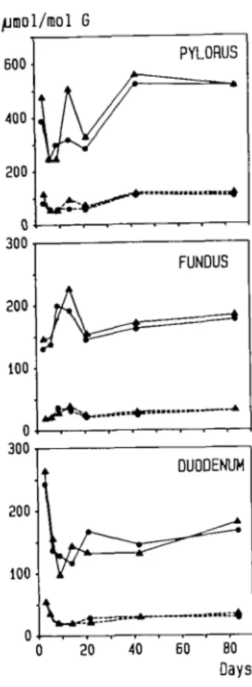
drinking water are shown in Figure 1. In all tissues examined,
levels of 7-methylguanine and C^-methylguanine showed
considerable variation during the first three weeks of exposure.
Later on, a steady state was reached which in the pylorus
amounted to ~520/xmol 7-methylguanine and HO^mol
C^-methylguanine/mol guanine. These values were three times
higher than those of fundus and duodenum which themselves
differed little from each other. Addition of sodium taurocholate
to the standard laboratory diet (0.25%) did not significantly affect
the extent of DNA methylation.
In a second experiment animals were given various
concen-trations of MNNG in the drinking water (10, 20, 40, 80 p.p.m.)
and killed after 21 days of exposure. The data obtained (Figure
2) clearly indicate that MNNG concentrations higher than 10-20
p.p.m. do not lead to a proportional increase in the formation
of 7-methylguanine. O^-Methylguanine levels showed a similar
lack of dose response. The amounts determined at 20 and 80
p.p.m. were 84 and 74 (pylorus), 21 and 24 (fundus), and 23
and 26 (duodenum) /imol/mol, respectively.
Immunohisto-chemical studies were carried out in animals exposed to MNNG
(40 or 80 p.p.m.) for 3 weeks. They showed that DNA
methylation is restricted to cells located near the gastric lumen.
Again, there was evidence of a regionally different extent of
alkylation. In the pyloric mucosa, immunoreactivity generally
yielded a stronger signal and C^-methylation of nuclear DNA
could easily be identified at levels down to 10 epithelial cells
below the luminal surface (Figure 3). In the fundus region,
Fig. 1. Concentrations of 7-methylguanine (solid line) and (Amethylguanine (dotted line) in DNA of stomach (pylorus, fundus) and duodenum during chronic exposure to MNNG in the drinking water (80 p.p.m.) over a period of 3 - 8 4 days One group of rats was treated with MNNG alone ( • ) ; the other group received MNNG plus a diet containing 0.25% (w/w) sodium taurocholate (A).
juraol 7-meG/mol G
eoo
Fig. 2. Levels of 7-methylguanine in DNA from pylorus, fundus and duodenum after 21 days of exposure of MNNG in the drinking water at concentrations ranging from 10—80 p.p.m.
unequivocal immunoreactivity to C^-methyldeoxyguanosine was
usually restricted to the cells directly bordering the gastric lumen.
No stained nuclei were detected in sections from untreated rats
(not shown).
DNA methylation after chronic exposure to MNNG It...
n-f •: • • • • " » • ' 5 #
Fig. 3< Immunohistochemical demonstration of O'-methyldeoxyguanosine in pyloric cell nuclei of rats which received MNNG in the drinking water (80 p.p.m.) over a period of 3 weeks. Note that immunoreactivity is restricted to a small layer of cells bordering the luminal surface. Nomarski interference contrast microscopy (X40 and X80).
Discussion
The objective of the present study was to determine levels of
methylpurines in stomach and duodenum of rats chronically
exposed to MNNG in the drinking water. Since epithelia of these
tissues have a high rate of cell turnover, we expected that steady
state levels be reached within 3 - 6 days after the onset of MNNG
administration. High concentrations were indeed observed within
three days of exposure (Figure 1). However, values varied
considerably during the initial stage of the experiment. The most
likely explanation for this phenomenon is that MNNG causes
stomach toxicity with erosions and subsequent cellular repair and
thus larger group-to-group variations in the level of DNA
alkylation than observed at later stages of continuous MNNG
administration. Histopathological analyses have shown that these
lesions occur most frequently during the first 1 - 2 weeks of
exposure (15). After - 3 weeks, levels of DNA methylation
reached a steady state and this probably reflects an adaptation
of the gastric mucosa to the toxic effects of MNNG. The lack
of a dose response during chronic exposure to MNNG
concentrations beyond 10—20 p.p.m. was unexpected since this
carcinogen does not require enzymic bioactivation and would thus
be expected to cause DNA alkylation proportional to dose. We
have considered the possibility that voluntary water intake is
reduced at high MNNG concentrations. Measurements showed
considerable day-to-day fluctuations. Although there was a
tendency towards reduced uptake at high MNNG concentrations,
these differences were too small to account for the observed lack
of dose response. Since thiols markedly enhance the
decompo-sition of MNNG, their presence at high concentrations in the
gastric mucosa has been linked to the organ-specific
carcino-genicity of MNNG (5). Our immunohistochemical studies showed
that the reaction of MNNG with target DNA is restricted to a
small number of epithelial cells bordering the gastric lumen. A
similar distribution was observed autoradiographically following
a single dose of [3H-methyl]MNNG (16). The possibility,
therefore, exists that free thiols in pyloric epithelia are easily
consumed in this reaction and thereby limit the extent of DNA
alkylation. Attempts to histochemically demonstrate an
MNNG-induced depletion of free thiols in the superficial layers of the
gastric mucosa failed although this effect could easily be shown
after intragastric administration of the thiol-blocking agent,
N-ethylmaleimide (unpublished results).
Since C^-methylguanine and related O-alkylated bases
represent critical DNA lesions responsible for the initiation of
malignant transformation by simple alkylating agents (17), our
results would suggest that the carcinogenic efficiency of MNNG
in this tumour model is largely independent of the concentration
of MNNG in the drinking water. This view is supported by a
report by Sugimura et al. (18) in which rats were chronically
exposed to MNNG at levels of 33 or 83 /ig/ml in the drinking
water. This considerable difference in dose was not paralleled
by either differences in the incidence of stomach carcinomas or
the time interval at which 50% of rats had died from tumours
( - 3 3 0 and 380 days).
When MNNG and related methylating nitroso compounds react
with DNA in vitro, i.e. in the absence of repair enzymes, the
ratio of the amounts of C^-methylguanine and 7-methylguanine
O.Kobori ft al.
formed is 0.11 (17). In the present study we observed a
consistendy higher ratio of —0.16 (Figure 1). Since the loss of
methylpurines due to mucosal cell turnover is similar for botfi
adducts, this finding indicates tfiat removal of 7-methylguanine
by spontaneous depurination and glycosylase-mediated excision
repair (19) is somewhat faster than the repair of
C^-methylguan-ine by me C^-alkylguanC^-methylguan-ine-DNA alkyltransferase. During the
course of the experiment, the ratio remained stable, suggesting
that in contrast to rat liver (20) carcinogen-induced toxicity does
not lead to an induction of the alkyltransferase in the stomach
mucosa.
In a previous study (5), we reported that DNA methylation
by MNNG in the glandular stomach is nine times higher than
in the forestomach and 20 times higher than in the esophagus.
The present study provides evidence that within the target tissue,
too, the extent of DNA alkylation closely correlates with the site
of tumour induction. Morphological analyses have shown that
after oral administration in the drinking water MNNG-induced
adenocarcinomas are selectively located in the gastric pylorus
(2), in particular at the lesser curvature (21). We found that levels
of 7-methylguanine and C^-methylguanine in this region were
3-fold higher than in DNA of the gastric fundus and the
duodenum and this was also apparent on immunohistochemical
stains for 06-meuiylguanine (not shown). This phenomenon,
too, remains unexplained. Thiol levels are known to be
considerably higher in the glandular stomach than in the squamous
epithelium of the forestomach (5) but no data are available on
the concentration of free thiols in different regions of the glandular
stomach. One may also argue that the bulk of fluid volume
consumed runs along the small curvature and that a homogeneous
distribution within the stomach lumen does not occur under
physiological conditions. In contrast, MNNG administration by
gavage produces carcinomas in various stomach regions including
the forestomach (22) and autoradiographic studies have shown
that following [I4C]MNNG application by stomach tube,
14C-labelled reaction products are distributed over the entire stomach
wall, rather than being concentrated in the pyloric region (5).
It has recently been reported that bile acid (taurocholic acid)
gready enhances MNNG-induced stomach tumorigenesis
(6,23,24). In these studies, even small amounts of MNNG
induced a high incidence of stomach tumours when taurocholic
acid was simultaneously or subsequently added to the diet or in
the drinking water, thus indicating a promoting effect. The present
study supports this view. Co-administration of taurocholate did
not affect the extent of interaction of MNNG with DNA in the
target tissue.
Acknowledgements
This work was in part supported by the Swiss National Science Foundation. The technical assistance of Brigitta Walker and Isabel Cackett is grateftilly acknowledged.
References
l.Kogure.K., Sasadaira,H., Kawachi,T., Shimosato.Y., Tokunaga.A., Fujimura.S. and Sugimura,T. (1974) Further studies on induction of stomach cancer in hamsters by yV-rnethyl-N'Hurro-A'-nitrosoguanidine. Br. J. Cancer, 29, 132-142.
2. Sugimura.T. and Kawachi,T. (1978) Experimental carcinogenesis. In Lipkin.M. and Good.R.A. (eds), Gastrointestinal Tract Cancer, Plenum Publishing Corp., New York, pp. 327-341.
3. Ohgalri.H. and Sugimura.T. (1988) Experimental stomach cancer. In DouglassrIr.,H.O. (ed.), Gastric Cancer, Churchill Livingstone, New York, Edinburgh, London, Melbourne, pp. 2 7 - 5 4 .
4. Kobori.O., Gedigk.P. and Totovic.V. (1976) Early changes of glandular stomach in Wistar rats ingesting A'-memyl-A/'-nitro-A/-nitrosoguanidine (MNNG). ZKrebsforsch., 87, 127-138.
2274
5. Wiestler.O., von Deimling.A., Kobori.O. and Kleihues.P. (1983) Location of /V-mcthyl-N'-nitro-^-nitrosoguanidine-induced gastrointestinal tumors correlates with thiol distribution. Carcinogenesis, 4, 879 — 883. 6. Kobori.O., Shimizu.T., Maeda.M., Atomi.Y., WatanabeJ., Shoji.M. and
Morioka.Y. (1984) Enhancing effect of bile and bile acid on stomach tumorigenesis induced by W-methyl-AT-nitro-A'-rutrosoguanidine. J. Nail.
Cancer Inst., 73, 853-861.
7. Sugimura.T., Fujimura.S., Kogure.K., Baba.T., Saito.T., Nagao.M., Hosoi.H., Shimosato.Y. and Yokoshima.T. (1969) Production of adeno-carcinomas in glanrftilnr stomach of experimental animals by W-methyl-A/'-nitro-A'-nitrosoguanidine. Gann Monogr. Cancer Res., 8, 157—196.
8. von Hofe.E., Grahmann.F., Keefer.L.K., Lijinsky.W., Nelson,V. and Kleihues.P. (1986) Methylation versus ethylation of DNA in target and non-target tissues of Fischer 344 rats treated with A/-nitrosomethylethylamine.
Cancer Res., 46, 1038-1042.
9. von Hofe,E. and Kleihues.P. (1986) Comparative studies on hepatic DNA alkylation in rats by A'-nitrosomethylethylamine and A'-nitrosodimethylamine plus W-nitrosodiethylamine. J. Cancer Res. Clin. Oncol., 112, 205-209. 10. SwenbergJ.A. and Bedell.M.A. (1982) Cell-specific DNA alkylation and repair: Application of new fluorometric techniques to detect adducts. In Magee.P.N. (ed.), Banbury Report 13: Indicators of Genotoxic Exposure. Cold Spring Harbor Laboratory, p. 205-220.
11. Ludeke.B.I. and Kleihues.P. (1988) Formation and persistence of O^-^-hydroxyethyl^'-deoxyguanosine in DNA of various rat tissues following a single dose of A'-nitroso-A'-<2-hydroxyethyl)urea. An immuno-slot-blot study. Carcinogenesis, 9, 147—151.
12. Heyting.C., Van Der Laken.C J., Van Raamsdonk.W. and Pool.C.W. (1983) Immunohistochemical detection of O^-ethyldeoxyguanosine in the rat brain after in vivo applications of N-ethyl-N-nitrosourea. Cancer Res., 43, 2935-2941.
13. MenkvekLG.J., Van Der Laken.CJ., Hermsen,T., Kriek,E., Scherer.E. and Den Engelse.L. (1985) Immunohistochemical localization of C^-ethyldeoxyguanosine and deoxyguanosin-8-yl-(acetyl)aminofluorene in liver sections of rats treated with diethylnitrosamine, ethylnitrosourea or N-acetylaminofluorene. Carcinogenesis, 6, 263—270.
14. Ordronneau.P., Lindstrom.P.B.M. and Petrusz.P. (1981) Four unlabelcd antibody bridge techniques: a comparison. /. Histochem. Cytochem., 29, 1397-1404.
15.Ohgaki,H., Kusama.K., Hasegawa.H., Sato.S., Takayama.S. and Sugimura,T. (1986) Sequential histcJogic changes during gastric carcinogenesis induced by Ar-methyl-A/'-nitro-A/-nitrosoguanidine in susceptible ACI and resistant Buffalo rats. /. Natl. Cancer Inst., TJ, 747-755.
16. Ohgaki.H., Tomihari.M., Sato.S., Kleihues.P. and Sugimura.T. (1988) Differential proliferative response of gastric mucosa during carcinogenesis induced by /V-methyl-Ap-nitro-Af-nitrosoguanidine in susceptible ACI rats, resistant Buffalo rats, and their hybrid Fl cross. Cancer Res., 48,5275-5279. 17. Singer.B. and Grunberger.D. (1983) Molecular Biology of Mutagens and
Carcinogens, Plenum Press, New York and London.
18. Sugimura.T., Fujimura.S. and Baba.T. (1970) Tumor production in the glandular stomach and alimentary tract of the rat by Af -methyl-A''-nitro-Ap'-nitrosoguanidine. Cancer Res., 30, 455-465.
19. Margison.G.P. and Pegg.A.E. (1981) Enzymatic release of 7-methylguanine from methylated DNA by rodent liver extracts. Proc. Nail. Acad. Sci. USA, 78, 861-865.
20. Margison.G.P. (1982) Chronic or acute administration of various dialkylnitrosamines enhances the removal of C^-methylguaninc from rat liver DNA in vivo. Chem-Biol. Interactions, 38, 189-201.
21. Kobori.O. (1980) Analytical study of precancerous lesions in rat stomach mucosa induced by A'-methyl-A''-nitro-Ar-nitrosoguanidine. Gann Monogr., 25, 141-150.
22. Hirono.I. and Shibuya.C. (1972) Induction of stomach cancer by a single dose of W-memyl-Ap-rutro-A'-nitrosoguanidine through a stomach tube. In Nakahara.W., Takayama.S., Sugimura.T. and Odashima.S. (eds), Topics in
Chemical Carcinogenesis, University of Tokyo Press, pp. 121-132.
23. Salmon,R.J., Laurent,M. and Thierry J.P. (1984) Effect of taurocholic acid feeding on methyl-nitro-A'-nitroso-guanidine induced gastric tumors. Cancer
Lett., 22, 315-320.
24. Cohen.A., Geller.S.A. and Horowtiz.I., Toth.L.S. and Werthcr.LJ. (1984) Experimental models for gastric leiomyosarcoma. The effects of /V-methyl-A^-nitro-Mnitrosoguanidine in combination with stress, aspirin, or sodium taurocholate. Cancer, 53, 1088-1092.
Received on June 2, 1988; revised on August 7, 1988; accepted on September 8, 1988