A new source for cardiovascular tissue engineering:
human bone marrow stromal cells
q
Alexander Kadner
a,*, Simon P. Hoerstrup
a, Gregor Zund
a, Karim Eid
b, Christine Maurus
a,
Serguei Melnitchouk
a, Jurg Grunenfelder
a, Marko I. Turina
aaClinic for Cardiovascular Surgery, University Hospital, Raemistrasse 100, CH 8091 Zurich, Switzerland bClinic for Trauma Surgery, University Hospital, Raemistrasse 100, CH 8091 Zurich, Switzerland
Received 1 October 2001; received in revised form 8 January 2002; accepted 30 January 2002
Abstract
Objective: Vascular-derived cells represent an established cell source for tissue engineering of cardiovascular constructs. Previously, cell isolation was performed by harvesting of vascular structures prior to scaffold seeding. Marrow stromal cells (MSC) demonstrate the ability to differentiate into multiple mesenchymal cell lineages and would offer an alternative cell source for tissue engineering involving a less invasive harvesting technique. We studied the feasibility of using MSC as an alternative cell source for cardiovascular tissue engineering. Methods: Human MSC were isolated from bone marrow and expanded in culture. Subsequently MSC were seeded on bioabsorbable polymers and grown in vitro. Cultivated cells and seeded polymers were studied for cell characterization and tissue formation including extracellular matrix production. Applied methods comprised flow cytometry, histology, immunohistochemistry, transmission (TEM) and scanning electron micro-scopy (SEM), and biochemical assays. Results: Isolated MSC demonstrated fibroblast-like morphology. Phenotype analysis revealed positive signals for alpha-smooth muscle actin and vimentin. Histology and SEM of seeded polymers showed layered tissue formation. TEM demon-strated formation of extracellular matrix with deposition of collagen fibrils. Matrix protein analysis showed production of collagen I and III. In comparison to vascular-derived cell constructs quantitative analysis demonstrated comparable amounts of extracellular matrix proteins in the tissue engineered constructs. Conclusions: Isolated MSC demonstrated myofibroblast-like characteristics. Tissue formation on bioabsorbable scaffolds was feasible with extracellular matrix production comparable to vascular-cell derived tissue engineered constructs. It appears that MSC represent a promising cell source for cardiovascular tissue engineering. q 2002 Elsevier Science B.V. All rights reserved.
Keywords: Tissue engineering; Cardiovascular; Marrow stromal cells; Polymer scaffold
1. Introduction
Currently valve replacement is an effective treatment using mechanical or biological protheses [1]. However, there are certain limitations regarding long-term benefit. Mechanical valves are associated with a substantial risk of thromboembolism while biological valves lack the ability to repair, grow, or remodel resulting in structural dysfunction [2,3].
Tissue engineering offers a promising approach to over-come these limitations by trying to create viable valve struc-tures with a thromboresistant surface and a living interstitium with repair and remodeling capabilities [4]. Several groups reported the feasibility of creating
autolo-gous living cardiovascular structures by seeding cells on synthetic polymer, collagen or xenogenic scaffolds [5–8]. Previously our group demonstrated the feasibility of tissue engineering living heart valve constructs by seeding bioab-sorbable polymer scaffolds [9]. However, all of these approaches are based on vascular-derived cell sources with certain shortcomings. Harvesting of the cells prior to scaffold seeding necessitates the sacrifice of an intact vessel. Furthermore, vascular-derived cells demonstrate different characteristics compared to valve interstitial cells, qualities which may be vital to the development and function of a tissue engineered heart valve [10]. Due to these limitations we investigated the feasibility of applying marrow stromal cells (MSC) as an alternative cell source for tissue engineer-ing. The usage of MSC may offer several advantages by: (i) showing characteristics of multipotent progenitor cells which are able to differentiate into a variety of mesenchy-mal cell types; (ii) easy collection and isolation methods avoiding the sacrificing of intact cardiovascular structures;
1010-7940/02/$ - see front matter q 2002 Elsevier Science B.V. All rights reserved. PII: S 1 0 1 0 - 7 9 4 0 ( 0 2 ) 0 0 0 7 9 - 9
www.elsevier.com/locate/ejcts
q
Presented at the joint 15th Annual Meeting of the European Association for Cardio-thoracic Surgery and the 9th Annual Meeting of the European Society of Thoracic Surgeons, Lisbon, Portugal, September 16–19, 2001.
* Corresponding author. Tel.: 141-1-255-3644; fax: 141-1-255-4775. E-mail address: [email protected] (A. Kadner).
and (iii) demonstrating immunological unique characteris-tics allowing persistence in an allogenic setting [11–13].
2. Materials and methods 2.1. Cell isolation
MSC were isolated from human bone marrow by washing in Dulbecco’s phosphate buffered saline (DPBS, Gibco) for 10 min at 1500 rpm. The cells were recovered after centri-fugation and resuspended in DPBS. A low-density cell frac-tion of bone marrow was obtained by centrifugafrac-tion of the cell suspension over a Ficoll step gradient (density 1.077 g/ ml) (Ficoll-Histopaque 1077, Sigma) at 1500 rpm for 10 min. The nucleated cells were collected from the interface, diluted with two volumes of DPBS and centrifuged at 1500 rpm for 10 min. Following, the cells were resuspended, counted and plated at 200 000 cells/cm2.
2.2. Cell cultivation
The isolated cell fraction was cultured in Dulbecco’s modified Eagle’s medium (DMEM, Gibco) supplemented with 10% fetal bovine serum (HyClone), penicillin (Gibco), and streptomycin (Gibco) in tissue flasks (Corning, Inc.) and left to adhere at 378C for 4–5 h. The non-adherent cells floated off, while mesenchymal cells adhered, spread, and grew. Medium was replaced at 24 and 72 h and every 6 days following. Daily growth progress was monitored by phase-contrast microscopy. The cells were serially passaged and expanded in a humidified incubator at 378C with 5% CO2. Sufficient cell numbers for cell seeding on
bioabsorb-able polymer scaffolds were obtained after 21–28 days. 2.3. Bioabsorbable polymer scaffolds
Non-woven polyglycolic-acid mesh (PGA, thickness: 1.0 mm, specific gravity: 69 mg/cm3, Albany Int.) was coated
with poly-4-hydroxybutyrate (P4HB, MW: 1 £ 106, PHA
4400, TEPHA Inc., Cambridge, MA). From the PGA/ P4HB composite scaffold material strips (20 £ 15 mm) were cut and cold gas sterilized with ethylene oxide. 2.4. Cell seeding and in vitro culture of polymer constructs
MSC were seeded onto the polymer scaffolds (n ¼ 6) with an approximate cell density between 4.5–5.5 £ 106 per cm2and cultured in nutrient medium (DMEM, Gibco) for 14 days in a humidified incubator (378C, 5% CO2).
2.5. Analysis of MSC cultures 2.5.1. Flow cytometry (FACS)
A single cell suspension of MSC was prepared for FACS. 0.5–1 £ 106cells in 100 ml phosphate buffered saline (PBS) plus BSA were incubated with saturating concentrations of monoclonal antibodies CD 31-FITC (Sigma, St. Louis),
LDL-Dil (Biomedical Technologies Inc, Stoughton, MA), CD 14-FITC (Beckon Dickinson, San Jose, CA). For intra-cellular staining, cells were permeabilized with ethanol for 30 min and incubated with monoclonal antibodies against ASMA (Sigma, St. Louis) and vimentin (NeoMarkers, Fremont). Following washing, staining with a secondary FITC-conjugated IgG goat-anti-mouse antibody (Chemi-con, Temecula, CA) was performed for 30 min. Forward and side scatter gates were set to exclude debris and 10 000 gated events were counted per sample. Correspond-ing isotype and positive controls were performed for each antibody. Cells were analyzed with the flow cytometer
FACS-Calibur (Becton Dickinson Immunocytometry
Systems, San Jose, CA). Data analysis was performed with the CELL QUEST software program (Becton Dickin-son Immunocytometry Systems, San Jose, CA). Expression levels were calculated as mean fluorescence intensity ratio (MFIR) defined as mean fluorescence intensity of the studied antibodies divided by mean fluorescence intensity of corresponding isotype controls.
2.5.2. Histology and immunohistochemistry
Isolated MSC were cultivated onto glass coverslips in nutrient medium (DMEM, Gibco). After 2–3 days cells were washed with PBS and fixed in 4% paraformaldehyde for 10 min. Cells were examined histologically by hematox-ylin & eosin (H & E) and Trichrome-masson stain.
Immuno-histochemistry was performed by incubation with
monoclonal mouse antibodies for ASMA (Sigma, St. Louis), vimentin (NeoMarkers, Fremont), desmin (NeoMar-kers, Fremont), collagen I–IV (Oncogen, Boston), and elastin (Sigma, St. Louis). Incubation with a secondary biotin-labeled goat-anti-mouse IgG antibody (Sima, St. Louis) was performed and the signal was developed with the avidin-peroxidase system (ABC kit, Vector Lab, Burlingame CA). Prior to intracellular staining, permeabilization of the cells was performed by incubation with 0.1% Triton (Sigma, St. Louis) for 10 min.
2.6. Analysis of MSC-seeded polymer constructs 2.6.1. Histology and immunohistochemistry
After 2 weeks in culture, sections of MSC seeded polymer scaffold strips were fixed in 4% phosphate-buffered formalin and embedded in paraffin. Paraffin sections were cut at 5-mm thickness and studied by H & E and Trichrome-masson stain. Immunohistochemistry was performed as described above by incubation with monoclonal mouse antibodies for ASMA, vimentin, desmin, collagen I–IV and elastin. 2.6.2. Scanning and transmission electron microscopy
Additional samples of MSC-seeded polymer strips were fixed in 2% glutaraldehyde (Sigma, St. Louis) and dehy-drated in acetone followed by drying in carbon dioxide for scanning electron microscopy (SEM) and transmission elec-tron microscopy (TEM).
2.6.3. Biochemical assays
Cellular and extracellular components of the MSC-seeded constructs were analyzed by biochemical assays and compared to vascular-derived cell (VC) seeded constructs. VC seeded constructs were identically generated as MSC-seeded constructs by using jugular venous fibro-blasts. Total DNA was isolated and purified by sequential organic extractions with phenol and phenol/chloroform/ isoamyl alcohol and quantitated by spectrophotometry. For determination of total collagen content, the tissue was completely acid-digested and total 5-hydroxyproline was measured. Total proteoglycan/glycosaminoglycan (GAG)
and elastin content were quantitated with a BLYSCANe
and FASTINe
assay (Biocolor, Belfast, Ireland) after tissue extraction [14]. Data of biochemical testing was statistically analyzed by SPSS 8.0 Software and expressed as mean ^ standard error of the mean. An unpaired test (Student t-test) was performed, considering a P-value ,0.05 as statis-tically significant. Six samples of each group were studied per test.
3. Results
3.1. Cell morphology
Isolated cells appeared small and round with a tendency to grow in clusters. Non-adherent cells were removed by medium change at 24 h and every 4 days thereafter. Elon-gated cells with fibroblast-like morphology appeared after 72 h and reached confluence after 10–14 days.
3.2. Flow activated cell scanning
Table 1 shows the results of FACS analysis for MSC. Flow cytometry characterization of MSC demonstrates no significant difference in expression of ASMA (MFIR 3.66) and vimentin (MFIR 12.59) compared to vascular-derived myofibroblasts. No positive signal was detected for CD 14 (MFIR 1.13), CD 31 (MFIR 1.1), and LDL (MFIR 1.94) among the isolated cell population (Table 1).
3.3. Histology and immunophenotyping of MSC
H & E and Trichrome-masson staining of fixed cells demonstrated the deposition of extracellular matrix through-out the cell culture. Immunohistochemistry showed the expression of ASMA and vimentin by MSC. The deposition
of collagen I and III was detected by positive staining. In contrast, no signal was observed following antibody stain-ing for desmin, collagen II, IV, and elastin.
3.4. Histology and immunohistochemistry of MSC-seeded polymer constructs
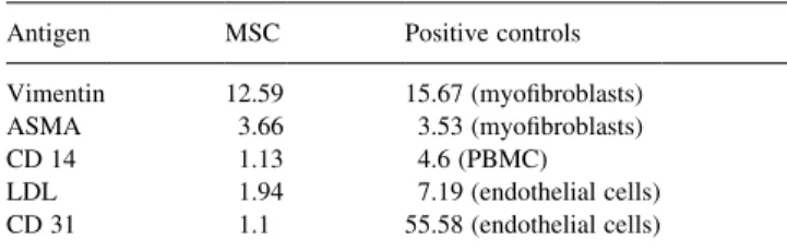
H & E and Trichrome-masson staining of the MSC-seeded polymer constructs showed a layered tissue forma-tion and a dense upper layer with deposiforma-tion of extracel-lular matrix proteins. Irregular celextracel-lular ingrowth was observed into less cellularized deeper parts of the polymer strips (Figs. 1A,B). Immunohistochemistry of MSC-seeded constructs showed positive staining for ASMA and vimen-tin (Figs. 1C,D). Extracellular matrix analysis demon-strated the deposition of collagen I and III (Figs. 1E,F). No positive staining was studied for desmin, collagen II, IV, and elastin.
3.5. Transmission and scanning microscopy
TEM of sections of MSC polymer constructs showed secretionally active fibroblasts with deposition of collagen fibrils (Fig. 2A). SEM revealed loose tissue formation and demonstrated the degradation of the polymer scaffold by multiple hydrolytic breakages and fragmentation of the polymer fibers (Fig. 2B).
3.6. Biochemical assays
Results of the biochemical assays are shown in Fig. 3. DNA content of the MSC constructs (2.7 mg/mg dry tissue ^0.6) was significantly superior (P , 0:01) compared to VC constructs (0.9 mg/mg dry tissue ^0.5). Biochemical assays detected a hydroxyproline content of MSC constructs of 3.1 mg/mg dry tissue ^0.4 which was comparable to VC constructs (2.8 mg/mg dry tissue ^0.5). The quantitative analysis of the glycosaminoglycan amount revealed 4.9
mg/mg dry tissue ^0.7 not significantly different
(P . 0:05) to VC constructs (5.8 mg/mg dry tissue ^0.6). Elastin was not detectable in MSC- and VC-constructs.
4. Discussion
Presently, vascular-derived cells represent an established cell source for seeding of tissue engineered cardiovascular constructs. By using venous or arterial vascular cells, func-tional tissue engineered patches, vascular grafts, and heart valves were generated [5,7,15]. In prior in-vitro and in-vivo studies Hoerstrup et al. demonstrated the feasibility of tissue engineering a complete and living autologous heart valve by using myofibroblasts of carotid artery origin [9]. Following 5 months implantation in sheep the tissue engineered valves functioned satisfactorily in-vivo while tissue formation gradually evolved to resemble native pulmonary valve morphology and histology. However, there is evidence that native heart valves consist of a mixed population of Table 1
MFIR of FACS analysis
Antigen MSC Positive controls
Vimentin 12.59 15.67 (myofibroblasts)
ASMA 3.66 3.53 (myofibroblasts)
CD 14 1.13 4.6 (PBMC)
LDL 1.94 7.19 (endothelial cells)
interstitial cells (IC) which show specific and unique char-acteristics different to vascular-derived fibroblasts [16,17]. Recently, Taylor et al. demonstrated the feasibility of using IC for tissue engineering [8]. A possible clinical application of creating a tissue engineered valve with IC is limited by using non-autologous IC.
Given these problems it appears that the appropriate cell source for tissue engineering of heart valves is still unclear. In the present study we evaluated the feasibility of using an alternative cell source for tissue engineering heart valves – mesenchymal stromal cells.
Isolation of MSC was easy to perform avoiding the sacrifi-cing of an intact vascular structure. Isolated cells appeared small, round, and elongated with a fibroblast-like morphol-ogy. After 72 h cells appeared to grow in a colony-forming
pattern. Identical morphological characteristics and growth pattern are reported for mesenchymal precursor cells by other studies using a similar isolation procedure [18,19]. Analysis of the cell population showed characteristics of a myofibro-blast-like differentiation. MSC expressed ASMA, vimentin and the deposition of collagen I and III was observed. MSC did not stain positive for the monoclonal antibody against desmin, a muscle cell marker. A similar staining pattern is reported for valve IC by Taylor et al. [8]. FACS analysis did not detect any CD 14, CD 31, or LDL positive cells, indicat-ing the absence of myeloid and endothelial cell differentia-tion of MSC. Furthermore, we did not observe any positive staining for collagen II, implying the absence of an osteo-blastoid differentiation of the isolated cells.
The formation of tissue by MSC showed identical results Fig. 1. Histology and Immunohistochemistry of MSC culture and polymer constructs. H & E (A); and Trichrome-masson (B) staining show cells with a fibroblast morphology and layered tissue formation with deposition of extracellular matrix proteins. MSC-seeded polymer constructs show a irregular cellular growth into less cellularized deeper layers. Immunohistochemical staining demonstrated the expression of ASMA (C); and vimentin (D). Extracellular matrix analysis demonstrated the deposition of collagen I (E); and III (F).
compared to vascular-derived cells seeded on polymer constructs after 14 days in culture. We observed a layered tissue structure formation with a dense upper layer. Overall, morphology and ultra-structural analysis showed good cell-polymer adhesion and growth of MSC into deeper parts of the polymer strips. Furthermore, the gradual biodegradation of the scaffold and the replacement by viable tissue was observed. Results of biochemical assays demonstrated no significantly different amount of extracellular matrix proteins of MSC polymer constructs compared to VC seeded constructs.
In previous studies it was demonstrated that exposure of tissue engineered heart valve constructs to a pulsatile flow significantly improved tissue development and mechanical properties [7,20]. A homogenous, dense tissue developed resembling native valve morphology. In addition,
biochem-ical analysis of the tissue engineered heart valves showed comparable values to native tissue. Given these results we anticipate that exposure of MSC-seeded constructs to a
biomimetic pulsatile flow system will demonstrate
improved tissue formation and differentiation. We also believe that future animal experiments are required to eval-uate the remodelling capacity, the growth potential, and long-term function of MSC-seeded tissue engineered constructs under physiologic conditions.
In conclusion, this study demonstrates the feasibility of using MSC as a new alternative cell source for tissue engi-neering. Cell isolation was easy to perform without the need to sacrificing intact vascular structures. MSC showed char-acteristics of myofibroblast-like differentiation with neo-tissue formation comparable to neo-tissue engineered constructs based on vascular-derived cells.
Fig. 2. Transmission and scanning electron microscopy. (A) TEM of sections of MSC polymer constructs shows cell elements typical of viable, secretionally active myofibroblasts with deposition of collagen fibrils (arrow). (B) SEM revealed loose tissue formation with good cell-polymer-attachments (arrow) and demonstrated the degradation of the polymer scaffold by multiple hydrolytic breakages and fragmentation of the polymer fibers (*).
Acknowledgements
The authors wish to thank Jay Tracy and Manfred Welti, Laboratory for Tissue Engineering and Cell Transplanta-tion, University Hospital Zurich, for their valuable technical assistance. We further thank Klaus Marquard, Department of Surgical Research, University Hospital Zurich, for providing the SEM pictures. Finally we are grateful to Dr Jeroen F. Visjager, Swiss Institute of Technology, Zurich, for performing the biomechanical testing.
References
[1] Braunwald E. Valvular heart disease. In: Braunwald E, editor. Heart Disease, 4th ed.. Philadelphia, PA: WB Saunders, 1992. pp. 1007–1077. [2] Vongpatanasin W, Hillis D, Lange RA. Prosthetic heart valves. N
Engl J Med 1996;335:407–416.
[3] Schoen FJ, Levy RJ. Tissue heart valves. Current challenges and future research. J Biomed Mater Res 1999;47:439–465.
[4] Mayer Jr JE. In search of the ideal valve replacement device. J Thorac Cardiovasc Surg 2001;122:8–9.
[5] Shinoka T, Shum TD, Ma PX, Tanel RE, Isogai N, Langer N, Langer R, Vacanti JP, Mayer Jr JE. Creation of viable pulmonary artery autografts through tissue engineering. J Thorac Cardiovasc Surg 1998 Ma;115(3):536–545 discussion 545–546.
[6] Bader A, Steinhoff G, Strobl K, Schilling T, Brandes G, Mertsching H, Tsikas D, Froelich J, Haverich A. Engineering of human vascular aortic tissue based on a xenogeneic starter matrix. Transplantation 2000 Ju;70(1):7–14.
[7] Hoerstrup SP, Zund G, Sodian R, Schnell AM, Grunenfelder J, Turina MI. Tissue engineering of small caliber vascular grafts. Eur J Cardi-othorac Surg 2001 Ju;20(1):164–169.
[8] Taylor PM, Allen SP, Dreger SA, Herbage D, Yacoub MH. Biological matrices (collagen) human aortic valve interstitial cells cultured on 3-day collagen sponge: relevance to tissue engineering. Presented at the First Symposium on Tissue Engineering for Heart Valve Substitutes, London, UK, 15th June 2001.
[9] Hoerstrup SP, Sodian R, Daebritz S, Wang J, Bacha EA, Martin DP, Moran AM, Guleserian KJ, Sperling JS, Kaushal S, Vacanti JP, Schoen FJ, Mayer Jr JE. Functional living trileaflet heart valves grown in vitro. Circulation 2000 No;102(19 Suppl. 3):III44–III49. [10] Roy A, Brand NJ, Yacoub MH. Molecular characterization of
inter-stitial cells isolated from human heart valves. J Heart Valve Dis 2000 Ma;9(3):459–464 discussion 464–465.
[11] Prockop DJ. Marrow stromal cells as stem cells for non-hematopoie-tic tissues. Science 1997 Apr 4;276(5309):71–74.
[12] Young HE, Mancini ML, Wright RP, Smith JC, Black Jr AC, Reagan CR, Lucas PA. Mesenchymal stem cells reside within the connective tissues of many organs. Dev Dyn 1995 Fe;202(2):137–144. [13] Liechty KW, Mackenzie TC, Shaaban AF, Radu A, Mosely AB,
Deans R, Marshak DR, Flake AW. Human mesenchymal stem cells engraft and demonstrate site-specific differentiation after in utero transplantation in sheep. Nat Med 2000 No;6(11):1282–1286. [14] Bergman I, Loxley R. Two improved and simplified methods for the
spectophotometric determination of hydroproline. Anal Chem 1963;35:1961–1965.
[15] Stock UA, Sakamoto T, Hatsuoka S, Martin DP, Nagashima M, Moran AM, Moses MA, Khalil PN, Schoen FJ, Vacanti JP, Mayer
Jr JE. Patch augmentation of the pulmonary artery with bioabsorbable polymers and autologous cell seeding. J Thorac Cardiovasc Surg 2000 De;120(6):1158–1167 discussion 1168.
[16] Taylor PM, Allen SP, Yacoub MH. Phenotypic and functional char-acterization of interstitial cells from human heart valves, pericardium and skin. J Heart Valve Dis 2000 Ja;9(1):150–158.
[17] Messier RH, Bass BL, Aly HM, Jones JL, Domkowski PW, Wallace RB, Hopkins RA. Dual structural and functional phenotypes of the porcine aortic valve interstitial population: characteristics of leaflet myofibroblasts. J Surg Res 1994 Ju;57(1):1–21.
[18] Zvaifler NJ, Marinova-Mutafchieva L, Adams G, Edwards CJ, Moss J, Burger JA, Maini RN. Mesenchymal precursor cells in the blood of normal individuals. Arthritis Res 2000;2(6):477–488.
[19] Bucala R, Spiegel LA, Chesney J, Hogan M, Cerami A. Circulating fibrocytes define a new leukocyte subpopulation that mediates tissue repair. Mol Med 1994 No;1(1):71–81.
[20] Hoerstrup SP, Sodian R, Sperling JS, Vacanti JP, Mayer JE. New pulsatile bioreactor for in vitro formation of tissue engineered heart valves. Tissue Eng 2000;6:75–79.
Appendix A. Conference discussion
Mr S. Stoica (Cambridge, UK): A very elegant concept. I thought the histology slides were most convincing. Did you use a negative and positive control for your monoclonal antibodies?
Dr Kadner: Certainly, we were performing both negative and positive controls.
Dr G. Gerosa (Padova, Italy): I congratulate you. You did very excellent work. I would like to ask you, the myofibroblasts that you identified with your immunohistochemistry were fetal type or adult type?
Dr Kadner: The bone marrow stromal cells were adult type, and the vascular-derived cells came from saphenous vein.
Dr A. Haverich (Hannover, Germany): I have a more general question, and that is that many other researchers in other fields use the same sort of cells and they are making liver and they are making pancreas, so they are using pluripotent neural stem cells and cellularized tissues out of that. What do you think will be the necessary environment for these cells when used for cardiovascular applications?
Dr Kadner: I would have to speculate about this. It appears that initially these cells take spontaneously a fibroblast-like lineage. However, other groups have shown that by using such cell medium supplements as dexa-methasone, insulin or transforming growth factor beta 3 that you can induce chondrocytic, osteocytic or adipocytic lineages. What would be very inter-esting for cardiovascular tissue engineering is the differentiation of these cells in a endothelial lineage.
Dr B. Messmer (Aachen, Germany): I think one of the most important things is the expression of collagen. Can you explain why you have an inhomogenous expression of collagen 1 and 2, which you have shown; you didn’t show 3 and 4? This is very inhomogenous, and this may be a disadvantage.
Dr Kadner: I agree with you, and I think the reason is that these constructs were cultured only for a very brief period, for 7 days under steady conditions. In this context I would like to refer to a talk my collea-gue, Simon Hoerstrup, is giving tomorrow. He will present our data for using these cells for creating tissue engineered heart valve constructs, which were cultured under dynamic conditions for an extended period of time.