Generation of a vancomycin-intermediate Staphylococcus aureus (VISA)
strain by two amino acid exchanges in VraS
Anne Berscheid
1†, Patrice Franc¸ois
2, Axel Strittmatter
3‡, Gerhard Gottschalk
3, Jacques Schrenzel
2,
Peter Sass
4§ and Gabriele Bierbaum
1*§
1
Institute of Medical Microbiology, Immunology and Parasitology (IMMIP), University of Bonn, Bonn, Germany;
2Genomic Research
Laboratory, Infectious Diseases Service, Geneva University Hospitals and the University of Geneva, Geneva, Switzerland;
3Institute of Microbiology and Genetics, University of Go¨ttingen, Go¨ttingen, Germany;
4Institute for Pharmaceutical Biology,
University of Du¨sseldorf, Du¨sseldorf, Germany
*Corresponding author. Tel:+49-(0)228-287-19103; Fax: +49-(0)228-287-14808; E-mail: [email protected] †Present address: Institute for Pharmaceutical Biology, University of Du¨sseldorf, Du¨sseldorf, Germany.
‡Present address: Eurofins MWG Operon, Anzingerstr. 7a, 85560 Ebersberg, Germany. §These authors share senior authorship.
Received 14 March 2014; returned 30 May 2014; revised 18 June 2014; accepted 10 July 2014
Objectives: Staphylococcus aureus is a notorious bacterial pathogen and antibiotic-resistant isolates complicate
current treatment strategies. We characterized S. aureus VC40, a laboratory mutant that shows full resistance to
glycopeptides (vancomycin and teicoplanin MICs
≥32 mg/L) and daptomycin (MIC¼4 mg/L), to gain deeper
insights into the underlying resistance mechanisms.
Methods: Genomics and transcriptomics were performed to characterize changes that might contribute to
devel-opment of resistance. The mutations in vraS were reconstituted into a closely related parental background. In
addition, antimicrobial susceptibility testing, growth analyses, transmission electron microscopy,
lysostaphin-induced lysis and autolysis assays were performed to characterize the phenotype of resistant strains.
Results: Genome sequencing of strain VC40 revealed 79 mutations in 75 gene loci including genes encoding the
histidine kinases VraS and WalK that control cell envelope-related processes. Transcriptomics indicated the
increased expression of their respective regulons. Although not reaching the measured MIC for VC40,
reconstitu-tion of the L114S and D242G exchanges in VraS(VC40) into the susceptible parental background (S. aureus NCTC
8325) resulted in increased resistance to glycopeptides and daptomycin. The expression of VraS(VC40) led to
increased transcription of the cell wall stress stimulon, a thickened cell wall, a decreased growth rate, reduced
autolytic activity and increased resistance to lysostaphin-induced lysis in the generated mutant.
Conclusions: We show that a double mutation of a single gene locus, namely vraS, is sufficient to convert the
vancomycin-susceptible strain S. aureus NCTC 8325 into a vancomycin-intermediate S. aureus.
Keywords: glycopeptides, teicoplanin, daptomycin, antibiotic resistance, two-component regulatory systems
Introduction
Methicillin-resistant Staphylococcus aureus (MRSA) is among the
leading causes of mortality by any single infectious agent in the
USA and the European Union.
1,2The options for the treatment
of MRSA are limited. Since the early 1990s, vancomycin and
related glycopeptide antibiotics have been employed as therapy
for serious MRSA infections.
3,4However, S. aureus strains with
intermediate or full resistance to vancomycin emerged in recent
decades
5–7and clinical MRSA strains with additional resistance
to daptomycin and linezolid have been isolated.
6,8,9Since these
pathogens are able to sensitively respond to antibiotic stress,
it may be predicted that the resistance situation will not
substantially relax in the future.
10,11Understanding the bacterial
response to antibiotics as well as the established resistance
mechanisms to clinically used drugs is mandatory to evaluate
alternatives to commonly applied treatment strategies.
Here, we set out to characterize the vancomycin-resistant
laboratory mutant S. aureus VC40,
12which had previously been
generated by 30 serial passages of strain RN4220DmutS in the
presence of increasing concentrations of vancomycin.
13S. aureus
VC40 has a vancomycin MIC of 64 mg/L, a concentration higher
than the MIC for clinical vancomycin-intermediate S. aureus
(VISA) strains. Actually, the measured MIC for strain VC40 meets
the criteria for a vancomycin-resistant S. aureus (VRSA) strain
rather than for a VISA strain; however, it lacks the vanA gene
#The Author 2014. Published by Oxford University Press on behalf of the British Society for Antimicrobial Chemotherapy. All rights reserved.
For Permissions, please e-mail: [email protected]
J Antimicrob Chemother 2014; 69: 3190 – 3198
cluster,
12which represents the common VRSA resistance
deter-minant.
14Comparative genomic and transcriptomic analyses of
S. aureus VC40 revealed non-synonymous single nucleotide
poly-morphisms (SNPs) in the sensor histidine kinase genes vraS and
walK as well as significantly altered expression levels of their
respective regulons. We reconstituted VraS(VC40) into the
paren-tal background (S. aureus NCTC 8325), characterized the
expres-sion of the respective regulon via quantitative real-time PCR
(qRT–PCR) and examined the phenotypes of the different mutant
strains by transmission electron microscopy (TEM), autolysis
assays and growth behaviour studies to elucidate the specific
role of VraS in antibiotic resistance. Our data show that VraS is a
central factor in S. aureus NCTC 8325 that can confer reduced
sus-ceptibilities to clinically applied drugs including the last-resort
antibiotics vancomycin and daptomycin.
Materials and methods
Strains, plasmids and growth conditions
Bacterial strains and plasmids are listed in Table S1 (available as Supplementary data at JAC Online). Unless otherwise indicated, S. aureus strains were grown in brain heart infusion (BHI) broth or Mueller– Hinton (MH) broth (Oxoid) and Escherichia coli strains were grown in lysogeny broth at 378C or 308C when using temperature-sensitive plasmids. For plasmid maintenance, the required media were supplemented with ampi-cillin (50 mg/L) or erythromycin (25 mg/L).
Growth curve analyses and antimicrobial
susceptibility testing
For growth curve analyses, three parallel cultures of the desired S. aureus strains were grown at 378C in BHI or MH broth with or without the addition of antibiotics in 96-well polystyrene round-bottomed microplates (Greiner). The optical density at 600 nm (OD600) was monitored kinetically using the Tecan SunriseTMplate reader equipped with an incubation
cham-ber (Tecan Group). The microplate was read every 10 min for 8 h with shak-ing in between and prior to every read. The data were evaluated usshak-ing MagellanTMsoftware (Tecan Group). MIC testing was performed using
the broth microdilution method in 96-well microplates with an inoculum of 1×10⁵–5×10⁵
cfu/mL according to CLSI standards. Glycopeptide MICs were evaluated in MH and BHI broth, since BHI is commonly used in vanco-mycin susceptibility testing as it was described to be more sensitive for the expression of the VISA type than MH.15MICs were determined after 24 h and after 48 h for the slowly growing strain VC40. For MIC determinations, CaCl2was added to the medium to a final concentration of 1.25 mM for daptomycin (Cubicinw
; Novartis Pharma) and friulimicin (MerLion Pharmaceuticals) or 1 mM for mersacidin (Sanofi-Aventis).
Genome sequencing and identification of SNPs
Genome sequencing including gap closure of S. aureus RN4220DmutS and S. aureus VC40 was performed as previously described.12After contig assembly, the resulting genome sequences were aligned against each other as well as to the reference genome sequence of the closely related S. aureus NCTC 8325 (NC_007795) using Mulan software (http://mulan. dcode.org/). SNPs, insertions and deletions unique to S. aureus VC40 were confirmed by Sanger sequencing. The genome sequence of S. aureus VC40 was deposited in NCBI GenBank under accession number CP003033.12
Preparation of total RNA
Extraction of total RNA was done as previously described.16Briefly, strains were grown in BHI broth without addition of antibiotics to an OD600of 1.0 and were then stabilized by incubation with RNAprotect (Qiagen) for 5 min
at 378C before harvesting. Cells were lysed in the presence of 200 – 400 mg/L lysostaphin (Genmedics) and total RNA was extracted using the PrestoSpin R bug Kit including DNase I treatment (Molzym). The quality and quantity of total RNA were determined by agarose gel electrophoresis and measured by using the Nanodrop spectrophotometer (Nanodrop Technologies). For each strain, the RNA of at least two independently grown cultures was analysed.
Microarray analyses
A validated S. aureus-specific microarray platform was employed contain-ing 10 807 60-mer oligonucleotide probes17,18(Agilent Technologies) covering .95% of ORFs annotated in strains N315, Mu50, MW2, COL, NCTC 8325, USA300, MRSA252 and MSSA476 including their respective plasmids. After DNase treatment of RNA samples, the absence of remain-ing DNA was evaluated by qRT–PCR (SDS 7700; Applied Biosystems) as pre-viously described.5,19Cy3-dCTP-labelled cDNA was synthesized from 5 mg batches of total RNA using SuperScript II reverse transcriptase (Invitrogen) and was purified via QiaQuick columns (Qiagen). Microarrays were normal-ized using Cy5-dCTP-labelled genomic DNA from the different sequenced strains used for the microarray design as previously described.17,20 Cy5-and Cy3-labelled cDNA mixtures were then diluted in 50 mL of Agilent hybridization buffer and were hybridized at 608C for 17 h in a hybridization oven (Robbins Scientific). Slides were washed, dried under nitrogen flow and scanned using a microarray scanner (Agilent Technologies). The microarray data were processed and evaluated as previously described17 and are posted in the Gene Expression Omnibus database (http:// www.ncbi.nlm.nih.gov/geo/) under accession numbers GPL10537 and GSE46887.
qRT– PCR
Quantitative transcription data were obtained by measuring sample amp-lification during the log-linear phase of the PCR using the Stratagene Mx3005P instrument (Agilent Technologies). Total RNA preparations (3 mg) were transcribed into cDNA using BioScript reverse transcriptase (Bioline) and pd(N)6random hexamers (GE Healthcare) as previously described.16qRT – PCR was performed using the Brilliant III Ultra-Fast SYBRwGreen QPCR Master Mix (Agilent Technologies). For all experiments, the amount of transcripts was determined from the appropriate standard curve and the target concentration was expressed in relation to the con-centration of the constitutively expressed housekeeping gene gyrB. Each standard curve was generated by assaying gene-specific PCR products. For each strain, two different cDNA probes were synthesized employing RNA preparations from independent cultures. The PCR products were veri-fied by melting curve analyses. All qRT–PCR runs were at least performed in duplicate.
Genetic manipulations of S. aureus
To reconstitute vraS of S. aureus VC40 into the susceptible parent strain NCTC 8325, vraS(VC40) was cloned into the temperature-sensitive shuttle vector pMAD that allows for markerless double homologous recombin-ation.21 To this end, a PCR product spanning the T1973900C and A1974284G mutations in vraS(VC40) was cloned into the pMAD vector using the primers vraS_for (5′-ACCGAATTCATGACGCAATGTATTCGAA-3′) and vraS_rev (5′-ATTGTCGACTCAATGGAAGGCGAAACAG-3′), thereby ge-nerating pMAD-vraS(VC40), which was then electro-transformed into S. aureus NCTC 8325 to finally yield S. aureus NCTC 8325 VraS(VC40) after recombination had occurred. The presence of the vraS(VC40) muta-tions after allelic exchange was confirmed by Sanger sequencing.
TEM
Strains were grown in BHI broth and cells were harvested in the exponen-tial growth phase (OD600of 1.0). Cells were fixed using 3% glutaraldehyde
and 2% osmium tetroxide in 0.1 M sodium phosphate buffer (pH 7.2), fol-lowed by a progressive dehydration and embedding in Epon resin (Embed-812; Electron Microscopy Sciences). Ultrathin sections of 40 – 60 nm were cut using an ultramicrotome (Leica) equipped with a diamond knife (ultra 458; Diatome AG) and further contrasted using uranyl acetate (3%) and lead citrate as described previously.22The samples were visua-lized using an EM 900 transmission electron microscope (Carl Zeiss Microscopy) at magnifications of 30000- to 50000-fold.
S. aureus lysis assays
For Triton X-100-induced autolysis, cells were grown to the exponential growth phase (OD600of 1.0) and chilled on ice before harvesting. Cells were washed once with ice-cold MilliQ ultrapure water (Merck Millipore) and then resuspended to an OD600of 1.0 in 50 mM Tris-HCl buffer contain-ing 0.05% Triton X-100. Autolytic activity was measured durcontain-ing incubation at 378C as a decrease in OD600over time using a spectrophotometer (UV-160; Shimadzu). For lysostaphin-induced lysis assays, strains were grown to an OD600of 1.0 and were then harvested by centrifugation. Cells were washed with MilliQ water and resuspended to an OD600of 0.8 in PBS including 200 ng/mL lysostaphin (Genmedics). Cell lysis was mea-sured as described above.
Results
S. aureus VC40 shows decreased susceptibility to various
cell wall biosynthesis inhibitors
S. aureus VC40 could grow on agar plates containing vancomycin
at concentrations up to 40 mg/L as indicated by population
ana-lyses
13and it exhibited increased MICs of the glycopeptides
vancomycin and teicoplanin (≥32 mg/L), of the lipopeptides
dap-tomycin and friulimicin, of the lantibiotic mersacidin as well as of
several b-lactams (Table
1
). Noteworthy, the MIC of daptomycin
reached 4 mg/L in strain VC40 and thus clearly exceeded the
MIC breakpoint for daptomycin susceptibility according to
EUCAST and CLSI criteria (resistant .1 mg/L). Obviously, S. aureus
VC40 had acquired specific mutations that affected the
suscepti-bilities to antibiotics with different individual targets, pointing at a
more universal resistance mechanism.
The S. aureus VC40 genome carries mutations that can be
linked to antibiotic resistance
Genome sequencing of S. aureus VC40 revealed a total of 79
mutations in 75 different loci compared with the genomes of its
parent strains RN4220DmutS, RN4220 and NCTC 8325,
12,23–25including 52 SNPs, 14 deletions and 13 insertions. The SNPs
could be divided into 34 non-synonymous SNPs, 13 synonymous
SNPs and 5 intergenic SNPs. The mutated gene loci comprised
regulatory genes (e.g. vraS and walK), genes related to cell
enve-lope metabolism and transport (e.g. lytD/SAOUHSC_01895, ssaA,
mraZ, vraG and mprF), chaperone-like genes (e.g. prsA, dnaK
and clpX) and genes related to cell metabolism (e.g. relP/
SAOUHSC_02811, rpoD and glmM). A complete list of the detected
mutations in S. aureus VC40 is presented in Table S2.
Increased expression of the VraSR and WalKR regulons
in S. aureus VC40
We compared the gene expression levels of untreated S. aureus
VC40 with those of its parent strain RN4220DmutS during the
exponential growth phase using S. aureus full genome
micro-arrays. Here, strain VC40 showed an increased expression of the
cell wall stress genes vraS, sgtB, cwrA, fmtA, murZ and tcaA
(Table
2
). Two genes that have been reported to be coregulated
with the cell wall stress stimulon showed transcription levels
that were opposite to those expected from published results, as
the expression of atlA was increased and that of prsA was
decreased, and sle1 showed no difference in regulation between
strains VC40 and RN4220DmutS. The 2-fold increased
transcrip-tion of atlA might be explained by the result that the WalKR
reg-ulon genes are up-regulated in strain VC40 as well, e.g. sceD and
ssaA showed higher transcript levels in strain VC40 (Table S3). The
expression of genes controlled by VraS and/or WalK was also
con-firmed by qRT –PCR (Figure
1
). Furthermore, various genes involved
in the transport and metabolism of carbohydrates, amino acids
and nucleotides were divergently expressed. For example, genes
of the lactose, histidine and urease operons exhibited higher
tran-script levels in strain VC40. Moreover, genes encoding serine
pro-teases (splABCDEF), cysteine protease (sspBC) as well as the ABC
transporter encoding genes vraFG and vraDE were more highly
expressed in strain VC40. In contrast, the cidAB genes, encoding
positive modulators of autolysin activity, showed significantly
decreased expression. The expression of genes encoding
riboso-mal proteins, amino-acyl-tRNA synthetase genes, F1Fo
ATP
syn-thase genes (atpABCDEFGH) and other genes of the respiratory
chain such as the succinate dehydrogenase genes (sdhABC) was
also lower in strain VC40, thereby resembling the expression
pat-tern of the stringent response of S. aureus.
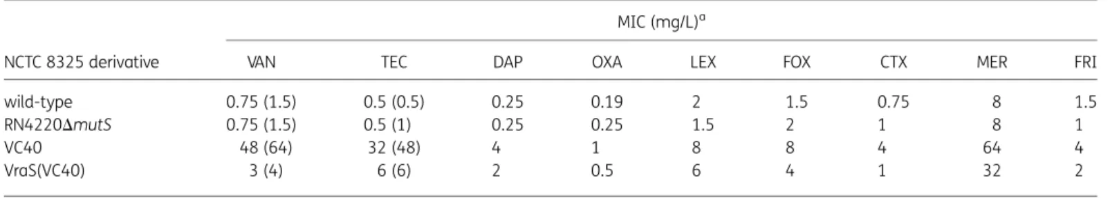
Table 1. Susceptibility testing of S. aureus NCTC 8325 derivatives used in this study
NCTC 8325 derivative
MIC (mg/L)a
VAN TEC DAP OXA LEX FOX CTX MER FRI
wild-type 0.75 (1.5) 0.5 (0.5) 0.25 0.19 2 1.5 0.75 8 1.5
RN4220DmutS 0.75 (1.5) 0.5 (1) 0.25 0.25 1.5 2 1 8 1
VC40 48 (64) 32 (48) 4 1 8 8 4 64 4
VraS(VC40) 3 (4) 6 (6) 2 0.5 6 4 1 32 2
VAN, vancomycin; TEC, teicoplanin; DAP, daptomycin; OXA, oxacillin; LEX, cefalexin; FOX, cefoxitin; CTX, cefotaxime; MER, mersacidin; FRI, friulimicin. aMICs in MH; numbers in parentheses represent the vancomycin and teicoplanin MICs in BHI. MICs were read after 24 and 48 h considering the slow growth of strain VC40. Noteworthy, MIC values for NCTC 8325 wild-type, RN4220DmutS and NCTC 8325 VraS(VC40) were unchanged after 24 and 48 h. MIC values represent the mean of at least four independent determinations.
Mutations in vraS affect antibiotic resistance in S. aureus
The L114S and D242G exchanges in VraS(VC40) were
reconsti-tuted into the genome of S. aureus NCTC 8325, the parental
background of strain VC40. Although not reaching the high
MIC values of S. aureus VC40, the resulting mutant, S. aureus
NCTC 8325 VraS(VC40), was characterized by an increased MIC
of vancomycin (MIC of 3 mg/L in MH broth and 4 mg/L in BHI
broth), teicoplanin (MIC of 6 mg/L in MH and BHI broths)
as well as daptomycin (MIC of 2 mg/L) (Table
1
). Noteworthy,
the presence of VraS(VC40) also affected the susceptibilities to
the cell wall-active antibiotics mersacidin and different
b-lactams (Table
1
). Reconstitution of the I544M exchange in
WalK was also attempted; however, despite repeated
experi-ments, the correct mutant was not obtained.
VraS(VC40) accounts for an increased expression
of the VraSR regulon
To study the gene regulatory impact of VraS(VC40) in strain NCTC
8325, we measured the relative transcript quantities of the vraS,
sgtB and lytM genes by qRT– PCR and, indeed, these genes were
more highly expressed in the S. aureus NCTC 8325 VraS(VC40)
mutant compared with its parent strain (Figure
1
), indicating an
increased expression of the VraSR regulon. Noteworthy, we did
not observe a significant alteration of atlA expression in strain
NCTC 8325 VraS(VC40).
VraS(VC40) contributes to an altered phenotype
of S. aureus
In growth kinetic experiments, a strongly reduced growth rate of
S. aureus VC40 was observed compared with its parent strain
(Figure S1). The NCTC 8325 VraS(VC40) mutant was also
charac-terized by a slightly lowered growth rate (Figure S1), indicating
that VraS(VC40) might to some extent contribute to the low
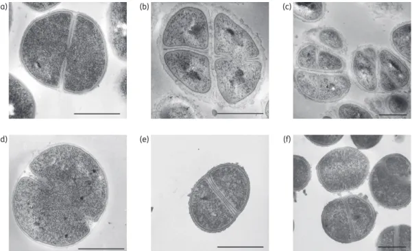
growth rate observed for strain VC40. To gain further insights
into the morphologies of strain VC40 and the NCTC 8325
VraS(VC40) mutant, we performed TEM of exponentially growing
cells (Figure
2
). Compared with the parent strains S. aureus NCTC
8325 (Figure
2
d) and RN4220DmutS (Figure
2
a), S. aureus VC40
was characterized by a significantly thickened cell wall with a
roughened surface and irregular appearance (Figure
2
b and c).
Furthermore, a pseudomulticellular phenotype probably caused
by a retarded cell separation was observed in strain VC40, since
new septa were formed before the cell had completed the
previ-ous cell division event. Similarly, the thickness of the cell wall was
also increased in strain NCTC 8325 VraS(VC40) (Figure
2
e and f).
Although the cell wall of the NCTC 8325 VraS(VC40) mutant also
displayed a more roughened and uneven surface, the
pseudomul-ticellular phenotype of strain VC40 was not observed here. The
measured cell wall thicknesses were 16.71 (+3.00) nm for strain
NCTC 8325 wild-type, 16.97 (+3.18) nm for strain RN4220DmutS,
62.36 (+13.98) nm for strain VC40 and 38.25 (+11.31) nm for the
NCTC 8325 VraS(VC40) mutant.
S. aureus strains VC40 and NCTC 8325 VraS(VC40) show
significantly reduced autolytic activities and an altered
susceptibility to lysostaphin
The autolytic activities of the S. aureus strains VC40 and NCTC
8325 VraS(VC40) were characterized by Triton X-100-induced
autolysis assays and were compared with their respective
paren-tal strains. Here, strains VC40 and NCTC 8325 VraS(VC40) both
exhibited a similarly severe reduction of autolytic activity
(Figure S2a). In addition, we determined lysostaphin-induced
lysis of the different strains over time (Figure S2b). Here, strains
RN4220DmutS and NCTC 8325 showed full susceptibility to
lysos-taphin treatment. Strain NCTC 8325 VraS(VC40) initially displayed
reduced cell lysis in the presence of lysostaphin, but the majority
of the cells were lysed after 60 min. Strikingly, strain VC40 was
char-acterized by a significantly increased resistance to lysostaphin.
Discussion
Genome sequencing of the vancomycin-resistant laboratory
mutant S. aureus VC40 identified SNPs in the histidine kinase
genes vraS and walK and we further investigated the impact of
VraS(VC40) on gene regulation, phenotypic characteristics and
antibiotic resistance. Mutations in regulatory genes are specifically
important, since these changes may alter the expression patterns
of large regulons and thus help the bacteria to adjust certain
Table 2. DNA microarray-based expression ratios of cell wall stress stimulon genes including those under VraSR control26,27of untreated S. aureus strains VC40 versus RN4220DmutS
ORF
IDa Gene Comments
Ratio VC40/ DmutS 00427 sle1 N-acetylmuramoyl-L-alanine amidase 0.90
00994 atlA bifunctional autolysin 2.09
00998 fmtA autolysis and methicillin resistance-related protein
2.07 01838 htrA serine protease, heat-shock protein
homologue
2.02 01972 prsA peptidyl-prolyl cis/trans isomerase 0.32 02012 sgtB monofunctional glycosyltransferase 4.67 02099 vraS two-component sensor histidine
kinase
1.66 02100 yvqF/vraT conserved hypothetical protein 1.71
02101 hypothetical protein 2.12
02112 conserved hypothetical protein 1.71 02365 murZ UDP-N-acetylglucosamine
enolpyruvyl transferase
1.92 02583 similar to lyt divergon expression
attenuator LytR
1.90 02635 tcaA teicoplanin resistance-associated
protein
2.46
02723 glycerate kinase 4.09
02724 hypothetical protein 4.17
02811 relP putative (p)ppGpp synthetase 5.10 02872 cwrA CwrA protein, cell wall responsive
for antibiotics
21.32
aORF IDs correspond to ‘SAOUHSC_’ locus tags of S. aureus NCTC 8325 (NC_007795).
8.0E+06 3.2 fold 2.1 fold vraS 6.0E+06 4.0E+06 2.0E+06 C o pies vr aS /10 6 copies gyrB 0.0E+00 VC40 8325 wt 8325 VraS(VC40) RN4220 DmutS VC40 RN4220 DmutS 8.0E+06 5.1 fold 4.7 fold sgtB 6.0E+06 4.0E+06 2.0E+06 C o pies sgtB /10 6 copies gyrB 0.0E+00 VC40 8325 wt 8325 VraS(VC40) RN4220 DmutS 8.0E+06 4.5 fold 3.1 fold lytM 6.0E+06 4.0E+06 2.0E+06 C o pies lytM /10 6 copies gyrB 0.0E+00 8325 wt 8325 VraS(VC40) 2.0E+07 3.4 fold 1.1 fold atlA 1.2E+07 1.6E+07 8.0E+06 4.0E+06 C o pies atlA /10 6 copies gyrB 0.0E+00 VC40 8325 wt 8325 VraS(VC40) RN4220 DmutS 1.2E+07 6.8 fold sceD 6.0E+06 8.0E+06 1.0E+07 4.0E+06 2.0E+06 C o pies sceD /10 6 copies gyrB 0.0E+00 VC40 RN4220 DmutS 1.2E+07 7.3 fold ssaA 1.0E+07 4.0E+06 6.0E+06 8.0E+06 2.0E+06 C o pies ssaA /10 6 copies gyrB 0.0E+00 VC40 RN4220 DmutS
Figure 1. Gene expression analyses of VraSR and WalKR regulon members. Transcript levels of vraS, sgtB, lytM, atlA, sceD and ssaA were determined by qRT–PCR in relation to gyrB expression. Values represent the mean of results obtained for two independently grown cultures for each strain and are representative for at least two separate qRT–PCR experiments.
(b) (c)
(a)
(e) (f)
(d)
Figure 2. Morphological analyses by transmission electron microscopy. Ultrathin sections of exponentially grown S. aureus strains RN4220DmutS (a), VC40 (b and c), NCTC 8325 wild-type (d) and NCTC 8325 VraS(VC40) (e and f) were visualized. Scale bars represent 0.5 mm.
physiological networks that allow them to counteract antibiotic
stress. In S. aureus VC40, the vraS gene harbours two
non-synonymous SNPs that lead to the amino acid exchanges L114S
and D242G in VraS. The introduction of these mutations into a
susceptible parental background resulted in noticeable MIC
increases of several cell wall-active antibiotics, including
vanco-mycin, teicoplanin and daptomycin. VraS is part of the VraSR
two-component regulatory system (TCRS) and conveys the so-called
cell wall stress response, which coordinates the regulation of
mostly cell wall biosynthesis-related genes such as sgtB or the
vraSR genes themselves.
26–29Various mutations in the vraSR/
yvqF(vraT) locus have been reported in VISA strains
30–33and,
interestingly, the location of the SNPs in vraSR/yvqF in different
strains is very diverse and there seems to be no specific hotspot
for mutations in certain domains of these genes.
Only a few of the SNPs in vraSR/yvqF have been further
investi-gated to elucidate their putative impact on gene regulation and
antibiotic resistance development so far.
33,34In our study,
VraS(VC40) was sufficient to convert the vancomycin-susceptible
strain S. aureus NCTC 8325 into a strain with a VISA type of
resist-ance. An earlier study described the effect of the I5N amino acid
exchange in VraS of strain Mu50.
34Here, the introduction of the
respective VraS(I5N) into the S. aureus Mu50V background
com-pared with the VraS wild-type showed a lower resistance increase
(MIC increase from 3.5 to 4.5 mg/L) compared with the effect that
is shown by VraS(VC40) in the NCTC 8325 background (MIC
increase from 0.75 to 3 mg/L).
34Furthermore, a study by
Gardete et al.
33detected a Y220C amino acid exchange in YvqF
in addition to mutations in vraG, yycH and lspA in strain SG-R, a
clinical VISA isolate of the USA300 clone, and identified YvqF as
a negative regulator of vraSR. However, a direct impact of
YvqF(Y220C) on vancomycin resistance could not be determined
by introducing the mutated yvqF gene on a plasmid into the
vancomycin-susceptible strain SG-S, which still harbours a
chromosomal copy of the wild-type yvqF gene. In contrast, the
introduction of a plasmid with the wild-type version of yvqF into
strain SG-R led to a decreased vancomycin MIC. In that study, a
chromosomal reconstitution of the yvqF mutation into strain
SG-S was not shown.
33Noteworthy, since the mutations in vraS
or yvqF mentioned above have each been reintroduced into
differ-ent strain backgrounds, a strain dependency of the observed
effects cannot be excluded.
In our study, the VraSR-controlled cell wall stress stimulon
26,27was characterized by a considerably increased expression in
S. aureus NCTC 8325 VraS(VC40) as well as in S. aureus VC40.
Remarkably, the lytM gene also showed a higher transcript level
in the NCTC 8325 VraS(VC40) mutant, which confirms the
state-ment by Gardete et al.
33that lytM appears to be an additional
member of the VraSR regulon. Obviously, the increased expression
of members of the VraSR regulon contributes to the observed
reduced susceptibility to vancomycin, teicoplanin, daptomycin,
mersacidin and oxacillin in our study. Noteworthy, these
antibio-tics have also been described to induce the VraSR
regu-lon.
16,26,28,35,36In this context, an accelerated peptidoglycan
biosynthesis, which may be mediated by an activated VraSR
reg-ulon, is likely to account for this decrease in antibiotic
susceptibil-ity by supplying an increased number of cell wall precursors or by a
faster incorporation of these precursors into the nascent
peptido-glycan meshwork, eventually also leading to a thickening of the
cell wall. Although commonly down-regulated upon vancomycin
treatment, we did not observe a significant alteration of atlA
expression in strain NCTC 8325 VraS(VC40), which is in accordance
with previous observations that the regulation of atlA is most
probably not mediated by VraSR.
26Of note, mutations in the
vraSR/yvqF genes are predominantly discussed regarding their
impact on vancomycin resistance. However, we detected a strong
influence of VraS(VC40) on daptomycin non-susceptibility, as the
daptomycin MIC for the NCTC 8325 VraS(VC40) mutant reached
2 mg/L. It has to be noted that despite the effect of the
vraS(VC40) mutations as well as the observation of Mehta
et al.
37concerning the contribution of deletion or up-regulation
of vraSR in daptomycin-non-susceptible strains, which share a
sig-nificant impact on daptomycin susceptibility, to our knowledge
daptomycin treatment has not been described to select for
muta-tions in the vraSR/yvqF operon so far.
An increased cell wall thickness as observed in strains VC40
and NCTC 8325 VraS(VC40) is likely to be a major contributor to
the increased resistance to glycopeptides and daptomycin of
these strains. Here, a cell wall thickness of 62 nm was reached
in strain S. aureus VC40 and 38 nm in strain NCTC 8325
VraS(VC40). Thickening of the cell wall has been previously
described as a common phenotypic feature of VISA strains
6,38,39and has also been related to daptomycin cross-resistance;
40how-ever, the cell wall thickness of the typical VISA strain grown in BHI
does not exceed 35 nm.
39Noteworthy, up-regulation of the VraSR
regulon and concomitant thickening of the cell wall have also
been reported in daptomycin-non-susceptible S. aureus strains
that were generated by serial daptomycin selection and that
add-itionally showed a heterogeneous VISA phenotype.
41Our study
supports a prominent role of the VraSR regulon in the build-up
of a thickened cell wall in S. aureus. Although the up-regulation
of the cell wall biosynthesis machinery by VraSR and/or a possible
deregulation of the bacterial autolytic system by the exchange
present in WalKR in strain VC40 would both provide plausible
explanations for the accumulation of excess cell wall material,
the explicit mechanism that leads to cell wall thickening remains
unclear. Interestingly, the pseudomulticellular phenotype of
strain VC40 was not observed in strain NCTC 8325 VraS(VC40),
indicating that VraSR is at least not solely responsible for this
phe-nomenon and that probably more than one mutation may
even-tually lead to this phenotype in S. aureus VC40.
Although many of the autolysin genes including atlA were
more highly expressed in strain VC40, Triton X-100-induced
autolysis of strain VC40 was significantly decreased. This
phenom-enon has also been observed in the VISA strain JH9 and the
vancomycin-resistant mutant VP32;
27,42,43however, the reason
for this remained elusive. The similarly reduced autolytic activities
of strain NCTC 8325 VraS(VC40) and strain VC40 might indicate
that an activated VraSR regulon generates modifications in the
peptidoglycan structure of these strains, eventually leading to
an increased resistance against Triton X-100-induced autolysis.
In accordance with this assumption, it has previously been
sug-gested that quantitative (or qualitative) changes in the teichoic
acids of VISA strains might decrease peptidoglycan degradation
by autolytic enzymes.
43The clearly increased cell wall thickness
of strain VC40 most probably accounts for the more severe
decrease in lysostaphin susceptibility compared with strain NCTC
8325 VraS(VC40) and it may also play a role in the autolytic
behav-iour of the affected strains, but it is most likely not the sole factor
in resistance against Triton X-100-induced autolysis. Since the
JAC
strains in our study and other VISA strains share the common
phenotype of a reduced autolytic activity,
6,43–47this feature is
likely important for the development of resistance.
Strain NCTC 8325 VraS(VC40) did not reach the measured MICs
and cell wall thickness of strain VC40, indicating that additional
mutations add to the high level of resistance in the latter strain.
In addition to the role of VraS(VC40) in contributing to the
devel-opment of glycopeptide and daptomycin resistance described
above, we identified an SNP in the walK gene leading to an
I544M amino acid exchange located in the HATPase_c domain
of the histidine kinase in strain VC40. The WalKR TCRS is an
essen-tial master regulator in cell wall metabolism and homeostasis.
48Deregulation of this system may have important consequences
for the bacteria and might enforce further adaptive changes, i.e.
concerning membrane or cell wall composition. Microarray
profil-ing revealed an increased expression of the WalKR regulon in
S. aureus VC40 including the ssaA, lytM, sceD and atlA autolysin
genes, which have been described to be under direct positive
WalR control.
49,50Mutations in the walKR operon have previously
been linked to vancomycin or daptomycin non-susceptibility in
several studies;
31,32,51–57however, only a few of these have
been analysed concerning their effect on antibiotic resistance,
which might also be related to the complexity of genetic
manipu-lation of the walKR locus due to its essential nature. Thus, it has
remained unclear so far if an induced or a repressed WalKR
regulon might finally cause a development of antibiotic
resistance.
27,32,58Other amino acid exchanges observed in strain VC40 included
MprF(H224Y) that may alter the activity of this protein and thus
support daptomycin cross-resistance. In this context, mprF showed
higher expression levels in VISA compared with
vancomycin-susceptible S. aureus (VSSA) and decreased levels of mprF have
also been correlated with increased vancomycin
susceptibil-ity.
27,59–61Furthermore, increased expression or mutation of
mprF have also been reported to occur frequently in
daptomycin-non-susceptible S. aureus isolates
54,57,62,63and depletion of mprF
was described to restore daptomycin susceptibility in
daptomycin-resistant S. aureus isolates.
64Daptomycin cross-resistance of strain
VC40 might be further supported by RpoD(D201N), which is the
housekeeping sigma factor A (s
A/s
70). Noteworthy, mutations in
rpoB and rpoC have been correlated with reduced vancomycin
and daptomycin susceptibility in S. aureus
54,57,65and the VISA
strains Mu50 and JH9 also harbour SNPs in the rpoB and rpoD or
rpoB and rpoC genes, respectively.
30,31In conclusion, our results prove the important role of the
histi-dine kinase VraS in the physiology of the bacterial cell envelope. In
our study, the amino acid exchanges in VraS led to an increased
expression of the associated regulon, thereby mediating an
increased thickening of the cell wall, alterations in autolytic
prop-erties and eventually resulted in significantly higher MICs of the
glycopeptides vancomycin and teicoplanin as well as
cross-resistance to daptomycin. Thus, VraS can act as an important
switch to turn on antibiotic resistance in the S. aureus NCTC
8325 background.
Funding
This work was supported by the Bundesministerium fu¨r Bildung und Forschung, Network PathoGenoMik-Plus (grant PTJ-BIO/0313801F to GB); the German Research Foundation (DFG; grant Bi 504/8-3 to GB); the
BONFOR program of the Medical Faculty, University of Bonn; and the Fonds National de la Recherche, Luxembourg (grant AFR PHD-09-114 to AB).
Transparency declarations
None to declare.
Supplementary data
Figures S1 and S2 and Tables S1 to S3 are available as Supplementary data at JAC Online (http://jac.oxfordjournals.org/).
References
1 DeLeo FR, Chambers HF. Reemergence of antibiotic-resistant Staphylococcus aureus in the genomics era. J Clin Invest 2009; 119: 2464– 74.
2 Ko¨ck R, Becker K, Cookson B et al. Methicillin-resistant Staphylococcus aureus (MRSA): burden of disease and control challenges in Europe. Euro Surveill 2010; 15: 19688.
3 Moellering RCJ. MRSA: the first half century. J Antimicrob Chemother 2012; 67: 4 –11.
4 Liu C, Bayer A, Cosgrove SE et al. Clinical practice guidelines by the Infectious Diseases Society of America for the treatment of methicillin-resistant Staphylococcus aureus infections in adults and children. Clin Infect Dis 2011; 52: e18–55.
5 Scherl A, Francois P, Charbonnier Y et al. Exploring glycopeptide-resistance in Staphylococcus aureus: a combined proteomics and tran-scriptomics approach for the identification of resistance-related markers. BMC Genomics 2006; 7: 296.
6 Howden BP, Davies JK, Johnson PD et al. Reduced vancomycin susceptibility in Staphylococcus aureus, including vancomycin-intermediate and heteroge-neous vancomycin-intermediate strains: resistance mechanisms, laboratory detection, and clinical implications. Clin Microbiol Rev 2010; 23: 99–139. 7 Ruef C. Epidemiology and clinical impact of glycopeptide resistance in Staphylococcus aureus. Infection 2004; 32: 315– 27.
8 Tenover FC, Sinner SW, Segal RE et al. Characterisation of a Staphylococcus aureus strain with progressive loss of susceptibility to vancomycin and daptomycin during therapy. Int J Antimicrob Agents 2009; 33: 564–8.
9 Tsiodras S, Gold HS, Sakoulas G et al. Linezolid resistance in a clinical iso-late of Staphylococcus aureus. Lancet 2001; 358: 207– 8.
10 Bro¨tz-Oesterhelt H, Sass P. Postgenomic strategies in antibacterial drug discovery. Future Microbiol 2010; 5: 1553–79.
11 Sass P, Bro¨tz-Oesterhelt H. Bacterial stress responses to antimicrobial agents. In: Wong HC, ed. Stress Responses in Foodborne Microorganisms. Hauppauge, NY: Nova Science, 2012; 131–72.
12 Sass P, Berscheid A, Jansen A et al. Genome sequence of Staphylococcus aureus VC40, a vancomycin- and daptomycin-resistant strain, to study the genetics of development of resistance to currently applied last-resort antibiotics. J Bacteriol 2012; 194: 2107–8.
13 Schaaff F, Reipert A, Bierbaum G. An elevated mutation frequency favors development of vancomycin resistance in Staphylococcus aureus. Antimicrob Agents Chemother 2002; 46: 3540– 8.
14 Perichon B, Courvalin P. Synergism between b-lactams and glycopep-tides against VanA-type methicillin-resistant Staphylococcus aureus and heterologous expression of the vanA operon. Antimicrob Agents Chemother 2006; 50: 3622– 30.
15 Hiramatsu K. Vancomycin resistance in staphylococci. Drug Resist Updates 1998; 1: 135–50.
16 Sass P, Jansen A, Szekat C et al. The lantibiotic mersacidin is a strong inducer of the cell wall stress response of Staphylococcus aureus. BMC Microbiol 2008; 8: 186.
17 Charbonnier Y, Gettler B, Francois P et al. A generic approach for the design of whole-genome oligoarrays, validated for genomotyping, dele-tion mapping and gene expression analysis on Staphylococcus aureus. BMC Genomics 2005; 6: 95.
18 Koessler T, Francois P, Charbonnier Y et al. Use of oligoarrays for char-acterization of community-onset methicillin-resistant Staphylococcus aur-eus. J Clin Microbiol 2006; 44: 1040–8.
19 Renzoni A, Barras C, Francois P et al. Transcriptomic and functional analysis of an autolysis-deficient, teicoplanin-resistant derivative of methicillin-resistant Staphylococcus aureus. Antimicrob Agents Chemother 2006; 50: 3048–61.
20 Talaat AM, Howard ST, Hale W et al. Genomic DNA standards for gene expression profiling in Mycobacterium tuberculosis. Nucleic Acids Res 2002; 30: e104.
21 Arnaud M, Chastanet A, Debarbouille M. New vector for efficient allelic replacement in naturally nontransformable, low-GC-content, gram-positive bacteria. Appl Environ Microbiol 2004; 70: 6887–91.
22 Venable JH, Coggeshall R. A simplified lead citrate stain for use in elec-tron microscopy. J Cell Biol 1965; 25: 407–8.
23 Nair D, Memmi G, Hernandez D et al. Whole-genome sequencing of Staphylococcus aureus strain RN4220, a key laboratory strain used in viru-lence research, identifies mutations that affect not only viruviru-lence factors but also the fitness of the strain. J Bacteriol 2011; 193: 2332– 5. 24 Berscheid A, Sass P, Weber-Lassalle K et al. Revisiting the genomes of the Staphylococcus aureus strains NCTC 8325 and RN4220. Int J Med Microbiol 2012; 302: 84– 7.
25 Gillaspy AF, Worrell V, Orvis J et al. The Staphylococcus aureus NCTC 8325 genome. In: Fischetti V, Novick R, Ferretti J et al., eds. Gram Positive Pathogens. Washington, DC: ASM Press, 2006; 381–412.
26 Kuroda M, Kuroda H, Oshima T et al. Two-component system VraSR positively modulates the regulation of cell-wall biosynthesis pathway in Staphylococcus aureus. Mol Microbiol 2003; 49: 807– 21.
27 McAleese F, Wu SW, Sieradzki K et al. Overexpression of genes of the cell wall stimulon in clinical isolates of Staphylococcus aureus exhibiting vancomycin-intermediate-S. aureus-type resistance to vancomycin. J Bacteriol 2006; 188: 1120– 33.
28 Utaida S, Dunman PM, Macapagal D et al. Genome-wide transcrip-tional profiling of the response of Staphylococcus aureus to cell-wall-active antibiotics reveals a cell-wall-stress stimulon. Microbiology 2003; 149: 2719– 32.
29 Sobral RG, Jones AE, Des Etages SG et al. Extensive and genome-wide changes in the transcription profile of Staphylococcus aureus induced by modulating the transcription of the cell wall synthesis gene murF. J Bacteriol 2007; 189: 2376– 91.
30 Ohta T, Hirakawa H, Morikawa K et al. Nucleotide substitutions in Staphylococcus aureus strains, Mu50, Mu3, and N315. DNA Res 2004; 11: 51 –6.
31 Mwangi MM, Wu SW, Zhou Y et al. Tracking the in vivo evolution of mul-tidrug resistance in Staphylococcus aureus by whole-genome sequencing. Proc Natl Acad Sci USA 2007; 104: 9451– 6.
32 Howden BP, McEvoy CR, Allen DL et al. Evolution of multidrug resistance during Staphylococcus aureus infection involves mutation of the essential two component regulator WalKR. PLoS Pathog 2011; 7: e1002359. 33 Gardete S, Kim C, Hartmann BM et al. Genetic pathway in acquisition and loss of vancomycin resistance in a methicillin resistant Staphylococcus aureus (MRSA) strain of clonal type USA300. PLoS Pathog 2012; 8: e1002505.
34 Cui L, Neoh HM, Shoji M et al. Contribution of vraSR and graSR point mutations to vancomycin resistance in vancomycin-intermediate Staphylococcus aureus. Antimicrob Agents Chemother 2009; 53: 1231–4. 35 Muthaiyan A, Silverman JA, Jayaswal RK et al. Transcriptional profiling reveals that daptomycin induces the Staphylococcus aureus cell wall stress stimulon and genes responsive to membrane depolarization. Antimicrob Agents Chemother 2008; 52: 980–90.
36 Dengler V, Meier PS, Heusser R et al. Induction kinetics of the Staphylococcus aureus cell wall stress stimulon in response to different cell wall active antibiotics. BMC Microbiol 2011; 11: 16.
37 Mehta S, Cuirolo AX, Plata KB et al. VraSR two-component regulatory system contributes to mprF-mediated decreased susceptibility to daptomycin in in vivo-selected clinical strains of methicillin-resistant Staphylococcus aureus. Antimicrob Agents Chemother 2012; 56: 92 –102. 38 Cui L, Murakami H, Kuwahara-Arai K et al. Contribution of a thickened cell wall and its glutamine nonamidated component to the vancomycin resistance expressed by Staphylococcus aureus Mu50. Antimicrob Agents Chemother 2000; 44: 2276– 85.
39 Cui L, Ma X, Sato K et al. Cell wall thickening is a common feature of vancomycin resistance in Staphylococcus aureus. J Clin Microbiol 2003; 41: 5 –14.
40 Cui L, Tominaga E, Neoh HM et al. Correlation between reduced daptomycin susceptibility and vancomycin resistance in vancomycin-intermediate Staphylococcus aureus. Antimicrob Agents Chemother 2006; 50: 1079– 82.
41 Camargo IL, Neoh HM, Cui L et al. Serial daptomycin selection gener-ates daptomycin-nonsusceptible Staphylococcus aureus strains with a heterogeneous vancomycin-intermediate phenotype. Antimicrob Agents Chemother 2008; 52: 4289– 99.
42 Mongodin E, Finan J, Climo MW et al. Microarray transcription analysis of clinical Staphylococcus aureus isolates resistant to vancomycin. J Bacteriol 2003; 185: 4638– 43.
43 Sieradzki K, Tomasz A. Alterations of cell wall structure and metabol-ism accompany reduced susceptibility to vancomycin in an isogenic series of clinical isolates of Staphylococcus aureus. J Bacteriol 2003; 185: 7103– 10.
44 Sieradzki K, Tomasz A. Inhibition of cell wall turnover and autolysis by vancomycin in a highly vancomycin-resistant mutant of Staphylococcus aureus. J Bacteriol 1997; 179: 2557–66.
45 Boyle-Vavra S, Carey RB, Daum RS. Development of vancomycin and lysostaphin resistance in a methicillin-resistant Staphylococcus aureus iso-late. J Antimicrob Chemother 2001; 48: 617–25.
46 Koehl JL, Muthaiyan A, Jayaswal RK et al. Cell wall composition and de-creased autolytic activity and lysostaphin susceptibility of glycopeptide-intermediate Staphylococcus aureus. Antimicrob Agents Chemother 2004; 48: 3749– 57.
47 Howden BP, Johnson PD, Ward PB et al. Isolates with low-level vanco-mycin resistance associated with persistent methicillin-resistant Staphylococcus aureus bacteremia. Antimicrob Agents Chemother 2006; 50: 3039–47.
48 Dubrac S, Bisicchia P, Devine KM et al. A matter of life and death: cell wall homeostasis and the WalKR (YycGF) essential signal transduction pathway. Mol Microbiol 2008; 70: 1307–22.
49 Dubrac S, Gomperts Boneca I, Poupel O et al. New insights into the WalK/WalR (YycG/YycF) essential signal transduction pathway reveal a major role in controlling cell wall metabolism and biofilm formation in Staphylococcus aureus. J Bacteriol 2007; 189: 8257–69.
50 Dubrac S, Msadek T. Identification of genes controlled by the essential YycG/YycF two-component system of Staphylococcus aureus. J Bacteriol 2004; 186: 1175– 81.
51 Hafer C, Lin Y, Kornblum J et al. Contribution of selected gene mutations to resistance in clinical isolates of vancomycin-intermediate Staphylococcus aureus. Antimicrob Agents Chemother 2012; 56: 5845–51. 52 Fischer A, Yang SJ, Bayer AS et al. Daptomycin resistance mechanisms in clinically derived Staphylococcus aureus strains assessed by a combined transcriptomics and proteomics approach. J Antimicrob Chemother 2011; 66: 1696–711.
53 Shoji M, Cui L, Iizuka R et al. walK and clpP mutations confer reduced vancomycin susceptibility in Staphylococcus aureus. Antimicrob Agents Chemother 2011; 55: 3870– 81.
54 Friedman L, Alder JD, Silverman JA. Genetic changes that correlate with reduced susceptibility to daptomycin in Staphylococcus aureus. Antimicrob Agents Chemother 2006; 50: 2137– 45.
55 Baltz RH. Daptomycin: mechanisms of action and resistance, and bio-synthetic engineering. Curr Opin Chem Biol 2009; 13: 144–51.
56 Patel D, Husain M, Vidaillac C et al. Mechanisms of in-vitro-selected daptomycin-non-susceptibility in Staphylococcus aureus. Int J Antimicrob Agents 2011; 38: 442– 6.
57 Mishra NN, Rubio A, Nast CC et al. Differential adaptations of methicillin-resistant Staphylococcus aureus to serial in vitro passage in daptomycin: evolution of daptomycin resistance and role of membrane carotenoid content and fluidity. Int J Microbiol 2012; 2012: 683450. 58 Jansen A, Tu¨rck M, Szekat C et al. Role of insertion elements and yycFG in the development of decreased susceptibility to vancomycin in Staphylococcus aureus. Int J Med Microbiol 2007; 297: 205– 15.
59 Ruzin A, Severin A, Moghazeh SL et al. Inactivation of mprF affects vancomycin susceptibility in Staphylococcus aureus. Biochim Biophys Acta 2003; 1621: 117–21.
60 Wootton M, MacGowan AP, Walsh TR. Expression of tcaA and mprF and glycopeptide resistance in clinical glycopeptide-intermediate Staphylococcus aureus (GISA) and heteroGISA strains. Biochim Biophys Acta 2005; 1726: 326–7.
61 Sass P, Bierbaum G. Native graS mutation supports the susceptibility of Staphylococcus aureus strain SG511 to antimicrobial peptides. Int J Med Microbiol 2009; 299: 313–22.
62 Yang SJ, Xiong YQ, Dunman PM et al. Regulation of mprF in daptomycin-nonsusceptible Staphylococcus aureus strains. Antimicrob Agents Chemother 2009; 53: 2636– 7.
63 Boyle-Vavra S, Jones M, Gourley BL et al. Comparative genome sequencing of an isogenic pair of USA800 clinical methicillin-resistant Staphylococcus aureus isolates obtained before and after daptomycin treatment failure. Antimicrob Agents Chemother 2011; 55: 2018– 25.
64 Rubio A, Conrad M, Haselbeck RJ et al. Regulation of mprF by antisense RNA restores daptomycin susceptibility to daptomycin-resistant isolates of Staphylococcus aureus. Antimicrob Agents Chemother 2011; 55: 364– 7.
65 Cui L, Isii T, Fukuda M et al. An RpoB mutation confers dual heteroresis-tance to daptomycin and vancomycin in Staphylococcus aureus. Antimicrob Agents Chemother 2010; 54: 5222– 33.