CD8+ cytolytic T cell clones derived against
the Plasmodium yoelii circumsporozoite
protein protect against malaria
Mauricio M. Rodrigues, Anne-Sophie Cordey1, Gladys Arreaza,
Giampetro Corradin1, Pedro Romero2, Janet L. Maryanski2,
Ruth S. Nussenzweig, and Fidel Zavala
Department of Medical and Molecular Parasitology, New York University School of Medicine, New York, NY 10010, USA
institute of Biochemistry, University of Lausanne, and 2Ludwig Institute for Cancer Research, Lausanne
Branch, 1066 Epalinges, Switzerland
Key words: cytotoxic epitopes, malaria immunity, CS protein
Abstract
Immunization of BALB/c mice with radiation-attenuated Plasmodium yoelll sporozoites induces cytotoxic T lymphocytes (CTL) specific for an epitope located within the amino acid sequence 2 7 7 - 2 8 8 of the P. yoelll circumsporozoite (CS) protein. Several CD8+ CTL clones were derived from the spleen cells of sporozolte-immunlzed mice, all displaying an apparently Identical epitope specificity. All the clones Induced high levels of cytolysls In vitro upon exposure to peptide-Incubated MHC-compatlble target cells. The adoptive transfer of two of these clones conferred complete protection against sporozotte challenge to naive mice. This protection Is species and stage specific. Using P. yoelll specific rlbosomal RNA probes to monitor the In vivo effects of the CTL clones, we found that their target was the Intrahepatocytlc stage of the parasite. The
protective clones completely Inhibited the development of the liver stages of P. yoelll. Some CTL clones were only partially Inhibitory In vivo, while others failed completely to alter liver stage development and to confer any detectable degree of protection. The elucidation of the effector mechanism of this CTL mediated protection against rodent malaria should facilitate the design of an effective malaria vaccine. From a broader perspective this model may provide further Insight Into the mechanlsm(s) of CTL mediated killing of intracellular non-viral pathogens In general.
Introduction
Protective immunity against rodent, simian and human malaria can be achieved by immunization with radiation-attenuated sporozoites (1). This protection appears to be mediated by a combination of humoral and T-cell mediated mechanisms (2,3). The extent to which any of these individual immune mechanisms can confer protection, in the absence of the others, remains to be determined.
Epitopes located within the circumsporozoite (CS) protein that are recognized by CD8+ cytolytic T lymphocytes (CTL) have been defined for several species of Plasmodia (3 - 6). Immuniza-tion of BALB/c mice with irradiated Plasmodium berghei sporozoites induces CD8+ CTL which recognize a synthetic peptide comprising amino acids 249-260 (NDDSYPSAEKI) of its CS protein (3). Immunization with irradiated Plasmodium yoelii sporozoites of BALB/c mice also induces CD8+ CTL, which recognize an epitope contained in a peptide with a sequence
(P. yoelii CS 277 - 288, NEDSYVPSAEQI) highly homologous to that of the P. berghei CTL epitope. Both of these CS peptides are presented to T cells in the context of the major histo-compatjbility complex (MHC) H-2 kd molecule (6). Another study
(5) defined a similar, overlapping P. yoelii CS epitope (CS 281-296).
It has been established that the adoptive transfer of cloned CD8+ CTLs specific for the P. berghei CS epitope can confer complete protection against sporozoite-induced infection (3). However, it has been suggested that CD8+ T cells against the
P. yoelii CS protein may not have a similar protective role (5). The availability of cloned CD8+ CTL, specific for the P. yoelii cytotoxic CS epitope, did permit us to establish the broader validity of the protective role of anti-CS CD8+ CTL in rodent malaria and, also, determine their specificity. Using a recently described method for measuring plasmodial ribosomal RNA
Correspondence to: F. Zavala, 341 East 25th Street New York, NY 10010, USA
sequences in infected mouse liver, we evaluated quantitatively the protective in vivo activity of the CTL clones.
Methods
Parasites and animals
Plasmodium yoelii (17X NL strain) and P. berghei (NK 65 strain)
were maintained by repeated cyclic passage of the parasites through Anopheles stephensi mosquitoes, and BALB/c mice or hamsters, respectively. BALB/c mice and hamsters were purchased from Jackson Laboratories, Bar Harbor, ME and from Charles River Laboratories, Wilmington, MA, respectively. Sporozoites were collected by dissecting the mosquito salivary glands in medium 199, - 2 weeks after the infective blood meal. Female BALB/c mice aged 4 - 8 weeks were used for the immunological studies. Mice were challenged intravenously with the indicated dose of viable sporozoites. Their parasitemia was monitored daily by peripheral blood smears obtained from the 3rd to the 10th day after challenge.
Peptides
Two peptides containing the CTL epitopes of the CS protein of
P. berghei (Pb 2 4 9 - 2 6 0 NDDSYIPSAEKI) and of P. yoelii (Py
2 7 7 - 2 8 8 NDESYVPSAEQI) were synthesized as described (3).
Cell culture
As culture medium we used DMEM - high glucose (Grand Island Biological Co., Long Island, NY) supplemented with 0.2 g/l of arginine (Calbiochem Co., La Jolla, CA), 0.036 g/l of L-asparagine (GIBCO), 10 mM HEPES, 2 mM L-glutamine, 5 x 10⁻⁵ M 2-mercaptoethanol, 10% fetal calf serum (Hyclone,
Logan, Utah) and 1 . 5 - 2 % of T cell growth factor medium, derived from phorbol myristate-acetate activated EL-4 cells (7). The cultures were maintained at 37°C in an atmosphere containing 5% CO2.
Stimulation and cloning of the cytotoxic T lymphocytes (CTL)
Two BALB/c mice were injected four times, i.v., with 105
radiation-attenuated (15 x 103 rads) P. yoelii sporozoites, every
15 days. Two weeks after the last injection, immune spleen cells were stimulated in culture with P815 cells, that had been pulsed with peptide Py 277-288, as previously described (6). After the third cycle of in vitro stimulation, cells were cloned, 0.5-1000 cells per well, in the presence of irradiated spleen cells and peptide pulsed P815 cells, as previously described (8).
Cells were also cloned directly from immune spleen cells using 300 -10,000 spleen cells per well. Once established, the clones (0.125 x 10⁶ per ml) were re-stimulated weekly with irradiated
feeder cells (2.5 x 1 06 per ml), and irradiated P815 cells
(1.25 x i O5 per ml) pulsed with 1 /iM of either peptide Py
277 - 288 or Pb 249 - 260. Four days later, and after the addition of an equal volume of medium, the cultures were divided and grown under the same conditions for 2 - 3 more days.
Adoptive transfer of CTL clones
CTL clones were harvested 6 - 7 days after re-stimulation with peptide-pulsed target cells and feeder cells. The desired number of cells from the CTL clones or spleens were centrifuged and resuspended in DMEM medium containing 2 x 10³ units per ml
of human recombinant interieukin 2 (IL-2) (kindly provided by Hoffmann-LaRoche), immediately before transfer. A total volume of 0.5 ml of the cell suspension or IL-2 alone was injected i v. into each mouse. All mice were challenged with sporozoites, 16 - 20 h after the cell transfer, unless otherwise indicated. These mice also received 103 units of human recombinant IL-2, 1 day
after the challenge. In some experiments the mice were also treated with mouse monoclonal antibody DB-1, which neutralizes murine interferon gamma (IFN-y) (9), or hamster monoclonal antibody TN3.19.12, anti-murine tumor necrosis factor (TNF) (10). These antibodies were injected 2 h before the CTL clones. In these experiments, the mice were challenged 5 - 6 h after the injection of the T cells.
Quantification of P. yoelii rRNA in the liver of infected mice
Total RNA was isolated from the livers of mice killed 42 h after they had been injected with 2 x 10⁵ sporozoites of P. yoelii. At
this time, i.e. 42 h after challenge, the intra-hepatocytic parasites (schizonts) have undergone maximal nuclear division and growth. The sporozoite dose was selected based on the finding that it consistently generates a strong signal, permitting the measure-ment of P. yoelii rRNA in the livers of control animals (11). Details of the method of isolation of RNA, the composition of the oligonucleotide probes specific for P. yoelii ribosomal RNA, and the hybridization procedure have been described elsewhere (11). Briefly, RNA was prepared by the method of Chomczynski and Sacchi (12). One-tenth of the whole liver RNA was precipitated with isopropanol. The RNA pellet was dissolved in 10 mM Tris - HCI pH 8.0,1 mM EDTA, denatured at 65°C in 20 x SSC (1 xSSC contains 150 mM NaCI, 15 mM Na citrate):37% formaldehyde (1:1). This solution was diluted 45 times and 0.2 ml were blotted onto nylon membranes. The RNA was fixed to the filters by UV crosslinking. Hybridization was performed overnight in 5 xSSPE (1 xSSPE contains 180 mM NaCI, 10 mM Na phosphate, 1 mM EDTA, pH 7.7), 1 % sodium dodecyl sulfate (SDS) and 500 /tg/ml heparin at 42°C, containing several oligonucleotide probes labeled with ^ P (106 c.p.m./ml; specific
activity > 2 x 1 08 c.p.m.//xg). Filters were washed twice in
6 xSSC + 0 . 1 % SDS and once with 1 xSSC + SDS, at room temperature for 15 min. The membranes were dried, the spots on the filters were cut out and the radioactivity measured in a liquid scintillation counter. A standard curve, for comparison purposes, was prepared using purified RNA from P. yoelii infected erythrocytes, and blotted under the same conditions. The minimum amount of rRNA that this assay can detect is 1 ng per sample.
Chromium release assay
For this in vitro cytotoxicity assay we used P815, a histo-compatible (H-2^ cell line, standardly used as target cell, labelled with 250 ^Ci of 51Cr (ICN Biomedicals, Irvine, CA). After
washing, 2 x 10³ cells per well were cultured with 104 CTL, in
the presence of the desired concentration of the peptide of choice. After 4 h at 37°C, the supernatants were collected with the aid of a semi-automatic harvester (Skatron, Sterling, VA). The results are expressed as the mean values obtained from duplicate cultures. The percentage of specific lysis was calculated as follows: experimental spontaneous release c.p.m./total -spontaneous release x100.
to o o UJ Q . CO CLONE YB8 7 8 9 10 11 0 CLONE YA23 C 7 8 9 10 11 0 CLONE YA15 - 1 0 7 8 9 10 11 0 CLONE YA26 9 0 - • 10 11 0 CLONE CS.C7 - 1 0 7 8 9 10 11 0 PEPTIDE CONCENTRATION ( - L 0 G1 0 M)
Fig. 1. Specificity of the in vitro lytc activity of CD8+ anti-CS clones. CTL clones (YB8, YA15, YA23 and YA26), generated by immunization with irradiated P yoeiii sporozoites (panels A - D) or P. berghet sporozoites (clone CS C7, panel E) were incubated with 51Cr-labelled P815 target cells, in the presence of various concentrations of the P. yoeiii peptide 2 7 7 - 2 8 8 ( • ) or P. berghei peptide 2 4 6 - 2 6 0 (O). The effector to target ratio (E/T) used in these experiments was 5:1. The release of 51Cr was measured after 4 h of incubation.
Results
Cytotoxic anti-P. yoeiii CD8+ T cell clones protect against a challenge with P. yoeiii sporozoites
Five CTL lines were obtained after cloning cells from one immune spleen (YA) that had been stimulated in mass culture for three weekly cycles: YA11, YA15, YA20, YA23, and YA26. Two others (YA2 and YB8) were cloned directly from immune spleen cells, i.e. without first undergoing in vitro stimulation. All these CTL clones recognize a synthetic peptide comprising the amino acid sequence 277 - 288 of the P. yoeiii CS protein. As shown in Fig. 1 (A to D), these CTL clones lyse the target cells in the presence of the P. yoeiii peptide 277-288, but do not recognize the cytotoxic epitope of P. berghei (peptide 249 - 260). In contrast, the CTL clone CS.C7 (3), derived from P. berghei immunized mice, recognizes the P. berghei CTL epitope and also displays a low, but significant, cross-reactivity with the P. yoeiii epitope (Fig. 1E). All these clones express CD3 and CD8, but not CD4 surface markers and are restricted by H-2kd (6).
To investigate the in vivo anti-plasmodial activity of the CD8+ T cell clones, a series of adoptive transfer experiments were performed in naive mice which, 20 h later, were challenged with
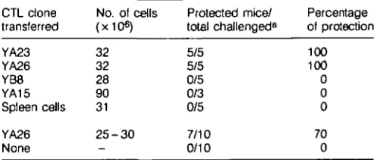
Table 1. Effect of adoptive transfer of CTL clones on the outcome
of a challenge with Plasmodium yoeiii sporozoites
CTL clone transferred YA23 YA26 YB8 YA15 Spleen ceils YA26 None No. of cells (x10«) 32 32 28 90 31 2 5 - 3 0 -Protected mice/ total challenged8 5/5 5/5 0/5 0/3 0/5 7/10 0/10 Percentage of protection 100 100 0 0 0 70 0 "All mice were challenged i.v. with 100 P. yoeiii sporozoites. The protected animals remained negative, while all the control mice developed parasitemia.
viable P. yoeiii sporozoites. As shown in Table 1, all the mice which received the cytotoxic clones YA23 and YA26 were protected, i.e. failed to develop parasitemia after sporozoite challenge. In contrast, adoptive transfer of CTL clones YB8 and
YA15, which appear to have the same epitope specificity as YA23 and YA26, failed to confer protection, even when as many as 9 x 10⁷ cells of clone YA15 were transferred to each mouse.
The protection conferred by clones YA23 and YA26 did depend on the size of the sporozoite challenge. Protection was consistently achieved when the mice were challenged with 100
P. yoelii sporozoites, which resulted in 100% infection in the
control group. The number of sporozoites used for challenge represents five times the minimum dose required to achieve infection of 100% of normal BALB/c mice (data not shown). When the challenge dose consisted of 1000 or more sporozoites, the experimental animals were no longer protected. They developed parasitemia, usually 1 day later than the controls (data not shown). When blood stages of P. yoelii were used to challenge the mice which had received the clone YA26, no inhibition of parasite development occurred, indicating that the protective activity of the CTL clones is stage specific (data not shown).
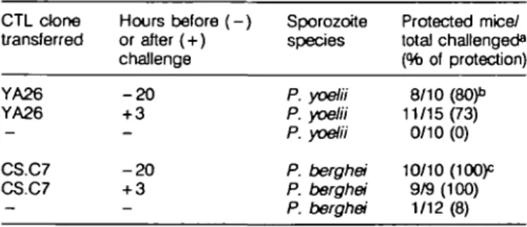
While the protective CTL clones inhibited parasite development when transferred into mice 20 h before their challenge with sporozoites, we observed a similar degree of protection when the CTL were transferred after sporozoite invasion of the hepatocytes has taken place (13). As seen in Table 2, when the anti-P. yoelii clone (YA26) was transferred 3 h after challenge with P. yoelii sporozoites, it conferred full protection to 73% of the mice. This same clone, transferred 20 h before challenge, conferred protection to 80% of the mice. Similar results were obtained with the anti-P. berghei CTL clone, CS.C7, which pro-tected all the mice, regardless of whether it was transferred before or after sporozoite challenge (Table 2).
Species-specificity of the protection mediated by anti-P. yoelii and anti-P. berghei CTL clones
The in vitro results did indicate that the CTL clones YB8, YA15, YA23, and YA26 (anti-P. yoelii) do not recognize the P. berghei epitope (Fig. 1A - D). As for the protective anti-P. berghei clone, CS.C7, it displays in vitro, a weak cross-reactivity with the P. yoelii epitope (Fig. 1E).
To characterize further the species-specificity of the protection
mediated by the anti-P. yoelii CTL clones, we performed a series of cross-protection experiments. When the CTL done YA26 (anti-P. yoelii) was transferred to mice and these were challenged with P. yoelii sporozoites, 90% of the mice were protected, i.e. did not develop parasitemia. In contrast, 80% of the mice which received the same YA26 clone developed parasitemia after they were challenged with P. berghei sporozoites (Table 3). Con-versely, when mice were injected with the anti-P. berghei clone, CS.C7, 100% protection was obtained in the group of mice challenged with P. berghei sporozoites, while no protection was observed when the challenge consisted of P. yoelii sporozoites (Table 3). All control mice injected with P. yoelii and more than 90% of those infected with P. berghei sporozoites developed parasitemia.
CTL clones inhibit the development of the liver stages of P. yoelii
To gain further and more quantitative insight into the inhibitory effect of these CTL clones on the development of liver stages of P. yoelii, we measured the amount of parasite-specific rRNA present in the liver of mice which had received CTL clones and were subsequently injected with P. yoelii sporozoites. The livers of several mice were excised 42 h after sporozoite challenge and the total RNA from each mouse liver was partially purified. The amount of parasite rRNA was determined on the basis of a dot-hybridization assay, using parasite-specific ribosomal oligonucleotide probes, as described elsewhere (11).
We found that mice which had received 2.5 x 10⁷ cells of the
protective clone YA26, and had been challenged with 2 x 10s
P. yoelii sporozoites, had very little or no detectable parasite rRNA in their livers. In contrast, the levels of parasite rRNA present in the livers of mice which had received the non-protective clone YA15 were comparable to those present in control mice, injected with parasites, but not with cytotoxic T cell clones (Fig. 2A). Identical results were obtained when the transfer of CTL clones was performed 20 h after sporozoite challenge (data not shown). Noteworthy was the finding that mice injected with clone YB8, considered to be non-protective on the basis of the parasitemia which the mice developed after cell transfer and challenge
Table 2. Protective effect of the adoptive transfer of CTL clones
performed prior to and after sporozoite challenge
Table 3. Effect of the adoptive of CTL clones YA26 and CS.C7
on the outcome of challenge with either P. yoelii or P. berghei sporozoites CTL done transferred YA26 YA26 CS.C7 CS.C7 Hours before ( - ) or after (+) challenge - 2 0 + 3 - 2 0 + 3 Sporozoite species P. yoelii P. yoelii P. yoelii P. berghei P. berghei P. berghei Protected mice/ total challenged8 (% of protection) 8/10 (80)" 11/15(73) 0/10 (0) 10/10 (lOOp 9/9 (100) 1/12 (8) CTL clones transferred8 YA26 CS.C7 None YA26 CS.C7 None Sporozoite challenge P. yoelii P. yoelii P .yoelii P. berghei P. berghei P. berghei Protected mice/ total challenged13 9/10 0/10 0/5 4/20c 19/20 0/20 Percentage of protection 90 0 0 20 95 0 were challenged with 100 sporozoites of P. yoelii or 104 of
P. berghei sporozoites. These doses represent five times the minimum amount of sporozoites to produce patent parasitemia in BALB/c mice. All P. yoelii controls and 11 of the 12 P. berghei control mice developed patent infections. None of the 'protected' mice developed parasitemia b0 . 5 - 2 . 5 x10⁷T cells were transferred per mouse. Combined results of two experiments.
C2 x 107 T cells were transferred per mouse.
a2 - 4 x 1 07 of CTL clone YA26, and 2 - 3 x 1 07 cells of CTL clones CS.C7 were transferred per mouse.
bAII mice were challenged with 100 P. yoelii or 104 P. berghei sporozoites. These doses represent five times the minimum amount of sporozoites to produce patent parasitemia in BALB/c mice. AO the control mice developed patent infections, while the protected mice remained negative.
(Table 1), showed a 59% reduction of the amount of P. yoelii rRNA in the liver. The adoptive transfer of CTL clone CS.B83, which is specific for the P. berghei epitope and does not cross-react in vitro with the P. yoelii epitope (3), did not have any signifi-cant effect on the levels of P. yoelii rRNA (Fig. 2A).
We also tested the effect on P. yoelii of the CTL clone CS.C7 which in vivo is protective against P. berghei sporozoites. As shown in Fig. 2B, the transfer of 2.5 x 10⁷ cells results in partial
decrease (40%) of the liver stage rRNA of P. yoelii. This partial inhibition may be due to the fact that CS.C7, differently from CS.B83, recognizes not only the P. berghei but also the P. yoelii peptide 277-288 in vitro (Fig. 1E). It is noteworthy that the inhibitory effect of the cross-reactive clone CS.C7 is much less efficient than the one mediated by the anti-P. yoelii clone YA26. The later clone (YA26), reduced by 85% the parasite rRNA when 5 xiO6, i.e. five times less cells, were transferred (Fig. 2B).
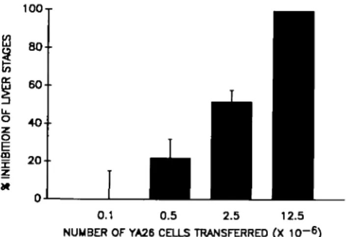
The capacity of clone YA26 to inhibit the development of liver stages was found to be dose-dependent. Reduction of the rRNA by 50% could be achieved after transferring as few as 2.5 x 10⁶ cells, while none or very low levels of parasite rRNA
were consistently observed after the adoptive transfer of
3OT
1.2 x 10⁷ of these cells. No inhibition of parasite development
was seen upon transfer of 1 x 10s cells of this clone (Rg. 3).
Antibodies against murine IFN-y and TNF fail to neutralize the anti-plasmodial activity of these CD8+ T cell clones
Earlier studies had shown that IFN-7 has a potent inhibitory effect on the development of the liver stages of malaria parasites (14). Furthermore it has been reported that neutralizing anti-IFN-7 antibodies can reverse the immunity induced by immunization with irradiated sporozoites (15). To determine whether IFN-y plays an important role in the in vivo anti-liver stage activity of the CTL clones, the neutralizing anti-IFN-7 monoclonal antibody DB-1 was
10OT
£3
CTL CLONE NONE CS.B&3 YA26 YB8 YA15 TRANSFERRED
0.1 0.5 2.5 12.5 NUMBER OF YA26 CELLS TRANSFERRED (X 10~6) Fig. 3. Dose dependence of the inhibitory activity of the CTL clone YA26 in vivo. Shown are the number of T cells adoptively transferred to naive mice, 6 h before their infection with P. yoelii sporozoites. The percentage inhibition was calculated in relation to the amount of parasite rRNA present in the liver of control mice, challenged with sporozoites but not injected with T cells. The bars represent the mean value obtained from three mice
± SE. 100% inhibition signifies that the sample contained less than 1 ng of parasite rRNA.
CTL CLONE NONE TRANSFERRED
YA26 YA26* CS.C7
R g . 2. In vivo inhibition of liver stage development by adoptively transferred CTL clones. 2.5 x 10⁷ cells of various anti-P. yoelii (YA26, YB8, and YA15) and anti-P. berghei (CS.B83 and CS.C7) CTL clones were adoptively transferred to naive mice 6 h before challenge with P. yoelii sporozoites. The amount of P. yoelii rRNA present in the liver of infected mice was measured 42 h after challenge. The bars represent the mean value obtained from three mice ± standard error of the mean (SE). The asterisk at YA26, in B, indicates that 5 X10⁶ cells were transferred to each mouse. Absence of parasite rRNA, as determined in this assay, means less than 1 ng in the sample.
100-r 8 0 - 60-o E m x z 40- 20-NEUTRAUZING ANTIBODY
NONE ANTI-IFN ANTI-TNF
Fig. 4. Neutralizing antibodies to murine IFN-y and TNF fail to reverse the in vivo inhibitory effect of a CTL clone. Monoclonal antibodies to murine IFN-7 (0.5 mg/mouse) or TNF (0.3 mg/mouse) were injected into naive mice, 2 h before they received the CTL clone YA26 (7.5 x 10⁶ cells per mouse). 6 h later, the animals were challenged with P. yoelii sporozoites. The percentage inhibition was calculated as in Rg. 3.
injected into mice, as previously described (15), prior to the adoptive transfer of the CTL clone YA26. The dose of antibody administered in different experiments ranged between 0.5 to 1.0 mg per mouse. As seen in Fig. 4, the degree of inhibition of parasite development mediated just by the injection of clone YA26 did not differ significantly from that seen in mice which had, in addition, been injected with the IFN-y neutralizing antibody. We were also unable to reverse the protective activity of clone YA26 by injecting monoclonal antibody TN3.19.12(0.3-1.0mg per mouse) which is specific for and inhibits the biological activity of murine TNF in vivo (16). Furthermore, the inhibitory activity of this CTL clone was not reversed when the mice simultaneously received both of these monoclonal antibodies (data not shown).
Discussion
The present study demonstrates that adoptive transfer, into naive mice, of certain CD8+ CTL clones which recognize a defined epitope in the CS protein of P. yoelii, protects against sporozoite-induced malaria. These results resemble those obtained earlier in the P. berghei system (3), and thus confirm the more general validity of the protective role of anti-CS CD8+ CTL in sporozoite-induced rodent malaria.
The experiments described in the present report also demonstrate that the anti-parasite activity of these protective T cell clones is species specific. In fact, the results show that while certain anti-P. berghei and anti-P. yoelii CD8+ clones confer a high degrees of protection against sporozoites of the respective species, the same clones induce no or only a small inhibitory effect when the mice are challenged with sporozoites of the heterologous species.
Still, it is noteworthy that parasitemia did not develop in 20% of the mice which had received the anti-P. yoelii CTL done YA26 and were challenged with P. berghei sporozoites (Table 3). In view of the fact that clone YA26 does not appear to cross-react
in vitro with a synthetic peptide containing the P. berghei epitope,
the partial cross-protection we observed suggests that this clone may possibly mediate a certain 'non-specific' inhibition of liver stage development. Alternatively, it is possible that clone YA26 cross-reacts in vivo with an epitope presented upon processing by the parasitized liver cells, which may differ somewhat in size and therefore in sequence from the peptide we used in the in vitro assay (17,18).
The most likely targets of the inhibitory effects of these cytotoxic T cells are the liver stages of the parasite. This view is based on the high degree of protection observed upon transfer of protective T cell clones 3 h after challenge, at a time when sporozoite invasion of hepatocytes is certain to have been completed (13). It is further corroborated by the fact that transfer of these clones did not protect mice against a challenge with blood stages of the same parasite. Furthermore, recent studies have shown that sporozoite-immune spleen cells, stimulated with a synthetic peptide comprising the P. yoelii 281 - 296 epitope, inhibit the development of P. yoelii liver stages in cultured mouse hepatocytes (5).
In the present report we demonstrate that the transfer of protective cytotoxic clones greatly reduces the level of parasite rRNA, thus providing direct evidence of the fact that these CD8+ T cell clones inhibit, in a dose-dependent manner, the
in vivo development of the liver stages of this parasite. Significant
decrease of rRNA was seen upon transfer of only 2.5 x i O6
cells of the YA26 clone. This is noteworthy since only a fraction of these cells reach the liver, most transferred cells being entrap-ped in the lung. Migration studies performed during a 48-h period with radiolabeled YA26 cells revealed that the amount of T cells which migrate to the liver reaches a maximum 24 h after transfer and represents 17.8 ± 5 . 1 % of the total amount of transferred cells. The pattern of migration is not altered by a concurrent sporozoite infection (manuscript in preparation).
The individual anti-P. yoelii CD8+ T cell clones we assayed differed in their capacity to confer protection against a sporozoite challenge, resembling our earlier observations in the P. berghei system (3). These functional differences among CTL clones were highlighted in experiments in which the protective effect of the different clones was determined by measuring the parasite rRNA in the liver of the challenged mice. Parasite rRNA was undetectable (less than 1 ng per sample) and therefore reduced by more than 20-fold in the livers of mice injected with the protective clone YA26. The fact that parasite rRNA was not detectable under these conditions cannot be interpreted as indicating the complete absence of parasites in the liver, namely that sterile immunity has been achieved. The lack of detection of parasite rRNA under these conditions most likely reflects the limit of sensitivity of the assay (11).
Mice injected with clone YA15 had parasite rRNA levels similar to those found in control mice injected with sporozoites but not with CTL clones. These results were unexpected, in view of the results of the in vitro cytotoxic assays in which clone YA15 lysed target cells as efficiently as clone YA26, and produced similar or even larger amounts of IFN-7. These findings indicate that the capacity of CTL clones to induce protection upon passive transfer requires additional properties, besides in vitro cytotoxicity.
Based on earlier studies (19), mice which had been injected with CTL clones, and their controls, were also injected with recombinant IL-2. Experiments in which we measured P. yoelii rRNA revealed that the administration of IL-2 did not alter the protective nor the non-protective effects of the transfer of clones YA26 and YA15, respectively (data not shown).
We are currently undertaking a detailed characterization of the fine epitope-specificity of these clones, of their profile of lymphokine production, and their patterns of migration and homing in mice. This will, hopefully, clarify the basis of the observed functional differences and cross-reactivities.
The mechanisms of the inhibitory activity of cytotoxic CD8+ T cell clones on the liver stages of malaria parasites has not yet been identified. It is possible that these clones exert a direct cytotoxic effect on the parasitized hepatocytes. Another possibility is that this inhibitory effect may be mediated by lymphokines produced locally by the CTL clones, after recognizing the corresponding epitope on the surface membrane of antigen-presenting cells (Kupffer cells or other mononuclear phagocytes). It was reported earlier that IFN-7 and TNF inhibit the development of plasmodial liver stages in vitro (14,20). In vivo treatment of sporozoite-immunized A/J mice with anti-IFN-7 monoclonal antibody was reported to reverse protection against P. berghei sporozoites (15). However, a similar treatment of sporozoite-immunized BALB/c mice appears to have no detectable effect (21). Our current results regarding the unchanged in vivo anti-parasitic activity of these CTL clones, upon
administration of neutralizing anti-IFN-7 and anti-TNF antibodies, suggest that systemic levels of these lymphokines may not play a significant role in the inhibitory effector mechanisms. These results do not rule out the possibility that these cytokines might be produced and acting locally, in the micro-environment of the CD8+ T cells and of the infected hepatocytes.
Taken together these results indicate that the rodent malarias provide a rather unique experimental model for the investiga-tion of the effector mechanisms by which CD8+ T cells can prevent the development of non-viral intracellular pathogens. The elucidation of these protective mechanisms should benefit ongoing attempts to design an effective malaria vaccine.
Acknowledgements
The authors would like to thank Dr Victor Nussenzweig for stimulating discussions and critical review of the manuscript. We also thank Mrs Rita Attzuler, Frank Gagliardi, and Yi Zhao for excellent technical assistance. M.M.R is supported by a fellowship from the Brazilian National Research Council F.Z. is a recipient of the Irma T.Hirschl Career Development Award. P.J.R. is supported by a fellowship from Hoffmann-LaRoche This work was supported by grant AI-27458-02 from NIH and grant DPE-0453-A-00-5012-00 from the Agency of International Development. Abbreviations CTL CS IFN IL MHC TNF cytolytic T lymphocytes circumsporozorte interferon interleukin
major histocompatibdity complex tumor necrosis factor
References
1 Nussenzweig, V. and Nussenzweig, R. S. 1989. Rationale for the development of an engineered sporozoite malaria vaccine. Adv. Immunol 45:283.
2 Tsuj, M., Romero, P., Nussenzweig, R , and Zavala, F. 1990. CD4 + cytolitic T cell clone confers protection against murine malaria. J. Exp. Med. 172:1353.
3 Romero, P., Maryasnki, J., Corradin, G., Nussenzweig, R. S., Nussenzweig, V., and Zavala, F. 1989. Cloned cytotoxic T cells recognize an epitope in the circumsporozoite protein and protects against malaria. Nature 341:323.
4 Kumar, S., Miller, L. H., Quakyi, I A., Ketster, D. B., Houghton, R. A., Matoy, W. L, Moss, B., Berzofsky, J. A., and Good, M. 1988. Cytotoxic T cells specific for the circumsporozoite protein of Plasmodium falciparum. Nature 334:258.
5 Weiss, W. R., Mellouk, S., Houghten, R. A., Sedegah, M., Kumar, S., Good, M , Berzofsky, J. A., Miller, L. H., and Hoffman, S. J. 1990.
Cytotoxic T cells recognize a peptide from the circumsporozoite protein on malana infected hepatocytes. J. Exp. Med. 171:763.
6 Romero, P., Maryanski, J., Cordey, A S., Corradin, G., Nussenzweig, R. S., and Zavala, F. 1990. Isolation and characteriza-tion of protective cytolytic T cells in a rodent malaria model system. Immunol. Lett. 25:27.
7 Farrar, J. J., Fuller-Farrar, J., Simon, P. L, Hilfiker, M. L , Stadler, B. M , and Farrar, W. L. 1980. Thymoma production of T cell growth factor (Interleukin 2). J. Immunol. 125:2555.
8 Maryanski, J. L, Acolla, R. S., and Jordan, B. 1986. H-2 restricted recognition of cloned HLA class I gene products expressed in mouse cells J. Immunol. 136:4340.
9 Van Der Meide, P. H., Dubbetd, M., Vijverberg, K , Kos, T., and Schellekens, H. 1986. The purification and characterization of rat gamma interferon by use of two monoclonal antibodies J. Gen. Virol. 67:1059.
10 Sheehan, K , Ruddle, N., and Schreiber, R. 1989. Generation and characterization of hamster monoclonal antibodies that neutralize murine tumor necrosis factor. J. Immunol. 142:3884.
11 Arreaza, G., Corredor, V., and Zavala, F 1991. Plasmodium yoelii: quantification of exoerythrocytic stages based on the use of nbosomal RNA probes Exp Parasitol. 72.103
12 Chomczynski, P. and Sacchi, N. 1987. Single step method of RNA isolation by guanidinium thiocyanate-phenol-chloroform extraction. Anal. Biochem 162-156.
13 Shin, S C , Vanderberg, J P , and Terzakis, J. A. 1982. Direct infection of hepatocytes by sporozortes of Plasmodium berghei J. Protozool. 29:448.
14 Ferreira, A., Schofield, L., Enea, V., Schellenkens, H., van der Meide, P., Collins, W. E., Nussenzweig, R. S., and Nussenzweig, V. 1986. Inhibition of development of exoerythrocytic forms of malaria parasites by gamma-lnterferon. Science 232881. 15 Schofield, L., Villaqiran, J., Ferreira, A., Schellekens, H., Nussenzweig, R. S., and Nussenzweig, V 1987. Interferon-gamma, CD8+ T cells and antibodies required for immunity to malaria sporozortes. Nature 330664
16 Ruddle, N. H., Bergman, C. M., McGrath, K. M., Lingenheld, E. G , Grunnet, M. L, Padula, S J., and Clark, R. B. 1990. An antibody of lymphotoxin and tumor necrosis factor prevents transfer of experimental allergic encephalomyelitis. J. Exp Med. 1721193. 17 Van Bleek, G. M. and Nathanson, S. G. 1990. Isolation of
endogenous^ processed immunodominant viral peptide from class 1 H-2Kb molecule. Nature 348.213.
18 Rdtzschke, O., Falk, K , Deres, K., Schild, H., Norda, M., Metzger, J., Jung, G., and Rammensee, H.-G. 1990. Isolation and analysis of naturally processed viral peptide as recognized by cytotoxic T cells. Nature 348:252
19 Taytor, P. M. and Askonas, B. A. 1986. Influenza nucteoprotein specific cytotoxic T cell clones are protective in vivo. Immunology 58:417. 20 Mazier, D., Renia, L, Nussler, A., Pied, S., Marussig, M., Goma, J., Gnllot, D., Miltgen, F., Drapier, J C , Corradin, G., Del Giudice, G., and Grau, G. E. 1990 Hepatic phase of malaria is the target of cellular mechanisms induced by the previous and the subsequent stages. A crucial role for liver nonparenchymal cells. Immunol. Lett 25:65. 21 Hoffman, S. L, Isenbarger, D., Long, G. W., Sedegah, M., Szarfman, A., Waters, L, Hollingdale, M. R., van der Meide, P. H., Finbloorn, D. S., and Ballow, W. R. 1989. Sporozoite vaccine induces genetically restricted T cell elimination of malaria from hepatocytes. Science 244:1078.