European Journal of Orthodontics 20 (1998) 389-397 © 1998 European Orthodontic Society
The initial effects of the treatment of Class II,
division 1 malocclusions with the van Seek activator
compared with the effects of the Herren activator
and an activator-headgear combination
Elisabeth Altenburger and Bengt Ingervall
Department of Orthodontics, University of Bern, Switzerland
SUMMARY The effects of the van Seek activator in the correction of Class II, division 1
malocclusions were studied in 39 children, aged 9-13 years (median 11 years), and com-pared with the effects of treatment with an activator according to Herren and with those of an activator-headgear combination. Profile cephalograms were made before treatment and at the attainment of a Class I molar relationship (median observation time 9 months).
The median improvement of the overjet was 4.7 mm and of the molar relationship 3.6 mm. This was largely achieved skeletally by an increase in mandibular prognathism while the skeletal effect on the maxilla was clinically insignificant. The maxillary incisors retroclined and the mandibular incisors proclined moderately. In general, no intrusion of the maxillary incisors was found and the eruption of the molars could not be stopped. The effects differed partly between the sexes, with a larger mandibular skeletal and molar reaction in boys, and a larger maxillary molar movement in girls. The larger mandibular reaction in the boys might have been due to the on average 4.5 months longer treatment time. The skeletal effects of the treatment were similar with all three activator types. The control of the incisors was, however, superior with the van Seek activator, especially when compared with the Herren activator.
Introduction
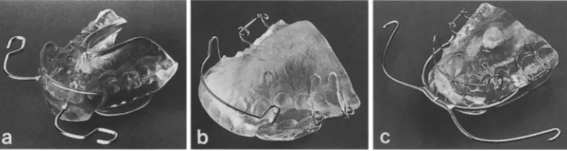
The activator is a widely used appliance for the treatment of Class II, division 1 malocclusions in the mixed dentition. Many different types of activators and activator derivatives are known. Some years ago a special type of activator was described by van Beek (1982, 1984). The van Beek activator differs from previously known activators by the full coverage of the labial sur-faces of the maxillary anterior teeth by acrylic (Figure 1a). Therefore, no labial bow is used. The idea is to hold the maxillary anterior teeth as rigidly as possible in order to prevent them from retroclination by the dorsally directed forces on the maxilla induced by the activator. Retro-clination of the maxillary anterior teeth by activator treatment is a well-known and often
undesirable phenomenon. To prevent maxillary incisor retroclination, the activator described by Teuscher (1988) uses torquing springs.
Proclination of the mandibular incisors is an almost unavoidable and, as a rule, undesirable sequela to activator treatment To minimize such proclination with the van Beek activator, the mandibular incisors and the alveolar process are relieved from acrylic on the lingual side. The labial surfaces of the mandibular incisors are, however, covered in acrylic in order to hold the teeth in a comparable way to the maxillary anterior teeth.
The van Beek activator has bows bilaterally for the attachment of a high-pull headgear force. The bows are incorporated in the acrylic in the anterior part of the activator and are short The point of force application is at the level of
Figure 1 The three activator types studied. (a) van Beek activator, (b) Herren activator, and (c) activator-headgear combination.
the maxillary canines. The outer parts of the bows are normally inclined upwards relative to the occlusal plane. The direction of the force together with the anterior application allows the intrusion of the maxillary anterior teeth. In high-angle cases the bows are kept parallel to the occlusal plane in an endeavour to retard the maxillary vertical growth in the posterior part of the dental arch.
The construction bite for the van Beek activator is taken with moderate protrusion of the mandible. In most cases the mandible is protruded to the edge-to-edge position of the incisors (about 7 mm). The bite raising in the construction bite is recommended to be fairly high, about 6-8 mm in the molar area. In cases with a tendency to anterior skeletal open bite an attempt can be made to intrude the mandibular molars by the application of gutta-percha to the activator over their occlusal surfaces.Itis thought that this load will intrude the molars and/or cause a distraction in the temporomandibular joints that promotes additional condylar growth. Both mechanisms would enhance autorotation of the mandible to bite closure.
The present study was undertaken to compare the efficacy of the van Beek activator with that of other types of activators used in our clinic. One of these is the activator developed by Herren (1953, 1959, 1980). Unlike the van Beek activator, the Herren activator does not take advantage of an extra-oral headgear force. The forces for growth modification are instead achieved exclusively by a large (minimum 8 mm)
protrusion of the mandible in the construction bite. In the construction bite, the distal occlusion is over-compensated by 3-5 mm. The antero-posterior forces are therefore larger than those produced by activators using less protrusion. The bite raising in the construction bite is 2-4 mm opening between the incisal edges. The Herren activator is fixed to the maxillary first molars with clasps (Figure 1b).Ithas a conventional maxillary labial bow and the edges of the mandibular incisors' are embedded in the acrylic to 2-3 mm without any relief on the lingual side. In the case of a deep bite the occlusal surfaces of the mandibular posterior teeth are relieved from the acrylic.
Another type of activator used in our clinic is similar to the Herren activator, but with slightly less protrusion, and combined with a high-pull headgear force of approximately 300 g per side. Tubes for the insertion of a conventional head-gear bow are embedded in the acrylic at the level of the second premolars (Figure lc). The point of force application on the headgear bow is more posterior, at the level of the molars, than with the van Beek activator. The effects of the Herren activator and of the activator-headgear com-bination in the treatment of Class II, division 1 malocclusions have been described in a previous article (Weiland et al.,1997).
The purpose of the study was to compare the effects of the van Beek activator with those of two other types of activators, the Herren acti-vator and an actiacti-vator-headgear combination. Specifically, possible advantages of the van Beek
EFFECTS OF THE VAN BEEK ACTIVATOR
activator with regard to control of the incisor inclination and the possibilities of intruding the maxillary incisors and of controlling the eruption of the molars were to be evaluated.
Subjects and methods
Twenty-two boys and 17 girls were included in the study. Their ages varied between 8 years, 9 months and 13 years, 4 months (median 11 years, 4 months). All subjects had a Class II, division 1 malocclusion that was treated with a van Beek activator. The subjects were enrolled consecutively in the study. The only criteria for the enrolment were a Class II malocclusion, a suitable age and characteristics of the dentition suitable for activator treatment. At the time of the study the van Beek activator was the only type of activator used in the clinic. The activator treatment was discontinued in four patients because of lack of co-operation. The effect of the activator was evaluated on profile cephalograms taken before the insertion of the activator and at the attainment of a Class I relationship of the posterior teeth. This phase of the treatment varied between 4 and 31 months (median 9 months). Only three patients had a treatment time exceed-ing 19 months. In the majority of the cases only one activator was used, but two patients had two activators and two had their activator adjusted by further protrusion of the mandible using cold-curing acrylic. In five cases, gutta-percha was applied to the activator over the occlusal surfaces of the mandibular molars. The high-pull headgear force used was 150-200 g per side. As with the other two types of activators mentioned in the introduction, the children were instructed to wear the appliance 12 hours per day and night. In practice, however, most children probably used their activator only at night.
The group treated with a Herren activator consisted of 15 boys and 12 girls with a median age of 10 years, 4 months (range 9 years, 2 months-12 years, 3 months) and with a median observation time of8 months (range 5-11 months). Nine boys and 11 girls were treated with the activator-headgear combination. Their ages varied between 8 years, 11 months and 12 years, 4 months (median 10 years, 1 month) and the
391 observation time between 6 and 9 months (median 7 months).
For ethical reasons, no control group of untreated Class II cases could be included. Data from the literature were collected and the changes of the cephalometric variables during a 9-month period calculated. These reference values for untreated Class II malocclusions were used in a previous study (Weiland et ai.,1997). They were collected from many sources in the literature. In all, 15 studies were used. Partly dif-ferent reference points and measurements were used, especially for linear variables. No correc-tion for linear enlargement was made as many studies lacked these data. The observation periods varied from 6 months to 6 years. As the growth rate during a longer period is not constant, the growth rates for an observation period of 9 months are just indicative.
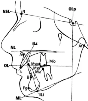
The cephalograms were taken with the man-dible in the intercuspal position. The reference point and lines shown in Figure 2 were used in the cephalometric analysis. The computerized cephalometric analysis system of Gebauer (1977) was used. Distances were reduced to zero magnification.
Figure 2 Reference points and lines used in the cephalo-metric analysis.
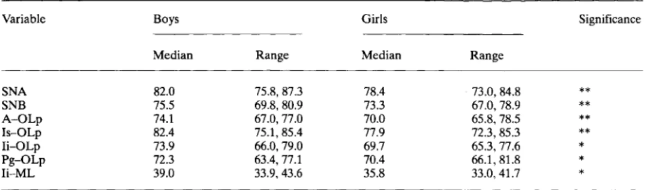
Table 1 Median and range of variables differing significantly between the sexes at the start of treatment. Variable SNA SNB A-OLp Is-OLp Ii-OLp Pg-OLp Ii-ML
Boys Girls Significance
Median Range Median Range
82.0 75.8,87.3 78.4 73.0,84.8 ** 75.5 69.8,80.9 73.3 67.0,78.9 ** 74.1 67.0,77.0 70.0 65.8,78.5 ** 82.4 75.1,85.4 77.9 72.3,85.3 ** 73.9 66.0,79.0 69.7 65.3, 77.6 * 72.3 63.4, 77.1 70.4 66.1,81.8 * 39.0 33.9,43.6 35.8 33.0,41.7 * *0.01<P<0.05; **0.001<P<0.01.
The analysis of antero-posterior linear changes was undertaken with the method of Pancherz (1982). A co-ordinate system, consisting of the occlusal line (OL) and a perpendicular to this line through the point sella (OLp), was drawn on the tracing of the pre-treatment cephalogram. The co-ordinate system was transferred to the subsequent cephalograms of the series by super-imposing on structures of the anterior cranial base as described by Bjork (1968). The overjet was recorded as the difference between the variables Is-OLp and Ii-OLp (Figure 2). The overbite was recorded as the vertical distance between the points Is and Ii, measured perpen-dicular to a constructed horizontal line, angu-lated 6 degrees to the nasion-sella line (NSL). The molar relationship was recorded as the difference between the variables Msa-OLp and Mia-OLp. Thus, a positive sign denotes a distal and a negative sign a neutral molar relationship.
Errors of the method and statistical analysis
The errors of the method used have been studied in a previous investigation (Stucki and Ingervall, 1998). The accidental errors of the cephalometric variables were at most 0.61 mm or 0.72 degrees with the exception of the angles ILi/ML and ILs/ILi, which had errors of 1.4 and 1.8 degrees, respectively. Therefore, in the present investiga-tion, the angles ILs/NL, ILi/ML, and ILs/ILi were measured twice and the mean used. This reduces the error by a factor of 0.70.
Differences between the sexes were tested with the Mann-Whitney V-test, and differences between the cephalograms taken before and after the treatment with the Wilcoxon matched-pairs, signed-ranks test.
Results
Differences between the sexes at the start of treatment
The variables that differed significantly between the sexes at the start of treatment are given in Table 1. The faces of the girls were more retro-gnathic than those of the boys. Six of the seven variables that differed between the sexes at the start of the treatment are an expression of the difference in facial prognathism. Inaddition, the variable Ii-ML showed that the height of the alveolar process was smaller in the girls than in the boys. There was no difference in age between the sexes at the start of treatment.
Effect of treatment
Four variables showed significant differences between the sexes in their changes with the treat-ment. The median changes of these variables are given in Table 2. Two of these variables (Ii-OLp and Mia-OLp) provide evidence of a larger for-ward movement of the mandibular dentition in boys than in girls. The difference of the changes of the variable Ar-OLp points to a larger
EFFECTS OF THE VAN BEEK ACTIVATOR 393
Table 2 Median and range of changes with the treatment for variables with significantly different changesin
boys and girls.
Variable
Ii-OLp Mia-OLp Ar-OLp Mio-ML
Boys Girls Significance
Median Range Median Range
3.5*** 1.1,5.7 2.5*** 1.4,4.9 *
3.8*** 1.0,6.4 2.8*** 0.7,5.0 *
0.9*** -0.6,2.1 -0.1 -1.3, 1.4 **
1.4*** -0.7,3.0 0.5* -0.7,2.4 **
*0.01<P<0.05; **0.001<P<0.01;***p<0.001.
mandibular growth in boys than in girls. The treatment time was longer in the boys than in the girls (median 12.5 versus 8 months, 0.01<P
<0.05).
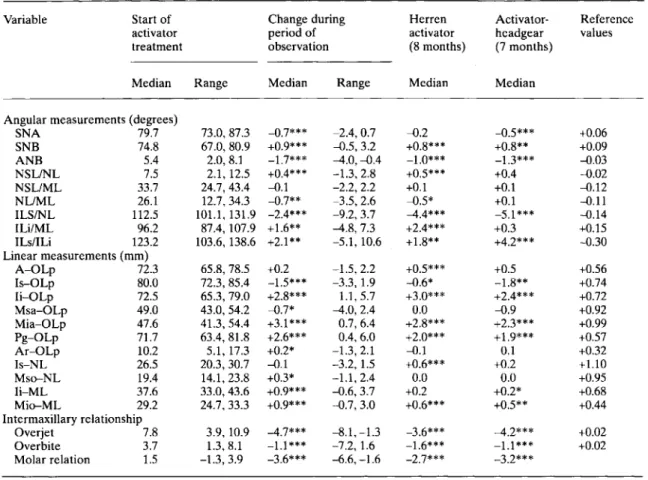
Since the great majority of the variables did not change significantly with treatment in the two sexes, the results are given for the sexes combined (Table 3). The table also gives the changes for cases treated with the Herren activator (median observation time 8 months) and for those treated with the activator-headgear combination (median observation time 7 months). The reference values in the table refer to the growth during 9 months of untreated Class II malocclusions.
During treatment the maxillary prognathism (SNA) decreased but the mandibular pro-gnathism (SNB, Pg-OLp) increased. Therefore, the sagittal jaw relationship (ANB) improved. There was a significant increase of the length of the mandible (Pg-OLp + Ar-OLp), P < O.OOL The maxilla rotated slightly posteriorly (NSLI
NL) with a small decrease of the vertical jaw relationship (NL/ML). The inclination of the mandible (NSL/ML) was. however, unchanged.
The maxillary incisors retroclined (ILs/NL, Is-OLp) and the mandibular incisors proclined (ILi/ML). The mandibular incisors and molars moved forward (Ii-OLp, Mia-OLp), but the maxillary incisors and molars backwards (Is-OLp, Msa-OLp). This resulted in a substantial decrease of the overjet and normalization of the molar relationship. For all variables, there
was a large inter-individual variation. Thus, for example, in one case the maxillary molar was relocated backwards 4 mm while in another case the molar moved 2.4 mm anteriorly (Msa-OLp). Likewise, there was a large variation in the increase in mandibular prognathism, up to a 6 mm increase in the distance Pg-OLp.
The height of the maxillary incisors (Is-NL) was unchanged, but the mandibular incisors erupted (Ii-ML). Because of the eruption of the molars (Mso-NL, Mio-ML), there was still a slight decrease of the overbite. Five patients had gutta-percha placed over the occlusal surfaces of the lower molars in order to intrude these teeth or to arrest their eruption. These patients had a mandibular inclination above the average (high angle cases). The five patients did not react dif-ferently than the other patients, i.e. the eruption of the molars, the inclination of the mandible, the change in vertical jaw relationship, the increase in face height and the decrease in overbite were similar to those of the other patients.
Components of Class II correction
With the method of Pancherz (1982), it is pos-sible to calculate the part of the correction of the overjet and of the molar relationship, respect-ively, that is due to skeletal and to dento-alveolar factors. The results are shown in Figures 3 and 4. Both the improvement of the overjet and those of the molar relationship were largely due to skeletal changes. The skeletal component for
Table 3 Median and range for the cephalometric variables at the start of treatment and for the change during treatment, as well as median changes in cases treated with a Herren activator or an activator-headgear combination. The table also gives growth changes in untreated Class II malocclusions during 9 months.
Variable Start of Change during Herren Activator- Reference
activator period of activator headgear values
treatment observation (8months) (7months)
Median Range Median Range Median Median
Angular measurements (degrees)
SNA 79.7 73.0,87.3 -0.7*** -2.4,0.7 -0.2 -0.5*** +0.06 SNB 74.8 67.0,80.9 +0.9*** -0.5,3.2 +0.8*** +0.8** +0.09 ANB 5.4 2.0,8.1 -1.7*** -4.0, -0.4 -1.0*** -1.3*** -0.03 NSL/NL 7.5 2.1, 12.5 +0.4*** -1.3,2.8 +0.5*** +0.4 -0.02 NSL/ML 33.7 24.7,43.4 -0.1 -2.2,2.2 +0.1 +0.1 -0.12 NL/ML 26.1 12.7,34.3 -0.7** -3.5,2.6 -0.5* +0.1 -0.11 ILSINL 112.5 101.1,131.9 -2.4*** -9.2,3.7 -4.4*** -5.1 *** -0.14 ILi/ML 96.2 87.4, 107.9 +1.6** -4.8,7.3 +2.4*** +0.3 +0.15 ILs/ILi 123.2 103.6, 138.6 +2.1** -5.1, 10.6 +1.8** +4.2*** -0.30 Linear measurements (mm) A-OLp 72.3 65.8,78.5 +0.2 -1.5,2.2 +0.5*** +0.5 +0.56 Is-aLp 80.0 72.3,85.4 -1.5*** -3.3,1.9 -0.6* -1.8** +0.74 Ii-OLp 72.5 65.3, 79.0 +2.8*** 1.1,5.7 +3.0*** +2.4*** +0.72 Msa-OLp 49.0 43.0,54.2 -0.7* -4.0,2.4 0.0 -0.9 +0.92 Mia-aLp 47.6 41.3, 54.4 +3.1*** 0.7,6.4 +2.8*** +2.3*** +0.99 Pg-OLp 71.7 63.4,81.8 +2.6*** 0.4,6.0 +2.0*** +1.9*** +0.57 Ar-OLp 10.2 5.1,17.3 +0.2* -1.3,2.1 -0.1 0.1 +0.32 Is-NL 26.5 20.3,30.7 -0.1 -3.2,1.5 +0.6*** +0.2 +1.10 Mso-NL 19.4 14.1,23.8 +0.3* -1.1,2.4 0.0 0.0 +0.95 Ii-ML 37.6 33.0,43.6 +0.9*** -0.6,3.7 +0.2 +0.2* +0.68 Mio-ML 29.2 24.7,33.3 +0.9*** -0.7,3.0 +0.6*** +0.5** +0.44 Intermaxillary relationship Overjet 7.8 3.9,10.9 -4.7*** -8.1, -1.3 -3.6*** -4.2*** +0.02 Overbite 3.7 1.3,8.1 -1.1 *** -7.2, 1.6 -1.6*** -1.1 *** +0.02 Molar relation 1.5 -1.3,3.9 -3.6*** -6.6, -1.6 -2.7*** -3.2*** * 0.01<P<0.05; ** 0.001<P<0.001; ***P<0.001. Overjet correction boys5.0 mm girls 4.0 mm / ... Skeletal Dental boys 2.7mm 54% boys2.3 mm girls 2.3mm 58% girls 1.7 mm
Maxilla Mandible Maxilla Mandible
boys-0.1 mm boys2.8 mm boys 1.6 mm boys0.7 mm girls -0.2 mm girls 2.5 mm girls 1.6 mm girls 0.1 mm
Figure 3 Mean maxillary and mandibular skeletal and dental changes contributing to overjet correction. A minus sign indicates unfavourable change for overjet correction.
Molar correction boys4.1 mm girls 3.4 mm / ... Skeletal Dental boys 2.7mm 66% boys1.3 mm girls 2.3mm 68% girls 1.2mm
Maxilla Mandible Maxilla Mandible
boys-0.1 mm boys2.8 mm boys0.3mm boys1.0 mm girls·0.2 mm girls 2.5 mm girls 1.lmm girls 0.1 mm
Figure 4 Mean maxillary and mandibular skeletal and dental changes contributing to correction of the molar relationship. A minus sign indicates unfavourable change
EFFECTS OF THE VAN BEEK ACTIVATOR
the correction of the overjet was 54 per cent in the boys and 58 per cent in the girls. Skeletal components accounted for 66 and 68 per cent of the correction of the molar relationship in the boys and girls, respectively. The changes in over-jet and in molar relationship were numerically larger in the boys than in the girls, and close to significance. The skeletal changes were similar in the two sexes with the exception that the mandible increased more in length (Pg-OLp +
Ar-OLp) in the boys than in the girls (median values 3.1 and 2.0 mm, 0.01<P <0.05). There was a difference between the sexes in the move-ment of the molars on their apical bases (Msa-OLp - A-(Msa-OLp and Mia-(Msa-OLp - Pg-(Msa-OLp). The maxillary molars moved more in the girls (0.01<
P<0.05) and the mandibular molars more in the boys (0.001<P<0.01).
Discussion
As the median ages of the three series of chil-dren treated with the different types of activators were not the same, comparisons between the groups should be made with caution. The chil-dren of the two series treated with the Herren activator and with the activator-headgear combina-tion were 1 year (median) younger than those of the van Beek group. The observation times also differed somewhat, with a median time of 7 months in the activator-headgear group, 8 months in the Herren activator group, and 9 months in the van Beek activator group. All the children were, however, treated in the same clinic, received the same instructions and surveillance, and were analysed with the same methods.
With this reservation in mind, the skeletal effects of the van Beek activator seem to be similar to those of the other two types of activators. A small decrease of the maxillary prognathism as measured by the angle SNA was found for all activators. This decrease was signifi-cant for the two activator types combined with headgear. A significant increase of the man-dibular prognathism of the same size was found for all types (variables SNB and Pg-OLp). The changes of the angles SNA and SNB in the van Beek activator group were almost identical with
395 those in a previous study of the effect of this type of activator (Dermautet al.,1992). Likewise, the inclination of the maxilla (NSL/NL) increased similarly, while the inclination of the mandible (NSL/ML) was unchanged with all activator types.
With regard to the dento-alveolar effects, the treatment with all three types of activators resulted in retroclination of the maxillary incisors. In the van Beek activator group this retroclin-ation was, however, moderate and numerically only half of that in the other two groups (ILsl NL). The proclination of the mandibular incisors was numerically smaller in the van Beek than in the Herren activator group (ILi/ML) but larger than in the activator-headgear combination group, where no significant proclination was found. The proclination of the mandibular incisors in the van Beek group was identical with that reported by Dermaut et al. (1992). The retroclination of the maxillary incisors, on the other hand, was in this study only half of that found by Dermaut
et al.(1992). In order to improve the control of
the maxillary incisor inclination, it is recom-mended to modify the activator by the use of torquing springs as described by Aelbers and Dermaut (1995).
With the van Beek activator the maxillary molars were moved slightly distally (Msa-OLp), an effect that was not noted for the Herren activator and not significant for the activator-headgear combination, although of the same mag-nitude as with the van Beek activator. The effect of the headgear used with two of the activator types is noticeable as a slight decrease of the maxillary prognathism and a retrusion of the maxillary dentition in comparison with the Herren activator.
The maxillary incisors were vertically unchanged (Is-NL) by the treatment with the van Beek activator while an increased height was noted in the Herren activator group. In untreated Class II cases the maxillary anterior alveolar height might be expected to increase by at least 1 mm per year (d.reference values). The unchanged alveolar height in the van Beek group must therefore be considered to be an effect of the treatment although, on average, no intrusion was recorded. Intrusion has, however, been
reported by Dermaut et al. (1992). In se;en indi-vidual cases the maxillary incisors were mtruded more than 1 mm, while only three cases had an increase in alveolar (incisor) height exceeding 1 mm. The vertical control of the maxillary incisors might explain the numerically greater increase of the mandibular anterior alveolar height in the van Beek group than with the other two activator types. Retardation of the eruption of the maxillary incisors allows increased space for eruption of the mandibular in~isors. The results in this study (on average no mtru-sion) are at variance with those of Dermaut et al. (1992), who found an average intrusion of more than 1 mm.
As mentioned in the introduction, one of the objectives of the van Beek activator in high angle cases is to hold the molars vertically by stopping their eruption. In the few cases where this was attempted, this goal was not achieved as both maxillary and mandibular molars erupted. The eruption was, however, small. The inability to control the molar eruption was also noted by Dermaut et al.(1992).
There were some differences between the sexes in their reaction to the treatment with the van Beek activator. More mandibular growth and more intramandibular tooth movement were found in the boys, while the girls had more move-ment of the maxillary molars. It is unclear whether these differences were due to different reactions to the activator therapy or merely reflect the longer treatment time in the boys or a different degree of skeletal maturation (growth potential). Different reactions to activator therapy have, however, been described, with more mandibular growth in boys and more maxillary retardation in girls (Luder, 1981, 1982; lakobsson and Paulin, 1990). In this study, the skeletal contribution to the Class II correction with the van Beek activator was practically the same for the two sexes. The main part of the correction was achieved .skeletall.y (increase of the mandibular prognathism), Th~s is in contrast to the effect of the Herren acti-vator and the actiacti-vator-headgear combination, which had a lower percentage skeletal contri-bution to the Class II correction (Weiland et al., 1997).
Conclusions
The van Beek activator is an effective appliance for the correction of Class II, division 1 maloc-clusions. The correction is mainly achieved by a skeletal mandibular reaction. The skeletal maxil-lary reaction is of no clinical significance. The treatment results in moderate proclination of the mandibular incisors and moderate retroclination of the maxillary incisors. The amount of intru-sion of the maxillary incisors is variable. On average, no intrusion was found in this study. The eruption of the molars could not be stopped. The skeletal effects were similar with all three activator types. The retroclination of the maxil-lary incisors is smaller with the van Beek activator than with the other activator types (Herren activator and activator-headgear com-bination) and the proclination of the mandibular incisors is smaller than with the Herren activator. The vertical control of the maxillary incisors is also better with the van Beek activator than with the Herren activator.
Address for correspondence
Professor Bengt Ingervall Klinik fur Kieferorthopadie Freiburgstrasse 7
CH-3010 Bern Switzerland
References
Aelbers C, Dermaut L 1995 Incisor root torque by means of a modified Teuscher-activator. European Journal of Orthodontics 17: 330 (Abstract)
Beek H van 1982 Overjet correction by a combined head-gear and activator. European Journal of Orthodontics 4: 279-290
Beek H van 1984 Combination headgear-activator. Journal of Clinical Orthodontics 18: 185-189
Bjork A 1968 The use of metallic implants in th~ st~dy
of facial growth in children: method and application. American Journal of Physical Anthropology 29: 243-254 Dermaut L R, Eynde F van den, Pauw G de 1992Skelet~l
and dento-alveolar changes as a result of headgear acti-vator therapy related to different vertical growth patterns. European Journal of Orthodontics 4: 140-146
Gebauer U 1977 Elektronische Mess- und Rechenanlage zur arcogrammetrischen Modelldiagnostik und zum
EFFECTS OF THE VAN BEEK ACTIVATOR
Auswerten von Fernrontgenbildem. Schweizerische .Monatsschrift fur Zahnheilkunde 87: 1170-1180 Herren P 1953 Die Wirkungsweize des Aktivators.
Schweizerische Monatsschrift fur Zahnheilkunde 63: 829-838
Herren P 1959 The activator's mode of action. American Journal of Orthodontics 45: 512-527
Herren P 1980 Das Wirkungsprinzip des Distalbiss-Aktivators. Fortschritte der Kieferorthopadie 41: 308-329 Jakobsson S-O, Paulin G 1990 The influence of activator treatment on skeletal growth in Angle Class 11:1 cases. A roentgencephalometric study. European Journal of Orthodontics 12: 174-184
Luder H U 1981 Effects of activator treatment-evidence for the occurrence of two different types of reaction. European Journal of Orthodontics 3: 205-222
397
Luder H U 1982 Skeletal profile changes related to two patterns of activator effects. American Journal of Orthodontics 81: 390-396
Pancherz H 1982 The mechanism of Class II correction in Herbst appliance treatment. A cephalometric investiga-tion. American Journal of Orthodontics 82: 104-113 Stucki N, Ingervall B 1998 The use of the Jasper Jumper for
the correction of Class II malocclusion in the young permanent dentition. European Journal of Orthodontics 20:271-281
Teuscher U 1988 Quantitative Behandlungsresultate mit der Aktivator-Headgear-Kombination. Huthig Verlag, Heidelberg
Weiland F J, Ingervall B, Bantleon H -P, Droschl H 1997 Initial effects of treatment of Class II malocclusion with the Herren activator, activator-headgear combination and with the Jasper Jumper. American Journal of Orthodontics and Dentofacial Orthopedics 112: 19-27