O R I G I N A L A R T I C L E
Epigenetic changes as a common trigger of muscle
weakness in congenital myopathies
Ori Rokach
1
, Marijana Sekulic-Jablanovic
1
, Nicol Voermans
2
, Jo Wilmshurst
3
,
Komala Pillay
4,5
, Luc Heytens
6
, Haiyan Zhou
7
, Francesco Muntoni
7
,
Mathias Gautel
8
, Yoram Nevo
9
, Stella Mitrani-Rosenbaum
10
, Ruben Attali
10
,
Alessia Finotti
11
, Roberto Gambari
11
, Barbara Mosca
12
, Heinz Jungbluth
13,14,†
,
Francesco Zorzato
1,12,†
and Susan Treves
1,12,†,
*
1
Department of Biomedicine and Anesthesia, Basel University Hospital, Basel, Switzerland,
2Department of
Neurology, Roadboud University Medical Center, Nijmegen, The Netherlands,
3Department of Paediatric
Neurology and Child Health,
4Department of Paediatric Pathology, NHLS, Cape Town, South Africa,
5Department
of Paediatrics and Child Health, University of Cape Town, Red Cross Children
’s Hospital, Cape Town, South Africa,
6
Department of Anesthesiology and Neurology, Antwerp University Hospital, Antwerp, Belgium,
7Dubowitz
Neuromuscular Centre and MRC Centre for Neuromuscular Diseases, Institute of Child Health, London, UK,
8
Randall Division of Cell and Molecular Biophysics and Cardiovascular Division, King
’s College London, London,
UK,
9The Unit of Neuropediatrics and Child Development, Division of Pediatrics,
10Goldyne Savad Institute of
Gene Therapy, Hadassah Hebrew University Medical Center, Jerusalem, Israel,
11Department of Life Sciences,
Section of Biochemistry and Molecular Biology,
12Department of Life Sciences, General Pathology Section,
University of Ferrara, Ferrara, Italy,
13Department of Paediatric Neurology, Neuromuscular Service, Evelina
Children’s Hospital, St. Thomas’ Hospital, London, UK and
14Department of Basic and Clinical Neuroscience,
Institute of Psychiatry, Psychology and Neuroscience (IoPPN), King
’s College London, London, UK
*To whom correspondence should be addressed at: LAB 408, Department of Biomedizin and Anesthesia, Hebelstrasse 20, 4031 Basel, Switzerland. Tel: +41 612652373; Fax: +41 612653702; Email: [email protected]
Abstract
Congenital myopathies are genetically and clinically heterogeneous conditions causing severe muscle weakness, and mutations in the ryanodine receptor gene (RYR1) represent the most frequent cause of these conditions. A common feature of diseases caused by recessive RYR1 mutations is a decrease of ryanodine receptor 1 protein content in muscle. The aim of the present investigation was to gain mechanistic insight into the causes of this reduced ryanodine receptor 1. We found that muscle biopsies of patients with recessive RYR1 mutations exhibit decreased expression of muscle-specific microRNAs, increased DNA methylation and increased expression of class II histone deacetylases. Transgenic mouse musclefibres over-expressing HDAC-4/HDAC-5 exhibited decreased expression of RYR1 and of muscle-specific miRNAs, whereas acute knock-down of RYR1 in mouse musclefibres by siRNA caused up-regulation of HDAC-4/HDAC-5. Intriguingly, increased class II HDAC expression and decreased ryanodine receptor protein and miRNAs expression were also observed in muscles of patients with nemaline myopathy, another congenital neuromuscular disorder. Our results indicate that a common
†H.J., F.Z. and S.T. contributed equally to this study.
Received: March 27, 2015. Revised and Accepted: May 22, 2015
© The Author 2015. Published by Oxford University Press. All rights reserved. For Permissions, please email: [email protected] doi: 10.1093/hmg/ddv195
Advance Access Publication Date: 27 May 2015 Original Article
pathophysiological pathway caused by epigenetic changes is activated in some forms of congenital neuromuscular disorders.
Introduction
Congenital myopathies constitute a genetically and phenotypic-ally broad spectrum of disorders characterized clinicphenotypic-ally by mus-cle weakness and atrophy, joint contractures, spinal deformities and variable cardiorespiratory involvement. Congenital myop-athies have been historically defined by their most predominant histopathological feature, with major entities being central core disease (CCD), multi-minicore disease (MmD), nemaline myop-athy (NM) and congenitalfibre type disproportion (CFTD) (1–4). Their severe complications require patients to receive continual medical attention, resulting in a substantial individual, familial, and social disease burden. Each congenital myopathy can be caused by mutations in more than one gene, and mutations in the same gene can cause different pathological phenotypes. The prime examples are ryanodine receptor 1 (RYR1)-related my-opathies, caused by mutations in the gene encoding the RyR1, the calcium release channel of the skeletal muscle sarcoplasmic re-ticulum. Physiologically, activation of the RyR1 leads to release of calcium from the sarcoplasmic reticulum, leading to muscle contraction by a process called excitation–contraction coupling (ECC) (5). Excitation–contraction coupling occurs at the triad, a structure made up of two membrane compartments: the trans-verse tubules containing the voltage-gated dihydropyridine re-ceptors and the sarcoplasmic reticulum terminal cisternae containing the RyR1. ECC requires the proper distribution and assembly of sarcoplasmic reticulum proteins, and tight regula-tion of calcium homeostasis is critical for proper muscle func-tion. Indeed, mutations in RYR1 lead to calcium dysregulation and are the underlying cause of several neuromuscular disor-ders. While most dominant mutations associated with CCD and malignant hyperthermia susceptibility are missense (6), reces-sive mutations associated with the pathological phenotypes of MmD, centronuclear myopathy (CNM) and CFTD (1–4) are often compound heterozygous, with one allele presenting a non-sense, intronic splice site or a frameshift mutation, and the other allele presenting a missense mutation (3,4). As to their mode of action, dominant missense mutations affect the bio-physical properties of the RyR Ca2+channel (6), whereas for
reces-sive mutations, the mechanism is still elureces-sive, though a common finding is the low levels of RyR1 and of other SR proteins in biop-sied muscles (2–4,7). Intriguingly, this decrease occurs only in mature muscle and not in other tissues expressing RyR1 such as B-lymphocytes (8).
Because of their heterogeneity, one of the major aims of research in congenital myopathies is tofind a common target in order to develop a pharmacological tool to help improve muscle function and thus quality of life in this group of pa-tients. In fact, though the number of patients with a given gen-etic form of a disease is small (1:3000), the number of patients suffering from inheritable congenital myopathies worldwide is ∼286 million (9) with CCD accounting for 16% of cases, nema-line rod myopathy for 20%, CNM for 14% and multicore myop-athy for 10% (http://www.muscular-dystrophy.org/research/ patient_registries) (1). Thus, discovering a common target downstream of the primary genetic defect could potentially benefit a large number of patients. The findings of the present investigation indicate that common epigenetic changes conse-quent to the primary genetic defect are activated in different congenital myopathies.
Results
Calcium homeostasis in myotubes from patients with mutations leading to decrease RyR1 content
Ourfirst approach was to study calcium homeostasis in myotubes derived from biopsies of four patients initially diagnosed as having MmD, three of whom carry recessive RYR1 mutations and one car-rying the heterozygous mutation p.G297D but who exhibited re-duced RyR1 protein on western blot. Subsequent whole-exome sequencing later revealed that the patient also harboured two compound heterozygous NEB mutations and is therefore identi-fied as NEM UK06 in Figure1. Figure1shows that the resting fura-2fluorescence ratio (340/380 nm) as well as the KCl-depend-ent calcium release curves were not differKCl-depend-ent, except for patiKCl-depend-ent Minicore UK 07, who showed a significantly reduced sensitivity to KCl (EC50for KCl was 55.3 ± 8.2 in myotubes from Minicore
UK07 compared with 13.9 ± 8.4 in myotubes from controls) (Fig.1A and B). The KCl-dependent calcium release curve for pa-tient Minicore UK08 (dotted trace with asterisk symbol Fig.1B) had also been previously reported to be similar to that of control myotubes (10). These results were surprising as western blots of muscle biopsies from all four patients showed a very large reduc-tion of RyR1 protein content (Fig.1C) leading us to expect a large effect on calcium homeostasis in myotubes. Because of these rea-sons, we hypothesized that the mechanism leading to reduced RyR1 expression is only operative in more mature tissues, such as myofibres, and not in cultured myotubes, even though the latter express the main protein components of the ECC machinery (7).
Epigenetic down-regulation of the ryanodine receptor 1
We focussed the next series of experiments on epigenetic me-chanisms that may be responsible for regulating RyR1 expression levels and in particular on the content of microRNAs (miRs). These endogenous∼22 nucleotide small non-coding RNAs are known to control gene expression by repressing translation or en-hancing RNA degradation. Because of the limited amount of bio-logical material available from patients, we decided to measure the expression levels of a selected group of muscle-specific miR transcripts, namely miR-1, miR-133, miR-206, and the muscle en-riched miR-486 (11–13). miR-22 and miR-124 were also measured as bioinformatics analysis showed that the 3′ UTR of the RYR1 gene contains binding sites for these two miRs. As controls, we selected miR-126 and miR-221 that are reported to be expressed in many different tissues (14,15). We analysed muscle biopsies from 5–9 controls, from 4–5 patients with CCD with dominant RYR1 mutations and from 12–16 patients with mutations leading to a decrease of RyR1 protein expression with pathological fea-tures of either MmD or CNM. The latter patients are categorized as‘Minicore’, and in addition to muscle weakness, they show decreased RyR1 protein in their muscle biopsy, and all have the following common pathological features: several minicores and increased number of internal nuclei in their muscle biopsy as well as a myopathic face often accompanied by ophthalmoplegia (16). All but patients Minicore NL01, UK02, UK05 and UK09 har-boured recessive RYR1 mutations (see Supplementary Material, Table S1 for patient’s diagnosis, genotypic and phenotypic charac-teristics). Figure2shows that the relative contents of 22, miR-124, miR-1 and miR-133 were reduced to almost undetectable
levels in biopsies from >60% of Minicore patients with decreased RyR1 protein expression, whereas the reduction was not as consist-ent in biopsies from CCD paticonsist-ents. Our results also indicate that the observed changes in miRs are specific to some muscle transcripts and not caused by a more global defect in the miR-synthesizing ma-chinery, as miR-206, miR486 as well as miR-126 and miR-221 were not decreased (Fig.2and Supplementary Material, Fig. S1).
RYR1 methylation
The results obtained so far point to a potential role of regulation of skeletal muscle gene expression by additional factors. We next verified whether changes of DNA methylation occur within the RYR1 gene. This experiment is essential as methylation-depend-ent expression of the RYR1 gene was not unexpected considering the presence of several CpG-rich regions. Genomic DNA was ex-tracted from biopsies of four controls and four Minicore patients, and the methylation of CpG regions was studied using the me-thyl-sensitive HpaII and the methyl-insensitive MspI restriction enzymes. After cleavage of genomic DNA, quantitative real-time PCR was performed and compared with a control PCR amp-lifying a proximal RYR1 gene region lacking HpaII/MspI cleavage sites (see schematic representation in Fig. 3A). The results obtained clearly indicate that the CpG-III region of the RYR1 gene (from nucleotide 6790 to nucleotide 7035) of all the Minicore patients analysed is hypermethylated compared with that of controls (Fig.3B) suggesting that RYR1 mutations are associated with deep changes in the pattern of DNA methylation. Additionally,
we analysed biopsies from Minicore patients and controls for DNA methyltransferase (DNM) expression and found that in the former group DNMT1 and DNMT2 are significantly up-regulated (Fig.3C), whereas the expression of DNMT3 did not vary significantly be-tween controls and patients (results not shown).
HDAC expression levels in muscle biopsies of patients with minicores with recessiveRYR1 mutations
The levels of expression of HDAC-4 and HDAC-5 in muscle biop-sies from controls and patients were subsequently determined for the following reasons: (i) class II histone deacetylases (HDACs) can be recruited in association with DNA methylation, (ii) these enzymes repress transcription by deacetylating core histones (17), (iii) they affect myogenesis by binding to the muscle transcription factor mef2 (18), (iv) they are predominantly ex-pressed in those tissues expressing mef2, that is skeletal muscle, heart and brain (18) and (v) HDAC-4 is a target of miR-22 whose down-regulation potentiates HDAC-4 expression (19). Figure4A shows a representative western blot of total muscle homogenate stained with anti-HDAC-4 and HDAC-5 antibodies; the bottom lane shows a loading control of the same blot stripped and probed with anti-myosin heavy chain (MHC) antibodies recogniz-ing all MHC isoforms. The control biopsy shows low levels of class II HDACs (Lane 1 Fig.4A), whereas samples from the Minicore pa-tients contain abnormally high levels of HDAC-4 and HDAC-5. Figure4B shows the relative content of HDAC-4 and HDAC-5 normalized for MHC content in biopsies from all the available
Figure 1. Myotubes from Minicore patients harbouring recessive RYR1 mutations do not show alterations of the resting [Ca2+] nor decreased Ca2+release after in vitro
stimulation. (A) Fura-2-loaded myotubes were imaged in Krebs Ringer solution containing 2 m Ca2+. No difference in the resting [Ca2+]
iwas observed between
controls (white bar), cells from patients with recessive mutations (grey bars) or a patient initially diagnosed as MmD carrying the heterozygous p.G297D RYR1 mutation but who also carries two compound heterozygous NEB mutations (black bar). Bars represent the mean (± SEM)fluorescence (340/380 nm) from the indicated number of cells. (B) KCl-dependent peak Ca2+release in Krebs Ringer containing 100 µ La3+. Each point represents the mean (±SEM) increase in fura-2fluorescence
ratio (340/380 nm) of at least 10 myotubes. The data were analysed through Bolzmann equation using Origin 6.0. (C) Western blot analysis of total protein extracts from muscle biopsies shows major decrease in RyR1 protein expression in Minicore patients. Desmin served as loading control.
patients. Biopsies from patients with recessive compound het-erozygous RYR1 mutations, with a non-sense, intronic splice site or a frameshift mutation in one allele and a missense muta-tion in the other allele show a 6- to 15-fold increase in class II HDAC content, and the increase in HDAC-4 expression was also detectable at the transcriptional level (Fig.4D). Interestingly, HDAC-4 was not detectable in any of the myotube cultures (Fig.4C) supporting the observed lack of effect of the mutations on the ECC characteristics of myotubes from the Minicore patients (Fig.1). Figure4E shows a photomicrograph taken by confocal microscopy on a sample from Minicore SA06; as can be seen, though the vast majority of HDAC-4 is distributed throughout the musclefibre, the amount co-localizing with nuclei is higher in the patient’s biopsy than in the control biopsy. Analysis of two Minicore patients and two controls confirmed that the percentage of HDAC-4 co-localizing with nuclei is∼10 times higher in muscles from Minicore patients than that from controls (9.2 ± 3.8% versus 1.1 ± 0.3%, respectively). We also compared HDAC-4 and HDAC-5 up-regulation and RYR1 hypermethylation in the samples of the four Minicore patients and found a positive correlation between high HDAC-4/5 levels and hypermethylation of the studied RYR1 CpG-III gene sequence (Fig.5A and B).
Effect of HDAC4/5 over-expression andRYR1 silencing in mouse musclefibres
To demonstrate a causative link between RYR1 mutations and the above-described epigenetic changes, we manipulated gene expres-sion by creating transgenic intact adult mouse skeletal muscle flex-or digitflex-orum brevis (FDB)fibres by either (i) over-expressing HDAC-4 and HDAC-5 or (ii) knocking down RyR1 by siRNA silencing. When compared with acute transfection with an empty plasmid, acute over-expression of HDAC-4 and HDAC-5 directly recapitulates the effects observed in muscle biopsies from Minicore patients (Fig.6). That is, acute over-expression of HDAC-4 and HDAC-5 in mouse FDBfibres decreases RYR1 transcript expression by ∼70% (Fig.6) and RyR1 protein content by 75% (Supplementary Material, Fig. S3), down-regulates muscle-specific miRs and down-regulates the expression of myomesin-1, a muscle-specific gene whose ex-pression is regulated by the transcription factor mef2 (20,21) (Fig.6). No changes were observed in mef2, miR-126, miR-221 and miR-486 expression (Supplementary Material, Table S2). On the other hand, silencing RYR1 for 8 days with siRYR1 significantly increased HDAC-4 and HDAC-5 expression levels but did not change the expression of muscle-specific miRs (Fig.7).
Figure 2. Muscle-specific miR expression levels differ in biopsies from patients with dominant and recessive RYR1 mutations. Each symbol represents the mean relative expression of the indicated miR from a single patient normalized to RNU44 content and to the muscle-specific housekeeping genes (DES/ACTN2). Control healthy individuals, circles; CCD with dominant RYR1 mutations, squares; Minicore, triangles. Statistical analysis was performed using ANOVA and Bonferroni multiple comparison test (95% confidence interval). miR-22 *P < 0.0005, CTRL n = 9, CCD n = 4, Minicore n = 15; miR-124 **P < 0.05, CTRL and CCD n = 5, Minicore n = 12; miR-1*P < 0.0005, ***P < 0.009, CTRL n = 8, CCD n = 5, Minicore n = 16; miR133 ****P < 0.002, CTRL n = 10, CCD n = 4, Minicore n = 16; miR-206; no statistical significance change between groups. CTRL n = 9, CCD n = 5, Minicore n = 16. Statistical analysis was performed using ANOVA and Bonferroni multiple comparison test (95% confidence interval).
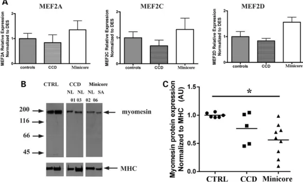
Increasing class II HDAC expression does not affect mef2 expression
Having established that down-regulation of RYR1 causes an in-crease in HDAC-4/5 expression, we reasoned that this elevation could lead to downstream effects on mef2, a master trans-activa-tor of skeletal muscle gene expression (22). Mef2 is sequestered by class II HDACs resulting in blockage of mef2-dependent gene transcription (23), and the RYR1 and miR-1/miR-133 genes contain intragenic mef2-dependent enhancer sequences that regulate their transcription in muscle (24,25). We therefore examined the patient’s muscle biopsies for: (i) mef2 content, to ascertain that there was no compensatory up-regulation of its expression owing to the increased expression of HDAC-4/5 and (ii) myomesin a protein that is transcriptionally regulated by mef2 (20,21). Figure8shows that the transcript levels of MEF2A, MEF2C and MEF2D are not significantly different between controls and biop-sies from patients. On the other hand, myomesin-1 protein levels normalized to MHC are significantly decreased (by ∼50% in biop-sies isolated from patients with minicores (Fig.8B and C).
HDAC-4/5 are also up-regulated in other congenital myopathies
To verify whether these observed effects (that is increased levels of HDACs, decreased levels of RyR1 and decreased levels of mus-cle-specific miRs) are specific for congenital myopathies owing to recessive RYR1 mutations or a more general response occurring in patients with other congenital myopathies, we analysed biop-sies from 11 patients with NM harbouring mutations in KBTBD13,
ACTA1 or NEB (NEM 6, NEM3 and NEM2, respectively). A similar decrease in muscle-specific miR-22, miR-133 and miR-1s was observed (Supplementary Material, Fig. S2). We also tested the muscle biopsies from the patients with NM for RyR1 content and HDAC-4 and HDAC-5 expression. Surprisingly, RyR1 protein content was significantly reduced (control versus NEM was 100 ± 27.9% versus 0.03 ± 0.02%, P < 0.01, Student’s t-test), and HDAC-4 and HDAC-5 protein levels were significantly increased (Supple-mentary Material, Fig. S2).
Discussion
Here, we identify a novel pathophysiological mechanism occur-ring in skeletal muscles of patients with congenital myopathies whereby activation of a cascade of events leads to the down-regu-lation of muscle-specific genes. We report that recessive com-pound heterozygous RYR1 mutations are accompanied by the following changes in skeletal muscle: (i) hypermethylation of the RYR1 gene, (ii) a 6- to 15-fold increase in class II HDAC expres-sion and (iii) reduction in muscle-specific miRs. Our results re-present a major advancement in thefield as to date the mode of action of recessive RYR1 mutations identified in patients with MmD, CNM and CFTD has been elusive and the functional characterization of cells harbouring such mutations has failed to yield a mechanism compatible with the disease phenotype (8,10,26). A regularfinding in muscle biopsies of patients with re-cessive RYR1 mutations has been a reduced expression level of RyR1 protein and transcript (2–4,26–28). This reduction appears to be muscle-specific and has not been observed in other tissues
Figure 3. The RYR1 is hypermethylated, and DNA methyltransferases 1 and 2 are up-regulated in muscles from Minicore patients. (A) Schematic representation showing the location of the CpG region III within the RYR1 gene, the position of the CpG sites (indicated by arrowheads) and the location of the 5′ CCGG 3′ HpaII/MspI site (arrowed); the location of the internal control region lacking HpaII/MspI sites is also shown, as well as the location of the PCR primers used to amplify the DNA. (B) Hypermethylation of CpG region III of the RYR1. Each symbol represents the mean relative methylation value from a patient (CTRL n = 4; Minicore n = 4). Experimental details are outlined in Materials and Methods. (C) DNA methyltransferase 1 (DNMT1) and DNMT2 are significantly up-regulated in muscles of Minicore patients. Each symbol represents the mean relative expression of DNMT1 (CTRL n = 5; Minicore n = 9) and DNMT2 (CTRL n = 4, Minicore n = 8) from a single patient normalized to the muscle-specific housekeeping gene DES (*P < 0.02, **P < 0.025, Student’s t-test).
ectopically expressing RYR1, such as B-lymphocytes (8). Because of these results, we set out to test the hypothesis whereby the mechanism leading to reduced RyR1 muscle expression may be
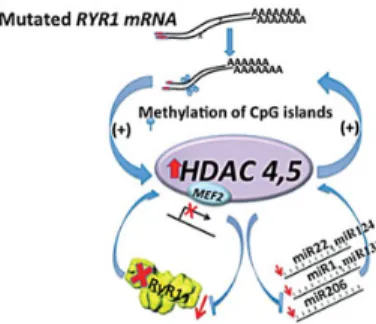
epigenetically regulated and the schematic representation de-picted in Figure9summarizes the results of the present study. The key point is that RYR1 mutations are accompanied by an
Figure 4. Class II HDACs are significantly up-regulated in muscle biopsies from patients with Congenital Muscle Disorders. (A) Western blot analysis of biopsies from a control muscle (Lane 1) and Minicore patients (Minicore SA03 and Minicore SA05). Fifty micrograms of total muscle protein extracts were separated on a 6% SDS–PAGE, blotted onto nitrocellulose and probed with anti-HDAC-4 and HDAC-5 antibodies. Lower portion of thefigure, loading control; the same blot was probed with anti-MHC recognizing all isoforms. (B) Quantification of HDAC-4 and HDAC-5 normalized to MHC in muscle biopsies of controls (circles), CCD patients (squares) and Minicore patients (triangles). HDAC-4 *P < 0.001, CTRL n = 9, CCD n = 5, Minicore n = 12; HDAC-5**P < 0.01 CTRL, n = 4, CCD n = 3, Minicore n = 9; ANOVA and Bonferroni multiple comparison test were performed. Each symbol represents results from a single patient. (C) No HDAC-4 protein is detectable in myotubes. (D) HDAC-4 transcript levels as assessed by qPCR in muscle biopsies from controls and Minicore patients ***P < 0.02, CTRL n = 4, Minicore n = 14; statistical analysis performed using Student t-test. (E) Confocal microscopy showing distribution of HDAC-4 in a muscle biopsy from a control (left panel) and Minicore patient. Arrows indicate co-localization of HDAC-4 and DAPI. Bar indicates 10 µm.
increased expression of class II HDACs. We are aware that an in-crease in HDAC expression has been reported in other conditions, including denervation, muscle atrophy, ALS and Huntington’s disease (29–34). However, we would like to point out that (i) such high levels of expression of class II HDACs have not been re-ported in any other human neuromuscular abnormalities inves-tigated so far; (ii) such an increase of HDAC is specific because the signal to background ratio of our set of data is 6–16 times greater than that reported in other muscle disorders and (iii) over-expression of HDAC-4 and HDAC-5 in mouse FDBfibres results in the down-regulation of RYR1 and of muscle-specific miRs. High levels of HDACs not only lead to chromatin condensation thereby decreasing gene transcription (21,35,36), but also seques-ter mef2 (23,37–39). In this context, it should be pointed out that there is no compensatory up-regulation of mef2 in muscles from the patients, so that up-regulation of HDACs would lead to the down-regulation of mef2-dependent proteins. That this is the case is supported by the fact that RYR1, myomesin and muscle-specific miRs containing mef2-dependent binding domains (21,24,25) are significantly down-regulated in the muscles of Minicore patients. Though few studies have focussed on the me-chanisms regulating RYR1 expression, the 5′ region of the human and porcine RYR1 gene contains, aside a mef2-binding domain (24), consensus sequences for the transcription factor SP1, for muscle-specific promoter elements as well as for a number of transcriptional activators (40). SP1 is a zincfinger transcription factor that binds to CG-rich regions present in many promoters.
Infibroblasts, SP1 interacts with HDAC-2 leading to the transcrip-tional silencing of the human telomerase reverse transcriptase (hTERT) gene in normal somatic cells (41). Whether SP1 can also interact with class II HDACs and whether this interaction is modified by CpG methylation also leading to repression of RYR1 gene transcription remains to be investigated.
Interestingly, the 3′ UTR of HDAC-4 and that of HDAC-5 have binding sites for miR-22/mir-124/miR-1/miR-206 and miR-206, respectively, and HDAC-4 is a target of miR-22 whose down-regu-lation potentiates its expression (19). Thus, it follows that a de-crease of miR-22, miR-124 and miR-1 activates a pathological loop leading to the further up-regulation of class II HDACs (Fig.9). This mechanism is compatible with and gives mechanis-tic insight to two previous observations: (i) other muscle-specific genes besides the RYR1 are down-regulated in patients with con-genital myopathies owing to RYR1 mutations (7) and (ii) the RYR1 was reported to be imprinted in some patients with MmD be-cause of epigenetic factors (42). The former observation is likely due to the sequestration of mef2 by class II HDACs (37–39). The latter observations on the other hand can be explained by the finding that the DNA methyltransferases DNMT1 and DNMT2 are over-expressed in muscle biopsies of Minicore patients, bringing about RYR1 hypermethylation. DNMT1 is a maintenance methyltransferases preserving methylation patterns but also has de novo activity (43). DNMT2 on the other hand is thought to par-ticipate in the recognition of damaged DNA and mutation repair (44). In fact, the two observations are mechanistically linked as hypermethylation goes hand in hand with HDAC activation and gene down-regulation (23,24,35,36,45); furthermore, DNA dam-age can activate DNA-methylation via activation of DNMT1 (46), resulting in a pathological loop that will ultimately shut down gene transcription of mef2-dependent genes.
As to the role miRs in neuromuscular diseases, this is still un-clear: over-expression of miR-22 is sufficient to cause cardiomyo-cyte hypertrophy (47) and several miRs are up-regulated in muscular dystrophies (48–50) but depending on the disease, some miRs may appear to be down-regulated (50). Interestingly, a recent study demonstrated that mice lacking miR-133 develop an adult onset CNM in type-2fibres, and this is accompanied by impaired mitochondrial function, fast to slow myofiber conver-sion and disarrangement of triads (51), histopathological changes very similar to those observed in recessive human RYR1-related myopathies. Though in the paper the authors con-clude that this is principally due to the dysregulation of dyna-min-2, one of miR-133′s targets, the similarities between the phenotype of the miR-133a knockout mice and that of patients with RYR1 mutations is striking and is indicative of a common pathophysiological pathway.
The main point emerging from our studies is that epigenetic factors are central culprits in recessive RYR1-linked myopathies. Our results also show that these factors are likely to also play a major role in other congenital muscle diseases such as NM. In support of a role of epigenetics, moderate exercise has been shown to improve the muscle function of some patients with congenital myopathies (52,53) and there is increasing evidence that physical activity influences DNA methylation in humans (53,54). Taken together our results suggest that a common patho-physiological mechanism is activated in skeletal muscles of patients with some congenital myopathies. The presence of mutations in muscle-specific genes (in this case RYR1) activates factors that lead to the up-regulation of DNMs and of class II HDACs, the master regulator of chromatin structure. Though the primary mechanism causing HDAC up-regulation is at the moment unclear, our data provide the proof of concept that
Figure 5. Correlation between DNA methylation and HDAC-4/HDAC-5 expression and up-regulation of DNA methyltransferases in muscles of patients with Minicore. The data shown in thisfigures were obtained from biopsies from four control individuals (empty squares) and from biopsies from four Minicore patients (filled squares) (Minicore NL02, Minicore NL03; Minicore SA03, Minicore SA05). (A) Correlation between HDAC-4 and RYR1 hypermethylation (correlation coefficient r = 0.4695). (B) Correlation between HDAC-5 and RYR1 hypermethylation (correlation coefficient r = 0.7925).
DNM and HDAC are potential pharmacological targets to treat a wide range of inherited neuromuscular conditions with different genetic backgrounds that as a common feature lead to a decrease in RyR1.
Materials and Methods
Quantitative PCR
Total RNA was extracted using Trizol (Life Technologies, #15596018). cDNA was synthesized with the High Capacity cDNA synthesis kit or Taqman microRNA Reverse Transcription kit (Applied Biosystems, #4366596). Transcript levels were quantified using Syber-Green reagent on an Applied Biosystem platform (7500 fast real-time PCR system); levels of expression from tripli-cate replicas were averaged and normalized to the content of the muscle-specific gene desmin (DES). In the case of human samples, because of the limited amount of biological material, not all biop-sies could be investigated for all genes. The sequences of the pri-mers used for qPCR are listed in Supplementary Material, Table S3.
MicroRNA determination
Quantification of selected miRs was performed using TaqMan master mix no-UNG 2 (Life Technologies, # PN 4427788) and the following miR assays (Life Technologies, # PN 4427975): miR-22, miR124a, miR-133a, miR-1, miR-206, miR-486-3p, miR-221 and miR-126. Each reaction was performed in triplicate, and the re-sults from each muscle sample were analysed and averaged. In human muscle biopsies, miR expression levels were normalized to RNU44 and to the muscle-specific genes DES and Actinin2 (ACTN2) that show similar Ct values in patients and healthy indi-viduals. In mouse FDBs, miR expression levels were normalized to U6 snRNA. In the case of human samples, because of the limited amount of biological material, not all biopsies could be investigated for all microRNAs.
DNA methylation
Total genomic DNA was isolated using the GeneElute mammalian genomic DNA Miniprep kit (Sigma Genosys). DNA methylation
Figure 6. In vivo over-expression of HDAC-4 and HDAC-5 causes down-regulation of RYR1 and of muscle-specific miRs. Each symbol shows the mean triplicate relative expression value of the indicated transcript normalized to the indicated gene. Circles, control FDBfibres mock transfected with the empty pIRES2-dsRed2 plasmid; squares, FDBfibres transfected with a plasmid encoding mouse HDAC-4 and HDAC-5. HDAC-4; *P < 0.0001, n = 6; HDAC-5 **P < 0.005, n = 6; RYR1 **P < 0.005, CTRL n = 4, HDAC-4 and 5 n = 6; miR-22 and miR-1; ***P < 0.045, n = 6; miR-133 ****P < 0.035, n = 6; miR-206 ****P < 0.035, n = 5; Myomesin ****P < 0.035, n = 4; miR-124 was quantified and no significance change was observed. Statistical analysis was performed using the Student’s t-test.
was assessed by PCR amplification of genomic DNA digested with the methyl-sensitive HpaII and MspI restriction enzymes (55) (see schematic representation in Fig.3A). Restriction enzyme digestion reactions were carried out overnight at 37°C, with HpaII or MspI (New England BioLabs), infinal volume of 20 µl. The primers used for PCR amplification are listed in Supple-mentary Material, Table S3 as: Human RYR1 CG-rich (F and R) amplifying the CpG-III region of the RYR1 gene and Human
RYR1 Control (F and R), amplifying a control region of the RYR1 gene lacking HpaII/MspI sites (see Fig.3A). To determine the extent of DNA methylation, theΔCt values were first ob-tained comparing the CpG-III and control PCRs of HpaII diges-tions, and then the ΔΔCt values were generated using as reference the samples exhibiting the higherΔCt. Higher ΔΔCt values indicate higher extent of DNA methylation at the CpG-III RyR1 MspI/HpaII cleavage sites.
Figure 7. Down-regulation of RYR1 by siRNA leads to up-regulation of HDAC-4 and HDAC-5. Each symbol shows the mean triplicate value relative expression level of the indicated transcript normalized to U6 snRNA (miR expression) or DES (RYR and HDAC expression), infibres isolated from a single mouse. Circles, control (mock) transfection with a scrambled siRNA; squares FDB transfected with siRYR RNA (see Materials and Methods for details). RYR1 *P < 0.0001, n = 7; HDAC-4 **P < 0.04, CTRL n = 6, siRYR1 n = 7. HDAC-5; **P < 0.04, CTRL n = 6, siRYR1 n = 9. miR-22, miR-133, miR-124 and miR-1 show no statistical difference. Statistical analysis was performed using the Student’s t-test.
Figure 8. Increased expression of HDAC-4/HDAC-5 leads to a decrease in the content of myomesin, without affecting mef2. (A) mef2A, mef2C and mef2D transcript levels are similar in muscle biopsies of patients and controls (expression levels normalized to DES). (B) Representative western blot showing that the protein content of myomesin is lower in patients with Minicore compared with healthy individuals (letters and numbers refer to patient no.; see Supplementary Material, Table S1); (C). Quantification of myomesin protein content in muscle biopsies: controls, circles (n = 6); CCD, squares (n = 5); Minicore, triangles (n = 9). *P < 0.02. Statistical analysis was performed using ANOVA and Bonferroni multiple comparison test (95% confidence interval).
Electrophoresis and immunoblotting
Total muscle proteins were extracted in 10 m Hepes pH 7.0, 150 m NaCl, 1 m EDTA and anti-protease (Roche, # 11873580001). Pro-tein concentration was determined using ProPro-tein Assay Kit II (Bio-Rad Laboratories) using BSA as a standard. SDS–PAGE, pro-tein transfer on to nitrocellulose membranes and immunostain-ing were performed as described previously (2,8). The following primary antibodies were used: mouse anti-RyR1 (Ryanodine 1 Re-ceptor, Thermo Scientific, # MA3-925), mouse anti-MHC (MHC, Millipore, #05-716), Rabbit anti-HDAC-4 (Histone Deacetylase 4, Cell Signaling, #2072) and rabbit anti- HDAC-5 (Histone Deacety-lase 5, Abcam #1439), and rat anti-myomesin was a generous gift of Prof. Mathias Gautel, King’s College, London, UK (56). Sec-ondary peroxidase conjugates were Protein G–peroxidase (Sigma, #P8170) and peroxidase-conjugated goat anti-mouse IgG (Sigma, #A2304). The immunopositive bands were visualized by chemilu-minescence using the Super Signal West Dura kit (Thermo Scien-tific). In order to perform statistical analysis, the intensity of the immunopositive bands was determined using ImageJ/FIJI. The intensity values were normalized to the intensity of the indicated muscle-specific housekeeping protein. The value (arbitrary units) obtained from the patient’s biopsies were divided by the mean value obtained from control biopsies and are expressed as 100%.
Mouse musclefibre electroporation and isolation
The procedure was as described by DiFranco et al. (57). Briefly, 8- to
14-week-old mice were anaesthetized using isofluorane, and 7.5 µl of 2 mg/ml Hyaluronidase in RNase-free Tyroide’s Buffer (Sigma Fine Chemicals, #H3506) was injected under the footpad. The mice were left 1 h under supervision, and subsequently, the fol-lowing constructs were injected into the footpad: pCMV6-HDAC4 (Origene #MR211598) and pCMV6-HDAC5 (Origene # MC202550) whereas control mice (mock transfected) received 20 µg of pIRES2-dsRed2 plasmid (Clonetech #632420). For siRNA silencing experiments, 6 nmol of RNA either specific for the RYR1 (Ambion; siRNA RyR1-#4390771) or a scrambled siRNA sequence (Negative control 2-#4390845) were used. siRNA-transfected FDBs were also injected with lipofectamine RNAiMAX (Invitrogen, #13778-030). Ten minutes post-injection, FDBs were electroporated using acupuncture needles placed parallel and perpendicular to the long axis of the foot (with 1 cm distance), and twenty pulses (100v/cm, 20 ms duration and 1 Hz of frequency) were given. Six
to ten days post–transfection, the mice were sacrificed and FDBs were isolated by enzymatic dissociation at 37°C for 60 min in Krebs Ringer solution no Ca2+( pH 7.4), containing 0.2%
collage-nase I (Sigma Fine Chemicals, C-0130). Enzymatic digestion was terminated by washing the muscle with Tyrode’s solution (pH 7.4), and singlefibres were isolated and total protein extracts prepared or RNA was extracted and analysed by qPCR.
Ca2+measurements
Primary skeletal muscle cultures and cell imaging were per-formed as previously described (58).
Confocal microscopy and immunofluorescence
Biopsies were embedded for pathological examination and sliced using a cryostat (10 µm thickness). Cryosections werefixed with methanol: acetone (1:1) for 30 min and then incubated in the fol-lowing solutions for 90 min at room temperature: blocking solu-tion (Roche, #115000694011), rabbit anti-HDAC-4 (Cell Signaling, #2072) and Alexa Fluor 647-conjugated anti-Rabbit IgG (Life Tech-nologies, #A21245). Nuclear staining was performed using DAPI (Invitrogen, #D21490), and slides were mounted with mounting medium (Sigma, #1000-4) and sealed hermetically with 1.5-mm-thick coverslip. A Nikon A1R Confocal microscope was used for 3D image acquisition with a 40× oil objective (N.A. = 1.3). Images were analysed using threshold co-localization function in ImageJ2/FIJI program.
Compliance with ethical standards
All procedures performed in studies involving human partici-pants were in accordance with the ethical standards of the insti-tutional and/or national research committee and with the 1964 Helsinki declaration and its later amendments or comparable ethical standards. This study was approved by the Ethikkommis-sion beider Basel ( permit No. EK64/12); all subjects gave written informed consent to carry out this work.
All applicable international, national and/or institutional guidelines for the care and use of animals were followed. All pro-cedures performed in studies involving animals were in accord-ance with the ethical standards of the institution or practice at which the studies were conducted. Experiments on mouse mus-cles were approved by the local Cantonal Veterinary authorities ( permit No. 2658).
Statistical analysis and graphical software
Statistical analysis was performed using the Student’s t-test; means were considered statistically significant when the P-value was < 0.05. When more than two groups were compared, analysis was performed using the ANOVA test followed by the Bonferroni post hoc test using the statistical package included in GraphPad Prism 6.0 software. Origin 6 was used to generate dose–response curves. Images were assembled using Adobe Photoshop CS (version 8.0).
Supplementary Material
Supplementary Material is available at HMG online.
Figure 9. Cartoon depicting how mutations in RYR1 lead to a decrease in RyR1 content thereby leading to weak muscles. Mutations lead to DNA hypermethylation and HDAC-4/HDAC-5 over-expression. This causes mef2 sequestration thereby inhibiting transcription of genes regulated by mef2, including the RYR1 and muscle-specific miRs. A decrease in RyR1 would severely affect muscle excitation–contraction coupling because this calcium channel is a central player in this mechanism, releasing the calcium necessary for muscle contraction from the sarcoplasmic reticulum.
Acknowledgements
We thank Dr Joery Molenaar for contacting the patients, gather-ing their written permission and organizgather-ing the transport of the material from the Dutch patients.
Conflict of Interest statement. None declared.
Funding
This work was supported by a grant from the Swiss National Science Foundation (SNF No. 31003A-146198), by a grant from the Myotubu-lar Myopathy (grant No. 12KCL 01-MT) and by a grant from the Basel Neuromuscular Association (NeRAB). O.R. was supported by a grant from the Botnar Stiftung. The support of the Department of Anaesthesia Basel University Hospital and the technical support of Anne-Sylvie Monnet are gratefully acknowledged.
References
1. Maggi, L., Scoto, M., Cirak, S., Robb, S.A., Klein, A., Lillis, S., Cullup, T., Feng, L., Manzur, A.Y., Sewry, C.A. et al. (2013) Con-genital myopathies–clinical features and frequency of indi-vidual subtypes diagnosed over a 5-year period in the United Kingdom. Neuromuscul. Disord., 23, 195–205.
2. Zhou, H., Jungbluth, H., Sewry, C.A., Feng, L., Bertini, E., Bush-by, K., Straub, V., Roper, H., Rose, M.R., Brockington, M. et al. (2007) Molecular mechanisms and phenotypic variation in RYR1-related congenital myopathies. Brain, 130, 2024–2036. 3. Wilmhurst, J.M., Lillis, S., Zhou, H., Pillay, K., Henderson, H.,
Kress, W., Müller, C.R., Ndondo, A., Cloke, V., Cullup, T. et al. (2010) RYR1 mutations are a common cause of congenital my-opathies with central nuclei. Ann. Neurol., 68, 717–726. 4. Clarke, N.F., Waddell, L.B., Cooper, S.T., Perry, M., Smith, R.L.,
Kornberg, A.J., Muntoni, F., Lillis, S., Straub, V., Bushby, K. et al. (2010) Recessive mutations in RYR1 are a common cause of Con-genital Fiber Type disproportion. Hum. Mutat., 31, E1544–E1550 5. Franzini-Armstrong, C. and Protasi, F.G. (1997) Ryanodine
re-ceptors of striated muscles: a complex channel capable of multiple interactions. Physiol. Rev., 77, 699–729.
6. Treves, S., Jungbluth, H., Muntoni, F. and Zorzato, F. (2008) Con-genital muscle disorders with cores: the ryanodine receptor calcium channel paradigm. Curr. Opin. Pharmacol., 8, 319–326. 7. Zhou, H., Rokach, O., Feng, L., Munteanu, I., Mamchaoui, K.,
Wilmshurst, J.M., Sewry, C., Manzur, A.Y., Pillay, K., Mouly, V. et al. (2013) RYR1 deficiency in congenital myopathies disrupts excitation-contraction coupling. Hum. Mutat., 34, 986–996. 8. Attali, R., Aharoni, S., Treves, S., Rokach, O., Becker-Cohen, M.,
Fellig, Y., Straussberg, R., Do, T., Daana, M., Mitrani-Rosen-baum, S. and Nevo, Y. (2013) Variable myopathic presentation in a single family with novel skeletal RYR1 mutation. PLoS One, 8, e69296. doi:10.1371/journal. pone. 0069296.
9. Emery, A.E. (1991) Population frequencies of inherited neuromus-cular diseases—a world survey. Neuromuscul. Disord., 1, 19–29. 10. Zhou, H., Yamaguchi, N., Xu, L., Wang, Y., Sewry, C.,
Jung-bluth, H., Zorzato, F., Bertini, E., Muntoni, F., Meissner, G. and Treves, S. (2006) Characterization of recessive RYR1 mu-tations in core myopathies. Hum. Mol. Genet., 16, 2791–2803. 11. Chen, J.F., Mandel, E.M., Thomson, J.M., Wu, Q., Callis, T.E.,
Hammond, S.M., Conlon, F.L. and Wang, D.Z. (2006) The role of microRNA-1 and microRNA-133 in skeletal muscle prolif-eration and differentiation. Nat. Genet., 38, 228–233.
12. Kim, H.K., Lee, Y.S., Sivaprasad, U., Malhotra, A. and Dutta, A. (2006) Muscle-specific microRNA miR-206 promotes muscle differentiation. J. Cell. Biol., 174, 677–687.
13. Small, E.M., O’Rourke, J.R., Moresi, V., Sutherland, L.B., McAn-ally, J., Gerard, R.D., Richardson, J.A. and Olson, E.N. (2010) Regulation of PI3-kinase/Akt signaling by muscle enriched microRNA-486. Proc. Natl Acad. Sci. USA., 107, 4218–4223. 14. Wang, M., Zhao, C., Shi, H., Zhang, B., Zhang, L., Zhang, X.,
Wang, S., Wu, X., Yang, T., Huang, F. et al. (2014) Deregulated microRNAs in gastric cancer tissue-derived mesenchymal stem cells: novel biomarkers and a mechanism for gastric cancer. Br. J. Cancer., 110, 1199–1210.
15. Sun, X., Wang, Z.M., Song, Y., Tai, X.H., Ji, W.Y. and Gu, H. (2014) MicroRNA-126 modulates the tumor microenviron-ment by targeting calmodulin-regulated spectrin-associated protein 1 (Camsap1). Int. J. Oncol., 44, 1678–1684.
16. Jungbluth, H., Sewry, C. and Muntoni, F. (2011) Core myop-athies. Semin. Pediatr. Neurol., 18, 239–249.
17. Workmann, J.L. and Kingston, R.E. (1998) Alteration of nucleo-some structure as a mechanism of transcriptional regulation. Annu. Rev. Biochem., 67, 545–579.
18. Lu, J., McKinsey, T.A., Zhang, C.L. and Olson, E.N. (2000) Regu-lation of skeletal myogenesis by association of the mef2 tran-scription factor with class II Histone deacetylases. Mol. Cell., 6, 233–244.
19. Zhang, J., Yang, Y., Yang, T., Liu, Y., Li, A., Fu, S., Wu, M., Pan, Z. and Zhou, W. (2010) microRNA-22, downregulated in hepato-cellular carcinoma and correlated with prognosis, suppresses cell proliferation and tumourigenicity. Br. J. Cancer, 103, 1215–1220.
20. Potthoff, M.J., Arnold, M.A., McAnally, J., Richardson, J.A., Bas-sel-Duby, R. and Olson, E.N. (2007) Regulation of skeletal muscle sarcomere integrity and postnatal muscle function by Mef2c. Mol. Cell Biol., 27, 8143–8151.
21. Hinits, Y. and Hughes, S.M. (2007) Mef2s are required for thick filament formation in nascent muscle fibres. Development, 134, 2511–2519.
22. Naya, F.J. and Olson, E. (1999) MEF2: a transcriptional target for signaling pathways controlling skeletal muscle growth and differentiation. Curr. Opin. Cell Biol., 11, 683–688. 23. McKinsey, T.A., Zhang, C.L., Lu, J. and Olson, E.N. (2000)
Signal-dependent nuclear export of a histone deacetylase regulates muscle differentiation. Nature, 408, 106–111. 24. Schmoelzl, S., Leeb, T., Brinkmeier, H., Brem, G. and Brenig, B.
(1996) Regulation of tissue-specific expression of the skeletal muscle ryanodine receptor gene. J. Biol. Chem., 271, 4763–4769. 25. Liu, N., Williams, A.H., Kim, Y., McAnally, J., Bezprozvannaya, S., Sutherland, L.B., Richardson, J.A., Bassel-Duby, R. and Olson, E.N. (2007) An intragenic MEF2-dependent enhancer di-rects muscle-specific expression of microRNAs 1 and 133 Mef2 and myomesin. Proc. Natl Acad. Sci. USA, 104, 20844–20849. 26. Zhou, H., Lillis, S., Loy, R.E., Ghassemi, F., Rose, M.R., Norwood,
F., Mills, K., Al-Sarraj, S., Lane, R.J., Feng, L. et al. (2010) Multi-minicore disease and atypical periodic paralysis associated with novel mutations in the skeletal muscle ryanodine recep-tor (RYR1) gene. Neuromuscul. Disord., 20, 166–173.
27. Monnier, N., Marty, I., Faure, J., Castiglioni, C., Desnuelle, C., Sac-coni, S., Estournet, B., Ferreiro, A., Romero, N., Laquerriere, A. et al. (2008) Null mutations causing depletion of the type 1 ryanodine receptor (RYR1) are commonly associated with recessive struc-tural congenital myopathies with cores. Hum. Mutat., 29, 670–678. 28. Bevilacqua, J.A., Monnier, N., Bitoun, M., Eymard, B., Ferreiro, A., Monges, S., Lubieniecki, F., Taratuto, A.L., Laquerrière, A., Claeys, K.G. et al. (2011) Recessive RYR1 mutations cause un-usual congenital myopathy with prominent nuclear internal-ization and large areas of myofibrillar disorganinternal-ization. Neuropathol. Appl. Neurobiol., 37, 271–284.
29. Bongers, K.S., Fox, D.K., Ebert, S.M., Kunkel, S.D., Dyle, M.C., Bullard, S.A., Dierdorff, J.M. and Adams, C.M. (2013) Skeletal muscle denervation causes skeletal muscle atrophy through a pathway that involves both Gadd45a and HDAC4. Am. J. Physiol. Endocrinol. Metab., 98, 4089–4096.
30. Bruneteau, G., Simonet, T., Bauché, S., Mandjee, N., Malfatti, E., Girard, E., Tanguy, M.L., Behin, A., Khiami, F., Sariali, E. et al. (2013) Muscle histone deacetylase 4 upregulation in amyotrophic lateral sclerosis: potential role in reinnervation ability and disease progression. Brain, 136, 2359–2368. 31. Mielcarek, M., Landles, C., Weiss, A., Bradaia, A., Seredenina,
T., Inuabasi, L., Osborne, G.F., Wadel, K., Touller, C., Butler, R. et al. (2013) HDAC4 reduction: a novel therapeutic strategy to target cytoplasmic huntingtin and ameliorate neurodegen-eration. PLOS Biol., 11, e1001717.
32. Cohen, T.J., Waddell, D.S., Barrientos, T., Lu, Z., Feng, G., Cox, G.A., Bodine, S.C. and Yao, T.P. (2007) The histone deacetylase HDAC4 connects neural activity to muscle transcriptional re-programming. J. Biol. Chem., 282, 33752–33759.
33. Consalvi, S., Saccone, V., Giordani, L., Minetti, G., Mozzetta, C. and Puri, P.L. (2011) Histone deacetylase inhibitors in the treatment of muscular dystrophies: epigenetic drugs for genetic diseases. Mol. Med., 17, 457–465.
34. Falkenberg, K.J. and Johnstone, R.W. (2014) Histone de-acetylases and their inhibitors in cancers, neurological dieases and immune disorders. Nature Rev. Drug. Discov., 13, 673–691.
35. Wang, Z., Qin, G. and Zhao, T.C. (2014) HDAC4: mechanism of regulation and biological functions. Epigenomics, 6, 139–150. 36. Baylin, S.B., Esteller, M., Rountree, M.R., Bachmanm, K.E.,
Schuebelm, K. and Herman, J.G. (2001) Aberrant patterns of DNA methylation, chromatin formation and gene expression in cancer. Hum. Mol. Genet., 10, 687–692.
37. Lu, J., McKinsey, T.A., Nicolm, R.L. and Olson, E.N. (2000) Sig-nal dependent activation of the MEF2 transcription factor by dissociation from histone deacytlases. Proc. Natl Acad. Sci. USA, 97, 4070–4075.
38. Miska, E.A., Karlsson, C., Langley, E., Nielesen, S.J., Pines, J. and Kouzaridesm, T. (1999) HDAC4 deacetylase associates with and represses the MEF2 transcription factor MEF2 tran-scription factor. EMBO J., 18, 5099–5107.
39. Wang, A.H., Bertos, N.R., Vezmar, M., Pelletier, N., Crosato, M., Heng, H.H., Th’ng, J., Han, J. and Yang, X.L. (1999) HDAC4, a human histone deacetylase related to yeast HDA1, is a tran-scriptional corepressor. Mol. Cell. Biol., 19, 7816–7827. 40. Phillips, M.S., Fujii, J., Khanna, V.K., DeLeon, S., Yokobata, K.,
DeJong, P. and MacLennan, D.H. (1996) The structural organ-ization of the human skeletal muscle ryanodine receptor (RYR1) gene. Genomics, 34, 24–41.
41. Won, J., Yim, J. and Kim, T.K. (2002) Sp1 and Sp3 recruit his-tone deacetylase to repress transcription of human telomer-ase reverse transcripttelomer-ase (hTERT) promoter in normal human somatic cells. J. Biol. Chem., 277, 38230–38238. 42. Zhou, H., Brockington, M., Jungbluth, H., Monk, D., Stanier, P.,
Sewry, C.A., Moore, G.E. and Muntoni, F. (2006) Epigenetic al-lele silencing unveils recessive RYR1 mutations in core myop-athies. Am. J. Hum. Gen., 79, 859–868.
43. Denis, H., Ndlovu, M.N. and Fuks, F. (2011) Regulation of mammalian DNA methyltransferases: a route to new me-chanisms. EMBO Reports, 12, 647–656.
44. Subramaniam, D., Thombre, R.T., Dhar, A. and Anant, S. (2014) DNA methyltransferases: a novel target for prevention and therapy. Front. Oncol., 4. doi:10.3389/fonc.2014.00080. 45. Newell-Price, J., Clark, A.J. and King, P. (2000) DNA
methyla-tion and silencing of gene expression. Trends. Endocrinol. Metab., 11, 142–148.
46. Cuozzo, C., Porcellini, A., Angrisano, T., Morano, A., Lee, B., Di Pardo, A., Messina, S., Iuliano, R., Fusco, A., Santillo, M.R. et al. (2007) DNA damage, homology-directed repair, and DNA methylation. PLOS Genet, 3, e110.
47. Huang, Z.P., Chen, J., Seok, H.Y., Zhang, Z., Kataoka, M., Hu, X. and Wang, D.Z. (2013) MicroRNA-22 regulates cardiac hypertrophy and remodeling in response to stress. Circ. Res., 112, 1234–1243. 48. Eisenberg, I., Eran, A., Nishinom, I., Moggio, M., Lamperti, C.,
Amatom, A.A., Lidov, H.G., Kang, P.B., North, K.N., Mitrani-Ro-senbaum, S. et al. (2007) Distinctive patterns of microRNA ex-pression in primary muscle disorders. Proc. Natl Acad. Sci. USA, 104, 17016–17021.
49. Dmitriev, P., Stankevicins, L., Ansseau, E., Petrov, A., Barat, A., Dessen, P., Robert, T., Turki, A., Lazar, V., Labourer, E. et al. (2013) Defective regulation of microRNA target genes in myo-blasts from fascioscapulohumeral dystrophy patients. J. Biol. Chem., 288, 34989–35002.
50. Chen, J.F., Callis, T.E. and Wang, D.Z. (2009) microRNAs and muscle disorders. J. Cell Sci., 122, 13–20.
51. Liu, N., Bezprozvannaya, S., Shelton, J.M., Frisard, M.I., Hulver, M.W., McMillan, R.P., Wu, Y., Voelker, K.A., Grange, R.W., Rich-ardson, J.A., Bassel-Duby, R. and Olson, E.N. (2011) Mice lacking microRNA 133a develop dynamin 2-dependent centronuclear myopathy. J. Clin. Invest., 121, 3258–3268.
52. Vosin, S., Eynon, N., Yan, X. and Bishop, D.J. (2014) Exercise training and DNA methylation in humans. Acta. Physiol., doi:10.1111/apha.12414.
53. Ling, C. and Rönn, T. (2014) Epigenetic adaptation to regular exercise in humans. Drug Discov. Today, 19, 1015–1018. 54. Barrès, R., Yan, J., Egan, B., Treebak, J.T., Rasmussen, M., Fritz,
T., Caidahl, K., Krook, A., O’Gorman, D.J. and Zierath, J.R. (2012) Acute exercise remodels promoter methylation in human skeletal muscle. Cell Metabol., 15, 405–411.
55. Lee, W.H., Isaacs, W.B., Bova, G.S. and Nelson, W.G. (1997) CG island methylation changes near the GSTP1 gene in prostatic carcinoma cells detected using the polymerase chain reac-tion: a new prostate cancer biomarker. Cancer Epidemiol. Bio-markers Prev., 6, 443–450.
56. Obermann, W.M.J., Gautel, M., Steiner, F., van der Ven, P.F., Weber, K. and Fürst, D.O. (1996) The structure of the sarcomeric M band: localization of defined domains of myo-mesin, M-protein and the 250 kD carboxy-terminal region of titin by immunoelectron microscopy. J. Cell Biol., 134, 1441–1453.
57. DiFranco, M., Quinonez, M., Capote, J. and Vergara, J. (2009) DNA transfection of mammalian skeletal muscles using in vivo electroporation. J. Vis. Exp., pii: 1520. doi:10.3791/1520. 58. Ducreux, S., Zorzato, F., Müller, C., Sewry, C., Muntoni, F., Quinlivan, R., Restagno, G., Girard, T. and Treves, S. (2004) Ef-fect of ryanodine receptor mutations on interleukin-6 release and intracellular calcium homeostasis in human myotubes from malignant hyperthermia-susceptible individuals and patients affected by central core disease. J Biol. Chem., 279, 43838–43846.
![Figure 4B shows the relative content of HDAC-4 and HDAC-5 normalized for MHC content in biopsies from all the availableFigure 1.Myotubes from Minicore patients harbouring recessiveRYR1mutations do not show alterations of the resting [Ca2+] nor decreased Ca](https://thumb-eu.123doks.com/thumbv2/123doknet/14902935.654571/3.918.153.767.99.521/normalized-availablefigure-myotubes-minicore-harbouring-recessiveryr-mutations-alterations.webp)