With 2 figures in the text
Changes in the surface of Dipetalonema viteae (Filarioidea) during its
development as shown by comparative peptide mapping
WERNER BASCHONG*
Swiss Tropical Institute, Socinstrasse 57, O//-4051 Basel, Switzerland (Accepted 15 August 1984)
SUMMARY
The cuticle of parasitic nematodes, the main contact site with the host, plays an important role in host-parasite interaction and thus also in immunological control. We compared different surface-iodinated life-stages of the filarial worm Dipetalonema viteae (microfilariae, infective 3rd-stage larvae (L3), adult males and females) with respect to changes in their
surface composition. Autoradiographs of peptide maps show that all stages present an identical set of peptide spots reflecting common surface protein(s). Spots specific for larvae L3 show that the composition of the iodinated surface differs in microfilariae and adults i.e.
it changes during development. Adults show a spot typical for males or females. Identical spots are found in L3. This suggests that a surface component is also sex specific.
INTRODUCTION
Nematodes undergo a series of moults during their life-cycle, shedding the external cuticle. The latter plays an important role in hostr-parasite relationships, since immu-nological control of parasitic helminths depends mainly on the structure of the contact site with the host, namely the cuticle (Lumsden, 1975; Mackenzie, Preston & Ogilvie, 1978; Ogilvie, Philipp, Jungery, Maizels, Worms & Parkhouse, 1981; Maizels, Philipp & Ogilvie, 1982). The occurrence of stage-specific surface antigens as observed for larvae of Dipetalonema viteae (Filarioidea) by Weiss & Tanner (1981) could mean that the structure of the contact site changes during development into the adult worm. Stage-and environment-dependent alterations of the composition of the cuticle are also indicated for other parasitic nematodes (Philipp, Parkhouse & Ogilvie, 1980; Maizels
et al. 1982; and very recently, Canlas & Piessens, 1984; Ortega-Pierres, Chayen, Clark
& Parkhouse, 1984; Philipp & Rumjaneck, 1984). A consequence of these structural changes, in or on the surface, is the exposure of a different set of potential antigens to the host's immunological system at each stage. Resulting changes in the immune response of the host can become most valuable for immunodiagnosis but such changes might also enable the parasite to overcome the infected host's immunological defence (Maizels et al. 1982; Philipp & Rumjaneck, 1984). We wanted therefore to investigate whether alterations at the level of proteins take place in the cuticle of D. viteae.
We iodinated the surface of different life-stages of the filarial parasite D. viteae as described earlier (Baschong & Rudin, 1982) and subsequently submitted these different stages to peptide mapping. This method was adapted to the needs required for detecting developmental changes of this parasite's surface structure.
* Present address: Biozentrum of the University of Basel, Department of Microbiology, Klingel-bergstrasse 70, CH-4056 Basel, Switzerland.
unguiculatus and the soft tick, Ornithodorus moubata as described by Worms, Terry &
Terry (1961). Male golden hamsters (60-80 g) came from a randomly bred colony (strain LAKZ, Institut fur Zuchthygiene der Universitat Zurich, Switzerland).
Adult worms were harvested from hamsters according to the method described by Baschong, Tanner, Betschart, Rudin & Weiss (1982), 3rd-stage larvae (L3) from infected
ticks by the method of Gass, Tanner & Weiss (1979), and microfilariae from in vi<ro-maintained female worms as described by Weiss & Tanner (1979).
Surface iodination
The different life-stages of D. viteae were iodinated with Na125I (EIR, Wiirenlingen, Switzerland) using (10/tm/tube) chloroglycoluril (trade name IODO-GEN™, Pierce Chemicals Co., Rockford, Illinois, USA) as a catalyst as described by Baschong & Rudin (1982).
Peptide mapping
Iodinated organisms were homogenized (2 females, 5 males in a 5 ml glass homogenizer; approximately 50000 microfilariae and 1000 L3 by ultrasonication on ice) in 1 ml of
0 1 M NH4HCO3. The homogenates were incubated in centrifugation tubes overnight at
ambient temperature with 25 fi\ of bovine trypsin (2 mg/ml in 0 1 M NH4HCO3,
TPCK-treated, Merck, Darmstadt, FRG) with stirring. The reaction mixture was then centrifuged (27000 g, 90 min, 4 °C) and the supernatant fraction discarded. The sediment was resuspended in 200 fi\ of performic acid (1/10 H2O2 (30 %) in concentrated
formic acid, Merck, Darmstadt, FRG, incubated for 2h at ambient temperature) and thus oxidized for 1 h on ice. The performic acid was evaporated (approximately 3 h, 10~2 Torr) and the sediment resuspended in 1 ml of 0 1 M NH4HCO3. This suspension was
digested with trypsin and centrifuged as described above. Between 1 and 2 x 10⁶ c.p.m. from each supernatant fraction (males, females, L3 and microfilariae) were lyophilized,
dissolved in concentrated formic acid and applied to a 20 x 20 plastic foil coated with cellulose (Polygram CEL-300, Macherey-Nagel, D-5160 Diiren, FRG). Peptide mapping was carried out essentially as described by Gracy (1977). Electrophoresis was done at 1000 volts for 30 min at 10 °C on a flat-bed system (Camag, Muttenz, Switzerland) using pyridine acetic acid (1:1, each 2-5 % (v/v)) as a buffer. Chromatography was performed
after drying overnight in AcOH/BuOH/Pyr/H2O 1:5:4:4. S-5X-ray film (Kodak,
Rochester, NY) was used for autoradiography. Prior to autoradiography peptide maps were sprayed with POPOP (2, 2-p-phenylene-6is(5-phenyloxazol), Merck, Darmstadt, FRG) to enhance sensitivity. Films were developed after 14-40 days of exposure.
RESULTS
Comparing only the most evident spots (Figs 1 and 2), the autoradiographs of the peptide maps of different life-stages of surface iodinated D. viteae (microfilariae L3,
females and males) show a common set of peptide spots for all stages. Adults produced a sex-specific spot in the cathodic region. The male spot migrated in the electrophoretic field in a position very similar to the female one (1st dimension), but chromatographed differently (2nd dimension). Identical' sex spots' were found in the L3 pattern, reflecting
Common: 9, 6, L3, mf electrophoresis a S 2 00
I
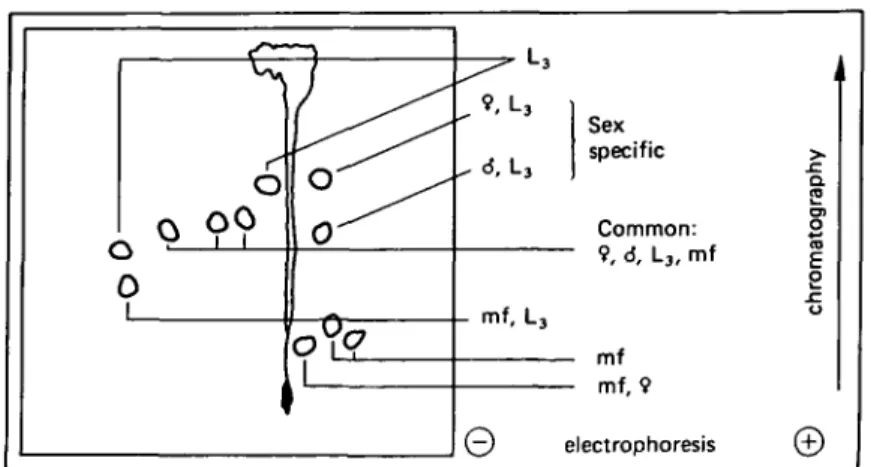
Fig. 1. Schematic representation of characteristic radioactive peptide spots of different stages of surface-iodinated Dipetalone.ma viteae analysed by peptide mapping, summarizing the major spots presented in Fig. 2 which indicates the peptide spot pattern shared by all stages and spots distinct for a stage or the sex, respectively,
mf, microfilariae. L3, infective 3rd-stage larvae;
the mixed population of L3 females and L3 males. They could not be detected in
microfilariae. A common spot between microfilariae and females was visible. Both microfilariae and L3 possess a peptide which was very positively charged. L3 showed
additional peptides, one being in this same region, whereas microfilariae showed two characteristic spots chromatographing low in the cathodic region. Spots specific for the adult stage exclusively could not be identified accurately.
DISCUSSION
Stage- and sex-dependent differences in protein extracts of D. viteae have been observed recently when analysing homogenates of whole worms (Priisse, Diesfeld & Vollmer, 1982; Lucius, Rauterberg& Diesfeld, 1983). However, changes in this parasite's cuticle have so far been indicated only by the observation of Weiss & Tanner (1981) who reported the occurrence of stage-dependent surface antigens of larvae. Precise knowledge of the macromolecules constituting the cuticle of parasitic nematodes is scarce and its surface composition is reported to vary considerably from species to species (Maizels et al. 1982). In different nematodes proteins of collagenous and non-collagenous origin have been reported to form a part of the surface structure. Some of these constituents are highly cross-linked by disulphide bridges and can be glycosylated (Lumsden, 1975; Adams, 1978; Bird, 1980; Clark, Philipp & Parkhouse, 1982; Maizels
et al. 1982; Murray, Waite, Tanzer & Hauschka, 1982; Philipp & Rumjaneck, 1984).
Moreover, a surface-specific antigen prepared from D. viteae (Baschong et al. 1982) binds strongly to lectins specific for oc iV-acetyl-galactosamine and for fucose (Baschong, unpublished results).
Specific iodination (Baschong & Rudin, 1982) was used to select for material exposed at or near the surface, and homogenates of the different stages of D. viteae were extracted with phosphate-buffered saline (PBS) to eliminate structures which raise mainly somatic antibodies (Baschong et al. 1982). We accounted for the possible presence of cuticular material extensively cross-linked by disulphide bridges (see above) as follows. In its
•
mf
Fig. 2. Autoradiographs of peptide maps from surface-iodinated different life-stages of
Dipetalonema viteae filarial worms: ($) adult females; (<J) adult males; (mf) microfilariae and
(L3) infective 3rd-stage larvae. © and 0 , Polarity of electrophoresis; (f) direction of
chromatography. (A) Spots common in all stages investigated, (D) common in mf and L3, (#)
common in $ and mf. Sex-specific spots (V) for $ and (A) for cJ. ( • ) Specific for L3 and (O)
specific for mf.
cross-linked state this should be susceptible to trypsin cleavage in a rather limited way and by digesting PBS-extracted worm homogenates and discarding the supernatant fractions after centrifugation we hoped to reduce our assay to mainly S—S cross-linked structures. The remaining sediment was treated with performic acid to cleave the disulphide bridges thus making structures accessible to the second trypsination. Centrifugation should give a supernatant fraction preferentially containing peptides generated by the trypsin digest after S-S cleavage; hence this supernatant fraction was used for peptide mapping. Other proteins should be solubilized already by the first trypsination and thus eliminated; or should not be soluble at all and therefore remain in the final sediment.
Two-dimensional peptide map analysis is very sensitive when used for looking for identities or differences in the primary structure of radio-isotope labelled proteins. The group of spots, common in all stages of D. viteae (Figs 1 and 2), represent in all probability a common surface protein(s) (cf. also Parkhouse, Philipp & Ogilvie, 1981; Maizels et al. 1982; Canlas & Piessens, 1984; Ortega-Pierres et al. 1984). Each stage shows additionally distinct radioactive spots, which can be shared with another stage as seen in adult females and microfilariae. Microfilariae are present in adult females. Therefore the very weak common spot may result from microfilariae which are labelled when iodinating female worms. In contrast to the other stages investigated (Baschong & Rudin, 1982), iodination of microfilariae is not restricted to the surface. Therefore the spots present only in microfilariae cannot be assigned indisputably to cuticular constituents. The infective 3rd-stage larvae demonstrate identities intermediate between microfilariae and adults but have also a specific pattern. The necessity of the infective larvae to adapt to the environment in the tick, and after transfer, to withstand the conditions in the rodent host, might account for the presence of surface elements from microfilariae and, depending on sex, from adults.
The same sex-specific spots, as detected for adult males and females, are found in L3
preparations. L3 represents obviously a mixed population of male and female larvae.
The very similar electrophoretic behaviour of the two 'sex spots' suggests common amino acid sequences. Distinct glycosylation of a common peptide composition could account for the different chromatographic behaviour. Further spots, common between L3 and adults or spots specific for adults alone, could not be assigned unequivocally.
On a molecular basis, we propose that the cuticle of D. viteae changes part of its constituents, i.e. potential antigens, during development and, in addition, also according to its sex. For the specific sample preparations, we assume that collagenous structures and/or highly S-S cross-linked matrix proteins, probably from the /?-mercaptoethanol-sensitive part of the cuticle (Betschart & Weiss, 1982; Cox, Kusch & Edgar, 1981) contribute to our findings. As a next step, identification of the constituents of the cuticle should help to elucidate the biochemical changes taking place during filarial development.
I thank the Stiftung fur Lehre und Forschung, the Geigy-Jubilaumsstiftung and the Guggenheim-Schnurr Stiftung of the Schweizerische Naturforschende Gesellschaft for sponsoring this work and the members of the Swiss Tropical Institute, Basel, for their hospitality and valuable discussions. I am indebted to Ms H. Ziegler for excellent technical assistance.
REFERENCES
ADAMS, E. (1978). Invertebrate collagens. Science 202, 591-8.
BASCHONG, W. & RUDIN, W. (1982). Comparison of surface iodination methods by electron microscopic autoradiography applied in vitro to different life-stages of Dipetalonema viteae (Filarioidea).
Parasitology 85, 559-65.
BASCHONG, W., TANNER, M., BETSCHART, B., RUDIN, W. & WEISS, N. (1982). Dipetalonema viteae (Filarioidea): Extraction and immunogenicity of cuticular antigens from adult female worms.
Journal of Experimental Parasitology S3, 262-9.
BETSCHART, P. & WEISS, N. (1982). Approaches to the isolation of the cuticle of filariae Dipetalonema
viteae for biochemical and immunological analysis. Proceedings of the 5th International Congress of Parasitology, Toronto, Canada, 1982. Molecular and Biochemical Parasitology (Suppl., 1982), 93.
BIRD, A. F. (1980). The nematode cuticle and its surface. In Nematodes as Biological Models, vol. 2 (ed. B. M. Zuckermann), pp. 213-36. New York and London: Academic Press.
CANLAS, M. M. & PIESSENS, W. F. (1984). Stage-specific and common antigens of Brugia malayi identified with monoclonal antibodies. Journal of Immunology 132, 3138-41.
characterization. Journal of Cell Biology 90, 1-17.
GASS, R. F., TANNER, M. & WEISS, N. (1979). Development of Dipetalonema viteae third stage larvae (Nematode; Filarioidea) in micropore chambers implanted into jirds, hamsters, normal and immunized mice. Zeitschrift fur Parasitenkunde 65, 488-96.
GRACY, R. VV. (1977). Two dimensional thin layer methods. In Methods in Enzymology, vol. 47 (ed.
C. H. W. Hirs and S. N. Timasheff), pp. 195-204. New York, San Francisco and London: Academic Press.
Lucius, R., RAUTERBERG, E. W. & DIESFELD, H. J. (1983). Identification of immunogenic proteins
of Dipetalonema viteae (Filarioidea) by the 'western blotting' technique. Tropenmedizin und
Parasitologie 34, 133-6.
LUMSDEN, D. L. (1975). Surface ultrastructureand cytochemistry of parasitic helminths. Experimental
Parasitology 37, 267-339.
MACKENZIE, C. D., PRESTON, P. M. & OOILVIE, B. M. (1978). Immunological properties of the surface of parasitic nematodes. Nature, London 276, 826-8.
MAIZELS, R. M., PHILIPP, M. & OGILVIE, B. M. (1982). Molecules on the surface of parasitic nematodes as probes of the immune response in infection. Immunological Reviews 61, 110-36.
MURRAY, L. W., VVAITE, J. H., TANZER, M. L. & HAUSCHKA, P. V. (1982). Preparation and charact-erisation of invertebrate collagens. In Methods in Enzymology, vol. 82 (ed. L. W. Cummingham and D. W. Frederiksen), pp. 65-96. New York, San Francisco and London: Academic Press.
OGILVIE, B. M., PHILIPP, M., JUNGERY, M., MAIZELS, R. M., WORMS, M. J. & PARKHOUSE, R. M. E.
(1981). The surface of nematodes and the immune response of the host. In Proceedings of the 3rd
Janssen Symposium, Biochemistry of Parasites and Host-Parasite Relationships (ed. M. Van der
Bosch), pp. 99-104. Amsterdam: Elsevier.
ORTEGA-PIERRES, G., CHAYEN, A., CLARK, N. W. T. & PARKHOUSE, R. M. E. (1984). The occurrence of antibodies to hidden and exposed determinants of surface antigens of Trichinella spiralis.
Parasitology 88, 359-69.
PARKHOUSE, R. M. E., PHILLIP, M. & OGILVIE, B. M. (1981). Characterization of surface antigens of
Trichinella spiralis infective larvae. Parasite Immunology 3, 339-52.
PHILIPP, M., PARKHOUSE, R. M. E. & OGILVIE, B. M. (1980). Changing proteins on the surface of a parasitic nematode. Nature, London 287, 538-40.
PHILIPP, M. & RUMJANECK, F. D. (1984). Antigenic and dynamic properties of helminth surface structures. Molecular and Biochemical Parasitology 10, 245-68.
PRUSSE, A., DIESFELD, H. J. & VOLLMER, S. (1982). Protein composition of various developmental stages of Dipetalonema viteae (Filarioidea). Journal of Helminthology 56, 251-5.
WEISS, N. & TANNER, M. (1979). Studies on Dipetalonema viteae (Filarioidea). 3. Antibody-dependent cell mediated destruction of microfilariae in vivo. Tropenmedizin und Parasitologie 30, 73-80.
WEISS, N. & TANNER, M. (1981). Immunogenicity of the surface of filarial larvae (Dipetalonema viteae).
Transactions of the Royal Society of Tropical Medicine and Hygiene 75, 179-81.
WORMS, M. J., TERRY, R. J. & TERRY, A. (1961). Dipetalonema witei, filarial parasite of the jird,
Meriones libycus. I. Maintenance in the laboratory. Journal of Parasitology 47, 963-70.