Brugada syndrome and fever: Genetic and molecular characterization
of patients carrying SCN5A mutations
Dagmar I. Keller
a,c,e,1, Jean-Se´bastien Rougier
b,1, Jan P. Kucera
d, Nawal Benammar
f,
Ve´ronique Fressart
f, Pascale Guicheney
e, Alois Madle
g, Martin Fromer
a,
Ju¨rg Schla¨pfer
a,*, Hugues Abriel
a,b,*
a
Service of Cardiology, CHUV, Lausanne, Switzerland
bDepartment of Pharmacology and Toxicology, University of Lausanne, Switzerland cDepartment of Cardiology, University Hospital, Basel, Switzerland
dDepartment of Physiology, University of Bern, Switzerland
eINSERM U582, Institut de Myologie, IFR14, Pitie´-Salpeˆtrie`re Hospital, Paris, France fService of Biochemistry B, IFR 14, Pitie´-Salpeˆtrie`re Hospital, Paris, France gSecond Department of Medicine, University Hospital, 30599 Pilzen, Czech Republic
Received 24 January 2005; received in revised form 25 March 2005; accepted 29 March 2005 Available online 10 May 2005
Time for primary review 23 days
Abstract
Objective: Brugada syndrome (BrS) is characterized by ventricular tachyarrhythmias leading to sudden cardiac death and is caused, in part, by mutations in the SCN5A gene encoding the sodium channel Nav1.5. Fever can trigger or exacerbate the clinical manifestations of BrS. The
aim of this work was to characterize the genetic and molecular determinants of fever-dependent BrS.
Methods: Four male patients with typical BrS ST-segment elevation in V1 – V3 or ventricular arrhythmias during fever were screened for mutations in the SCN5A gene. Wild-type (WT) and mutant Nav1.5 channels were expressed in HEK293 cells. The sodium currents (INa) were
analysed using the whole-cell patch clamp technique at various temperatures. Protein expression of WT and mutant channels was studied by Western blot experiments.
Results: Two mutations in SCN5A, L325R and R535X, were identified. Expression of the two mutant Nav1.5 channels in HEK293 cells
revealed in each case a severe loss-of-function. Upon the increase of temperature up to 42-C, we observed a pronounced acceleration of Nav1.5 activation and fast inactivation kinetics. Cardiac action potential modelling experiments suggest that in patients with reduced INa,
fever could prematurely shorten the action potential by virtue of its effect on WT channels. Further experiments revealed that L325R channels are likely misfolded, since their function could be partially rescued by mexiletine or curcumin. In co-expression experiments, L325R channels interfered with the proper function of WT channels, suggesting that a dominant negative phenomenon may underlie BrS triggered by fever.
Conclusions: The genetic background of BrS patients sensitive to fever is heterogeneous. Our experimental data suggest that the clinical manifestations of fever-exacerbated BrS may not be mutation specific.
D 2005 European Society of Cardiology. Published by Elsevier B.V. All rights reserved. Keywords: Arrhythmia (mechanism); Na-channel; Sudden death
0008-6363/$ - see front matterD 2005 European Society of Cardiology. Published by Elsevier B.V. All rights reserved. doi:10.1016/j.cardiores.2005.03.024
* Corresponding authors. H. Abriel is to be contacted at Service of Cardiology, and Department of Pharmacology and Toxicology, University of Lausanne, Bugnon, 27, 1005 Lausanne, Switzerland. Tel.: +41 21 6925364; fax: +41 21 6935355. J. Schla¨pfer, Service of Cardiology, CHUV and University of Lausanne, 1011 Lausanne, Switzerland. Tel.: +41 21 3140052; fax: +41 21 3140013.
E-mail addresses: Jurg.Schlaepfer@chuv.hospvd.ch (J. Schla¨pfer), Hugues.Abriel@unil.ch (H. Abriel).
1
Both authors contributed equally to this study.
1. Introduction
Brugada syndrome (BrS) is characterized by ST segment elevation in the right precordial leads V1 – V3 on the surface ECG, with incomplete or complete right bundle branch block and an increased risk for sudden cardiac death (SCD) caused by polymorphic ventricular tachycardia or fibrilla-tion (VT/VF) [1]. The BrS is a primary electrical cardiac disorder with no apparent underlying structural heart disease and is inherited by an autosomal dominant trait. Mutations have been identified in the SCN5A gene encoding the a-subunit of the voltage-gated sodium channel Nav1.5 [2].
Yet, only 10 – 30% of clinically affected individuals carry a mutation in this gene[3]. Most of SCN5A mutations lead to a ‘‘loss-of-function’’ by reducing the sodium current (INa)
available during the phases 0 (upstroke) and 1 (early repolarization) of the cardiac action potential (AP)[1].
The molecular and cellular mechanisms leading to the BrS are not yet completely understood. In some patients, the BrS is concealed on the surface ECG and can be unmasked using sodium channel blockers. Furthermore, fever has been reported to unmask or exacerbate the typical BrS ECG pattern on the surface ECG and to trigger VT[4]. During the past six years, several case reports of fever-induced BrS have been published[5 – 9].
In the present study, we screened SCN5A in four unrelated patients presenting typical BrS ECGs during episodes of fever. Two heterozygous mutations were identified: a novel missense mutation, L325R, and a non-sense mutation, R535X. We carried out molecular and functional analyses of the consequences of these two mutations, using classical biochemical and patch-clamp techniques. The two mutations markedly decreased the INa
after transfection of the mutant proteins, suggesting that reduced availability of INa during the early phase of the
cardiac AP may cause fever-induced tachyarrhythmias. We also provide experimental evidence for protein misfolding resulting from the L325R mutation, since the INamediated
by these mutant channels was partially rescued by curcumin and mexiletine. In addition, we present data suggesting for the first time that BrS mutant channels may exert a dominant negative effect on WT channels.
2. Methods
This study was performed according to the guidelines of the Swiss Society of Medical Genetics and conforms to the Declaration of Helsinki. All individuals gave written informed consent to participate in the study.
2.1. Molecular screening
Genomic DNA was extracted from peripheral lympho-cytes. In the four patients, the coding exons of SCN5A were amplified by polymerase chain reaction (PCR), using
primers designed in intronic flanking sequences, according to the gene sequences [10]. Denaturating high performance liquid chromatography (DHPLC) was performed on DNA-amplification products in at least one temperature condition. Abnormal DHPLC profiles were analysed by sequence reaction in both strands of the exon, using big dye termination mix, and analysed by cycle sequencing on an automated laser fluorescent DNA sequencer (ABI prism 3100, Applied Biosystems). The novel mutation L325R was absent in 200 normal alleles.
2.2. Functional and biochemical analysis of Na+channels SCN5A mutations were engineered into WT cDNA (clone hH1a received from Dr. R. Kass) cloned in pcDNA3.1 (Invitrogen), using the QuickChange Kit (Stra-tagene) and verified by sequencing. For electrophysiology studies, HEK293 cells were transiently transfected with 0.6 Ag WT or mutant Nav1.5 construct cDNAs or as indicated.
All transfections included 2.0 Ag pIRES-hh1-CD8 cDNA encoding hh1 subunit and CD8 antigen, as a reporter gene. Cells were transfected using calcium phosphate and INa
were measured after 48 h. Anti-CD8 beads (Dynal) were used to identify transfected cells. Patch-clamp recordings were carried-out using an internal solution containing (mmol/L) CsCl 60; Cs Aspartate 70; EGTA 11; MgCl21;
CaCl21; HEPES 10; and Na2-ATP 5, pH 7.2 with CsOH;
external solution NaCl 130; CaCl22; MgCl21.2; CsCl 5;
HEPES 10; and glucose 5, pH 7.4 with CsOH. Using these solutions, 5 min after rupturing the membrane, we observed no significant alteration of the availability curve and the peak current. Holding potentials were 100 mV and no leak subtraction was performed. Peak currents were measured during a current-voltage protocol and INadensities (pA/pF)
were obtained by dividing the peak INa by the cell
capacitance obtained from the pClamp function. Measure-ments were carried out at different temperatures using a control system (Cell MicroControls) heating the perfusion solution. Obtaining an adequate voltage-clamp control when measuring voltage-gated INain voltage-clamp conditions at
physiological temperatures may represent a difficult task. We took great care to ensure the best possible voltage-clamp control: (1) We compensated the series resistances by virtually 100% using a VE-2 amplifier (Alembic Instru-ments) based on a ‘‘state estimator’’ series resistance compensation approach [11]. (2) The resistance of the pipettes was in the range of 1.7 – 2.5 MV. (3) We used data only from cells where the access resistance remained stable over all the duration of the experiment. (4) Cells for which signs of poor voltage-clamp control such as delayed inflections of the current or discontinuities in the peak INa
vs. Vm curve were not analysed. As presented in theTable 1, despite the fact that activation kinetics were faster with increasing temperature (22-C up to 42 -C, see Fig. 4) the voltage dependence of activation characterized by the V1/2
not significantly influenced by the temperature, indicating that the quality of the voltage-clamp control was comparable at all temperatures tested. In some experiments, cells were pre-incubated with thapsigargin (Acros), curcumin (Acros), or mexiletine (Sigma). The cells were washed of the drugs 10 min prior to the recordings.
Western blotting conditions have been described pre-viously [12]. The anti-Nav1.5 affinity purified antibody
recognizes residues 493 – 511 of rat Nav1.5 (ASC-005,
Alomone). These residues are identical in the human sequence. Anti-actin antibody was from Sigma.
Data are presented as meansTSEM. Two-tailed Student t-test was used to compare means; p < 0.05 was considered significant.
2.3. Mathematical modelling of INa and of the action
potential
WT INawas modelled using the 6-state Markovian model
of Clancy and Rudy[13](errata onhttp://rudylab.wustl.edu/ research/cell/methodology/markov/ina.htm). Temperature-dependence of the transition rates was incorporated using a Q10factor approach according to the following equation:
a(T)=Q10(T T0) / 10Ua(T0), where T is absolute temperature,
a(T) is the rate at temperature T, and a(T0) the original
model rate at the reference temperature T0of 310.15-K. To
reconstruct the AP of the right ventricular epicardium, this INa formulation was introduced into the Luo – Rudy
dynamic (LRd) cell model[14], with the updates proposed by Faber [15]. As done previously [16], the transient outward K+ current (Ito) was incorporated according to
Dumaine et al. [17] with a maximal conductance of 1.35
mS/AF to simulate the high Ito density in the right
ventricular epicardium. The AP was elicited by a 0.5 ms current pulse of 80 AA/AF.
3. Results
3.1. Description of four index cases with fever-sensitive BrS This study included four male index cases of Caucasian origin in whom either typical BrS ECG pattern and/or VT was triggered by fever. Patient 1, 44-year-old, was admitted in the emergency room for investigation of a first episode of syncope during fever (up to 39 -C) caused by pneumo-nia. The ECG was suggestive of BrS (Fig. 1) and an ajmaline test was positive. No sustained ventricular arrhythmia was inducible during electrophysiological study (EPS). The patient received an implantable cardioverter defibrillator (ICD). Patient 2, 57-year-old, was referred for investigation of syncope while driving resulting in a car accident. Resting ECG showed clear ST segment elevation in V1 – V2 compatible with BrS, and an ICD was implanted (Fig. 1). During three fever episodes due to pneumonia, the patient had several sustained VT episodes successfully terminated by the ICD. Patient 3, a 45-year-old subject with no previous history of syncope, was admitted at the hospital for bacterial enteritis with fever (39.8 -C). The routine ECG showed a typical BrS pattern (Fig. 1). Two days later, when the patient was afebrile, the ECG was normalized. A flecainide test was positive, but no ventricular arrhythmia was inducible during EPS. The clinical presentation of the fourth patient, a 50-year-old
Table 1
The following biophysical parameters of WT and L325R channels were recorded at 22, 37 and 42-C: V1 / 2values and slope factor (K) of the
voltage-dependence of steady-state activation and inactivation
22-C 37-C 42-C Activation K (mV/e-fold) WT 5.4T 0.4 (n = 9) 4.9T 0.4 (n = 6) 5.3T 0.2 (n = 5) L325R 7.2T 0.3 (n = 7) * 7.9T 0.3 (n = 4) * 8.4T 1.0 (n = 4) * V1 / 2(mV) WT 41.0T 2.3 (n = 9) 42.8T 1.7 (n = 6) 42.0T 2.3 (n = 5) L325R 31.0T 1.1 (n = 7) ** 32.4T 3.5 (n = 4) * 33.3T 1.7 (n = 4) * Time to the peak (ms) WT 0.49T 0.03 (n = 9) 0.35T 0.06 (n = 5) # 0.36T 0.07 (n = 5) #
L325R 0.71T 0.03 (n = 7) *** 0.47T 0.02 (n = 4) n.s. ### 0.43T 0.04 (n = 4) n.s. ### Inactivation K (mV/e-fold) WT 6.1T 0.3 (n = 5) 5.8T 0.4 (n = 5) 5.5T 0.3 (n = 5) L325R 5.9T 0.2 (n = 18) n.s. 4.9T 0.3 (n = 9) n.s. 5.5T 0.7 (n = 8) n.s. V1/2(mV) WT 77.1T1.6 (n = 5) 77.7T 1.9 (n = 5) 77.9T 2.3 (n = 5) L325R 74.0T 1.0 (n = 18) n.s. 73.3T 1.6 (n = 12) n.s. 71.3T 0.9 (n = 8) ** s (ms) WT 0.52T 0.02 (n = 7) 0.29T 0.02 (n = 3) ### 0.25T 0.02 (n = 3) ### L325R 0.84T 0.05 (n = 7) *** 0.58T 0.05 (n = 4) ** ## 0.60T 0.04 (n = 4) ** # INaamplitude (%) WT 100T 19 (n = 11) 118T 3 (n = 9) ### 116T 4 (n = 6) ## L325R 100T 10 (n = 14) 119T 4 (n = 8) ### 124T 8 (n = 5) ##
Steady-state inactivation protocol used: 25-ms test pulse to 20 mV after a 500-ms conditioning pulse. The data from individual cells were fitted with Boltzmann relationship, y(Vm) = 1 / (1 + exp[(Vm V1 / 2) / K]), in which y is the normalized current or conductance, Vm is the membrane potential, V1 / 2is the
voltage at which half of the channels are activated or inactivated, and K is the slope factor.
Mean time to the peak INa, time constant (s) of onset of fast inactivation, and normalized peak INawere all measured at 20 mV, number of cells in
male, has been previously published[5]. Briefly, the patient was admitted at the hospital with pleuritis and fever (38.1 -C). The ECG showed downward sloping ST segment elevations in V1 – V3 and was normalized as soon as the patient was afebrile. However, a similar abnormal ECG was recorded after infusion of ajmaline. Only patient 3 had a family history of SCD; his half-brother died suddenly at age 48 for an unknown reason, with no demonstrable heart disease.
3.2. Identification of two SCN5A mutations
We screened all coding exons of SCN5A by a PCR-DHPLC-sequencing approach. A novel heterozygous mis-sense mutation was identified in patient 2. A T to G substitution at position 974 resulted in the L325R mutation with a positively charged arginine (Fig. 2A). The leucine-325 residue is located in the linker segment preceding the domain 1 P-loop (Fig. 2C). In patient 3, a heterozygous non-sense mutation was identified, R535X, resulting from a C to T substitution at position 1603. This mutation leads to a premature stop codon (Fig. 2B) and has been
previously described [18]. However, an investigation of the functional consequences of this mutation has not been performed, yet.
3.3. Electrophysiological characterization of the two mutant Nav1.5 channels
The functional consequences of the SCN5A mutations were studied, using HEK293 cells transiently transfected with mutant cDNAs. As illustrated in Fig. 3A, the level of expression of the L325R mutant channels analysed by Western blot was comparable to that of WT channels. In contrast, using the patch-clamp technique, L325R channels generated INathat was reduced by about 80%, compared to
WT channels (Fig. 3B – C). At room temperature (22-C), the steady-state inactivation parameters of L325R channels were not different from WT (Table 1). In contrast, the L325R mutation caused a ¨10 mV positive shift of the V1 / 2
steady-state activation curve (Table 1). Kinetics of activa-tion (assessed by the time to the peak) and onset of fast inactivation were slower for the L325R INa (Table 1 and
Fig. 4A). Index Case 1 ECG (38.0oC, pneumonia)
V1 V2 V3 V1 V2 V3 V1 V2 V3
Index Case 2 ECG (rest, no fever)
Index Case 4 ECG (38.1oC, pleuritis)
V1
V2
V3
Index Case 3 ECG (39.8oC, enteritis)
Fig. 1. Electrocardiograms of the four unrelated index cases showing different BrS patterns in leads V1 – V3. The index cases 1 – 3 are patients followed at the University Hospital of Lausanne (Switzerland). Patient 4 originated from Pilzen (Czech Republic).
Transient expression of R535X channels yielded a truncated protein with a molecular weight which, as predicted from the sequence, was about one fourth of that of the WT protein (Fig. 3D). No INacould be recorded from
cells expressing the truncated protein (Fig. 3E – F).
3.4. Effect of temperature on the properties of WT and L325R channels and on the action potential
In order to get further insight into the mechanisms underlying the effect of fever, we analysed the effect of temperature on WT and L325R channels. Data presented in theTable 1 (and see Methods) illustrates that we achieved an excellent voltage-clamp control for the recording of INa
at physiological temperatures. Increasing the temperature significantly accelerated both the activation and inactivation kinetics of WT and L325R INa(Fig. 4A andTable 1) and, in
agreement with a previous study[19], the resultant peak INa
was increased. The peak WT INawas 16T 4% larger at 42 -C
compared to 22 -C; L325R INawas increased by 24T 8%.
The steady-state inactivation and activation parameters were also measured at 37 -C and 42 -C and the difference in activation V1 / 2that was observed at room temperature was
still present (Table 1). No significant temperature-depend-ence of both V1 / 2and K-slope factor was observed, neither
for activation, nor for inactivation.
How to explain the increase in peak INa paralleling the
increasing temperature? If one assumes that all kinetic
processes governing INa follow an identical sensitivity to
temperature ( Q10factor), one would expect INato exhibit an
overall accelerated time course without any major change of its peak. This increase of peak INa could thus be the
consequence of a Q10, which is larger for activation than for
inactivation, leading to a relatively larger acceleration of activation, compared to inactivation. To test this hypothesis, we used, in a first step, a Hodgkin – Huxley formalism to estimate Q10 for activation and inactivation by fitting WT
INa recordings elicited by pulses to 10 mV with the
function INa= g NamaxU(1 exp( amt ))3U(exp( aht ))U
(V ENa), where am and ah are the rate constants for
activation and inactivation, respectively. Then, the Q10
factors were computed from the slope of the linear regression of ln(am) and ln(ah) as a function of temperature.
Q10 values of 2.11 (r = 0.97) and 1.90 (r = 0.99) were
obtained for amand ah, respectively. In a second step, we A WT L325R Contr ol Nav1.5 60 114 81 50 37 26 KDa WT L325R 0 50 100 150 200 250 300 350 INa pea k cur re nt (p A/ pF) *** 1 ms 1 nA WT L325R -100 mV 10 ms Contr ol WT R535X 114 81 WT R535X INa pea k cur re nt (p A/ pF) *** 1 ms 1 nA WT R535X Nav1.5 KDa Nav1.5 R535X B C -100 mV 10 ms 0 50 100 150 200 250 300 350 D E
F
Fig. 3. Biochemical and functional analysis of L325R and R535X mutant channels. (A) Western blot of HEK293 cell lysates expressing WT and L325R channels. Controls are non-transfected cells. (B) Current traces recorded from cells expressing WT or L325R channels in response to a series of 10-ms test pulses (inset). (C) Averaged peak currents of cells expressing WT and L325R channels, n = 15 cells from 3 independent experiments, ***p < 0.001. (D) Western blot of cells expressing WT and R535X mutant proteins. (E) Current traces recorded from cells expressing WT or R535X channels. (F) Averaged peak currents of cells expressing WT and R535X channels, n = 15 cells from 3 independent experiments, ***p < 0.001.
Fig. 2. Genetic analysis of the two index cases with SCN5A mutations. (A and B) Sequence analysis of SCN5A exons 8 and 12 with base changes leading to the L325R and R535X mutations in index cases 2 (A) and 3 (B), respectively. (C) Membrane topology of Nav1.5 and the location of the two
reconstructed INa, as shown in Fig. 4B and C, using a
Markovian model of the WT channel [13], in which we incorporated temperature-dependence using the Q10
formal-ism. When we assumed Q10 to be identical for all rate
constants (the value of 2.0 was chosen between 2.1 and 1.9), increasing temperature from 22 to 42-C resulted only in a modest peak INa increase of 5.2% (Fig. 4B). In the
simulation presented inFig. 4C, we incorporated Q10values
of 2.15 for all three activation transitions and 1.9 for both inactivation transitions. The absence of a temperature-dependence of V1 / 2 for steady-state activation and
avail-ability in our experiments indicates that there is no change in the steady-state occupancy of the different states with increasing temperature. Consequently, this suggests that for a given transition, the ratio of the forward and backward rates does not change with temperature. Therefore, the Q10’s
for the reverse transitions (deactivation and recovery from inactivation, respectively) were set equal to that of the forward transitions. The Q10’s for the transition between
states IF and C1 could not be determined from our experiments and an intermediate value of 2.0 was assumed
in both directions. Using this set of Q10parameters, peak INa
increased by 24.8% when temperature was increased from 22 to 42 -C (Fig. 4C), an increase which is similar to that observed experimentally. The effects of this temperature-dependent behaviour of WT INa on the right ventricular
epicardial AP were further investigated using the LRd model (Fig. 5). In the control simulation (100% INa, 37-C,
Fig. 5A), the AP exhibited a deep notch characteristic of the right ventricular AP. No significant changes in AP morphology were observed when either temperature was increased to 40 -C (Fig. 5B) or when INa was reduced to
50% to simulate a heterozygote BrS mutant cell (Fig. 5C). However, with 50% INa and at 40 -C, the AP exhibited
premature repolarization and a loss of the dome, consistent with the cellular model of BrS (Fig. 5D).
3.5. Rescue of the L325R Nav1.5 currents
The large decrease in peak current density caused by the L325R mutation (about 80%) cannot be explained by the alterations in the biophysical properties of L325R Nav1.5.
WT L325R 22
˚
C 27˚
C 32˚
C 37˚
C 42˚
C 2 nA A B C 22˚
C 27˚
C 32˚
C 37˚
C 42˚
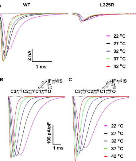
C 1 msFig. 4. INarecordings of WT and L325R channels at different temperatures, and mathematical modelling. (A) Representative current traces obtained after
depolarizing HEK293 cells expressing WT or L325R channels at 20 mV (holding potential 100 mV) at different temperatures. (B and C) Reconstruction of WT INa, using the Clancy – Rudy model under the assumption of an identical Q10of 2.0 for all transition rates (B) and of a Q10that is larger for activation rates
compared to inactivation rates (2.15 vs. 1.9, C). The Q10used are marked on the respective state transition diagrams. The voltage-clamp protocol is the same as
Because the mutation replaces an uncharged leucine by a positively charged arginine, we considered the hypothesis that this mutation influences the folding process of the protein, thus altering either biosynthesis or trafficking of the channel.Fig. 6A illustrates that, similarly to a recent report
[20], pre-incubation of the cells expressing L325R channels with 500 AM mexiletine rescued the mutant INaup to 55%
of the WT current. We also investigated the effect of curcumin, which has been shown to rescue misfolded mutant CFTR channels in a mouse model of cystic fibrosis
[21]. This drug is a low-toxic sarcoplasmic reticulum Ca2+ -ATPase inhibitor[22]. Incubation with 25 AM of curcumin during 24 h increased significantly the mutant currents by ¨150% (Fig. 6A). In contrast, thapsigargin, another drug known to rescue certain misfolded ion channels[23], had no effect. In addition to these treatments expected to improve trafficking of misfolded channels, we also incubated cells expressing L325R channels for 24 h at 28 -C. This condition also partially rescued the mutant channels by ¨300% (Fig. 6A).
3.6. Dominant negative effect of the L325R mutant allele Consistent with an autosomal dominant trait, both SCN5A mutation carriers were found to be heterozygous. In order to mimic this state in our cellular model, we investigated the co-expression of WT and mutant ‘‘alleles’’ by co-transfecting WT and mutant cDNAs.Fig. 6B shows that when reducing the amount of WT cDNA by 50%, the resultant INa was decreased by about 50%. This condition
represents the clinical situation were there would be no synthesis at all of the protein resulting from the mutant allele. However in our experimental model, both mutants were expressed (Fig. 3A and D). When WT and L325R channels were co-transfected in a 1 : 1 ratio, the peak INawas
reduced to only 18T 8% of the control condition where
100% of WT channels were expressed (Fig. 6B). This finding strongly suggests that the L325R allele exerts a dominant negative effect on WT channels. In contrast, this negative effect was not observed when the truncated R535X
A B ** *** INa pea k cur re nt (p A/ pF) 0 50 100 150 200 250 300 350 WT L325 R thap si -gar gin curcumin mexile tine n.s. Norm aliz ed INa pea k c u rr ent (% ) 100% WT ** 28 ˚C 50% L325R 50% WT + 50% R535X 50% WT + 0 20 40 60 80 100 120
*
*
n.s.**
50% empty vector 50% WT + 4 3 2 1 C 180 KDa 63 49 Nav1.5 WT/L325R Nav1.5 R535X actinFig. 6. Treatments rescuing L325R channels and dominant negative effect of L325R channels. (A) HEK293 cells were transfected with WT or L325R cDNAs and INawas measured after 48 h. L325R cells were treated with
either thapsigargin 1 AM for 12 h, curcumin 25 AM for 24 h, or mexiletine 500 AM for 24 h, or incubated for 24 h at 28-C; n = 15 – 20 cells from at least 2 independent experiments, **p < 0.01, ***p < 0.001 vs. L325R. (B) HEK293 cells were transfected with WT and mutant cDNAs. 100% corresponds to 0.3 Ag cDNA per transfection flasks. In the second condition, the WT cDNA was reduced to 50% (0.15 Ag cDNA) and complemented with empty cDNA vector. The condition mimicking the heterozygous expression of WT and mutant channels revealed an interference of the L325R proteins with the current generated by the WT channels. The R535X protein had no negative influence on the WT currents; n = 15 – 20 cells from at least 3 independent experiments, *p < 0.05, **p < 0.01. (C) Western blots of HEK293 cell lysates illustrating the levels of expression of WT and mutant channels in the four conditions analysed in (B). Protein loading was verified by anti-actin immunoblotting. Fig. 5. Mathematical modelling of the AP using the temperature-dependent
INamodel presented inFig. 4C. (A) Control AP (at 37-C and with 100%
INa). (B) Simulation of increased temperature (40-C, with 100% INa). (C)
Simulation with 50% INa, mimicking a heterozygote genotype, at 37-C.
(D) Simulation with 50% INaand at 40-C. Note the abbreviation of the AP
and the WT proteins were co-transfected (Fig. 6B). Since, the total pool of expressed Nav1.5 proteins is unchanged
(Fig. 6C), it can be postulated that the L325R channels interfere with the trafficking of WT channels.
4. Discussion
In this study, we identified two mutations in the SCN5A gene in four index patients with BrS pattern ECGs during episodes of fever. Our in vitro data indicate that the mutations cause an important loss-of-function of the Nav1.5 channels. Functional analyses of the L325R and
R535X mutant channels suggest that the manifestations of fever-exacerbated BrS may not be mutation specific. Furthermore, our experiments suggest novel molecular mechanisms underlying BrS, such as misfolding of mutant proteins leading to dominant negative effect of mutant channels.
4.1. Clinical and genetic findings
Fever can trigger VT/VF in BrS patients [4]. Here, we report four male cases in which SCN5A has been screened. Two mutations were found; R535X in an asymptomatic patient with a typical BrS ECC pattern during fever and L325R, a novel mutation, in a patient with fever-induced VT. The main clinical implications of these findings are that BrS has to be considered in patients with unexplained syncope during febrile state, and that genetic investigation should be performed in patients with an ECG suggestive of BrS during fever. Finally, BrS patients should be advised to take antipyretic drugs early in the course of any febrile illness.
4.2. Possible mechanisms of fever susceptibility in BrS patients
One of the current models explaining the ECG alterations seen in BrS is based on an imbalance between the depolarizing and repolarizing currents during the AP phase 1, mainly in cells expressing a large transient outward Ito
current as the epicardial cells of the right ventricle[1]. In patients with loss-of-function mutations of Nav1.5, resulting
in less INa during the phase 1, the large Ito current may
repolarize the membrane prematurely producing a loss of the dome (phase 2) of the AP. When such premature AP shortening happens heterogeneously in the myocardium, this may generate phase 2 re-entries that can cause VT/VF. Hence, the delicate balance of currents, mainly Ito
(repola-rizing), ICaand INa(both depolarizing), during the AP phase
1 is very critical.
How could an increase of temperature above 37-C alter this subtle balance in patients? It has been shown that the T1620M SCN5A mutation found in patients with ‘‘classi-cal’’ BrS alters the temperature sensitivity of fast
inactiva-tion of these channels [17]. However, our study suggests that in patients carrying a SCN5A mutation at the heterozygous state in whom the function of mutant channels is either reduced or abolished, the temperature-dependent properties of WT INaitself might also lead to the
typical ECG characteristics during fever. This concept is supported by the observation that the patient with the R535X mutation (channels yielding no measurable current) and the patient with the L325R mutation (channels mediating only a small INa) both exhibited sensitivity to
fever. Moreover, the investigation of the L325R channel properties at different temperatures was not consistent with a mutation specific effect of temperature that would lead to a further decrease of mutant INa. In consequence, we
postulate that the effect of elevated temperature on the remaining INa, which is mainly or totally mediated by WT
channels, could be responsible for the ECG phenotype in BrS patients.Fig. 4illustrates that the effects of increasing the temperature on WT INa are multiple and intricate,
suggesting that distinct temperature sensitivities govern the different state transitions of the sodium channel (e.g., activation and inactivation). As supported by the simula-tions presented in Fig. 5, this complex temperature-dependence of WT INamight then be crucial in determining
the balance of currents during depolarization and during the phase 1 of the AP. During phase 1, most of INa has
undergone fast inactivation and only a small fraction of INa
is still contributing to depolarization. Although small, our simulations indicate that this fraction is nonetheless critical. Therefore, accelerated inactivation under conditions of elevated temperature, combined with a decreased level of expression of Nav1.5 channels, might then be sufficient to
obliterate the ‘‘phase 1 depolarization reserve’’ and to shift the delicate balance of currents in favour of a premature repolarization, thus revealing a BrS phenotype. This hypothesis is supported by a recent model study by Gima and Rudy who demonstrated that the accelerated inactiva-tion of the T1620M Nav1.5 channel leads to a BrS ECG
phenotype through a decreased INa [16]. However, since
the differences in WT INaproperties measured at 37 and 42
-C were minimal (Table 1), one should be cautious in extrapolating our experimental data to the situation of BrS patients with fever. Furthermore, the influence of elevated temperature on the AP is complex and the role of fever in BrS is likely to be multifactorial. It is plausible that temperature-dependent changes of currents other than INa
could precipitate the BrS during fever. For example, fever may also increase the peak transient outward current (Ito)
through mechanisms similar to those we propose for INa
and precipitate repolarization in epicardial tissue. It was beyond the scope of this study to introduce temperature-dependent kinetics for currents other than INa and we are
aware that this represents a limitation of the model; however, it allows us to demonstrate that the mechanism we propose may be a realistic option that remains to be explored further.
4.3. Misfolding of Nav1.5 protein contributes to the L325R
phenotype
The consequences of the BrS SCN5A mutations on Nav1.5 function are multiple[1]. Recently, Chahine’s group
reported three BrS SCN5A missense mutations leading to a charge modification and generating channels that were not trafficked to the membrane[24 – 26]. The L325R mutation described here leads to a similar phenotype since the protein expression was not altered (Fig. 3A) and treatments with chemical chaperones, as well as expression at 28 -C, partially rescued INa. Misfolding of Nav1.5 therefore
appears to be one of the mechanisms underlying BrS, as it is the case for KvLQT1 and hERG channels in congenital
long QT syndrome [27], and CFTR channels in cystic fibrosis [21]. The significant mutant INa increase obtained
by treating the cells with curcumin may be of clinical importance since this drug has a very low toxicity profile and has already been tested in a clinical setting as an anti-cancer drug[28]. In contrast, thapsigargin, which similarly to curcumin can block the endoplasmic reticulum Ca2+ -pump, did not rescue the mutant INa. It may therefore be
possible that the positive effect of thapsigargin is restricted for specific channels[23].
4.4. Dominant negative effect of mutant Nav1.5
A dominant negative effect of mutant ion channel subunits has been frequently reported for channels formed by multiple a-subunits, such as the KvLQT1 channel in
LQTS[29]. In contrast, Nav1.5 is not known to oligomerize
with other a-subunits in order to form a minimal ion channel, and, therefore, the dominant negative effect of L325R channels on WT Nav1.5 (Fig. 6B) was unexpected. It
can be postulated that mutant proteins interfere directly or indirectly with either biosynthesis or trafficking of WT channels. It also remains to be shown whether this mechanism could be generalized to other mistrafficked BrS channels since, to our knowledge, no study directly addressed this issue.
5. Conclusions
In the past few years, a number of case reports described that fever triggers the clinical manifestations of BrS. Hence, based on these studies and the present work, BrS should be considered in any patient with syncope during febrile state. The genetic background of BrS is heterogeneous. In the small population of fever-susceptible cases presented here, two patients out of four did not carry any mutation in SCN5A. Additional investigations are therefore needed to identify the genetic bases of this disorder. Finally, as illustrated by our findings, the molecular and cellular mechanisms underlying the fever-dependent manifestations of BrS are complex. Further studies are needed in order to
elucidate this important issue since there is no curative treatment available for BrS to date.
Acknowledgments
We are grateful to the patients for their cooperation. This work was supported by grants of the Swiss National Science Foundation (632 – 66149.01 SNF professorship to HA and 3100A0-100285 to JPK), Fondations Vaudoise de Cardio-logie, Rita et Richard Barme, and Leducq. DK was supported by a grant of the Swiss National Science Foundation and the ADUMED-Foundation. Patch-clamp experiments were performed in the department of physiol-ogy (University of Lausanne) thanks to the help of Prof. P. Kucera. We would like to thank Dr. M.X. van Bemmelen for his useful comments on this manuscript.
References
[1] Antzelevitch C, Brugada P, Brugada J, Brugada R, Shimizu W, Gussak I, et al. Brugada syndrome: a decade of progress. Circ Res 2002;91: 1114 – 8.
[2] Napolitano C, Rivolta I, Priori SG. Cardiac sodium channel diseases. Clin Chem Lab Med 2003;41:439 – 44.
[3] Wilde AA, Antzelevitch C, Borggrefe M, Brugada J, Brugada R, Brugada P, et al. Proposed diagnostic criteria for the Brugada syndrome: consensus report. Circulation 2002;106:2514 – 9. [4] Antzelevitch C, Brugada R. Fever and Brugada syndrome. Pacing Clin
Electrophysiol 2002;25:1537 – 9.
[5] Madle A, Kratochvil Z, Polivkova A. The Brugada syndrome. Vnitr Lek 2002;48:255 – 8.
[6] Porres JM, Brugada J, Urbistondo V, Garcia F, Reviejo K, Marco P. Fever unmasking the Brugada syndrome. Pacing Clin Electrophysiol 2002;25:1646 – 8.
[7] Mok NS, Priori SG, Napolitano C, Chan NY, Chahine M, Baroudi G. A newly characterized SCN5A mutation underlying Brugada syn-drome unmasked by hyperthermia. J Cardiovasc Electrophysiol 2003; 14:407 – 11.
[8] Smith J, Hannah A, Birnie DH. Effect of temperature on the Brugada ECG. Heart 2003;89:272.
[9] Sanchez JM, Kates AM. Brugada-type electrocardiographic pattern unmasked by fever. Mayo Clin Proc 2004;79:273 – 4.
[10] Wang Q, Li Z, Shen J, Keating MT. Genomic organization of the human SCN5A gene encoding the cardiac sodium channel. Genomics 1996;34:9 – 16.
[11] Sherman AJ, Shrier A, Cooper E. Series resistance compensation for whole-cell patch-clamp studies using a membrane state estimator. Biophys J 1999;77:2590 – 601.
[12] van Bemmelen MX, Rougier JS, Gavillet B, Apotheloz F, Daidie D, Tateyama M, et al. Cardiac voltage-gated sodium channel Nav1.5 is regulated by Nedd4-2 mediated ubiquitination. Circ Res 2004;95:284 – 91. [13] Clancy CE, Rudy Y. Linking a genetic defect to its cellular phenotype
in a cardiac arrhythmia. Nature 1999;400:566 – 9.
[14] Luo CH, Rudy Y. A dynamic model of the cardiac ventricular action potential: I. Simulations of ionic currents and concentration changes. Circ Res 1994;74:1071 – 96.
[15] Faber GM, Rudy Y. Action potential and contractility changes in [Na(+)](i) overloaded cardiac myocytes: a simulation study. Biophys J 2000;78:2392 – 404.
[16] Gima K, Rudy Y. Ionic current basis of electrocardiographic wave-forms: a model study. Circ Res 2002;90:889 – 96.
[17] Dumaine R, Towbin JA, Brugada P, Vatta M, Nesterenko DV, Nesterenko VV, et al. Ionic mechanisms responsible for the electro-cardiographic phenotype of the Brugada syndrome are temperature dependent. Circ Res 1999;85:803 – 9.
[18] Smits JP, Eckardt L, Probst V, Bezzina CR, Schott JJ, Remme CA, et al. Genotype-phenotype relationship in Brugada syndrome: electrocardiographic features differentiate SCN5A-related patients from non-SCN5A-related patients. J Am Coll Cardiol 2002;40: 350 – 6.
[19] Murray KT, Anno T, Bennett PB, Hondeghem LM. Voltage clamp of the cardiac sodium current at 37 degrees C in physiologic solutions. Biophys J 1990;57:607 – 13.
[20] Valdivia CR, Tester DJ, Rok BA, Porter CB, Munger TM, Jahangir A, et al. A trafficking defective, Brugada syndrome-causing SCN5A mutation rescued by drugs. Cardiovasc Res 2004;62:53 – 62. [21] Egan ME, Pearson M, Weiner SA, Rajendran V, Rubin D,
Glockner-Pagel J, et al. Curcumin, a major constituent of turmeric, corrects cystic fibrosis defects. Science 2004;304:600 – 2.
[22] Logan-Smith MJ, Lockyer PJ, East JM, Lee AG. Curcumin, a molecule that inhibits the Ca2+-ATPase of sarcoplasmic reticulum but increases the rate of accumulation of Ca2+. J Biol Chem 2001; 276:46905 – 11.
[23] Egan ME, Glockner-Pagel J, Ambrose C, Cahill PA, Pappoe L, Balamuth N, et al. Calcium-pump inhibitors induce functional surface
expression of Delta F508-CFTR protein in cystic fibrosis epithelial cells. Nat Med 2002;8:485 – 92.
[24] Baroudi G, Pouliot V, Denjoy I, Guicheney P, Shrier A, Chahine M. Novel mechanism for Brugada syndrome: defective surface local-ization of an SCN5A mutant (R1432G). Circ Res 2001;88:E78. [25] Baroudi G, Acharfi S, Larouche C, Chahine M. Expression and intracellular localization of an SCN5A double mutant R1232W/T1620M implicated in Brugada syndrome. Circ Res 2002; 90:E11 – 16.
[26] Baroudi G, Napolitano C, Priori SG, Bufalo AD, Chahine M. Loss of function associated with novel mutations of the SCN5A gene in patients with Brugada syndrome. Can J Cardiol 2004;20:425 – 30. [27] Gouas L, Bellocq C, Berthet M, Potet F, Demolombe S, Forhan A, et al. New KCNQ1 mutations leading to haploinsufficiency in a general population; defective trafficking of a KvLQT1 mutant. Cardiovasc Res 2004;63:60 – 8.
[28] Cheng AL, Hsu CH, Lin JK, Hsu MM, Ho YF, Shen TS, et al. Phase I clinical trial of curcumin, a chemopreventive agent, in pa-tients with high-risk or pre-malignant lesions. Anticancer Res 2001; 21:2895 – 900.
[29] Chouabe C, Neyroud N, Guicheney P, Lazdunski M, Romey G, Barhanin J. Properties of KvLQT1 K+channel mutations in Romano – Ward and Jervell and Lange – Nielsen inherited cardiac arrhythmias. EMBO J 1997;16:5472 – 9.