www.elsevier.com / locate / cardiores
D
ifferential sensitivity of atrial and ventricular K
ATPchannels to
metabolic inhibition
*
Serge Poitry, Laurianne van Bever, Fabrice Coppex, Angela Roatti, Alex J. Baertschi
´ ´ `
Department of Physiology, Centre Medical Universitaire, Universite de Geneve, 1 rue Michel-Servet, 1211 Geneva 4, Switzerland Received 9 August 2002; accepted 26 September 2002
Abstract
Objective: The aim is to compare the activation of ATP-sensitive potassium channels (KATP channels) in intact and metabolically impaired atrial and ventricular myocytes. Methods: The KATPchannel current is measured by whole cell and gramicidin-perforated patch clamp recordings in 164 cultured neonate rat cardiomyocytes. Results: In whole cell recordings with 84 mmol / l ADP in pipette, spontaneous activity is significantly higher in atrium than ventricle, and EC50 for the KATP channel opener diazoxide is 0.13 mmol / l (atrium) versus 3.1 mmol / l (ventricle). With an ATP-regenerating system in pipette, EC50 for diazoxide is 19.7 mmol / l (atrium) versus 54.9 mmol / l (ventricle). In gramicidin-perforated patch recordings, atrial myocytes respond significantly to 100 nmol / l of the mitochondrial protonophore CCCP, while ventricular myocytes do not. EC50for diazoxide is 129 mmol / l (atrium) versus .2500 mmol / l (ventricle) for myocytes exposed to CCCP, and 676 versus .2500 mmol / l, respectively, without CCCP. Conclusions: (1) KATPchannels are significantly more sensitive to metabolic inhibition in atrial than ventricular myocytes. (2) Sensitivity of atrium versus ventricle to the channel opener diazoxide increases from 3:1 to $24:1 with ADP or metabolic inhibition. If extended to intact hearts, the results would predict a higher atrial sensitivity to ischemia, and a high sensitivity of the ischemic atrium to KATP channel openers.
2003 European Society of Cardiology. Published by Elsevier Science B.V. All rights reserved.
Keywords: Atrial function; K-ATP channel; Membrane currents; Myocytes
1 . Introduction Sarcolemmal KATP channels are composed of a
potas-sium channel pore, formed by four potaspotas-sium inward Sarcolemmal KATP channels were first discovered in rectifier subunits (KIR6.1 or KIR6.2), and by an associa-heart [1] and subsequently in most other organs. They tion of four regulatory sulfonylurea receptor subunits couple the membrane potential to the metabolic state of the (SUR1, SUR2A or SUR2B) [2–5]. There is convincing cell [2–5]. In pancreatic b-cells, the normally open KATP evidence from both native ventricular and artificially channels close in response to increased glucose influx, expressed KATPchannels that the ‘cardiac’ KATPchannel is causing membrane depolarization and calcium-induced a combination of KIR6.2 and SUR2A subunits [7]. These exocytosis of insulin secretory vesicles [3]. In cardiac channels typically open in response to the KATP channel myocytes, the normally closed KATP channels open in opener pinacidil but not to diazoxide [2,4,5]. In contrast, response to metabolic distress, and thus reduce the action atrial KATPchannels open in response to both pinacidil and potential duration, calcium influx, force of contraction and diazoxide [8], though species differences exist [9]. Further-possibly the ATP demand [5]. In heart as well as kidney more, the KATP channel blocker glibenclamide is highly and brain the sarcolemmal KATPchannels thus help protect effective in closing atrial but not ventricular KATPchannels the tissues from ischemia, though mitochondrial KATP [8]. These differences in pharmacological characteristics channels are also likely to be involved [6]. are important, since KATP channel modulators used in the treatment of diabetes (sulfonylureas) or other diseases *Corresponding author. Tel.: 5347; fax:
141-22-702-5402.
E-mail address: [email protected](A.J. Baertschi). Time for primary review 24 days.
0008-6363 / 03 / $ – see front matter 2003 European Society of Cardiology. Published by Elsevier Science B.V. All rights reserved. P I I : S 0 0 0 8 - 6 3 6 3 ( 0 2 ) 0 0 7 1 5 - 0
(diazoxide) could affect atrial myocytes, even though they apex of 2–3-day-old rats and cultured [19] similarly to the little affect the ventricular myocytes. method of Sadoshima et al. [20]. The cells were plated at Interestingly, in the presence of 100 mmol Mg-ADP, low density (10 000–50 000 cells / dish) on fibronectin– ventricular myocytes do become sensitive to diazoxide gelatin coated 8-mm glass slides. On the second and third [10]. Such ADP levels can be reached in hypoxia [11] and day of culture, when the recordings were made, at least presumably in ischemic heart disease. Interactions between two out of three cells are myocytes. Only initially contract-diazoxide and Mg-ADP may be explained by the absolute ing myocytes devoid of contact to neighboring cells were requirement, in SUR, of the second nucleotide binding fold examined.
for the action of both diazoxide and Mg-ADP [10,12,13],
though other SUR domains are also required for diazoxide 2 .2. Patchclamp recordings of KATP current action [14]. Mg-ADP antagonizes the ATP-induced
inhibi-tion of the KATP channel [15]. The low sensitivity of Whole cell recordings of the KATPcurrent were obtained ventricular myocytes to diazoxide may be explained by an similarly as described previously [8]. From a holding interaction of the C-terminal tail of SUR2A with the potential of 240 mV voltage ramps were imposed every 30 second nucleotide binding fold [13]. Interactions between s from 280 to 190 mV over a 10-s period. This resulted in diazoxide and Mg-ADP on the atrial KATP channel are still quasi-steady-state current–voltage curves. Membrane po-unknown, but the high sensitivity to diazoxide [8] raises tential was measured in current-clamp mode at 0 pA at the two questions. First, is this sensitivity mediated by an end of each ramp.
atrium-specific KATP-channel, or was it due to the presence For whole cell recordings, the pipette solution contained of ADP in the patch pipette? ATP-containing solutions are (in mmol / l) 120 KCl, 1.3 CaCl , 1.3 MgCl , 10 HEPES,2 2
usually contaminated by ADP. Second, is the atrial KATP 10 glucose, 10 BAPTA and either 1 mmol / l K -ATP and2
current more sensitive to ADP, and thus more sensitive to 10 mmol / l K-ADP (to partly simulate metabolic impair-hypoxia and ischemia than the ventricular KATP current? ment), or an ATP-regenerating system with 1 mmol / l Answering these question should help to better understand K -ATP, 3 mmol / l creatine-phosphate, 5 U / ml creatine2
the excitability of the normal and ischemic heart. kinase (to minimize the ADP concentration). BAPTA was
21
This study is conducted on neonate cultured myocytes, chosen rather than EGTA to minimize chelation of Mg . since the startling diazoxide sensitivity has been discov- The pH was adjusted to 7.3 with KOH, and osmolality to ered in neonate atrial myocytes in culture [8]. The first aim 290 mOsm / kg with KCl. Nucleotide containing solutions is to compare the sensitivity of atrial and ventricular KATP were aliquoted, frozen and thawed just before use, and currents to ADP and diazoxide in conventional whole cell kept on ice for a maximum of 2–4 h. The ATP and ADP recordings, using the same patch pipette solutions on either concentrations were assayed by HPLC in thawed aliquoted type of myocyte. The second aim is to compare the samples. Patch pipette solutions supplemented with 10 sensitivity of atrial and ventricular myocytes to CCCP and mmol / l ADP and 1 mmol / l ATP showed HPLC assay diazoxide in gramicidin-perforated patch clamp recordings, values of 84 mmol / l ADP and 1.08 mmol / l ATP. To an experimental method that best preserves the physiology minimize the ADP contamination, ATP was kept at around of intact cells [16,17]. The results indicate that atrial 1 mmol / l. Patch pipette solutions supplemented with the myocytes loaded with 84 mmol / l ADP are 24-times more ATP-regenerating system showed HPLC assay values of sensitive to diazoxide than ADP-loaded ventricular 0.0 mmol / l ADP and 1.16 mmol / l ATP. The bath solution myocytes. Furthermore, intact atrial myocytes respond to contained (in mmol / l): 5 KCl, 1 CaCl , 1 MgCl , 1182 2
simulated mild ischemia (100 nmol / l CCCP) while ven- NaCl, 10 HEPES, and 10 glucose. The pH was adjusted to tricular myocytes do not. The results support the hypoth- 7.4 with NaOH, and osmolality to 290 mOsmol / kg with esis of an atrium-specific KATPchannel with high sensitivi- sucrose.
ty to metabolic inhibition and diazoxide. For gramicidin-perforated patch recordings, the pipette contained (in mmol / l) 120 KCl, 1.3 CaCl , 1.3 MgCl , 102 2
HEPES, 10 glucose, 10 BAPTA and 1 K -ATP, and was2
2 . Methods supplemented with gramicidin D at a concentration of 5
mg / ml [16]. The bath solution was as described above.
2 .1. Cell culture The recording was made in the cell attached mode. Several
minutes elapsed until the gramicidin established a low The investigation conforms with the Guide for the Care resistance path between pipette and cytoplasm. The series and Use of Laboratory Animals published by the US resistance (mean6S.E.M.) was 35.262.9 MV (n541) in National Institutes of Health (NIH Publication No. 85-23, atrial myocytes and 36.664.8 MV (n516) in ventricular revised 1996). Atrial myocytes were dissociated from atrial myocytes. The resistance compensation range extends to appendages of 2–3-day-old rats and cultured for up to 3 100 MV in the Axopatch 200B amplifier, and is sufficient days in a 5% CO incubator, as described previously [18].2 for a precise voltage clamp control during maximal KATP
to measure the KATPcurrent and membrane potential while stored in ethanol as a 2 mmol / l stock solution, yielding a largely preserving the cellular protein and ion content maximal ethanol concentration of 0.05% in the external [16,17]. Membrane potentials (means6S.E.M.) reached buffer. Diazoxide, pinacidil and glibenclamide were stored 275.860.8 mV in atrial myocytes and 278.261.8 mV in in DMSO as 100, 100 and 10 mmol / l stock solution, ventricular myocytes, attesting to the capability of these respectively, yielding a maximal DMSO concentration of cells of maintaining normal potassium gradients. 0.1%. These concentrations of ethanol and DMSO were found previously to have no effect on KATP channel
2 .3. General protocols activity ([8,22]; and unpublished).
In whole cell recordings on 107 myocytes, spontaneous 2 .5. Data analysis activation of the KATP current was measured during a
control period of 8–12 min. Incremental concentrations of In order to exclude the possible contribution of chloride diazoxide were then applied (from 0.01 to 100 mmol / l) channels, the KATP current obtained during the voltage during periods of 6–10 min, followed by a maximal ramp was measured at 0 mV, close to the chloride stimulation with co-added 100 mmol / l pinacidil and by an equilibrium potential. The steady state current was ob-inhibition test with co-added 0.1–1 mmol / l glibenclamide. tained for the control period and each period of drug Not all concentrations were applied to all cells, as the total application and each myocyte. Steady state is defined here, recording period was limited to 45–50 min. Four groups for each cell and each drug application, as a current were examined: (1) and (2) atrial and ventricular varying less than 61% of the maximal current per min. myocytes, respectively, with 84 mmol / l ADP and 1.08 During diazoxide stimulation, steady state was reached in mmol / l ATP (measured values) in the pipette; (3) and (4) most cases for the gramicidin recordings in both cell types, atrial and ventricular myocytes, respectively, with an ATP- in 78.7% of all cases for whole cell atrial KATP current regenerating system in pipette. The rationale was to recordings, and in 64.9% of all cases for whole cell simulate mild hypoxic conditions with moderately elevated ventricular KATP current recordings. Overshoot with sub-ADP [11], and to compare these with conditions where sequent lower KATP current was usually produced in the cytoplasmic ADP levels were minimized by the ATP- cases where no steady state was achieved. The maximal regenerating system. An 8–12-min control period was current was measured in those situations. In a first analysis, deemed sufficient to dialyze ADP, ATP and creatine kinase these currents (in pA) were averaged over all cells for each into the cytoplasm. From the measurements of access group and plotted as means6S.E.M. in Figs. 2 and 5. resistance and cell capacitance of this study, the calculated Significance of the means and differences between means time constant of cell dialysis [21] for ADP was 14869 s were analyzed by ANOVA for repeated measures on ranks (n589) in atrial myocytes and 137613 s (n518) in (nonparametric statistics), with software from the SAS ventricular myocytes. The calculated cytoplasmic concen- Institute (Carey, NC, USA). To obtain the change in tration of creatine kinase reached 60% of the pipette current density (in pA / pF ), the current at the end of the concentration within a 10-min period. control period was subtracted and the difference divided by The general protocol was the same in gramicidin the cell capacitance. Averages were obtained over all cells perforated patch recordings on 57 myocytes, with the for each group and plotted as means6S.E.M. in Figs. 3 following exceptions. In groups 5–8, atrial and ventricular and 6. The EC50 and maximal slope of the resulting eight myocytes were stimulated at the end of the experiment sigmoid dose–response curves for diazoxide were analyzed with the mitochondrial protonophore CCCP (1 mmol / l) in by MicrocalORIGINsoftware (version 5), and the results are order to evoke a maximal activation. In groups 7 and 8, listed in Table 1.
atrial and ventricular myocytes were exposed, after the control period and throughout the experiment, to 100
nmol / l CCCP. The rationale was to compare channel 3 . Results activation by diazoxide in the presence or absence of a
mild metabolic impairment. Unlike the metabolic inhibitor 3 .1. Comparison of atrial and ventricular KATP channels oligomycin, CCCP increases the ATP consumption by the in whole cell recordings
F1F0-ATPase. Concentrations of diazoxide and CCCP
were similar to those used in previous studies [8,22,23], Using the same patch pipette solutions and recording and finalized by trial and error. conditions, atrial and ventricular myocytes were recorded either with 84 mmol / l ADP and 1.08 mmol / l ATP
2 .4. Chemicals and drugs (example Fig. 1) or with an ATP-regenerating cocktail in
the pipette. The slow ramp protocol (Fig. 1A) ensures that CCCP, diazoxide, glibenclamide, gramicidin D, creatine the voltage-dependent potassium channels are only mini-phosphate, creatine kinase, K -ATP; K-ADP, and BAPTA2 mally activated if at all. Activation by pinacidil and came from SIGMA, and pinacidil from Alexis. CCCP was diazoxide and inhibition by glibenclamide provide
pharma-Table 1
a
Statistical summary for dose–response curves of KATP channel opener diazoxide
Recording EC50 EC50 Maximal slope n
(mmol / l) log (mmol / l) (pA / pF) / decade
A V A V A V A V Whole-cell 0.13 3.1 20.8960.13 0.4960.28 63.6616.0 29.468.9 30 11 with ADP Whole-cell 19.7 54.9 1.2960.17 1.7460.16 20.863.5 41.869.2 59 7 with CrP Gramicidin 129 3715 2.1160.11 3.5760.79 29.663.4 30.3612.0 18 9 with CCCP-7 Gramicidin 676 2570 2.8360.01 3.4160.06 67.660.2 71.263.2 23 7 without CCCP a
Sigmoids were fitted to the dose–response curves to yield the EC50and maximal slope. In whole cell patch clamp recordings the pipette contained either 1.08 mmol / l ATP and 84 mmol / l ADP (ADP) or an ATP-regenerating system (CrP). In gramicidin-perforated patch recordings, the bath contained either 100 nmol / l or no CCCP. Note large differences in EC50between atrial (A) and ventricular (V) myocytes under the same recording conditions; n, number of myocytes.
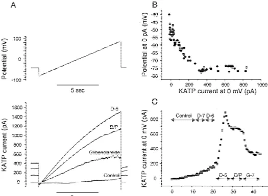
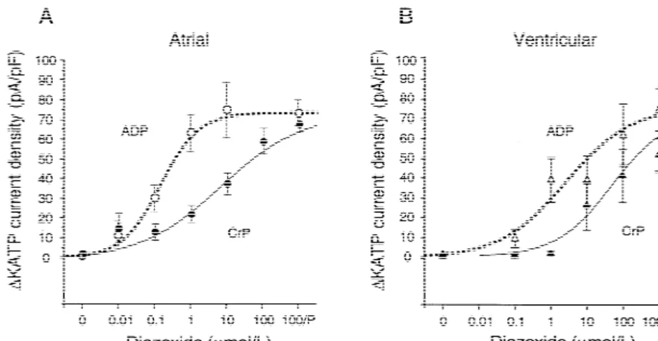
cological evidence that the channels are KATP-channels abolishes the spontaneous activation in the atrial myocytes. (Fig. 1C) (see also [8]). In contrast to ventricular In order to more precisely define the diazoxide sensitivity, myocytes, atrial myocytes display at rest a significant KATP the change in current density was calculated, mean values channel activation in the presence of Mg-ADP (Fig. 2), fitted by sigmoid dose–response curves (Fig. 3), and EC50
and a remarkable sensitivity to diazoxide with a threshold and maximal slopes determined and shown in Table 1. In concentration of 10 nmol / l. Under the same recording atrial myocytes, the presence of 84 mmol / l ADP increases conditions, the ventricular myocytes display a threshold the maximal slope and dramatically decreases the EC50(in concentration of 1 mmol / l. The ATP-regenerating cocktail mmol / l) by 150-fold. In ventricular myocytes, the presence decreases the diazoxide sensitivity in both cell types, as the of Mg-ADP shifts the dose–response curve to the left, but threshold concentration increases to 0.1–1 mmol / l in atrial the decrease of EC50 is only 18-fold. Under identical myocytes and to 10 mmol / l in ventricular myocytes. It recording conditions, atrial myocytes display 24-fold
Fig. 1. Example of whole cell recording on a ventricular myocyte with 84 mmol / l ADP and 1.08 mmol / l ATP in pipette. (A) Ramp protocol (10 s) of voltage clamp with current traces below; (B) relationship between membrane potential at 0 pA and current at 0 mV; (C) KATPcurrent at 0 mV as function of time. Concentrations in log molar, D, diazoxide; D/ P, D with 100 mmol / l pinacidil; G-7, 100 nmol / l glibenclamide on top of D/ P.
Fig. 2. Statistical summary of all whole cell recordings of KATPcurrent at 0 mV. Means are shown as light columns, and S.E.M. as black bars on top. (A) Atrial myocytes; (B) ventricular myocytes. ADP, 84 mmol / l ADP and 1.08 mmol / l ATP in pipette; CrP, ATP-regenerating system in pipette. *, P,0.05 relative to 0; c, P,0.05 relative to CrP; v, P,0.05 relative to ventricle; g, P,0.05 relative to no glibenclamide. S, spontaneous activity; for other abbreviations see Fig. 1.
(ADP) and 2.8-fold (ATP regenerating cocktail) lower CCCP (100 nmol / l) (example Fig. 4). This significantly
EC50 than ventricular myocytes. activates the atrial but not the ventricular myocytes.
Threshold concentrations of diazoxide are 1 and 10 mmol / l 3 .2. Comparison of atrial and ventricular KATP channels for atrial and ventricular myocytes, respectively, in the in gramicidin-perforated patch recordings presence of 100 nmol / l CCCP (Fig. 5), and 10–100 mmol / l in its absence. Maximal concentrations of 100 Both atrial and ventricular myocytes become remarkably mmol / l diazoxide and pinacidil were used in an attempt to insensitive to diazoxide when recorded in the gramicidin- maximally stimulate the KATP current, but result in only perforated patch mode. Thus mild metabolic impairment half-maximal activation or less when compared to values was simulated by exposing the myocytes to low dose obtained under whole cell recordings. Maximal activation
Fig. 3. Dose–response curves for increase in KATPcurrent density at 0 mV (current normalized by cell capacitance). (A) Atrial myocytes; (B) ventricular myocytes. For statistics see Table 1; 100 / P5100 mmol / l diazoxide with 100 mmol / l pinacidil (not included in sigmoid regression curves). For abbreviations see Fig. 2.
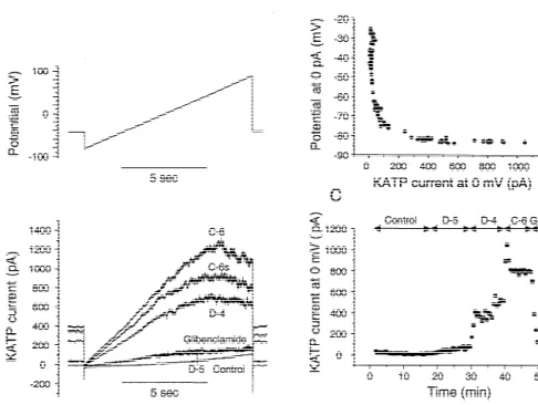
Fig. 4. Example of gramicidin-perforated patch recording on an atrial myocyte. (A) Ramp protocol (10 s) of voltage clamp with current traces below; (B) relationship between membrane potential at 0 pA and current at 0 mV; (C) KATPcurrent at 0 mV as function of time. For abbreviations see Fig. 1. C-6, C-6s51 mmol / l CCCP at peak and steady-state.
is attained at the end of the experiment with 1 mmol / l diazoxide, relative to no CCCP (Table 1). In contrast, the
CCCP (Fig. 5). EC50 for diazoxide in ventricular myocytes is not
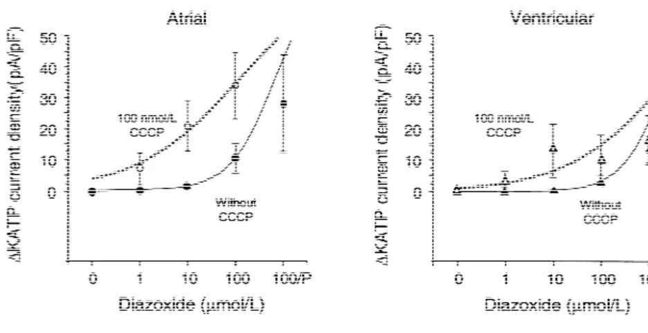
sig-In atrial myocytes that are mildly impaired with 100 nificantly changed by CCCP. When metabolically impaired nmol / l CCCP, sigmoid dose–response curves for the by 100 nmol / l CCCP, the atrial myocytes are 28-times current densities (Fig. 6) show a 5-fold lower EC50 for more sensitive to diazoxide than ventricular myocytes.
Fig. 5. Statistical summary of all gramicidin-perforated patch recordings of KATPcurrent at 0 mV. Means are shown as light columns, and S.E.M. as black bars on top. (A) Atrial myocytes; (B) ventricular myocytes. nt, not tested; for other abbreviations see previous figures. Note leftward shift of atrial KATP
Fig. 6. Dose–response curves for increase in KATPcurrent density at 0 mV. (A) Atrial myocytes; (B) ventricular myocytes. Sigmoid curves were fitted without including response to 100 / P. For statistics see Table 1. For abbreviations see previous figures.
When not metabolically impaired the atrial myocytes are ADP sharply increases the sensitivity to diazoxide of 3.8-times more sensitive than the ventricular myocytes. native ventricular as well as KIR6.2 / SUR2A KATP chan-Interestingly, the exposure to CCCP decreases the maximal nels [10], raising the question if contaminating ADP in the slope of the dose response curves (Table 1). Presumably, patch pipette confounds the interpretation of patch clamp in intact cells local gradients of ADP and other soluble experiments. Are the atrial and ventricular KATP channels cytoplasmic factors may contribute to the overall respon- one and the same, represented by KIR6.2 / SUR2A [5], and siveness of the sarcolemmal KATP channels. do they respond to diazoxide as a function of intracellular Thus both electrophysiological approaches yield similar ADP? The present study was designed to answer these ratios of sensitivity to diazoxide of atrial relative to questions.
ventricular KATP channels. These ratios are particularly ADP could be the confounding factor, since the ADP large (.24:1) when the myocytes are metabolically im- concentration measured by HPLC (84 mmol / l) is
sig-paired. nificantly higher than the concentration added (10 mmol / l)
to the ATP-containing pipette solution. Presumably, the additional ADP is a hydrolytic product of ATP. This factor
4 . Discussion is now controlled for by applying the same pipette
solutions to both atrial and ventricular myocytes. Under This study compares—in the same species and under these identical recording conditions, the atrial myocytes identical experimental conditions—the activation of atrial respond to mild metabolic inhibition while ventricular and ventricular KATP currents by metabolic inhibition and myocytes do not (see i) above). The atrial myocytes are diazoxide. The major new findings are: (i) the presence of highly sensitive to the interaction between diazoxide and Mg-ADP in the patch pipette, or low dose CCCP in the intracellular Mg-ADP, while the ventricular myocytes are extracellular medium, activates an atrial but not a ventricu- not (see ii) above). A critical test with gramicidin-per-lar KATP current; (ii) in the presence of Mg-ADP in the forated patch recordings (see iii) above), where the patch pipette the atrial KATP current is 24-times more myocytes are left largely intact [16,17], confirms the sensitive to diazoxide than the ventricular KATP current; results from whole cell recordings. Could other methodo-(iii) in the presence of low dose CCCP in the extracellular logical differences be involved? The myocytes stem from medium the atrial KATP current is 28-times more sensitive the same species (rat), strain (Sprague–Dawley), and age to diazoxide than the ventricular KATP current; (iv) in the (2–3 days), are dispersed on the same day and recorded on absence of ADP or CCCP, the atrial KATPcurrent is 3- to the same days, and exposed to similar culture media. 4-times more sensitive to diazoxide than the ventricular Although the serum concentration is 10% for the atrial and
KATP current. 5% for the ventricular culture medium [19], the cells
undergo the same hypertrophy as measured by b-actin 4 .1. Evidence for an atrium-specific KATP channel in cell mRNA [Schmidt et al., submitted for publication, 2002]. culture Thus hypertrophy per se, due to serum stimulation [24],
could not have been a factor either.
In a previous study we have shown that the KATP current Five other factors conceivably contribute to the overall in cultured rat atrial myocytes is highly sensitive to responsiveness of KATP channels: (1) cell geometry; (2)
21
diazoxide [8] as compared to published sensitivities of local pH; (3) cytosolic Mg-ATP and free Mg con-ventricular myocytes [2,4,5]. However, the application of centration; (4) nucleotide dialysis and (5) nucleotide
hydrolysis. Regarding geometrical factors, the reticular ventricular myocytes (Fig. 3; Table 1). A parallel shift of network and myofibrils are far less developed in neonatal the dose–response curve would have been expected for the than adult myocytes, thus it is unlikely that morphological atrial KATP channel if it was identical to KIR6.2 / SUR2A. differences could explain the large difference in diazoxide Overall, these results suggest that the atrial KATPchannels sensitivity of neonate atrial and ventricular myocytes. include SUR1, SUR2B and possibly KIR6.1 subunits. The Regarding local pH, CCCP may perhaps form proton leaks determination of the exact composition of the atrial KATP
in the plasmalemma. Since membranes have relatively channel will require immunoprecipitation techniques for uniform characteristics, a potential proton leak in plas- the small amounts of channel protein available from atrial malemma or other membranes should be similar in both myocyte cultures.
cell types. The very low concentrations of CCCP used in
this study to prime the cells (100 nmol / l) should therefore 4 .3. Significance have very little differential effects on both cell types.
21
Regarding cytosolic Mg-ATP and free Mg , the same Whole cell recordings allow for a precise characteriza-concentrations were applied to both cell types. They were tion of the KATPchannels, since the myocytes are dialyzed .150-times higher than in another study [10] where ADP with known ion and nucleotide concentrations. However, and diazoxide sensitivities were examined in guinea pig soluble cytoplasmic messengers escape into the patch ventricular myocytes. The reason is that our pipette pipette solution, and local gradients in ADP or other
21
solution contained BAPTA—which chelates mainly Ca , factors are almost totally dissipated. The results from
21 21
vs. EGTA [10]—which chelates both Ca and Mg . It is gramicidin-perforated patch recordings are more repre-possible, therefore, that Mg-ATPase activity on or near the sentative for predicting the behavior of intact cells [16,17]. KATP channels somewhat enhanced the sensitivity to Mild metabolic inhibition significantly increases the KATP
diazoxide. Regarding nucleotide dialysis in whole cell current in atrial but not ventricular intact myocytes, and recordings, the time constants were similar in both types of small increases in potassium current are sufficient to myocytes (see Methods), and short (,150 s) relative to the hyperpolarize the cell. One would predict that mild meta-10-min control period. However, the Mg-ATPase activity bolic inhibition could increase the risk of arrhythmia in of SUR would differ, if the KATP channel composition atrial but not ventricular myocytes. These experiments on differed between atrial and ventricular myocytes. Thus, the cultured neonate myocytes need to be extended to freshly present study indeed favors the existence of an atrium- isolated myocytes, in vivo, and to humans to further test specific KATP channel in myocyte culture. whether the atrium is more sensitive to ischemia than ventricle. Regional differences in the sensitivity of the 4 .2. Evidence for SUR1, SUR2B and /or KIR6.1 subunits KATP channels to Mg-ADP could have a profound in-in the atrial KATP channel fluence on impulse conduction in ischemic hearts.
In the presence of 84 mmol / l intracellular ADP, a
significant basal activity exists in atrial KATP channels, A cknowledgements while the ventricular KATP channels are totally inactive. It
is already known that KIR6.2 / SUR1 in b-cells and This study was supported by the Swiss National Science KIR6.1 / SUR2B in smooth muscle cells show significant Foundation (grants nos. 31-59551.99 and 31-066838.01). basal activity, while KIR6.2 / SUR2A channels in striated We thank Dr. Olivier Sorg (University of Geneva Hospi-muscle are virtually silent (reviewed in [5]). Possible tal) for the HPLC measurements, and the Louis Jeantet reasons include a higher hydrolytic activity of SUR1 and Foundation for support of medical student Fabrice Coppex. SUR2B relative to SUR2A, and a higher sensitivity to
Mg-ADP of KIR6.1 / SUR relative to KIR6.2 / SUR [25].
Furthermore, there is a significant difference in diazoxide R eferences sensitivity between atrial and ventricular KATP channels
1
(Table 1) that cannot be explained on methodological [1] Noma A. ATP-regulated K -channels in cardiac muscle. Nature 1983;305:147–148.
grounds (see above). This further suggests the involvement
1 [2] Yokoshiki H, Sunagawa M, Seki T, Sperelakis N. ATP-sensitive K of SUR1 and SUR2B, as these subunits are known to
channels in pancreatic, cardiac and vascular smooth muscle cells. confer the diazoxide sensitivity to pancreatic KIR6.2 / Am J Physiol 1998;274:C25–C37.
SUR1 and smooth muscle KIR6.1 / SUR2B channels [2–5]. [3] Seino S. ATP-sensitive potassium channels: a model of heteromul-Finally, the results demonstrate a striking synergy between timeric potassium channel / receptor assemblies. Annu Rev Physiol
1999;61:337–362. ADP and diazoxide in the activation of the atrial KATP
[4] Aguilar-Bryan L, Bryan J. Molecular biology of adenosine tri-channels. This is shown in whole cell recordings by the
phosphate-sensitive potassium channels. Endocr Rev sharp increase in the maximal slope of the diazoxide dose 1999;20(2):101–135.
response curves for atrial myocytes, while there is a [5] Babenko AP, Aguilar-Bryan L, Bryan J. A view of sur / KIR6 X, parallel left-ward shift of the dose–response curves for KATPchannels. Annu Rev Physiol 1998;60:667–687.
1 [6] Garlid KD, Paucek P, Yarov-Yarovoy V et al. Cardioprotective effect channels: regulation by intracellular nucleotides and K channel
of diazoxide and its interaction with mitochondrial ATP-sensitive opening drugs. Am J Physiol 1995;269:C525–545. 1
K channels. Possible mechanism of cardioprotection. Circ Res [16] Kyrozis A, Reichling DB. Perforated-patch recording with 1997;81:1072–1082. gramicidin avoids artifactual changes in intracellular chloride con-[7] Babenko AP, Gonzalez G, Aguilar-Bryan L, Bryan J. Reconstituted centration. J Neurosci Methods 1995;57:27–35.
human cardiac K-ATP channels: functional identity with the native [17] Tajima Y, Ono K, Akaike N. Perforated patch-clamp recording in channels from the sarcolemma of human ventricular cells. Circ Res cardiac myocytes using cation-selective ionophore gramicidin. Am J
1998;30:1132–1143. Physiol 1996;271:C524–532.
[8] Baron A, van Bever L, Monnier D, Roatti A, Baertschi AJ. A novel [18] Jiao JH, Baumann P, Baron A et al. Sulfonylurea receptor ligands KATP current in cultured neonatal rat atrial appendage car- modulate stretch-induced ANF secretion in rat atrial myocyte diomyocytes. Circ Res 1999;85:707–715. culture. Am J Physiol Heart Circ Physiol 2000;278:H2028–2038. [9] Ogbaghebriel A, Shrier A. Differential responsiveness of atrial and [19] Baertschi AJ, Monnier D, Schmidt U et al. Acid prohormone
ventricular myocytes to potassium channel openers. J Cardiovasc sequence determines size, shape, and docking of secretory vesicles Pharmacol 1995;25:65–74. in atrial myocytes. Circ Res 2001;89:E23–E29.
[10] D’hahan N, Moreau C, Prost AL et al. Pharmacological plasticity of [20] Sadoshima J, Jahn L, Takahashi T, Kulik TJ, Izumo S. Molecular cardiac ATP-sensitive potassium channels toward diazoxide re- characterization of the stretch-induced adaptation of cultured cardiac vealed by ADP. Proc Natl Acad Sci USA 1999;96(21):12162– cells. J Biol Chem 1992;267:10551–10560.
12167. [21] Push M, Neher E. Rates of diffusional exchange between small cells 1
[11] Vankatesh N, Lamp ST, Weiss JN. Sulfonylureas, ATP-sensitive K and a measuring patch pipette. Pflugers Arch 1988;411:204–211. 1
channels, and cellular K loss during hypoxia, ischemia, and [22] Baron A, Monnier D, Roatti A, Baertschi AJ. Pituitary adenylate metabolic inhibition in mammalian ventricle. Circ Res cyclase-activating polypeptide activates KATP current in rat atrial 1991;69:623–637. myocytes. Am J Physiol Heart Circ Physiol 2001;280:H1058– [12] Shyng S, Ferrigni T, Nichols CG. Regulation of KATP channel H1065.
activity by diazoxide and MgADP. Distinct functions of the two [23] Baumann P, Poitry S, Roatti A, Baertschi AJ. Plasmalemmal KATP
nucleotide binding folds of the sulfonylurea receptor. J Gen Physiol channels shape triggered calcium transients in metabolically im-1997;110(6):643–654. paired rat atrial myocytes. Am J Physiol Heart Circ Physiol 2002, in [13] Matsuoka T, Matsushita K, Katayama Y et al. C-Terminal tails of press.
sulfonylurea receptors control ADP-induced activation and diazox- [24] Schaub MC, Hefti MA, Harder BA, Eppenberger HM. Various 1
ide modulation of ATP-sensitive K channels. Circ Res hypertrophic stimuli induce distinct phenotypes in cardiomyocytes. J
2000;87:873–880. Mol Med 1997;75:901–920.
[14] D’Hahan N, Jacquet H, Moreau C, Catty P, Vivaudou M. A [25] Babenko AP, Bryan J. A conserved inhibitory and differential transmembrane domain of the sulfonylurea receptor mediates activa- stimulatory action of nucleotides on KIR6.0 / SUR complexes is tion of ATP-sensitive K(1) channels by K(1) channel openers. Mol essential for excitation–metabolism coupling by KATP channels. J Pharmacol 1999;56(2):308–315. Biol Chem 2001;276:49083–49092.
1 [15] Terzic A, Jahangir A, Kurachi Y. Cardiac ATP-sensitive K