All rights reserved
Lycopene and Myocardial Infarction Risk in the EURAMIC Study
Lenore Kohlmeier,1 Jeremy D. Kark,2 Enrique Gomez-Gracia,3 Blaise C. Martin,4 Susan E. Steck,5 Alwine F. M. Kardinaal,6 Jetmund Ringstad,7 Michael Thamm,1 Victor Masaev,8 Rudolf Riemersma,9 Jose M. Martin-Moreno,10 Jussi K. Huttunen,11 and Frans J. Kok6
A multicenter case-control study was conducted to evaluate the relations between antioxidant status assessed by biomarkers and acute myocardial infarction. Incidence cases and frequency matched controls were recruited from 10 European countries to maximize the variance in exposure within the study. Adipose tissue needle aspiration biopsies were taken shortly after the infarction and analyzed for levels of carotenoids and tocopherols. An examination of colinearity including all covariates and the three carotenoids, a-carotene, /3-carotene, and lycopene, showed that the variables were sufficiently independent to model simultaneously. When examined singularly, each of the carotenoids appeared to be protective. Upon simultaneous analyses of the carotenoids, however, using conditional logistic regression models that controlled for age, body mass index, socioeconomic status, smoking, hypertension, and maternal and paternal history of disease, lycopene remained independently protective, with an odds ratio of 0.52 for the contrast of the 10th and 90th percentiles (95% confidence interval 0.33-0.82, p = 0.005). The associations for a- and /3-carotene were largely eliminated. We conclude that lycopene, or some substance highly correlated which is in a common food source, may contribute to the protective effect of vegetable consumption on myocardial infarction risk. Am J
Epidemiol 1997;146:618-26.
adipose tissue; carotene; carotenoids; myocardial infarction
Coronary heart disease remains a major cause of mortality in developed countries and is increasingly recognized as an important cause of morbidity in the developing world as well. A number of important risk factors for coronary heart disease have been identified including hypertension, hypercholesterolemia, insulin resistance, and cigarette smoking. However, these fac-tors can only partly account for variations in the inci-Received for publication December 23, 1996, and accepted for publication June 27, 1997.
1 Institute for Social Medicine and Epidemiology, Berlin,
Ger-many.
2 Hadassah Medical Organization and Hebrew University
Hadas-sah School of Public Health, Jerusalem, Israel.
3 Department of Preventive Medicine, Hospital Universitario,
Facultad de Medicina, Malaga, Spain.
4 Institute for Social and Preventive Medicine, Zurich University,
Zurich, Switzerland.
5 University of North Carolina, Chapel Hill, NC.
6 TNO Toxicology and Nutrition Institute, Zeist, Netherlands. 7 Medical Department, Ostfold Central Hospital, Sarpsborg,
Nor-way.
8 Russian Institute for Preventive Medicine, Moscow, Russia. 9 Cardiovascular Research Unit, University of Edinburgh,
Edin-burgh, Scotland.
10 Escuela Nacional de Sanidad, Madrid, Spain. 11 National Public Health Institute, Helsinki, Finland.
Reprint requests to Dr. Lenore Kohlmeier, Departments of Nutri-tion and Epidemiology, Schools of Public Health and Medicine, The University of North Carolina at Chapel Hill, CB# 7400, McGavran-Greenberg, Chapel Hill, NC 27599-7400.
dence of coronary heart disease either between or within populations (1-3). Studies of lipid metabolism have suggested that oxidative modifications of low density lipoprotein accelerate atherogenesis (4-6), and supplements of the antioxidant vitamin E reduce the incidence of nonfatal myocardial infarction (7).
Hypothesized methods of promotion of atherogene-sis by oxidized low density lipoprotein include stim-ulation of monocyte and platelet adhesion to endothe-lium, inhibition of vasodilation, stimulation of synthesis of autoantibodies, and promotion of prolif-eration of smooth muscle cells leading to the promo-tion of foam cells and fatty streaks in the arterial intima (8-10). Natural antioxidants present in the diet may inhibit the oxidative modification of low density lipoprotein and slow the progression of atherosclerosis (11).
Observational epidemiologic studies that explored the antioxidant vitamin hypothesis using ecologic studies and cross-sectional studies (12), case-control studies (13-16), and cohort studies (17, 18) generally provide evidence supportive of the hypothesis that some antioxidant vitamins may reduce the risk of coronary heart disease. However, several large scale trials have not confirmed a protective effect of )3-carotene (19-21) and are inconsistent for vitamin E (7,
19). Ongoing randomized trials of primary (22) and secondary (23) prevention should elucidate the role of various doses of antioxidants.
A variety of nutrients have antioxidant activity (e.g., carotenoids, vitamin C, tocopherols). Carotenoids are fat-soluble pigments found in many fresh fruits and vegetables transported in the human body via lipopro-teins. They have been shown to have antioxidant abil-ities in vitro, being most effective as quenchers of singlet oxygen. /3-Carotene is by far the most widely studied carotenoid. Increased intake and tissue levels of j3-carotene have been shown in epidemiologic stud-ies to be associated with decreased risk of coronary heart disease (12, 15, 16). The EURAMIC Study sug-gested that dietary /3-carotene plays a protective role for myocardial infarction, especially in heavy smokers (15). However, the Finnish trial of /3-carotene and a-tocopherol failed to show a reduction in coronary heart disease in smokers with /3-carotene supplements (19). The authors inferred from the unexpected find-ings that "/3-carotene may not be the active . . . com-ponent of the fruits and vegetables identified as pro-tective in observational studies" (19, p. 1034). Similarly, both the CARET and Physician's Health studies failed to show a chemoprotective effect of /3-carotene on cardiovascular disease (18, 20, 21). Thus, the postulated protection of /3-carotene as an antioxidant against either cancer or cardiovascular dis-ease remains unproved.
Since supplementation has not been successful, whereas epidemiologic studies suggest a protective effect of high vegetable consumption, attention is fo-cusing on other compounds in vegetables that could account for their protective effect. For example, other carotenoids that are present in human tissues in sub-stantial concentrations are a-carotene and lycopene. a-Carotene is present in relatively high concentrations in pumpkins and carrots as is lycopene in tomatoes, guava, and watermelon. Intakes of these compounds are often closely correlated with /3-carotene intakes. This fact might explain why in observational studies /3-carotene appears to be protective, but in the largest reported randomized trial it was not (19). It is neces-sary therefore to assess if there are other micronutri-ents with antioxidant activity that may play a protec-tive role against coronary heart disease, especially other carotenoids apart from /3-carotene (e.g., a-carotene and lycopene).
The EURAMIC Study, a multicenter case-control study, reported on the relation between levels of a-tocopherol and /3-carotene in adipose tissue and first acute myocardial infarction (15). This has been expanded to examine other carotenoids available in large quantities. We analyzed the levels of a-carotene,
/3-carotene, and lycopene in adipose tissue of cases and controls of the EURAMIC Study with the aim of assessing the role of other carotenoids in explaining the protective association seen between /3-carotene and myocardial infarction.
MATERIALS AND METHODS Subject recruitment
The EURAMIC Study design has been described in detail elsewhere (24). Briefly, centers in 10 countries recruited incidence cases of first acute myocardial infarction in men under 70 years of age from coronary care units of participating hospitals. Of the eligible cases, 81 percent participated in the study, as did 57 percent of the controls, who consisted of men recruited from the population in the catchment area of the hos-pitals providing the acute myocardial infarction cases (15). The sampling of controls was frequency matched for age (5-year intervals). The study excluded all per-sons reporting within the past year a physician-prescribed change in diet, alteration in dietary vitamin supplement use, or weight change exceeding 5 kg. A history of alcohol or drug abuse or of major psychiat-ric disorder also served as grounds for exclusion. In-formed consent was obtained for all participants in accordance with the requirements of responsible com-mittees on human experimentation. A standard ques-tionnaire was used in all centers to maintain compa-rability. Anthropometric measures were taken directly from all subjects.
Fat aspirate
Subcutaneous adipose tissue was taken from the buttock by needle aspiration (25). To assist in acquir-ing the appropriate skills for samplacquir-ing and to ensure standardized procedures, a videotape showing the technique was distributed to all participating centers. The adipose samples were taken from most cases within 3 days and often on the day of the infarction. Samples were collected directly into Vacutainer adapt-ers (Becton, Dickinson & Company, East Rutherford, New Jersey) and, without further handling or exposure to light or air, immediately placed on dry ice or in liquid nitrogen. They were stored at — 70°C and trans-ported on dry ice. Quality control samples were in-cluded in the shipments. Carotenoids and tocopherol were analyzed using reverse-phase high-performance liquid chromatography and spectrophotometric detec-tion.
Carotenoid concentrations were based on the amount of fat in the sample. This was achieved by analysis of fatty acids from a split sample from each individual. After saponification and acidification, the
free fatty acids were extracted with hexanol and meth-ylated. Gas-liquid chromatography (HRGC 5300 Mega Series; Carlo Erba, Modena, Italy) with split injection was conducted in Zeist, Netherlands, using a 30-m-long DB-23 column, inner diameter = 0.253-mm phase layer, and helium as carrier gas, in a temperature-programmed run. Heptadecanoic acid was added as an internal standard to the sample prior to saponification. The addition of three reference sam-ples taken from a single large specimen of tissue allowed assessment of within and between run analytic variation. Approximately equal numbers of samples from cases and controls were analyzed in each run. Coefficients of analytic variation for the individual fatty acids ranged from 13 to 38 percent. The coeffi-cients of variation for a-carotene, j3-carotene, and lycopene were 9.0 percent, 6.7 percent, and 8.5 per-cent, respectively.
Data analyses
Analyses use the pooled data set MI002 from the EURAMIC Study. An algorithm identified unreliable assays by comparing fat mass estimated from assay values with actual tissue sample weight. In addition, complete chromatographic results were examined for samples showing extreme or inconsistent carotenoid or fatty acid values, and these were verified against the original chromatograms. Subjects with assays deemed unreliable or with evident chromatographic problems were excluded from all subsequent analyses. Two chromatograms showed no peaks for the carotenoids and were excluded. In another 91 individuals, the total fat content of the adipose samples, as estimated using a C17 standard in the chromatograms, was > 1 stan-dard deviation from the total biopsy weight. This could be due to unreliable biopsy weight measure-ments or flawed chromatography. Their samples were also excluded from the final analyses.
A total polyunsaturated fatty acid variable was cre-ated and includes the three omega-3 fatty acids and five omega-6 fatty acids measured in the adipose tis-sue samples.
Initial analyses used simple descriptive statistics to compare carotenoid levels among cases and controls in the different centers. The primary analyses were based on conditional logistic regression modeling after co-linearity diagnostics to ascertain the independence of the covariates. Models are conditioned on age and recruitment center unless otherwise noted. All statis-tical analyses used SAS version 6.10 software.
Factors included in the logistic regression models in this process included the known risk factors for car-diovascular disease: age, body mass index, current and past smoking, and family history of myocardial
infarc-tion. Other potential confounders from the initial array of demographic and health history variables were se-lected for inclusion based upon their statistical signif-icance and effect on improving the model fit in this data set. Models were then run with and without inclusion of other carotenoids simultaneously in the equation. All variables meeting a statistical signifi-cance criterion of p = 0.10 were selected for inclusion. Potential interactions were investigated by stratifica-tion. To ensure that the model estimates are not un-stable because of one covariate's being approximately linearly associated with a combination of the others, we conducted colinearity diagnostics on each set of variables entered into a model. The procedure Proc Reg, using the option "collin" in SAS, was used to estimate the variance inflation, conditional indices, and the eigen values (26). If either of the former were high, or the latter low, the set of variables causing the colinearity was not used simultaneously in one equa-tion. This was used, among other things, to test the modeling of carotenoids simultaneously (27).
Odds ratio measures of association were derived from the conditional logistic regression models. Odds ratios were also calculated separately for each center, conditioned on age. With the exception of quintile comparisons, all odds ratios represent estimates of the difference in risk between the 10th and 90th percen-tiles for each carotenoid, with the percenpercen-tiles based on the respective carotenoid's distribution in the control population.
RESULTS
The distribution of risk factors for myocardial in-farction in cases and controls is presented in table 1. The average age of the subjects was 54 years. Men with acute myocardial infarction were more over-weight than controls, and a greater proportion were current smokers who also smoked more heavily, were more likely to report a maternal and paternal history, and had a greater prevalence of self-reported hyper-tension.
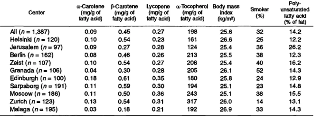
The median adipose tissue concentrations of the three carotenoids for cases and controls by center are shown in table 2. There was a sixfold difference in the concentrations of a-carotene among the controls be-tween centers, a threefold difference in /3-carotene, and less than a twofold difference in lycopene concen-trations. Higher a- and /3-carotene concentrations were found among controls in the northwest countries. Malaga had the lowest median levels of a-carotene (0.03 mg/g of fatty acid) and /3-carotene (0.18 mg/g of fatty acid in controls). The geographic distribution of lycopene was less consistent, with Helsinki and Malaga having the lowest mean lycopene
concentra-TABLE 1. Distribution
Overall (n = 1,379) Cases (n = 662) Controls (n = 717)
of risk factors by disease status, EURAMIC Study, 1991-1992
Age (years) Mean SDf 54.0 9.1 54.7* 9.0 53.3* 9.2 Body mass index Mean 26.3 26.5* 26.0 SD 3.7 3.9 3.5 Current smokers (%) 43 5 5 * 32 Risk factor No. of dgarett.es/day Mean 9.7 13.9* 5.8 SD 14.5 16.6 10.7 Exsmokers (%) 35 3 2 * 38 Maternal history of Mlt (%) 12 17* 7 Paternal history of Ml (%) 22 2 6 * 19 High blood pressure (%) 21 2 6 * 17
* Significantly different from controls at p < 0.05. t Ml, myocardial infarction; SD, standard deviation.
TABLE 2. Median adipose tissue carotenoid concentrations of controls by center, EURAMIC Study, 1991-1992
a-Carotene p-Carotene Lycopene a-Tocopherol Body mass Center (mg/g of (mg/g of (mg/g of (mg/g of Index
fatty add) fatty add) tatty acid) fatty acid) (kg/m*)
Smoker unsaturated Poly-fatty acid (% of fat) All (n= 1,387) Helsinki ( n = 120) Jerusalem (n = 97) Berlin ( n = 162) Zeist(n=107) Granada (n = 106) Edinburgh (n=100) Sarpsborg(n = 191) Moscow (n = 186) Zurich (/)= 123) Malaga ( n = 195) 0.09 0.10 0.09 0.08 0.10 0.04 0.18 0.11 0.11 0.13 0.03 0.45 0.54 0.27 0.46 0.54 0.30 0.61 0.59 0.50 0.54 0.18 0.27 0.23 0.28 0.26 0.27 0.28 0.35 0.30 0.36 0.31 0.21 198 161 124 213 206 205 180 194 243 317 192 25.6 26.6 25.4 25.5 25.4 26.1 25.8 25.1 25.1 26.0 26.9 32 25 36 38 40 52 24 23 38 14 33 14.2 12.2 26.2 12.3 16.2 14.3 12.9 14.8 15.5 13.1 14.3
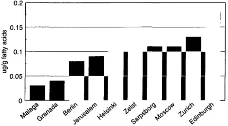
tions (0.23 mg/g of fatty acid and 0.21 mg/g of fatty acid, respectively). Moscow had the highest overall median concentrations of lycopene at 0.36 mg/g of fatty acid, followed closely by Edinburgh at 0.35 mg/g of fatty acid. The median carotenoid concentrations by center for controls are presented in figures 1-3.
Pearson's correlation coefficients were calculated to assess the relation between the carotenoids and myo-cardial infarction-associated risk factors. All three carotenoids were significantly (p < 0.05) negatively
correlated with body mass index (r = —0.22 for a-carotene, r = -0.23 for /3-carotene, and r = —0.24 for lycopene). a-Carotene and /3-carotene were also significantly negatively correlated with the number of cigarettes smoked per day (r = —0.14 for a-carotene,
r = —0.12 for /3-carotene). The carotenoids were
strongly correlated with each other. For a- and /3-carotene, r = 0.78 {p < 0.0001), for a-carotene and lycopene, r = 0.60 (p < 0.0001), and for /3-carotene and lycopene, r - 0.65 (p < 0.0001). Despite these
FIGURE 2. Median lycopene concentrations by center, EURAMIC Study, 1991-1992.
strong correlations between the carotenoids, colinear-ity diagnostics revealed that, in the complete models, individual carotenoids were adequately separated.
Results of conditional logistic regression (condition-ing on age and center) after adjustment for body mass index, socioeconomic status, smoking, family history of disease, and history of high blood pressure are presented in table 3. The odds ratios for myocardial infarction with the continuous carotenoid variables per unit of change as modeled both separately and simul-taneously revealed lycopene to be the only carotenoid with a significant independent association with lower risk of myocardial infarction. The odds ratio for lyco-pene, when modeled separately, was 0.50 (95 percent confidence interval 0.34-0.73) for the contrast be-tween the 10th and 90th percentiles in this population. When modeled simultaneously with a- and /3-carotene, the odds ratio was almost identical, 0.52 (95 percent confidence interval 0.33-0.82). /3-Carotene, on the other hand, was not significantly inversely
associated with protection against myocardial infarc-tion (odds ratio = 0.73, 95 percent confidence interval 0.55-0.96) prior to simultaneous modeling, but after adjustment for other carotenoids, the point estimate of 1.01 (95 percent confidence interval 0.66-1.55) re-mained insignificantly associated with myocardial in-farctions. Similarly, a protective association of a-carotene on its own was virtually eliminated when modeled together with the other carotenoids.
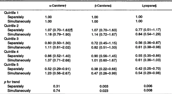
Table 4 reports the odds ratios for myocardial in-farction by quintiles of adipose tissue carotenoid con-centrations, based on the distributions in the control population, with the bottom fifth serving as the refer-ent category. Trend analyses showed all three carote-noids to be protective when modeled separately. In simultaneous analyses, accounting for all three caro-tenoids at the same time, no trend toward lower risk with higher a-carotene levels was seen {p = 0.74). The trend for /3-carotene remained statistically signif-icant after inclusion of the other carotenoids in the 0.2 0.15
s
I 0-1
I
0.05I
• • I
• • I I I
— — •
11
'S
S
< / /
</
TABLE 3. Conditional odds ratios for myocardial infarction by carotenoid adipose tissue sample concentration, EURAMIC Study, 1991-1992*
Adipose
concentration a-Carotene valueP ^Carotene
P value Lycopene P value Separately Simultaneously 0.68 (0.49-0.93)t 0.91 (0.58-1.41) 0.02 0.66 0.73 (0.55-0.96) 1.01 (0.66-1.55) 0.03 0.96 0.50 (0.34-0.73) 0.52 (0.33-0.82) 0.003 0.005 * The odds ratios are conditional on age and center; covariates are body mass index, age, center, smoking, maternal and paternal history of disease, and history of high blood pressure. The odds ratios are based on the contrast of the 10th and 90th percentiles of the carotenoid from controls in the population at large. All of the carotenoids are scaled in these models.
t Numbers in parentheses, 95% confidence interval.
TABLE 4. Conditional odds ratios of myocardial infarction associated with quintiles of adipose carotenoids compared with the lowest quintile, EURAMIC Study, 1991-1992*
Quintile 1 Separately Simultaneously Quintile 2 Separately Simultaneously Quintile 3 Separately Simultaneously Quintile 4 Separately Simultaneously Quintile 5 Separately Simultaneously p for trend Separately Simultaneously a-Carotenet 1.00 1.00 1.07(0.70-1.63)H 1.18(0.79-1.90) 0.80(0.50-1.30) 1.11 (0.61-2.02) 0.86(0.52-1.40) 1.37(0.71-2.66) 0.52 (0.29-0.91) 1.23(0.56-2.67) 0.31 0.74 p-Carotenef 1.00 1.00 1.07(0.70-1.63) 1.14(0.72-1.67) 0.72(0.45-1.15) 0.82(0.51-1.33) 0.90(0.56-1.45) 1.01 (0.60-1.67) 0.38 (0.22-0.66) 0.47 (0.26-0.99) 0.003 0.023 Lycopene§ 1.00 1.00 0.77(0.51-1.17) 0.84(0.54-1.29) 0.56 (0.36-0.87) 0.61 (0.38-0.98) 0.55 (0.35-0.86) 0.61 (0.36-1.03) 0.42 (0.25-0.70) 0.54 (0.29-0.98) 0.006 0.008
* The odds ratios are conditioned on age and center; covariates are body mass index, smoking, maternal and paternal history of disease, history of high blood pressure, age, and a-tocopherol. The odds ratios are based on the contrast of the 10th and 90th percentiles of the carotenoid from controls in the population at large. All of the carotenoids are scaled in these models.
t Median a-carotene levels are 0.03, 0.05, 0.09, 0.13, and 0.24 |ig/g of fat for quintiles 1-5, respectively. t Median p-carotene levels are 0.14, 0.29,0.44, 0.63, and 1.11 ng/g of fat for quintiles 1-5, respectively. § Median lycopene levels are 0.11, 0.20, 0.27, 0.38, and 0.62 ng/g of fat for quintiles 1-5, respectively. H Numbers in parentheses, 95% confidence interval.
model. However, the association within the upper four quintiles was not consistent, and only for the fifth quintile of the /3-carotene distribution was the odds ratio substantially protective in the simultaneous model at 0.47 (95 percent confidence interval 0.26-0.99). Lycopene showed the greatest protective effect with a tendency toward increasing inverse odds ratios with increased concentrations, evident in both the sep-arate and simultaneous models. The probability of the inverse association's arising by chance is 0.008 (from the test of trend).
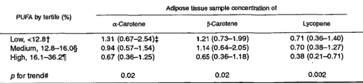
Polyunsaturated fats and carotenoid effects An examination of the carotenoid interactions with polyunsaturated fats and acute myocardial infarction using interaction terms and categorizing
polyunsatu-rated fat levels in adipose tissues by tertiles revealed a significant interaction (table 5). The protective effect of lycopene increased at each increasing level of poly-unsaturated fat and was significant in the individuals whose adipose tissue contained more than 16.1 percent of the fat in the form of polyunsaturates. /3-Carotene and a-carotene were never significantly associated with polyunsaturated fat levels but tended also to show a trend toward protection only in the presence of the highest concentrations of polyunsaturated fats.
Smoking status and carotenoid effects
Stratification by smoking status indicated that the effect of lycopene among nonsmokers (table 6) was strongest. ^-Carotene was not protective in any group.
TABLE 5. Conditional odds ratios for myocardial infarction and carotenoids by tertile of polyunsaturated fatty acid (PUFA), EURAMIC Study, 1991-1992*
Adipose tissue sample concentration of PUFA by tertile (%)
a-Carotene p-Carotene Lycopene
Low, <12.8f Medium, 12.8-16.0§ High, 16.1-36.211 p for trend* 1.31 (0.67-2.54)$ 0.94(0.57-1.54) 0.67(0.36-1.25) 0.02 1.21 (0.73-1.99) 1.14(0.64-2.05) 0.65(0.36-1.18) 0.02 0.71 (0.36-1.40) 0.70(0.38-1.27) 0.38 (0.21-0.71) 0.002
* Conditional on age and center; covariates are body mass index, socioeconomic status, smoking, maternal and paternal history of disease, and history of high blood pressure. The odds ratios are from controls of the 10th and 90th percentiles of the carotenoid in the population at large.
t Median PUFA for the low tertile is 11.3% of fat. i Numbers in parentheses, 95% confidence interval. § Median PUFA for the medium tertile is 14.3% of fat. H Median PUFA for the high tertile is 19.3% of fat. # Trend test weighted by tertile medians.
TABLE 6. Conditional odds ratios for myocardial infarction and carotenoids by smoking category, EURAMIC Study, 1991-1992* Modeled simultaneously by smoking Never smoker Exsmokers Smokers a-Carotenet 0.93 (0.49-1.76)H 0.70 (0.38-1.28) 1.28(0.68-2.40)
Adipose tissue concentration of
P-Carotenet 1.14(0.51-2.54) 0.92(0.53-1.59) 0.93(0.61-1.59) Lycopene§ 0.33 (0.13-0.85) 0.41 (0.21-0.83) 0.63(0.39-1.01) * Conditional on age and center; covariates are body mass index, socioeconomic status, maternal and patemal history of disease, and history of high blood pressure.
t Median values of a-carotene are 0.10, 0.09, and 0.07 ng/g of fat for never smokers, exsmokers, and smokers, respectively.
t Median values of p-carotene are 0.50, 0.41, and 0.34 u.g/g of fat for never smokers, exsmokers, and
smokers, respectively.
§ Median values of lycopene are 0.27,0.23, and 0.26 ng/g of fat for never smokers, exsmokers, and smokers, respectively.
11 Numbers in parentheses, 95% confidence interval.
DISCUSSION
Lycopene is present in the blood in concentrations roughly equal to those of /3-carotene. For unknown reasons, certain organs (28) selectively take up or concentrate lycopene so that tissue levels greatly ex-ceed those of /3-carotene. Although little is known about the uptake and turnover of carotenoids in adi-pose tissue, the adiadi-pose tissue levels of lycopene, as with other carotenoids, are derived largely from the diet and serve as a better indicator of long-term expo-sure than do serum or recent dietary records (29). Even if tissues such as adrenal glands or prostate concen-trate lycopene, the vast majority of the carotenoids will be found in the adipose tissue. It is important to note that lycopene was not available as a supplement to the individuals in this study prior to their recruit-ment. Major food sources appear to be tomatoes and tomato products, watermelon, grapefruit, and seafood such as lobster and crab. Foods rich in lycopene have recently been suggested to be inversely associated
with prostate cancer (30). There are indications in the literature that lycopene is a more potent antioxidant than /3-carotene. DiMascio et al. (31) showed the disappearance in vitro of lycopene in cells under sin-glet oxygen stress to be greater than that of a-carotene, /3-carotene, and a-tocopherol. A study of the sequen-tial order of antioxidant consumption in Cu2+
-medi-ated oxidation of low density lipoprotein found that lycopene was consumed first, with cryptoxanthin and lutein/zeaxanthin next, and /3-carotene following in last place (32). Furthermore, it has recently been dis-covered that skin exposure to ultraviolet radiation un-der controlled experimental conditions results in a strong (31-46 percent) and rapid reduction in skin lycopene levels but in no change in /3-carotene levels in the skin (33). Thus, lycopene may be the underlying carotenoid providing antioxidant protection.
Since individuals consuming diets high in vegeta-bles tend to have higher levels of all carotenoids, the strong association between lycopene and /3-carotene
can conceivably explain the /3-carotene-myocardial in-farction association evident in some observational studies. |3-Carotene may be a good marker of lycopene intake, and its effect disappears when both are consid-ered simultaneously. In the same line of argument, lycopene could be a marker of other active phyto-chemicals in tomatoes and possibly in other major food sources of this antioxidant. The protective poten-tial of lycopene was greatest among individuals with the highest polyunsaturated fat stores, consistent with an antioxidant effect. The interaction was not, how-ever, strongest among smokers.
Adipose tissue provides quantitative measures of prior exposure unbiased by problems of dietary assess-ment by questionnaire or imprecise food composition values. It is subject to individual differences in metab-olism and absorption. Despite these weaknesses, for components widely distributed in the diet with high day-to-day variation in intakes—as is the case with carotenoids—it remains a useful biomarker of expo-sure. In a case-control study, there is the potential for the disease condition to affect the biomarker. To pre-vent this, adipose tissue was used, and aspirations were done during hospitalization soon after the infarc-tion. Since adipose tissue is a stable depository of fat-soluble substances whose turnover tissue is quite slow (29, 34), and since it was collected shortly after the infarctions, it is unlikely to be significantly af-fected by the occurrence of myocardial infarction. In this case-control design, only patients who survived until admission to the hospital and until adipose tissue aspiration were included. This may be a potential source of bias if the association among the deaths differs from that in the surviving patients.
Our results are consistent with a protective effect of adipose tissue levels of lycopene, but not of /3-carotene or a-/3-carotene, on myocardial infarction risk. Alternately, lycopene may be a marker of another protective substance in a common food source. The protective association with lycopene was not seen in smokers. It did, however, interact with another major source of oxidative stress, stores of polyunsaturated fats, which are markers of consumption of high poly-unsaturated fat diets. This suggests that lycopene may be operating under a tissue-specific antioxidant mech-anism.
Research Foundation of the Israel Academy of Sciences and Humanities, the EC Milk Intervention Board, the Ulster Cancer Foundation, the Dutch Ministry of Health, Spanish FIS and the Ministry of Science and Education, the German Federal Health Office, the Norwegian Council, Swiss NRF (grant 32-31312-91), and the Yrjo Jahnsson Foundation, Finland.
ACKNOWLEDGMENTS
The EURAMIC Study was supported by an EC-Concerted Action by the Commission of European Com-munities. The national studies were financed by the Basic
REFERENCES
1. Stamler J. Opportunities and pitfalls in international compar-isons related to patterns, trends, and determinants of CEID mortality. Int J Epidemiol 1989;18(suppl 1):S3-18.
2. Artaud-Wild SM, Connor SL, Sexton G, et al. Differences in coronary mortality can be explained by differences in choles-terol and saturated fat intakes in 40 countries but not in France and Finland: a paradox. Circulation 1993;88:2771-9. 3. Heller RF, Chinn S, Tunstall-Pedoe TID, et al. How well can
we predict heart disease? Findings in the United Kingdom Heart Disease Prevention Project. Br Med J 1984;288: 110-23.
4. Steinberg D, Parthasarathy S, Carew TE, et al. Beyond cholesterol: modifications of low-density lipoprotein that in-crease its atherogenicity. N Engl J Med 1989;320:915-24. 5. Goldstein JL, Ho YK, Basu SK, et al. Binding site on
mac-rophages that mediates uptake and degradation of acetylated low density lipoprotein, producing massive cholesterol depo-sition. Proc Natl Acad Sci U S A 1979;76:333-7.
6. Slyper AH. Low-density lipoprotein density and atherosclero-sis. Unraveling the connection. JAMA 1994;272:305-8. 7. Stephens NG, Parsons A, Schofield PM, et al. Randomised
controlled trial of vitamin E in patients with coronary disease: Cambridge Heart Antioxidant Study (CHAOS). Lancet 1996; 347:781-6.
8. Frei B. Cardiovascular disease and nutrient antioxidants: role of low-density lipoprotein oxidation. Crit Rev Food Sci Nutr 1995;351:83-98.
9. Gaziano J, Michael C, Hennekens H. The role of /3-carotene in the prevention of cardiovascular disease. Ann N Y Acad Sci 1993;691:148-55.
10. Holvoet P, Collen D. Oxidized lipoproteins in atherosclerosis and thrombosis. FASEB J 1994;8:1279-84.
11. Halliwell B, Gutteridge JM. Lipid peroxidation, oxygen rad-icals, cell damage, and antioxidant therapy. Lancet 1984;1: 1396-7.
12. Gey KF, Moser UK, Jordan P, et al. Increased risk of cardio-vascular disease at suboptimal plasma concentrations of es-sential antioxidants: an epidemiological update with special attention to carotene and vitamin C. Am J Clin Nutr 1993; 57(suppl):787S-97S.
13. Riemersma RA, Oliver M, Elton RA, et al. Plasma antioxi-dants and coronary heart disease: vitamins C and E, and selenium. Eur J Clin Nutr 1990;44:143-50.
14. Riemersma RA, Wood DA, Maclntryre CCA, et al. Risk of angina pectoris and plasma concentrations of vitamins A, C, and E and carotene. Lancet 1991 ;337:1—5.
15. Kardinaal AF, Kok FJ, Ringstad J, et al. Antioxidants in adipose tissue and risk of myocardial infarction: the EURAMIC Study. Lancet 1993,342:1379-84.
16. Street DA, Comstock GW, Salkeld RM, et al. A population-based case-control study of the association of serum antioxi-dants and myocardial infarction. Am J Epidemiol 1991; 134: 719-20.
17. Kok FJ, De Bruijn AM, Vermeeren R, et al. Serum selenium, vitamin antioxidants, and cardiovascular mortality: a 9-year follow-up study in the Netherlands. Am J Clin Nutr 1987;45: 1368-77.
18. Kushi LH, Folsom AR, Prineas RJ, et al. Dietary antioxidant vitamins and death from coronary heart disease in postmeno-pausal women. N Engl J Med 1996;334:1156-62.
19. The Alpha-Tocopherol, Beta-Carotene Cancer Prevention Study Group. The effect of vitamin E and beta carotene on the incidence of lung cancer and other cancers in male smokers. N Engl J Med 1994;330:1029-35.
20. Ommen GS, Goodman GE, Thomquist MD, et al. Effects of a combination of beta carotene and vitamin A on lung cancer and cardiovascular disease. N Engl J Med 1996;334:1150-5. 21. Hennekens CH, Buring JE, Manson JE, et al. Lack of effect of long-term supplementation with beta carotene on the inci-dence of malignant neoplasms and cardiovascular disease. N Engl J Med 1996;334:1145-9.
22. Women's Health Study Research Group. The Women's Health Study: summary of the study design. J Myocardial Ischemia 1992;4:27-9.
23. Manson JE, Gaziano JM, Spelspberg A, et al. A secondary prevention trial of antioxidant vitamins and cardiovascular disease in women: rationale, design, and methods. Ann Epi-demiol 1995;5:261-9.
24. Kardinaal AFM, van't Veer P, Kok FJ, et al. EURAMIC Study: antioxidants, myocardial infarction, and breast cancer. Eur J Clin Nutr 1993;47(suppl 2): 64-71.
25. Beynen AC, Katan MB. Rapid sampling and long-term stor-age of subcutaneous adipose-tissue biopsies for determination of fatty acid composition. Am J Clin Nutr 1985;42:317-22.
26. Statistical Analysis System, Inc. SAS statistics, 4th ed. Ver-sion 6. Cary, NC: Statistical Analysis System, Inc, 1990:1364. 27. Kleinbaum DG, Kupper LL, Morgenstern H, eds. Epidemio-logic research. Principles and quantitative methods. Belmont, CA: Wadsworth, Inc, 1982.
28. Stahl W, Schwarz W, Sundquist AR, et al. cis-trans isomers of lycopene and beta-carotene in human serum and tissues. Arch Biochem Biophys 1992;294:173-7.
29. Kohlmeier L, Kohlmeier M. Adipose tissue as a medium for epidemiologic exposure assessment. Environ Health Perspect 1995;103(suppl 3):99-106.
30. Giovannucci E, Ascherio A, Rimm EB, et al. Intake of caro-tenoids and retinol in relation to risk of prostate cancer. J Natl Cancer Inst 1995;87:1767-76.
31. DiMascio P, Kaiser S, Sies H. Lycopene as the most efficient biological carotenoid singlet oxygen quencher. Arch Biochem Biophys 1989;274:532-8.
32. Esterbauer H, Striegl G, Puhl H, et al. Role of vitamin E and carotenoids in preventing oxidation of low density lipopro-teins. Ann N Y Acad Sci 1989;70:254-67.
33. Ribaya-Mercado JD, Garmyn M, Gilchrest BA, et al. Skin lycopene is destroyed preferentially over beta-carotene during ultraviolet irradiation in humans. J Nutr 1995; 125:1854-9. 34. Handelman GJ, Epstein WL, Peerson J, et al. Human adipose
alpha-tocopherol and gamma-tocopherol kinetics during and after 1 y of alpha-tocopherol supplementation. Am J Clin Nutr 1994;59:1025-32.