Human Reproduction vol.11 no.6 pp. 1173-1176, 1996
CASE REPORT
Aorto-subclavian thromboembolism: a rare complication
associated with moderate ovarian hyperstimulation
syndrome
Marc Germond, Daniel Wirthner,
Dominique Thorin1, Patrick Ruchat2, Axel Essinger3
and Pierre De Grandi
Fertility Unit, Department of Gynaecology and Obstetrics,
'Service of Anesthesiology, 2Service of Cardiovascular Surgery and 3Department of Radiology, CHUV, 1011 Lausanne, Switzerland
The case of an arterial aorto-subclavian thromboembolism associated with a moderate ovarian hyperstimulation syn-drome (OHSS) and following ovulation induction for in-vitro fertilization in a young woman is reported. Because of the lack of response to systemic thrombolysis, a left postero-lateral thoracotomy was performed on day 8 after embryo transfer. A fibrinocruoric embolus situated at the junction of the left subclavian artery from the aorta was removed through a left subclavian arteriotomy. The distal axillary embolus was removed by a retrograde balloon catheter embolectomy. A moderate OHSS was observed. The ovarian stimulation and OHSS-related risks of thrombo-embolism are discussed. We conclude that, in the absence of risk factors, counselling about possible complications resulting from stimulation must be emphasized.
Key words: arterial thromboembolism/IVF/menotrophin/
ovarian hyperstimulation syndrome
Introduction
Ovarian hyperstimulation syndrome (OHSS) is the most fre-quent and worrying complication following ovulation induc-tion. The incidence of this side-effect has increased since gonadotrophin releasing hormone analogues (GnRHa) have been used. For many authors, the different protocols which associate follicle stimulating hormone (FSH) and/or human menopausal gonadotrophin (HMG) with GnRHa seem to be related to a significantly higher rate of moderate or severe OHSS (Rizk and Smitz, 1992). Some authors (Rabau et al, 1967) have classified ovarian response in three clinical cat-egories (mild, moderate and severe) and six grades (two per category) based on the severity of the signs, symptoms and laboratory findings. The more recent definition of OHSS proposed by Golan et al. (1989) has offered a more clinical approach to this syndrome. Severe complications of this iatrogenic syndrome include thromboembolism, adult respira-tory distress syndrome, acute hydrothorax (Daniel et al, 1995) and, occasionally, death (Schenker and Ezra, 1994). During ovarian stimulation for in-vitro fertilization (TVF) and embryo © European Society for Human Reproduction and Embryology
transfer, the risk of moderate or severe OHSS after GnRHa/ HMG stimulation is evaluated as 0.6-14.0% (Rizk and Smitz, 1992). We report the case of. an arterial aorto-subclavian thromboembolism associated with a moderate OHSS in a young woman.
Case report
A non-Caucasian 32 year old woman was treated in our IVF and embryo transfer programme for a secondary infertility associated with a male factor and a bilateral tubal obstruction. Her past medical history reported a right salpingectomy for an ectopic pregnancy and a proximal left tubal occlusion. A mild essential hypertension was easily corrected with labetalol (200 mg/day).
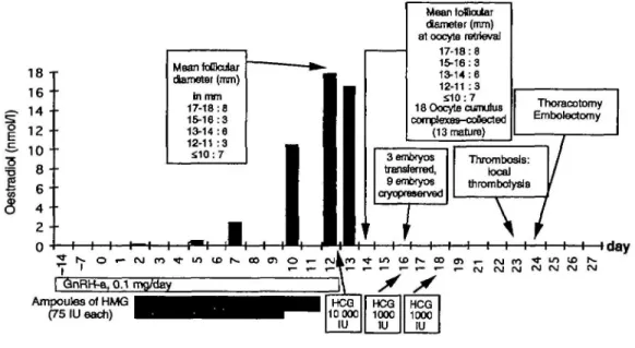
The treatment procedure consisted of a long protocol (Figure 1). After pituitary desensitization with GnRHa (0.1 mg Deca-peptyl; Ferring, Zurich, Switzerland) administered from day 23, multiple follicular development was induced using 225 IU HMG (Pergonal; Serono, Aubonne, Switzerland) daily. Follicular growth was monitored with serial ultrasound scans and repeated estimations of oestradiol blood concentrations. On day 10 of stimulation, the HMG dose was reduced to 150 IU because of the high oestradiol concentration (10.5 nmol/1). By day 12, 11 follicles > 1 6 mm in diameter and 16 smaller follicles were observed. The oestradiol concentration reached 17.8 nmol/1 when 10 000 IU human chorionic gonado-trophin (HCG; Profasi; Serono) were administered. Oocyte recovery took place 36 h later. In all, 18 oocytes were collected; 12 oocytes fertilized, three embryos were transferred to the uterus 2 days later and nine embryos were frozen. A dose of 1000 IU HCG was given i.m. on the day of embryo transfer and 48 h later for luteal phase support. This support was stopped because the patient complained of nausea and abdominal tension. No diarrhoea or vomiting was noticed. Abdominal ultrasonography showed enlarged ovaries (9 cm). As expected, multiple ovarian cysts and a small quantity of ascites were found. The haemoglobin concentration was 139 g/1, with a haematocrit of 0.44. Electrolytes and creatinine concentrations were within the normal range. A diagnosis of moderate OHSS was made, based on clinical, laboratory and echographic findings. On day 6 after transfer, the patient began to complain of pain and paraesthesia in the left arm which she had noticed 36 h earlier. Examination revealed pulseless radial and humeral arteries and a cold left arm. The aetiology of die acute ischaemia was determined by an arteriography, which showed a large clot in the left subclavian artery (Figure 2) 1173
M.Gennond et aL 5
I
1 8 - • 1 6 • • 14 .. 1 2 •• 1 0 •• 8 • 6 • • 4 • 2 -Mean fofflaiar diameter (mm) in mm 17-18:8 16-16:3 13-14:6 12-11 :3 £10:7 H 1—I 1 1 H MeanfoSaiar ttameter (mm) at oocyte retrieval 17-18:8 15-16:3 13-14:6 12-11 : 3 £10:7 18 Oocyte cumulus complexss-coBected (13 mature) 3embryos transferred, 9e<nbfyos cryopreserved • / Thoracotomy Emboloctomy # Thrombosis: local thrombotysls \ / O w- r j k r o •<)• QnRH-a, 0.1 mgAtey Ampoules of HMG (75 IU each)Figure 1. Chronology of events.
and an occlusion of the left axillary artery. Signs of slight haemoconcentration were present (haemoglobin 151 g/1, haem-atocrit 0.46). Clotting function tests were within the normal range [quick time (FT), partial thrombin time (PTT)]. As thrombolysis was initiated as an emergency procedure, more specific thrombosis risk factors such as protein C, protein S, Leiden V factor and antithrombin HI deficiencies were not checked. Immediate local thrombolysis was attempted with 100 000 and 200 000 IU urokinase without success. Anti-coagulation using i.v. heparin and oral aspirin was initiated. Given the lack of response, a left postero-lateral thoracotomy was performed on day 7 after transfer. Surgical exploration revealed a fibrinocruoric embolus situated at the junction of the left subclavian artery with the aorta. The embolus was removed through a left subclavian arteriotomy. The distal axillary embolus was removed by a retrograde balloon catheter embolectomy. The presence of a patent foramen oval (para-doxical emboli) was excluded during the operation by using a transoesophageal echocardiography with microbubbles test. The choice of this surgical strategy was motivated by the danger of left carotid arterial emboli after the use of retrograde balloon catheter embolectomy from the left humeral artery. At the end of the operation, the left radial artery was again palpable. Heparin administration was discontinued and di-coumarol administration introduced after the existence of a pregnancy had been excluded.
Discussion
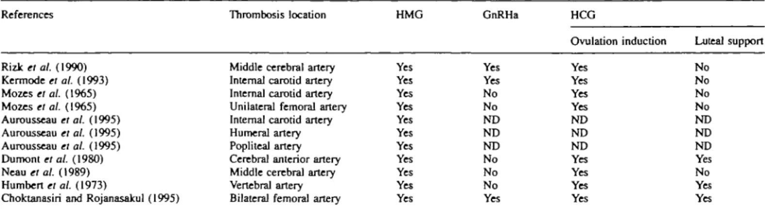
Arterial thromboembolism is a rare complication of OHSS. Only 11 cases of arterial thrombosis have been described following gonadotrophin stimulation: two cases of right middle cerebral artery thrombosis (Neau et aL, 1989; Rizk et aL, 1990), one of cerebral anterior artery thrombosis (Dumont
et aL, 1980), one of vertebral artery thrombosis (Humbert et aL, 1973), three of internal carotid artery thrombosis (Mozes et aL, 1965; Kermode et aL, 1993; Aurousseau et aL, 1995),
one of humeral artery thrombosis (Aurousseau et aL, 1995), one of unilateral femoral artery thrombosis (Mozes et aL,
I—i—I—I—i—i—I—I—i—I—I—(day HCG 10000 IU HCG 1000 HJ HCG 1000 IU
Figure 2. Selective angiogram of left subclavian artery showing proximal occlusion by a fibrinocruoric embolus and distal axillary obstruction. A radiological steal subclavian syndrome occurred.
1965), one of bilateral femoral artery thrombosis (Choktanasiri and Rojanasakul, 1995) and one of popliteal artery thrombosis (Aurousseau et aL, 1995). Most of these occlusions are characterized by their position in the superior limb or in the cerebral territory. The case reported here shows an unusual location of thrombosis occurring during a moderate OHSS.
No risk factor for thromboembolism had been detected, either by clinical observation or by cardiological and laboratory investigations performed before the treatment Three OHSS risk factors were present: a high oestradiol concentration induced by the simultaneous administration of GnRHa with HMG, ovulation induction with HCG and the use of HCG as a luteal phase support Only one of the previously described cases of arterial thrombosis presents all these risk factors (Table I).
In our reported case, OHSS risk was, in our opinion, not sufficient to cancel HCG administration or to avoid embryo transfer. In our practice, the cycle is cancelled when the oestradiol concentration is > 2 0 nmol/1 or when the number of follicles > 1 6 mm in diameter is >12. This attitude is supported by Morris et al. (1995). Our patient did not develop severe OHSS, although the retrospectively calculated predic-tion rate according to Delvigne et al. (1993) was 78.9%.
Many factors contribute to thrombogenesis during the evolu-1174
Aorto-subclavLan thromboembolism
Table I. Menotrophins and gonadotrophin-releasing hormone analogue (GnRHa) use in the 11 reviewed cases of arterial thrombosis associated with ovarian
hyperstimulation syndrome
References Thrombosis location HMG GnRHa HCG
Yes Yes Yes Yes Yes Yes Yes Yes Yes Yes Yes Yes Yes No No ND ND ND No No No Yes Ovulation induction Yes Yes Yes Yes ND ND ND Yes Yes Yes Yes Luteal support No No No No ND ND ND Yes No Yes Yes R i z k « al. (1990) Kermode el al. (1993) Mozes et al. (1965) Mozes et al. (1965) Aurousseau et al. (1995) Aurousseau et al. (1995) Aurousseau et al. (1995) Dumont et al. (1980) Neau et al. (1989) H u m b e r t s al. (1973)
Choktanasiri and Rojanasakul (1995)
Middle cerebral artery Internal carotid artery Internal carotid artery Unilateral femoral artery Internal carotid artery Humeral artery Popliteal artery Cerebral anterior artery Middle cerebral artery Vertebral artery Bilateral femoral artery
HCG = human chorionic gonadotrophin; HMG = human menopausal gonadotrophin; ND = not described. tion of an OHSS. Some of these seem to be relatively
non-specific and related to fluid shifting from the vascular space to the extracellular space. As demonstrated by McClure et al (1994), capillary permeability increases during spontaneous or induced ovulation, mainly caused by a vascular endothelial growth factor (VEGF). Usually, the angiogenesis of spon-taneous ovulation is self-limited to one follicle. In the context of induced hyperstimulation, the neovascularization involves multiple follicles. In this case, VEGF production may overflow the ovarian capillary bed, spill over into the peritoneal cavity and promote the formation of ascites. This phenomenon explains the moderate to extreme haemoconcentration and hyperviscosity observed in severe OHSS. As suggested pre-viously (McClure et al, 1994), the measurement of VEGF in the plasma or urine of patients at risk of OHSS would probably improve the safety of IVF and related procedures. Moreover, the stasis of venous blood flow in the pelvic brim and the lower limb is favoured by abdominal distension.
At present, many research groups are focusing their attention on endothelial cell changes and cascade coagulation. Stress-induced leukocytosis is a common finding during severe OHSS. As shown in the umbilical vein, endothelial monolayer cells may be injured by the activation of polymorphonuclear leuko-cytes, releasing a protease which could impair the fibrinolytic potential of the endothelium (Pintucci et al, 1993). These lesions favour the exposition of a highly thrombogenic extra-cellular matrix. Blood flow in the damaged vessels promotes adhesion and the spreading of platelets on the surface of this matrix (Kolpakov et al., 1994).
The extrinsic coagulation pathway is triggered by endothelial cell procoagulant activity. This activity is involved in thrombo-genesis associated with OHSS and seems to be independent of oestradiol and progesterone plasma concentrations.
Independent of the endothelium-blood interface, humoral modifications of haemostasis factors are associated with high plasma concentrations of oestradiol. When the coagulation cascade is triggered, an oestrogen-induced hypercoagulable state is generated by increased activities of several factors (factor Vm, von Willebrand factor and fibrinogen) and by decreased activities of antithrombin HI and protein C (Massafra
et al., 1993). The decreased activity of factor VII may be
interpreted as a relative protective mechanism operating at very high plasma concentrations of oestrogen (Bremme et al, 1994). Currently used laboratory tests such as Quick time, accelerated partial thrombin time, fibrinogen or platelet count have all failed to identify a hypercoagulable state. Whole blood clotting time and whole blood clot lysis time, as used by Aune et al (1993), are not available for routine investigation. In-vitro clot-based methods, testing the visco-elastic properties of the thrombus, are sensitive methods which are used to investigate the blood hypercoagulability associated with ovarian stimulation. However, they have failed to describe the platelet and vascular endothelium interface.
In conclusion, whether thrombogenesis is dependent upon important hormonal changes induced by stimulation, vascular damage, haemostasis modifications or all these factors is not clear.
Conclusion
In our reported case, arterial thrombosis occurred even though a moderate OHSS was present Although a moderate OHSS was predictable, the risk of arterial thrombosis was not This emphasizes the importance of informing patients about possible complications associated with stimulation.
References
Aune, B., Oian, P. and 0sterud, B. (1993) Enhanced sensitivity of the extrinsic coagulation system during ovarian stimulation for in-vitro fertilization.
Hum Reprod., 8, 1349-1352.
Aurousseau, M.H., Samana, M.M., Belhassen, A., Herve, F. and Hugues, J.N. (1995) Risk of thromboembolism in relation to an in-vitro fertilization programme: three case reports. Hum. Reprod., 10, 94-97.
Bremme, K., Wramsby, H., Andersson, O., Wallin, M. and Blomback, M. (1994) Do lowered factor VII levels at extremely high endogenous oestradiol levels protect against thrombin formation? Blood Coagul Fibrinolysis, 5, 205-210.
Choktanasiri, W. and Rojanasakul, A. (1995) Acute arterial thrombosis after gamete intrafallopian transfer a case report. J. Assist Reprod. Genet., 12, 335-337.
Daniel, Y, Yaron, Y, Oren, M., Peyser, M.R. and Lessing, J.B. (1995) Ovarian hyperstimulation syndrome manifests as acute bilateral hydrothorax. Hum.
Reprod., 10, 1684-1685.
Delvigne, A., Dubois, M., Battheu, B. et al (1993) The ovarian hyperstimulation syndrome in in-vitro fertilization: Belgian multicentric 1175
M.Germond et al
study, n . Multiple discriminant analysis for risk prediction. Hum. Reprod., 8, 1361-1366.
Dumont, M., Combet, A. and Domechini, Y. (1980) Thrombose arteVielle c£r£brale a la suite d'une hyperstimulation ovarienne: grossesse sextuple, avortement therapeutique. Nouv. Presse Med., 9, 3628-3631.
Golan, A., Ron-El, R., Herman, A. et al. (1989) Ovarian hyperstimulation syndrome: an update review. Obstet. GynecoL Sum., 44, 430-440. Humbert, G., Delaunay, P., Leroy, J. et al. (1973) Accident vasculaire cerebral
au cours d'un traitement par les gonadotrophines. Nouv. Presse Med., 2, 28-30.
Kermode, A.G., Churchyard, A. and Carroll, W.M. (1993) Stroke complicating severe ovarian hyperstimulation syndrome. Aust. N.Z. J. Med., 23, 219-220. Kolpakov, V., Adamo, M.C., Salvatore, L. et al. (1994) Neutrophil derived cathepsin G induces potentially thrombogenic changes in human endothelial cells: a scanning electron microscopy study in static and dynamic conditions.
Thromb. Haemost., 72, 140-145.
Massafra, C , Butini, P., Cavion, M.A. et aL (1993) Evaluation of risk of thrombosis during use of low-dose ethinylestradiol-desogestrel oraJ contraceptive. Adv. Contracept., 9, 195-203.
McClure, N., Healy, D.L., Rogers, P.A.W. et al. (1994) Vascular endothelial growth factor as capillary permeability agent in ovarian hyperstimulation syndrome. Lancet, 344, 235-236.
Morris, R.S., Paulson, RJ., Sauer, M.V. and Lobo, R.A. (1995) Predictive value of serum oestradiol concentrations and oocyte number in severe ovarian hyperstimulation syndrome. Hum. Reprod., 10, 811-814. Mozes, M., Bogokowsky, H. and Antebi, E. (1965) Thromboembolic
phenomena after ovarian stimulation with human gonadotrophins. Lancet,
2, 1213-1215.
Neau, J.P., Marechaud, M., Guitton, P. et al. (1989) Occlusion de l'artere cirtbrale moyenne lors d'une induction de I'ovulation par les gonadotrophines. Rev. NeuroL, 145, 859-861.
Pintucci, G., Iacoviello, L., Castelli, M.P. et al. (1993) Cathepsin G-induced release of PAI-1 in the culture medium of endothelial cells: a new thrombogenic role for polymorphonuclear leukocytes? J. Lab. Clin. Med.,
122, 69-79.
Rabau, E., David, A., Serr, D.M., Mashiach, S. and Lunenfeld, B. (1967) Human menopausal gonadotropins for anovulation and sterility. Am. J.
Obstet. Gynecol., 98, 92-98.
Rizlc, B. and Smitz, J. (1992) Ovarian hyperstimulation syndrome after superovulation using GnRH agonists for IVF and related procedures. Hum.
Reprod., 7, 320-327.
Rizk, B., Meagher, S. and Fischer, A.M. (1990) Severe ovarian hyperstimulation syndrome and cerebrovascular accidents. Hum. Reprod., 5, 697-698.
Schenker, J.G. and Ezra, Y. (1994) Complications of assisted reproductive techniques. Fertil. Steril., 61, 411-422.
Received on October 3, 1995; accepted on April 14, 1996