British Journal of Anaesthesia 1993; 70: 459-461
CASE REPORTS
SPONTANEOUS EXCITATORY MOVEMENTS DURING
RECOVERY FROM PROPOFOL ANAESTHESIA IN AN INFANT:
EEG EVALUATION
A. BORGEAT, O. H. G. WILDER-SMITH, P. A. DESPLAND AND P. RAVUSSIN
SUMMARY
Spontaneous excitatory movements have been observed during reco very from propofol anaesthesia in children. Epilepsy has been postulated as a possible mechanism to explain these movements.
We report the first case in which these spontaneous excitatory movements were studied using sim-ultaneous multichannel EEG recordings. (Br. J.
Anaesth. 1993; 70: 459-461)
KEY WORDS
Anaesthetic techniques: intravenous Anaesthetics intravenous: propofol. Complications: spontaneous excitatory movements.
Spontaneous excitatory movements have been ob-served during both induction and recovery from propofol anaesthesia [1-3]. Although there have been several case reports of "seizures" or opistho-tonus during recovery from propofol anaesthesia [4—7] none of these has been evaluated by sim-ultaneous EEG recordings.
An infant had exhibited spontaneous excitatory movements during recovery from previous propofol anaesthetics. In the course of a subsequent propofol anaesthetic, we were able to study these spontaneous excitatory movements using simultaneous multi-channel EEG recordings.
CASE REPORT
The patient was a 6-week-old, otherwise healthy, 4-kg young girl. She was admitted for radiotherapy (35 courses of 3 min duration) of her congenital bilateral retinoblastoma. During the first two treatments, anaesthesia was performed with propofol. In the course of each recovery, spontaneous excitatory movements (graded stage 3 according to the pre-viously described classification [3]) were observed, lasting 15—20 min until full awakening. The an-aesthetic technique used was as follows: pre-medication with oral atropine 0.15 mg; local an-aesthesia for venous cannulation (24-gauge) insertion using EMLA cream on the back of both hands. In the anaesthesia room, heart rate, arterial pressure and SpOj were recorded. Anaesthesia was induced
with a bolus of propofol 20 mg i.v. The lungs were ventilated manually with oxygen (FIO I 100%). For
the third course of radiotherapy, we decided to record the multichannel EEG during the phase of excitatory movements. After induction of anaesthesia
(as described above) and completion of radiotherapy treatment, the child received a supplementary bolus of propofol 5 mg i.v. to facilitate placement of the EEG electrodes. Twelve intradermal needle elec-trodes were placed according to the 10-20 inter-national system.
Electrode impedances were less than 6 kf2. A CEM EEG recording machine (Vickers, U.K.) was used to record eight channels. The high frequency filter was set at 70 Hz; bipolar longitudinal montages were used. An ECG was recorded simultaneously on one channel.
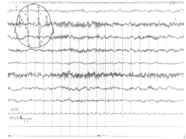
Four minutes after the last bolus of propofol, the EEG recordings analysed by our specialist electro-encephalographer, showed fast waves (10 Hz) with a mean voltage 50 uV coupled with theta waves (voltage 100-200 nV) (fig. 1). No spontaneous move-ments were noted. As the infant started to awake, excitatory movements involving the four limbs and the trunk appeared 7 min after the last dose of propofol. These movements were choreiform with extension, flexion and intermittent jerking of the limbs. They lasted up to 20 min until full awakening. The EEG recording during this period (7 min after last bolus of propofol) was characterized by blocking alpha-waves associated with symmetrical delta—theta waves (voltage 50-100 uV) (fig. 2). Spikes, spike-waves or other epileptogenic patterns were not observed. During the whole procedure, heart rate, arterial pressure and SpOt (98-99%) remained
stable. The postoperative course was otherwise uneventful. The next day, an awake and asleep EEG were performed. These recordings were unremark-able. The same anaesthetic procedure coupled with an EEG recording was performed during the 23rd course of treatment. The results were similar to those reported above.
DISCUSSION
This is the first report of simultaneous continuous EEG recordings during spontaneous excitatory movements in an infant recovering from propofol anaesthesia. In this patient, the EEG recordings demonstrated deep anaesthesia with no signs of
A. BORGEAT, M.D.; O. H. G. WILDER-SMITH, M.D. ; Department
of Anaesthesiology, University Hospital of Geneva, 1211 Geneva
14, Switzerland. P. A. DESPLAND, M . D . ; P. RAVUSSM, M . D . ;
Department of Anaesthesiology, University Hospital of Lausanne, Switzerland. Accepted for Publication: October 9, 1992.
460
BRITISH JOURNAL OF ANAESTHESIA
i I !
I ' i ! i ' • ' M I !
FIG. 1. EEG after induction demonstrating beta and theta activities: no spontaneous excitatory movements.
• p »•?•*• • • ivviv* If vvvvvv v* • • ! • * * • • • * * * • * • ! >»m»^«» • • • * • > * • * avv***^** . . * . * . . . . * ' " " ^ y ^
-/ ( ^ ^ A ^ , ^ 1 ^ J \ ^ r V ^ s A ^ ^
FIG. 2. EEG during spontaneous excitatory movements (arrow on ECG) demonstrating blocking alpha waves associated with symmetrical delta-theta waves.
PROPOFOL AND SPONTANEOUS EXCITATORY MOVEMENTS 461
epileptifonn activity. Although there have been several case reports of "seizures" or opisthotonus during recovery from propofol anaesthesia that might have implicated propofol as a convulsant [4—7], careful studies conducted in both animals and humans strongly suggest that, on the contrary, propofol possesses anticonvulsant properties [8-10]. Spontaneous movements occur during induction and recovery from propofol anaesthesia; they have been observed in adults [1,2] and in children, with a greater incidence in the latter group [3], suggesting an age-dependent differential sensitivity of this phenomenon. We demonstrated recently that, in children, larger induction doses of propofol are associated with a decreasing incidence of spon-taneous movements [3]. This child received a large induction dose (5 mg kg"1 i.v.—usual dose,
3-4 mg kg!-4;1), thus explaining the lack of spontaneous
excitatory movements during the induction phase. The origin of these movements remains unknown. However, recent evidence suggests involvement of subcortical structures. Indeed, recent studies have demonstrated that subhypnotic doses of propofol possess antiemetic and antipruritic properties [11, 12], which tend to lend support to other subcortical actions of propofol. Interestingly, in these studies the duration of action of propofol was much longer than the hypnotic or cortical durations of action of much greater doses of propofol. This would suggest a much greater sensitivity to propofol of non-cortical structures, compared with cortical areas.
Recently, Kalkman and colleagues showed that propofol 2 mg kg"1 i.v. caused significant depression
of both electrical and magnetic transcranial motor evoked responses which significantly outlasted any anaesthetic or sedative effects [13]. This marked disparity between the subcortical electrophysio-logical effects and the apparent (cortical) effects on consciousness is also consistent with significant disparities between the cortical and subcortical pharmacodynamics of propofol. In a study investi-gating the basic mechanisms associated with ex-citatory movements caused by propofol anaesthesia, Dolin and colleagues found that excitatory behaviour observed in mice receiving intraperitoneal propofol were augmented by strychnine, a glycine antagonist [14]. Glycine is a ubiquitous amino acid in the central nervous system; however, in its role as an inhibitory neurotransmitter it appears to be confined to sub-cortical areas [15].
In conclusion, this case report confirms that the spontaneous excitatory movements observed during recovery from anaesthesia with propofol are not associated with cortical epileptic activity and do not differ (nature and origin) from those observed during induction. Although the possibility of true non-cortical seizure activity cannot be excluded, the evidence increasingly points towards other sub-cortical mechanisms.
REFERENCES
1. Valanne J, Korttila K. Comparison of methohexitone and propofol ("Diprivan") for induction of cnflurane anaesthesia in outpatients. Postgraduate Medical Journal 1985; 61 (Suppl. 3): 138-143.
2. Fahy LT, Van Mourik GA, Utting JE. A comparison of the induction characteristics of thiopental and propofol.
An-aesthesia 1985; 40: 939-944.
3. Borgeat A, Dessibourg C, Popovic V, Meier D, Blanchard M, Schwander D. Propofol and spontaneous movements: an EEG study. Anesthesiology 1991; 74: 24-27.
4. Cameron AE. Opisthotonus again. Anaesthesia 1987; 42: 1124.
5. Hopkins CS. Recurrent opisthotonus associated with an-aesthesia. Anaesthesia 1988; 43: 904.
6. Jones GW, Boykett MH, Klok M. Propofol, opisthotonus and epilepsy. Anaesthesia 1988; 43: 905.
7. Collier C, Kelly K. Propofol and convulsions—the evidence mounts. Anaesthesia and Intensive Care 1991; 19: 573-575. 8. Rampton AJ, Griffin RM, Stuart CS, Durcan JJ, Huddy NC,
Abbott MA. Comparison of methohexital and propofol for electroconvulsive therapy: effects on hemodynamic responses and seizure duration. Anesthesiology 1989; 70: 412-417. 9. Lowson S, Gent JP, Goodchild CS. Anticonvulsant
proper-ties of propofol and thiopentone: Comparison using two tests in laboratory mice. British Journal of Anaesthesia 1990; 64: 59-63.
10. Wood PR, Browne GPR, Pugh S. Propofol infusion for the treatment of status epilepricus. Lancet 1988; 1: 480-481. 11. Borgeat A, Wilder-Smith OHG, Saiah M, Rifat K.
Sub-hypnotic doses of propofol relieve pruritus induced by epidural and intrathecal morphine. Anesthesiology 1992; 76: 510-512.
12. Borgeat A, Wilder-Smith OHG, Saiah M, Rifat K. Sub-hypnotic doses of propofol possess direct antiemetic proper-ties. Anesthesia and Analgesia 1992; 74: 539-541. 13. Kalkman Cor J, Drummond JC, Ribberink AA, Patel PM,
Sano T, Bickford RG. Effects of propofol, etomidate, midazolam and fentanyl on motor evoked responses to transcranial electrical or magnetic simulation in humans.
Anesthesiology 1992; 76: 502-509.
14. Dolin SJ, Smith MB, Soar J, Morris PJ. Does glycine antagonism underlie the excitatory effects of methohexitone and propofol? British Journal of Anaesthesia 1992; 68: 523-526.
15. Betz H. Biology and structure of the mammalian glycine receptor. Trends in Neuroscience 1987; 10: 113-117.