DOI 10.1007/s00402-007-0454-0
O R T H O P A E D I C S U R G E R Y
Propionibacterium spp. in prosthetic joint infections:
a diagnostic challenge
Björn Zappe · Susanne Graf · Peter E. Ochsner · Werner Zimmerli · Parham Sendi
Received: 21 May 2007 / Published online: 15 September 2007 © Springer-Verlag 2007
Abstract
Introduction Propionibacterium species are common inhabitants of the skin and usually non-pathogenic for humans. However, Propionibacterium spp. can occasion-ally cause infections, but are estimated to play a minor role in prosthetic joint infections (PJI). The relative frequency of these anaerobes and their potential to cause surgical site infection may be clinically underestimated. An unknown proportion of these infections might be missed, since little is known about their clinical presentation, and since growth of Propionibacterium spp. in diagnostic samples is often interpreted as contamination. Thus, a hypothesis is being tested, stating that Propionibacterium spp. is not as rare as often reported, and it can cause severe soft-tissue damages in PJI.
Materials and methods In this retrospective analysis, we reviewed all PJI that had been treated in our institution from 2000 to 2005, and assessed the relative frequency of those caused by Propionibacterium spp. In the identiWed cases, features that led to the diagnosis (clinical, laboratory, radiological, microbiological and histopathological charac-teristics) were analysed.
Results Of 139 cases of prosthetic joint infections, 8(6%) were caused by Propionibacterium spp. Seven patients complained of pain as the main symptom, and four had damaged soft-tissue. Analysis of the diagnostic procedures showed a median of 39% positive samples out of all cul-tured biopsies (median 9.5 biopsies per case), with a median time-to-positivity of 8 days. Results of histopatho-logical examinations of the periprosthetic tissue correlated well with the clinical courses.
Conclusions Our data suggest that Propionibacterium associated prosthetic joint infections occur at a relative fre-quency that is comparable to many other pathogens. Clini-cal signs are generally subtle, but the spectrum includes
also signiWcant soft-tissue damages. In this study, a median
of 9.5 biopsies per case, an incubation time of 14 days, and the aid of histopathological examinations proved to be helpful in establishing the diagnosis.
Keywords Prosthesis-related infections ·
Propionibacterium · Anaerobic bacteria · Replacement arthropasties · Postoperative complications
Introduction
Propionibacterium species are Gram-positive, non-spore forming, anaerobic rods and common inhabitants of the skin; they are usually considered as non-pathogenic for humans. However, these microorganisms can occasionally cause severe infections, including endocarditis, pneumonia, brain abscess, arthritis, osteomyelitis and prosthetic joint
associ-ated infections (PJI) [3, 4, 6, 10, 12, 14, 19, 31]. These
micro-organisms are estimated to play a minor role in PJI, being
isolated in only 2–4% of the cases [16, 20]. However, an
unknown proportion of Propionibacterium associated PJI No funding was available.
B. Zappe · P. E. Ochsner
Clinic of Orthopaedic Surgery, Kantonsspital, Liestal, Switzerland
W. Zimmerli · P. Sendi (&) Unit of Infectious Diseases,
Basel University Medical Clinic Liestal, Rheinstrasse 26, 4410 Liestal, Switzerland e-mail: [email protected]
S. Graf
may not be detected due to several reasons. First, isolation of these anaerobes by standard culture techniques is diYcult, due to their microbiological properties. Second, belonging to
the skin Xora, their growth in cultures of diagnostic
speci-mens is often interpreted as contamination [4]. Third, in
con-trast to other pathogens, such as Staphylococcus aureus, little is known about the spectrum of clinical presentation of Pro-pionibacterium associated PJI; this leads to doubts whether these anaerobes, normally causing low-grade infections, can by their own cause severe soft-tissue damages. Therefore, diagnosing Propionibacterium associated PJI represents a challenge in clinical practice.
However, based on our own observation, we raised the hypothesis that Propionibacterium spp. is not as rare as often reported, and indeed, can cause severe soft-tissue damages in PJI. The reason for that observation may be due to the use of a rational diagnostic concept that is applied in our centre, including multiple sampling and histopathologi-cal examinations. Thereby, a ratio of “culture-positive biopsies to totally taken biopsies” and the comparison with the clinical course, microbiological and histopathological results can help to distinguish between contaminants and causative pathogens.
In this study, we analysed retrospectively Propionibac-terium associated PJI that had been treated in our institution from 2000 to 2005, assessed their relative frequency and reviewed the clinical, radiological, microbiological and his-topathological characteristics that led to the diagnosis.
Materials and methods Diagnosis of PJI
In addition to clinical signs, such as pain, eVusion, ery-thema, induration, edema at the implant site or sinus tract communicating with the prosthesis, the growth of the same microorganism in at least two cultures of synovia and/or
periprosthetic tissue was required [2]. In case of a relapse
of PJI with the same species, one positive sample was su
Y-cient.
Study population
The cases were identiWed among a cohort of 139 patients with PJI that had been treated in our institution from Janu-ary 2000 to December 2005. This number corresponds to 39% of all revision arthroplasties (355) performed in our clinic during this time period; 65% of these 139 PJI were referred cases. Our clinic for orthopedic surgery is a 48-bed unit acting as a primary care centre for all types of orthope-dic surgery of the extremities, as well as a tertiary care cen-tre for patients needing revision arthroplasty.
Case deWnition
Patients were included in the study, if the above-mentioned
criteria for PJI were fulWlled, and a Propionibacterium spp.
was identiWed as the causative pathogen. In the case of polymicrobial infection, the same criteria were required for each single species.
Informed consent
At admission to our centre, all patients were asked whether they were willing to allow the use of their information for research purposes. In this study, consent has been received from all involved patients.
Clinical presentation
Patient charts were reviewed for signs of PJI in their history prior to proven diagnosis. Clinical signs and symptoms, as well as the time of onset were analysed. The condition of the soft tissue was evaluated and categorised as either intact/slightly damaged or moderately/severely damaged
[32]; the latter one included the presence of an abscess or a
sinus tract.
Laboratory diagnostics
C-reactive protein (CRP) and white blood cell (WBC) count were evaluated at presentation of clinical symptoms, and prior to any intervention (e.g. joint puncture).
Imaging diagnostics
At the time of diagnosis, plain radiographs of the prosthesis were compared with images taken shortly after implanta-tion. The condition of the implant was examined for signs of loosening and/or periprosthetic osteolysis, and graded as
stable or unstable [22, 25]. In case of inconclusive clinical
presentation and normal Wndings in the conventional X-ray,
a nuclear scintigraphy was performed [9].
Microbiological diagnostics
Analysis included culture results of synovial Xuid (obtained by preoperative joint aspiration) and of periprosthetic tis-sue.
Synovial Xuid was aspirated in an operation room under sterile conditions and transported to the microbiology labo-ratory within 2 h. Specimens were analysed by Gram stain and then directly inoculated in various culture media (Brain–heart bouillon, Columbia sheep blood, Chocolate and Brucella agar, bioMériux, France). Incubation was performed aerobically and anaerobically (Brucella agar,
incubator Wlled with 10% H2, 10% CO2 and 80% N2;
resid-ual O2 is removed through the use of a palladium catalyst;
incubation temperature 35 § 2°C).
According to our in-house practice, at least four biopsies of periprosthetic tissue were obtained from each patient at the site of suspected infection (e.g. macroscopic inXamma-tion). Pre- and intra-operative Wndings, such as sinus tract or pus, prompted a higher number of samples. Each speci-men was collected in a sterile and separate box to avoid cross-contamination, and transported to the microbiology laboratory within 2 h. Then each biopsy was inoculated in a separate tube with liquid medium (brain-heart bouillon, bioMérieux, France), and incubated for 14 days. As soon as growth in the liquid medium was visible, subcultures were performed on agar plates (Columbia sheep blood, Choco-late and Brucella, bioMériux, France). However, routine subculturing on agar plates was performed on days 2 and 10 of the 14-day incubation period.
Bacteria were identiWed by using all of the following methods: Gram-staining, catalase and agglutination test, as well as the VITEK II system (bioMérieux, France). Propi-onibacterium spp. in particular was identiWed by using Gram-staining, catalase and indol tests, as well as the RapID ANA II system (Remel, Lenexa, Kansas State, USA).
In the detected cases, the number of totally taken biopsies, the number of culture-positive biopsies, the time-to-positivity, the ratio of culture-positive to totally taken biopsies, and co-infections with other pathogens were evalu-ated. Minimal inhibitory concentrations (MICs) were examined using E tests (AB Biodisk, Solna, Sweden) on Muller-Hinton and Muller-Hinton blood agar (bioMériux, France).
Histopathological diagnostics
Through the following method every microbiological Wnd-ing could be assigned to a histopathological result. DurWnd-ing surgical procedures, each single biopsy was numbered and cut in two parts, one for microbiological, and one for histo-pathological examination. Thus, identical numbers corre-sponded to two parts from the same biopsy.
Tissue samples for histopathology were processed and embedded in paraYn. Sections were cut using aseptic tech-niques, and stained with haematoxylin and eosin. Speci-mens were analysed for polymorphonuclear leucocytes,
lymphocytes and tissue macrophages [5, 15]. The presence
of Wve or more neutrophils per high-power Weld at a magni-Wcation of 400 was deWned as acute inXammation and the presence of lymphocytes, plasma cells and/or macrophages, without neutrophils was deWned as chronic inXammation
[1]. In addition, sections were Gram-stained in case of
sus-picion of PJI by the clinician or by the pathologist (acute
inXammation in haematoxylin and eosin staining).
Results Patients
Of 139 patients, we identiWed 8 cases with PJI due to Propionibacterium spp. including 4 hip, 3 knee and 1 shoulder prostheses. The cases included one female and seven male patients with a median age of 59.5 (39–82) years. This
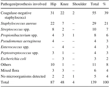
corresponded to 6% of the causing agents of all PJI (Table1).
In comparison to other pathogens, only staphylococcal and streptococcal infections were more frequent.
Clinical presentation
The data on clinical presentation prior to identiWcation of Propionibacterium spp. are presented in Table2. Onset of symptoms that were likely related to PJI occurred in four patients (50%) within the Wrst month and in three patients (37.5%) between 7 and 20 months of implanting the pros-thesis. However, in six of these seven patients, diagnosis was signiWcantly delayed, with a median duration of symp-toms of 10 months (IQR 4.5–19). In only one of these seven patients (case 6), diagnosis was also established with-out any delay. In this patient, serosanguinous discharge at the implant site occurred after primary wound healing. PJI was conWrmed by surgical revision, shortly thereafter. One out of eight patients complained of symptoms at a very late
stage (case 1). Six patients suVered from persistent pain,
and one patient (case 1) complained of pain only on move-ment. None of the patients reported fever or chills. Only one patient (case 5) was treated with antimicrobial agents prior to obtaining specimens for diagnosis (e.g. blood sam-ples, synovial Xuid, biopsies). Case 5 received fusidic acid, which was discontinued on the day of admission.
Table 1 Cultured microorganisms in prosthetic joint infection
Pathogen/prosthesis involved Hip Knee Shoulder Total %
Coagulase-negative staphylococci 31 22 2 55 39 Staphylococcus aureus 22 7 – 29 21 Streptococcus spp. 8 2 – 10 7 Propionibacterium spp. 4 3 1 8 6 Pseudomonas aeruginosa 4 – – 4 3 Enterococcus spp. – 4 – 4 3 Peptostreptococcus spp. 3 1 – 4 3 Escherichia coli – 3 – 3 2 Others 10 1 – 11 8 Mixed Xora 3 3 – 6 4 No microorganisms detected 2 2 1 5 4 Total 87 48 4 139 100
Tab le 2 Clin ic al p re se n ta tio n o f Pr opi o n ibac te ru m sp p. as so ci at ed P JI M ma le , F fe m ale , PJI p ro sth et ic jo in t in fe ct io n a S ero sa ng ui no us d is ch ar g e a t t h e i m p la nt si te oc cu rr ed a ft er pr im ary w o u n d h ea li n g b S k in su rf ace w as i n ta ct , bu t s in u s tr ac t w as de te ct ed b y ar th ro g ra phy c Fi ndi n g s o n t h e X -ra y w ere n o rm al (s ta bl e i m p la n t), but nu cl ea r s ci n ti g ra p h y su gg es te d PJ I P at ie n t 1 2 3 456 78 A g e ( y ea rs )/ Se x 51/ M 8 2 /M 3 9/ M 7 5/ M 6 3 /F 7 4 /M 4 6/ M 5 6/ M In fe ct ed p ro sth es is Hi p K n ee K n ee K n ee H ip S h o u ld er Hip H ip Time f ro m imp la n ta tio n to o n se t o f sy mp to ms (mon th s) 122 Im m ed ia te ly af te r s u rg er y Im m edi at el y af te r s u rg er y 0 .5 7 0. 5 1 0 2 0 Du ra tio n o f s y mp to m s (mon th s) 3 1 02 94 .5 1 9 0 1 6 6 Time f ro m imp la n ta tio n to di ag no si s o f PJ I (mon th s) 1 2 5 1 0 29 5 2 6 0 .5 26 26 S y m p to ms a n d sig n s P ain o n m ove m en t P er sis te n t p ain P ers ist en t p ai n P ers is te n t p ain P er sis te n t p ain S er o u s d is ch arg e a P ers is te n t p ain P ers is te n t p ain Co n d iti o n o f so ft-tis su e at time o f d ia g n o sis Si nu s t rac t, ab sc es s No W nd in gs N o W nd in gs Re dd in g, sw el li n g Si n u s t ra ct W ou nd a, si n u s t ra ct N o W ndi n g s S w el li n g , s inu s tr ac t b G ra d in g o f so ft -t is su e S ev er el y d am age d In ta ct In ta ct S lig h tly da m ag ed Se ve re ly da m ag ed M o d er at el y d am aged Int ac t S eve re ly da m ag ed b Co n d iti o n o f imp la n t ( ima g in g ) Un st ab le Un sta b le S ta b le c S ta b le Un st ab le S ta b le S ta b le Un st ab le
At the time of diagnosis, clinical and radiological exam-inations (including arthrography) showed a sinus tract and/ or an abscess in four patients; these cases were graded as “moderately/severely” damaged. In one patient, swelling and red discoloration was detected at the implant site, and in three patients no pathological Wndings of the soft-tissue were found; these were graded as “intact/slightly dam-aged”.
Laboratory diagnostics
The median CRP value was 17 mg/l (normal < 5 mg/l). In six patients the values ranged from 6 to 20 mg/l, whereas in the other two patients 68 and 75 mg/l were noted. The
median WBC count was 8.1 (IQR 6.2–16.7) £ 109cells/l
(normal 4–10 £ 109cells/l).
Imaging studies
Migration of the implant and/or periprosthetic osteolysis was detected by conventional X-ray in four cases. In one patient, Wndings on the X-ray were normal, but three-phase bone scintigraphy with technetium-99 m labeled methylene diphosphonate suggested PJI. In two patients imaging was unremarkable, but scintigraphy was not performed, because
clinical Wndings were strongly suggestive of PJI. In one
patient with stable implant (case 7), further imaging
analy-sis was not performed, despite inconclusive clinical
Wnd-ings.
Microbiological diagnostics
Propionibacterium acnes was identiWed in all but one case (case 5), in which the species was not identiWed. In four of the cases, synovial Xuid was Gram-stained and cultured, but showed no bacteria or growth, respectively. In two of the other cases, PJI was evident from clinical signs, and since a surgical procedure was inevitably required, cultur-ing of synovial Xuid was not performed (case 1: Wstula, case 6: persistent wound secretion 2 weeks after implantation). In the remaining two patients infection was primarily mis-diagnosed as aseptic implant loosening (case 2) or pain due to malpositioning of the implant components (case 7).
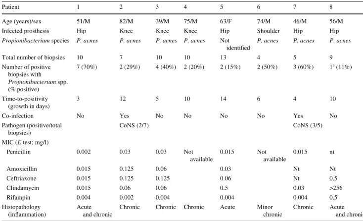
The median number of biopsies taken during surgery was 9.5 (IQR 6–10) with a median time-to-positivity of
8 days (IQR 4.5–11) (Table3). The median percentage of
positive cultures from the biopsy samples was 39% (IQR 17.5–55). In one patient (case 8), Propionibacterium spp. grew in only one out of nine biopsies. This patient was referred to our centre because he was suVering from a relapse of a Propionibacterium spp. associated PJI, and therefore included in the study. In addition, histopatho-logical examination in this case showed signs of acute
inXammation in all biopsies. In the Wrst episode of infection,
Propionibacterium spp. grew in four of four biopsies and the patient was treated with clindamycin. The MIC for
clin-damycin was not examined in the Wrst episode; however, it
proved to be at a resistance level in the culture specimen from the relapse.
Co-infection was present in two patients (cases 2 and 7), namely with coagulase-negative staphylococci (CoNS)
(Table3). In two other patients (cases 1 and 5),
microbio-logical results showed growth of CoNS in one single speci-men, but were interpreted as contaminants, according to the deWnition criteria for PJI.
MICs were tested for amoxicillin in four cases, for peni-cillin and ceftriaxone in Wve cases, and for clindamycin and rifampin in six cases. In two cases, these data were not available. All MICs tested proved to be in the susceptible range, except the one for clindamycin in one case (patient 8).
Histopathological diagnostics
Histopathological examination showed signs of acute inXammation in one case, chronic and acute inXammation in two cases, and signs of chronic inXammation in Wve cases. In all but two cases (cases 2 and 7) sections were Gram-stained but revealed no bacteria.
Discussion
The growth of Propionibacterium spp. in cultures of syno-vial Xuid and periprosthetic tissue often raises the question of relevance in PJI. Several reports have shown that these cases represented a diagnostic challenge in clinical
orthope-dics [11, 23]. These arguments may inXuence the
estima-tion that these microorganisms play a minor role in PJI. However, based on our own observation, we raised the hypothesis that Propionibacterium spp. is not as rare as often reported and can cause severe soft-tissue damages in PJI.
The relative frequency of Propionibacterium associated PJI in our retrospective study was higher than in most
pub-lished series [16, 20]. However, it may be even higher,
since our routine microbiological investigations did not include speciWc media for anaerobic cultures other than Brucella agar (e.g. thioglycolate, fastidious anaerobe broth), and since soniWcation of removed implants was not performed. Thus, PJI due to Propionibacterium spp. indeed occurred at a comparable rate in PJI than many other
micro-organisms [8, 21]. In this investigation, only staphylococcal
and streptococcal infections occurred more frequently. Propionibacterium spp. is mainly associated with low-grade infections, which typically present with subtle signs
and often occur within 24 months of implantation [32]. In this study, seven out of eight patients complained of pain
without more speciWc symptoms, and the infection
symp-toms presented in all but one case (case 1) within 24 months of implantation. Most of these low-grade infec-tions may be acquired during implantation and not by
hae-matogenous seeding [13, 17, 32], although this relationship
is diYcult to demonstrate conclusively. The delay in the diagnosis is often related to the low virulence and slow growth of these microorganisms. Yet, they still can cause severe tissue damage and implant failure as shown in 50% of our cases. An extensive sinus-tract in a Propionibacte-rium associated PJI is illustrated in Fig.1 (case 1). Aware-ness of this bacterial feature is important, in particular when interpreting culture results in PJI with moderately or severe soft-tissue damages.
Not surprisingly, neither WBC count nor CRP values were helpful in establishing or excluding the diagnosis of Propionibacterium associated PJI.
Preoperative joint puncture should be strived for, irre-spective of the pathogen. Culture of synovial Xuid (in pedi-atric blood culture bottles) has been reported to have a
sensitivity of 77% and a speciWcity of 99% [26]. In the case
of Propionibacterium associated PJI, this procedure may
not always be helpful [8]. In four of our cases, synovial
Xuid was cultured, but revealed no growth.
Table 3 Microbiological and histopathological results
NT not tested. Gram-staining was performed in all but two patients (case 2 and 7), but showed no bacteria
a This patient was suVering from a relapse of a Propionibacterium spp. associated PJI. In the Wrst episode of infection, Propionibacterium spp. grew in four of four biopsies. The patient was treated with clindamycin, but MIC for clindamycin was at a resistant level (>256 mg/l) in the culture specimen from the relapse
Patient 1 2 3 4 5 6 7 8
Age (years)/sex 51/M 82/M 39/M 75/M 63/F 74/M 46/M 56/M
Infected prosthesis Hip Knee Knee Knee Hip Shoulder Hip Hip
Propionibacterium species P. acnes P. acnes P. acnes P. acnes Not
identiWed
P. acnes P. acnes P. acnes
Total number of biopsies 10 7 10 10 13 4 5 9
Number of positive biopsies with Propionibacterium spp. (% positive) 7 (70%) 2 (29%) 4 (40%) 2 (20%) 2 (15%) 2 (50%) 3 (60%) 1a (11%) Time-to-positivity (growth in days) 3 12 5 10 14 6 4 10
Co-infection No Yes No No No No Yes No
Pathogen (positive/total biopsies) CoNS (2/7) CoNS (3/5) MIC (E test; mg/l) Penicillin 0.002 0.03 0.03 Not available 0.015 Not available 0.015 nt Amoxicillin 0.015 0.125 0.06 0.03 Nt Nt Ceftriaxone 0.015 0.125 0.125 0.06 Nt 0.5 Clindamycin 0.015 0.06 0.06 0.5 0.03 >256 Rifampin 0.004 0.002 0.004 0.004 0.004 0.5 Histopathology
(inXammation) Acute and chronic
Chronic Chronic Chronic Acute Minor chronic
Chronic Acute and chronic
Fig. 1 Case 1: Prosthetic joint infection with severely damaged
soft-tissue, including a large Wstula through the bone. In seven out of ten obtained biopsies growth of Propionibacterium acnes was detected
Several methods have been suggested to improve the detection rate of these infections, such as a long incubation
time [6, 12] or the use of soniWcation of removed implants
[28, 29]. The median time-to-positivity in this series was
8 days, and is comparable to Wndings of others [12].
How-ever, since these microorganisms grow slowly and need anaerobic conditions, the sensitivity of culture results remains unsatisfactory. To overcome this deWcit, several biopsies are obtained. The optimal number of biopsies has not yet been deWned. A thoroughly performed prospective study, looking at the detection rate of several microorgan-isms in periprosthetic tissue specimens, recommended six
samples [1]. In our study, a median of 9.5 biopsies per case
was taken which may explain the relative frequency of 6% Propionibacterium spp. associated PJI. Due to the retro-spective study-design and the lack of a control group, it remains speculative whether in certain cases fewer biopsies might have missed the diagnosis. Nevertheless, our data and those of Atkins et al. support the concept of culturing six (or even more) specimens, in particular in low grade PJI. Routine culture of removed implants is often associ-ated with contamination and shows a low sensitivity, prob-ably because bacteria are not dislodged from the bioWlm
[27–29]. Culturing sonicated Xuid, though, shows very
promising results in improving the diagnosis of infection
[28, 29]. Since in clinical practice this method is not yet
widely available, the sensitivity of culture results can be increased by obtaining a higher number of biopsies.
In two of our cases, co-infection with CoNS was detected, which raises diYculties to discriminate between pathogens and contaminants. However, Propionibacterium
spp. occurs not rarely in mixed Xora [12], and thus likely
represent true pathogens in these two cases. Many patho-gens grow faster than Propionibacterium spp. and are con-sequently isolated earlier. Thus, discontinuation of incubation includes the risk of incomplete diagnosis.
Thorough analyses of the involved pathogens, including antimicrobial susceptibility and MIC testing, is important for the medical management of these infections. Propioni-bacterium spp. are typically susceptible to penicillin, ceftri-axone and clindamycin. However, resistant strains do exist
[30], and antimicrobial treatment without previous MIC
testing might result in failure, despite correct surgical ther-apy. In combination with other antimicrobial agents, rifam-pin is often used in the treatment of PJI, and therefore was tested here also. It has bactericidal activity against surface-adhering, slow-growing and bioWlm-producing
staphylo-cocci [33], but to our knowledge its eYcacy has not been
tested for Propionibacterium spp. residing in bioWlms [18].
Yet, previous [7] and our investigations have demonstrated
low MIC values that might justify further clinical investiga-tions. Nevertheless, when rifampin is considered, MIC test-ing should be demanded and, importantly, the compound
never be administered alone, since some pathogens rapidly
develop resistance [24].
Histopathological examination of the periprosthetic tis-sue can support infection diagnosis, because the level of inXammation often correlates well with the clinical course
[5, 29]. In our series, four of eight patients had signs of
chronic infection, which are compatible with the long dura-tion of symptoms. In three cases signs of acute infecdura-tion were detected (including the cases with signs of acute and chronic infection), and in all of these, soft-tissue was mod-erately or severely damaged. In one patient (case 6), infec-tion was detected immediately, which might explain the
lack of speciWc histopathological signs.
In conclusion, isolation of Propionibacterium spp. from PJI should not a priori be interpreted as contamination, but requires further analyses. Our study suggests that these infections occur at a relative frequency that is comparable to many other pathogens (not including staphylococci and streptococci). Most of the cases in our series presented with a long duration of symptoms and subtle signs, but 50% revealed damaged soft-tissues and unstable implants. The diagnosis was established with a median of 9.5 biopsies per case, an incubation time of 14 days and the aid of histopath-ological examinations.
Acknowledgments We like to thank Docent Bengt Gårdlund (Department of Infectious Diseases, Karolinska University Hospital Huddinge, Stockholm, Sweden) for his helpful comments. We are grateful to the laboratory technician from our Unit of Microbiology and Anna Bächtold from our library for superb assistance. This study was presented at the 92nd Conference of the German Society of Ortho-pedics and Orthopedic Surgery, Berlin, Germany, 2–6 October 2006. ConXict of interest statement None of the authors has a conXict of interest. Ethical Board Review statement: In this retrospective analysis, all data are anonymously presented, and patient consent has been received from all involved humans prior to the study.
References
1. Atkins BL, Athanasou N, Deeks JJ et al (1998) Prospective evalu-ation of criteria for microbiological diagnosis of prosthetic-joint infection at revision arthroplasty. The OSIRIS Collaborative Study Group. J Clin Microbiol 36:2932–2939
2. Berbari EF, Hanssen AD, DuVy MC et al (1998) Risk factors for prosthetic joint infection: case-control study. Clin Infect Dis 27:1247–1254
3. Brook I, Frazier EH (1993) Anaerobic osteomyelitis and arthritis in a military hospital: a 10-year experience. Am J Med 94:21–28 4. Brook I, Frazier EH (1991) Infections caused by
Propionibacteri-um species. Rev Infect Dis 13:819–822
5. Fehring TK, McAlister JA Jr (1994) Frozen histologic section as a guide to sepsis in revision joint arthroplasty. Clin Orthop Relat Res 304:229–237
6. Günthard H, Hany A, Turina M et al (1994) Propionibacterium acnes as a cause of aggressive aortic valve endocarditis and
importance of tissue grinding: case report and review. J Clin Microbiol 32:3043–3045
7. HoeZer U, Ko HL, Pulverer G (1976) Antimicrobiol suscepti-bility of Propinibacterium acnes and related microbial species. Antimicrob Agents Chemother 10:387–394
8. Ince A, Seemann K, Frommelt L et al (2004) One-stage revision of shoulder arthroplasty in the case of periprosthetic infection. Z Ort-hop Ihre Grenzgeb 142:611–617
9. Ivancevic V, Perka C, Hasart O et al (2002) Imaging of low-grade bone infection with a technetium-99 m labelled monoclonal anti-NCA-90 Fab’ fragment in patients with previous joint surgery. Eur J Nucl Med Mol Imaging 29:547–551
10. Jakab E, Zbinden R, Gubler J et al (1996) Severe infections caused by Propionibacterium acnes: an underestimated pathogen in late postoperative infections. Yale J Biol Med 69:477–482
11. Launder WJ, Hungerford DS (1981) Late infection of total hip arthroplasty with Propionibacterium acnes: a case and review of the literature. Clin Orthop Relat Res 157:170–177
12. Lutz MF, Berthelot P, Fresard A et al (2005) Arthroplastic and osteosynthetic infections due to Propionibacterium acnes: a retro-spective study of 52 cases, 1995–2002. Eur J Clin Microbiol Infect Dis 24:739–744
13. Maderazo EG, Judson S, Pasternak H (1988) Late infections of to-tal joint prostheses. A review and recommendations for preven-tion. Clin Orthop Relat Res 229:131–142
14. Mathisen GE, Meyer RD, George WL et al (1984) Brain abscess and cerebritis. Rev Infect Dis 6(Suppl 1):S101–S106
15. Ochsner PE, Hailemariam S (2006) Histology of osteosynthesis associated bone infection. Injury 37(Suppl 2):S49–S58
16. Pandey R, Berendt AR, Athanasou NA (2000) Histological and microbiological Wndings in non-infected and infected revision arthroplasty tissues. The OSIRIS Collaborative Study Group. Oxford skeletal infection research and intervention service. Arch Orthop Trauma Surg 120:570–574
17. Poss R, Thornhill TS, Ewald FC et al (1984) Factors inXuencing the incidence and outcome of infection following total joint arthro-plasty. Clin Orthop Relat Res 182:117–126
18. Ramage G, Tunney MM, Patrick S et al (2003) Formation of
Propionibacterium acnes bioWlms on orthopaedic biomaterials
and their susceptibility to antimicrobials. Biomaterials 24:3221– 3227
19. Richards BS (1995) Delayed infections following posterior spinal instrumentation for the treatment of idiopathic scoliosis. J Bone Joint Surg Am 77:524–529
20. Segawa H, Tsukayama DT, Kyle RF et al (1999) Infection after total knee arthroplasty. A retrospective study of the treatment of eighty-one infections. J Bone Joint Surg Am 81:1434–1445 21. Senneville E, Savage C, Nallet I et al (2006) Improved
aero-anaer-obe recovery from infected prosthetic joint samples taken from 72 patients and collected intraoperatively in Rosenow’s broth. Acta Orthop 77:120–124
22. Stumpe KD, Notzli HP, Zanetti M et al (2004) FDG PET for diVer-entiation of infection and aseptic loosening in total hip replace-ments: comparison with conventional radiography and three-phase bone scintigraphy. Radiology 231:333–341
23. Sulkowski MS, Abolnik IZ, Morris EI et al (1994) Infectious arthritis due to Propionibacterium acnes in a prosthetic joint. Clin Infect Dis 19:224–225
24. Tenover FC (2006) Mechanisms of antimicrobial resistance in bacteria. Am J Med 119:S3–S10; discussion S62–S70
25. Tigges S, Stiles RG, Roberson JR (1994) Appearance of septic hip prostheses on plain radiographs. AJR Am J Roentgenol 163:377– 380
26. Trampuz A, Hanssen AD, Osmon DR et al (2004) Synovial Xuid leukocyte count and diVerential for the diagnosis of prosthetic knee infection. Am J Med 117:556–562
27. Trampuz A, Piper KE, Hanssen AD et al (2006) Sonication of ex-planted prosthetic components in bags for diagnosis of prosthetic joint infection is associated with risk of contamination. J Clin Microbiol 44:628–631
28. Trampuz A, Piper KE, Jacobson MJ et al (2007) Sonication of re-moved hip and knee prostheses for diagnosis of infection. N Engl J Med 357:654–663
29. Tunney MM, Patrick S, Gorman SP et al (1998) Improved detec-tion of infecdetec-tion in hip replacements. A currently underestimated problem. J Bone Joint Surg Br 80:568–572
30. Tyrrell KL, Citron DM, Warren YA et al (2006) In vitro activities of daptomycin, vancomycin, and penicillin against Clostridium
diYcile, C. perfringens, Finegoldia magna, and
Propionibacteri-um acnes. Antimicrob Agents Chemother 50:2728–2731 31. Yocum RC, McArthur J, Petty BG et al (1982) Septic arthritis
caused by Propionibacterium acnes. JAMA 248:1740–1741 32. Zimmerli W, Trampuz A, Ochsner PE (2004) Prosthetic-joint
infections. N Engl J Med 351:1645–1654
33. Zimmerli W, Widmer AF, Blatter M et al (1998) Role of rifampin for treatment of orthopedic implant-related staphylococcal infec-tions: a randomized controlled trial. Foreign-Body Infection (FBI) Study Group. JAMA 279:1537–1541