Received: 19 July 2002 Revised: 8 January 2003 Accepted: 3 March 2003 Published online: 24 July 2003 © Springer-Verlag 2003
Abstract Unenhanced helical com-puted tomography (UHCT) has evolved into a well-accepted alterna-tive to intravenous urography (IVU) in patients with acute flank pain and suspected ureterolithiasis. The pur-pose of our randomized prospective study was to analyse the diagnostic accuracy of UHCT vs IVU in the normal clinical setting with special interest on economic impact, applied radiation dose and time savings in patient management. A total of 122 consecutive patients with acute flank pain suggestive of urolithiasis were randomized for UHCT (n=59) or IVU (n=63). Patient management (time, contrast media), costs and radiation dose were analysed. The films were independently interpreted by four radiologists, unaware of previous findings, clinical history and clinical outcome. Alternative diagnoses if present were assessed. Direct costs of UHCT and IVU are nearly identical (310/309 Euro). Indirect costs are much lower for UHCT because it saves examination time and when performed immedi-ately initial abdominal plain film (KUB) and sonography are not nec-essary. Time delay between access to the emergency room and start of the imaging procedure was 32 h 7 min for UHCT and 36 h 55 min for IVU. The UHCT took an average in-room time of 23 min vs 1 h 21 min for IVU. Mild to moderate adverse reac-tions for contrast material were seen
in 3 (5%) patients. The UHCT was safe, as no contrast material was needed. The mean applied radiation dose was 3.3 mSv for IVU and 6.5 mSv for UHCT. Alternative diagnoses were identified in 4 (7%) UHCT patients and 3 (5%) IVU patients. Sensitivity and specificity of UHCT and IVU was 94.1 and 94.2%, and 85.2 and 90.4%, respec-tively. In patients with suspected renal colic KUB and US may be the least expensive and most easily accessable modalities; however, if needed and available, UHCT can be considered a better alternative than IVU because it has a higher diagnos-tic accuracy and a better economic impact since it is more effective, faster, less expensive and less risky than IVU. In addition, it also has the capability of detecting various addi-tional renal and extrarenal patholo-gies.
Keywords Acute flank pain · Intravenous urography · Helical computed tomography · Cost analysis · Patient management analysis · Radiation dose S. A. Pfister A. Deckart S. Laschke S. Dellas U. Otto C. Buitrago J. Roth W. Wiesner G. Bongartz T. C. Gasser
Unenhanced helical computed tomography vs
intravenous urography in patients with acute
flank pain: accuracy and economic impact in
a randomized prospective trial
S. A. Pfister (
✉
) · S. Dellas · U. Otto C. Buitrago · W. Wiesner · G. Bongartz Department of Radiology,University Hospital,
Petersgraben 4, 4031 Basel, Switzerland e-mail: [email protected]
Tel.: +41-41-2272030 Fax: +41-41-2272031
A. Deckart · S. Laschke · T. C. Gasser Department of Urology,
University Hospital,
Petersgraben 4, 4031 Basel, Switzerland J. Roth
Department of Radiologic Physics, University Hospital,
Petersgraben 4, 4031 Basel, Switzerland Present address:
S. A. Pfister, Medical Imaging Lucerne, Theaterstrasse 7, 6003 Lucerne, Switzerland
Introduction
Since its introduction in 1923 intravenous urography (IVU) has been regarded the technique of choice for ra-diographic evaluation of acute renal colic [1]. The IVU provides structural and functional information about kid-neys, ureters and urinary bladder, including site, degree and nature of obstruction, as well as presence or absence of various possible congenital anomalies; however, there are some undesirable aspects of IVU including the need for exposure of patients to intravenous contrast material with a risk of adverse reactions. Furthermore, bowel preparation can be helpful in order to achieve good diag-nostic quality in IVU, but it leads to a significant time delay between admission and diagnosis.
In the 1990s several authors suggested different alter-natives to urography. abdominal plain film (KUB) alone, the combination of KUB and ultrasonography [2], and UHCT were widely discussed. Since its first publication [3], UHCT became the first-line imaging modality in the clinical setting of suspected urolithiasis for diagnosis and treatment planning in many centres all over the world [4, 5, 6, 7, 8, 9, 10, 31].
Unenhanced helical CT reliably detects all stones in the collecting system by direct visualization because concrements possess sufficient density to be visualized by CT [11]. The two known exceptions are stones of pro-teases inhibitors, such as indinavir sulfate [12], or mu-coid matrix stones which are of low attenuation similar to soft tissue and, therefore, not visible directly by CT.
Unenhanced helical CT gives information about stones, obstructing and nonobstructing ones, clinically relevant and actually not relevant stones (stone burden; Fig. 1). It additionally can reveal signs associated with ureteral obstruction, even after recent stone passage.
These secondary signs, including hydronephrosis, hydro-ureter, perinephric stranding (Fig. 2) and “tissue rim sign” (Fig. 3), have been reported to have a positive pre-dictive value of >90% for the presence of a stone [13, 14, 15].
Accuracy and suitability of UHCT for acute renal col-ic has already been well demonstrated in the literature.
Advantages and disadvantages of different protocols (UHCT first choice, KUB and US and IVU, KUB and US and UHCT, IVU first choice) for imaging patients with flank pain concerning especially costs and doses have been discussed thoroughly [16]. Immediate UHCT alone has the advantage of reducing the time of diagno-sis and the overall management cost [17]. The combina-tion of CT and with KUB and US has the advantage of being cheaper and delivering a lower dose, although reaching a diagnostic conclusion may take longer.
Fig. 1 Calculus in the left ureteropelvic junction with rim sign.
Subtle perinephric stranding. Further stones are visible in the lower minor and major calyces of the left kidney (stone burden)
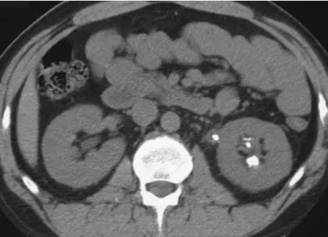
Fig. 2 a Swelling of the right kidney with stranding of perirenal
fat, thickening of Gerota’s fascia and moderately dilated intrarenal collecting system compared with normal left kidney and normal perirenal structures. b In this patient an obstructing calculus in the proximal ureter with rim sign is depicted
The goal of this study was to analyze and compare the diagnostic accuracy of UHCT vs IVU in the work-up of patients with clinically suspected acute urolithiasis in a university hospital without changing the current clinical setting. Especially analysed and compared were the eco-nomic impact, the applied radiation dose, time to diagno-sis and in-room time for both examinations.
Subjects and methods
One hundred twenty-two consecutive patients presenting at the University Hospital Basel Emergency Department with acute
flank pain suspicious for renal colic were prospectively enrolled in the study. The study design was approved by the local ethics com-mittee and a written informed consent was obtained from each pa-tient. Patients were recruited between November 1998 and March 2000.
All patients were examined by the emergency room physician. Urine and blood analysis as well as KUB and abdominal US were routinely performed in every patient as we do in all cases of acute unknown abdominal pain. If after this first work-up renal colic was the most likely diagnosis patients were randomized to receive either UHCT or IVU. Depending on the urgency UHCT was per-formed as soon as possible or the patient got a regular appoint-ment. The IVU patients got a bowel preparation as done regularly in our hospital.
Thereafter, all 122 patients were followed-up to establish the final diagnoses. The course of clinical symptoms, passage of a cal-culus, results of urological interventions and alternative diagnoses were noted.
Exclusion criteria were: known urolithiasis, impaired renal function (creatinine >150 mmol/l), signs of infection with fever, chills or CRP elevation (>5), patients younger than 17 years of age and pregnancy.
Computed tomography scans were obtained using a standard protocol performed on a helical single-slice CT scanner (Hi-Speed CT-i General Electric Medical Systems, Milwaukee, Wis.). The in-vestigation was performed without intravenous contrast material. The 5-mm-thick slices (at 120 kV, 260 mAs, with an increment of 7.5 mm and pitch 1.5) were obtained in a single helical acquisition extending from the top of the kidneys through the symphysis. To-tal scan time was 40–50 s during one or two breath holds. Slices
Fig. 3 a Perinephric stranding surrounding the right kidney with
fluid collection from the right sinus to the perinephric space poste-riorly (forniceal rupture) and subtle dilated extrarenal pelvis. Re-nal sinus cysts on the left side which could be misdiagnosed as di-lated pelvis, but there is no stranding. b Contrast material has been given because of a suspicious complex cyst on the right upper pole which turned out to be a simple cyst (not shown). These films con-firm the forniceal rupture and the renal sinus cyst. c In the same patient a stone in the distal right ureter with subtle rim sign is de-picted. Two small phleboliths, one on each side of the pelvis (pos-teriorly). d Postcontrast CT confirms the stone in the right ureter and the phleboliths in the normal small linear distal ureter
fied ureter above a dense or radiolucent structure (or both). Diag-nosis of ureteral obstruction was made if unilateral dilatation of the ureter at a specific level occurred or a unilateral delay in the time of ipsilateral contrast excretion in the renal collecting system was determined.
The spontaneous passage or the retrieval of a stone by cystos-copy and ureteroscystos-copy, as well as the identification of a stone dur-ing retrograde pyelography, was used as gold standard to confirm the diagnosis of ureterolithiasis.
Finally, all images were reviewed on films by three indepen-dent staff radiologists who were unaware of the previous findings and the clinical outcome.
A standardized scoring sheet was used. All four readers evalu-ated each case for the presence, location, and size of ureteral and renal calculi, dilatation of the collecting system, nephrographic and urographic pattern (IVU only), as well as perinephric and periureteral soft tissue stranding and soft tissue rim sign (UHCT only).
In addition, the readers were asked to categorize each case as follows:
1. No evidence of ureteral calculus or pyeloureterectasis 2. Low probability of ureteral calculus or pyeloureterectasis 3. Indeterminate probability of calculus or pyeloureterectasis 4. High probability of ureteral calculus or pyeloureterectasis 5. Positive for ureteral calculus causing obstruction or not Any other abdominal abnormalities not related to the urinary tract were reported.
We considered patients as true positive when all four readers considered the presence of a stone as very probable (4) or sure (5), which means that they all gave a score higher than 16 (4×4=16 of max 4×5=20). Patients without the confirmed diagnosis of uretero-lithiasis and without alternative diagnosis were considered to be true negatives.
The time interval between patient’s arrival at the emergency room and the specific examination and the examination time (in-room time) of UHCT or IVU, respectively, were measured and analysed. Furthermore, the direct costs of either group were com-pared. Radiation doses for UHCT and IVU were measured by our radiation physicist using thermoluminescence dosimetry (TLD: CaF2) measuring radiation exposure on surface (in vivo) for IVU and measuring radiation exposure in an Alderson-Phantom for UHCT. Supplementary organ doses and effective doses were cal-culated by Monte Carlo Computer Programs (ODS-60 and CT-Dose). Any adverse reaction to contrast material was noted.
Results
Nine of 122 patients had to be excluded since they were unable to be contacted for follow-up. The remaining 113
via cystoscopy, ureteroscopy and stone fragmentation, whereas 4 patients (9%, 4 of 42) got stone fragmentation by extracorporeal shock-wave lithotripsy.
Seven patients (16%, 7 of 42) had documented stones but became pain free without recognizing the stone pas-sage. These patients were not included as false positive.
One patient (2%, 1 of 55) had spontaneous stone pas-sage without stone documentation in UHCT (one false negative). The re-evaluation of the films showed a possi-bly faint, very little stone at the vesico-ureteral junction.
Thirteen patients (24%, 13 of 55) were stone free in UHCT.
Unsuspected diseases beyond the urinary tract were identified in 4 patients (7%, 4 of 55) including 1 tumour of uterus (uteral fibroid), 1 adnexal mass (teratoma) and 2 severe spondyloses. These findings were not supposed to be responsible for the clinical symptoms. Eight pa-tients (14%, 8 of 55) became pain free spontaneously without any specific diagnosis. A total of 12 patients were counted as true negatives.
The sensitivity and specificity of UHCT for the diag-nosis of acute urolithiasis among all readers were 85.1 and 98.1%, respectively, using a five-category scoring system (stone probability: 1=none; 2=low; 3=moderate; 4=high; 5=stone present) and raised up to 94.1 and 94.2% when a scoring system with three categories (stone probability: 1+2=none; 3=intermediate; 4 and 5=stone present) was used.
Fifty-eight patients (51%; 58 of 113, 15 women, mean age 46.1 years; and 43 men, mean age 44.9 years) were randomized for IVU.
Thirty-seven patients (64%, 37 of 58) had clinically proven stones. Spontaneous stone passage was docu-mented in 20 patients (54%, 20 of 37). A total of 10 pa-tients (27%, 10 of 37) underwent endoscopic removal of stones via cystoscopy, ureteroscopy and stone fragmen-tation, whereas 7 patients (19%, 7 of 37) got stone frag-mentation with extracorporeal shock-wave lithotripsy. Two patients (5%, 2 of 37) had more than one stone. Five (13%, 5 of 37) patients had a positive IVU but did not recognize the stone passage. We did not count them as false positive. There were 7 (19%, 7 of 37) false-nega-tive examinations. In 3 cases the stones were smaller
than 3 mm, they did not produce a significant ureter ob-struction and passed spontaneously. In 2 cases the stone passed during the preparation time and the IVU showed no obvious secondary signs. In 1 case the stone was not obstructive but radiolucent and therefore not detectable in the IVU, and in another case the IVU was misdiag-nosed because of subtle findings.
In 3 cases (5%, 3 of 58) alternative IVU findings were believed to be responsible for flank pain. Nephrop-tosis with intermittent obstruction, stenosis of distal ure-ter and stenosis of the ureure-teropelvic junction were diag-nosed in 1 case each. The remaining 11 patients (19%, 11 of 58) became pain free without any specific diagno-sis.
The sensitivity and the specificity of IVU for the di-agnosis of acute urolithiasis among all readers were 75 and 91.7%, when a scoring system with five categories (stone probability: 1=none; 2=low; 3=moderate; 4=high; 5=stone present) was used and raised up to 85.2 and 90.4% when a three-category scoring system (stone probability: 1+2=none; 3=intermediate; 4 and 5=stone present) was used.
At our institution the average charges for IVU are 309 Euro (range 270–361 Euro) depending on the amount of contrast material used and whether additional radio-graphs (oblique views, tomographies, delayed films) are obtained. For the initial abdominal radiograph (98 Euro) and sonography (82 Euro) the patient is charged 180 Euro, whereas the average charge for UHCT is 310 Euro. The average time interval between patient’s arrival at the emergency room and the radiographic examination was 36 h 55 min for IVU and 32 h 7 min for UHCT re-spectively; for outpatients 53 h 52 min and 43 h 30 min, for inpatients 26 h 34 min and 15 h 3 min, respectively.
The average examination time (in-room time) for IVU and UHCT was 1 h 21 min and 23 min, respectively [outpatients: 1 h 13 min (min. 42 min/max. 2 h 19 min) and 21 min (min. 8 min/max. 39 min), respectively; in-patients: 1 h 26 min (min. 35 min/max. 3 h 23 min) and 25 min (min. 7 min/max. 45 min)].
Radiation doses were evaluated by thermolumines-cence dosimetry in a 3D model for the helical CT
tech-nique (Alderson-Phantom) and with thermoluminescence dosimetry on the patient’s surface for the IVU technique. The average effective dose for the UHCT technique was 6.5 mSv and for the IVU technique 3.3 mSv.
Adverse reactions for contrast material were seen in 3 patients (5%, 3 of 58). One patient showed a mild (no medication was needed) and 2 patients showed a moder-ate cutaneous allergic reaction to intravenous contrast material with successful treatment by anti-allergic drugs. No severe allergic reaction to contrast material was ob-served. One patient suffered from strong colics after in-travenous contrast media administration and, therefore, needed spasmolytics and analgetics.
Discussion
We present the second published prospective randomized trial comparing UHCT vs IVU in patients with acute flank pain after the first such study with a similar struc-ture was published in Australasian Radiology [18].
Sensitivity of 85.1 and 75% and specificity of 98.1 and 91.7% of UHCT and IVU, respectively, for the diagnosis of urolithiasis are high in our study but lower than those from other groups; however, as mentioned previously, if a scoring system with three categories (stone probability: 1+2=none; 3=intermediate; 4 and 5=stone present) was ap-plied the sensitivity and specificity of UHCT raised up to 94.1 and 94.2% and for IVU 85.2 and 90.4%, respectively.
Overall, the sensitivity and the specificity of the two tests are comparable since using Mann-Whitney U test at the significance level of 5% revealed no significant dif-ferences.
These results reach the data of previously published studies (see Table 1).
A possible explanation of our inferior statistical re-sults may be found in the fact that in our department ra-diologists are not specialized exclusively on GU imaging as in many other Anglo-Saxon centres and, therefore, the level of uncertainty may be higher.
Another potential reason why we did not reach the same high sensitivity and specificity as other groups was
Table 1 Literature survey:
diagnostic value of unenhanced helical computed tomography in the diagnosis of ureterolithi-asis
Reference No. of Sensitivity Specificitiy Patients with
patients (%) (%) stones (%) [19] 292 97 96 48 [1] 100 98 100 55 [20] 126 100 96 39 [21] 417 95 98 45 [22] 106 96 100 71 [23] 105 98 98 47 [7] 66 100 100 79 [9] 125 99 97 73 [18] 228 100 100 159 Present study 55 94 94 76
such as lithotripsy, ureterorenoscopy, spontaneous stone retrieval and the mentioned scoring system were the base of our gold standard which enabled us to compare both examinations indirectly. Because the equivalent accuracy of UHCT to IVU has already been established, a direct comparison was not necessary and, therefore, we mini-mized the applied radiation dose obviating a second im-aging procedure in the same patient.
Furthermore, our hard clinical inclusion criteria were intended to keep the number of negative patients low (unnecessary examination and radiation exposure), and therefore, we received only a small number of 13 (24%, 13 of 55) UHCT-negative patients.
There is a substantial limitation in those studies (as in ours) that use reported spontaneous stone retrieval as gold standard. Not all patients recognize the spontaneous stone passage; therefore, the number of false-negative patients may be high and the specificity lower, accord-ingly.
In terms of radiation dose, we found a twofold dose for UHCT compared with IVU. These data are in con-cordance with literature, where radiation dose was mea-sured and found to be twice or three times for UHCT as compared with IVU (see Table 2).
Nevertheless, the discussion about radiation dose in this context is still ongoing and some authors are ques-tioning whether the radiation dose is justifiable [24].
The invention of multislice CT will surely additional-ly further increase the radiation dose, but newer data have shown that reduction of slice thickness to 3 mm and pitch to 1.5, and application of lower exposure factors, may reduce the delivered dose while maintaining the same diagnostic accuracy [26, 27, 28]. Further improve-ments concerning radiation exposure in CT can be ex-pected by 16-row CT.
Our study was conducted as a cost-effectiveness and patient management study. At our institution, a universi-ty hospital in Switzerland, direct costs for UHCT and IVU are very similar, being 310 and 309 Euro, respec-tively. Other authors report that IVU is even more expen-sive than unenhanced helical CT (see Table 3); however, these authors also included the indirect costs resulting from a longer examination time for doctors and technical
tems of the various countries. We compared the costs pa-tients were charged but we did not analyse real costs in details, which might be a limitation of our study; howev-er, it is well known that the charges do not reflect real cost. There is no doubt that indirect costs of IVU are far higher (room-occupying time, patient preparation time, contrast material administration and possible averse re-actions) than for UHCT.
The former work-up of patients with suspected renal colic in our institution has not changed during the study to receive the most authentic results concerning the time management. We could demonstrate that the time inter-vals between first admittance and diagnostic imaging were similar for both modalities and surprisingly high (36 h 55 min for IVU and 32 h 7 min for UHCT, respec-tively). This corresponds to the fact that our emergency doctors (after exclusion of really urgent cases by urine and blood test, US and KUB) were used to waiting for further diagnostic work-up because of the time-consum-ing bowel preparation for IVU as done in our institution routinely.
As shown by Patel et al. [17] a protocol with initial US and KUB is cheaper and delivers a lower radiation dose to the patient, although reaching a diagnostic con-clusion takes longer than immediate UHCT. On the other hand, UHCT (as the initial examination) reduces the time of diagnosis and the overall management cost. It is obvious that in many cases KUB and US can already provide the correct diagnosis, but many positive cases nevertheless additionally undergo IVU to form the basis for future follow-ups by urologists.
Ureteral calculi are generally treated on the basis of their size, location and composition [29, 30]. Except for composition, UHCT gives all-important information for treatment planning and forms a perfect basis of future fol-low-ups. The UHCT has a higher diagnostic accuracy, a better economic impact, is faster, less expensive and less risky than IVU, and in addition it also has the capability to detect various additional renal and extrarenal patholo-gies. Overall, UHCT might be considered a better alterna-tive imaging modality than IVU in patients with suspect-ed renal colic. Additionally, if UHCT is performsuspect-ed imme-diately, initial KUB and US might be skipped.
We have had few negative cases and alternative diag-noses were identified only in four UHCT patients (7%); moreover, none of them were supposed to be responsible for their clinical symptoms. In fact, large numbers of negative cases would be a strong indication for using UHCT (hence, an indication for looking for other causes of flank pain).
A possible disadvantage resulting from loss of func-tional information provided by IVU is the differential diagnosis of renal infarcts which can be easily missed by UHCT.
Conclusion
In patients with suspected renal colic KUB and US may of course still be the least expensive and most easily ac-cessible modalities; however, if needed and available, UHCT can be considered a better alternative than IVU because it has a higher diagnostic accuracy and a better economic outcome since it is more effective, faster, less expensive and less risky than IVU. In addition, it also has the capability to detect various additional renal and extrarenal pathologies.
References
1. Fielding JR, Steele G, Fox LA, Heller H, Loughlin KR (1997) Spiral comput-erized tomography in the evaluation of acute flank pain: a replacement for excretory urography. J Urol
157:2071–2073
2. Haddad MC, Sharif HS, Shaded MS et al. (1992) Renal colic: diagnosis and outcome. Radiology 184:83–88 3. Smith RC, Rosenfield AT, Choe KA,
Essenmacher KR, Verga M, Glickmann MG, Lange RC (1995) Acute flank pain: comparison of non-contrast-enhanced CT and intravenous urography. Radiology 194:789–794 4. Katz DS, Lane MJ, Sommer FG (1997)
Non-contrast spiral CT for patients with suspected renal colic. Eur Radiol 7:680–685
5. Fielding JR, Silvermann SG, Samuel S, Zou KH, Loughlin KR (1998) Unenhanced helical CT of ureteral stones: a replacement for excretory urography in planning treatment. AJR 171:1051–1053
6. Yilmaz S, Sindel T, Arslan G, Özkaynak C, Karaali K, Kabaalioglu A, Lüleci E (1998) Renal colic: comparison of spiral CT, US and IVU in the detection of ureteral calculi. Eur Radiol 8:212–217
7. Ruppert-Kohlmayr AJ, Stacher R, Preidler KW, Zigeuner R, Primus G, Ricabona M, Szolar DHM (1999) Nativ-spiral-computertomographie bei patienten mit akutem flankenschmerz: sinn oder unsinn? Fortschr Röntgenstr 170:168–173
8. Thomson JM, Glocer J, Abbott C, Maling TM, Mark S (2001) Computed tomography in diagnosis of acute flank pain from urolithiasis: a randomized study comparing imaging costs and radiation dose. Australas Radiol 45:291–297
9. Hamm M, Wawroschek F, Weckermann D, Knöpfle E, Häckel T, Krawzak G, Harzmann R (2001) Unenhanced helical computed tomography in the evaluation of acute flank pain. Eur Urol 39:460–465
10. Dalla-Palma L, Pozzi-Mucelli R, Stacul F (2001) Present-day imaging of patients with renal colic. Eur Radiol 11:4–17
11. Newhouse JH, Prien EL, Amis ES Jr, Dretler SP, Pfister RC (1984) Computed tomographic analysis of urinary calculi. AJR 142:545–548 12. Blake SP, McNicholas MMJ,
Raptopoulos V (1998) Nonopaque crystal deposition causing ureteric obstruction in patients with HIV undergoing indinavir therapy. AJR 171:717–720
13. Smith RC, Verga M, Dalrymple NC, McCarthy S, Rosenfield AT (1996) Acute ureteral obstruction: value of secondary signs on helical unenhanced CT. AJR 167:1109–1113
14. Kawashima A, Sandler CM, Boridy IC, Takahashi N, Benson GS, Goldmann SM (1997) Unenhanced helical CT of ureterolithiasis: value of the tissue rim sign. AJR 168:997–1000
15. Heneghan JP, Dalrymple NC, Verga M, Rosenfield AT, Smith RC (1997) Soft tissue “rim” sign in the diagnosis of ureteral calculi with use of the unenhanced helical CT. Radiology 202:709–711
16. Grisi G, Stacul F, Cuttin R,
Rimondini A, Meduri S, Dalla Palma L (2000) Cost analysis of different protocols for imaging a patient with acute flank pain. Eur Radiol 10:1620–1627
17. Patel M, Han SSY, Vaux K et al. (2000) A protocol of early spiral computed tomography for the detection of stones in patients with renal colic has reduced time to diagnosis and overall management costs. Aust NZ J Surg 70:39–42
18. Homer JA, Davies-Payne DL, Peddinti BS (2001) Randomized prospective comparison of non-contrast enhanced helical computed tomography and in-travenous urography in the diagnosis of acute ureteric colic. Australas Radiol 45:285–290
19. Smith RC, Verga M, McCarthy S, Rosenfield AT (1996) Diagnosis of acute flank pain: value of unenhanced CT. AJR 166:97–101
20. Boulay I, Holtz P, Foley WD, White B, Begun FP (1999) Ureteral calculi: diagnostic efficacy of helical CT and implications for treatment of patients. AJR 172:1485–1490
21. Dalrymple NC, Verga M, Anderson KR, Bove P, Covey AM, Rosenfield AT, Smith RC (1998) The value of unenhanced helical computerized tomography in the mangement of acute flank pain. J Urol 159:735–740 22. Miller OF, Rineer SK, Reichard SR,
Buckley RG, Donovan MS, Graham IR, Goff WB, Kane CJ (1998) Prospec-tive comparison of unenhanced spiral computed tomography and intravenous urogram in the evaluation of acute flank pain. Urology 52:982–987 23. Vieweg J, Teh C, Freed K, Leder RA,
Smith RHA, Nelson RH, Preminger GM (1998) Unenhanced helical com-puterized tomography for the evalua-tion of patients with acute flank pain. J Urol 160:679–684
24. Denton ER, Mackenzie A, Greenwell T, Popert R, Rankin SC (1999) Unenhanced helical CT for renal colic: Is the radiation dose justifiable? Clin Radiol 54:444–447
25. Bergman A, Engiund A, Magnusson A (2000) Comparison between radiation doses during non-contrast CT and intravenous urography in patients with acute urinary tract colic. Eur Radiol 10 (Suppl 1):360