Treating Intracranial Hypertension in Patients
with Severe Traumatic Brain Injury during
Neurointensive Care
New Features of Old Problems?
John F. Stover, Peter Steiger, Reto Stocker
1Ab stract
Despite the envisioned breakthrough prophesied for the end of the past century in healing brain injured pa-tients, both clinicians and basic scientists are still strug-gling with this burden. In the past decades, intensive research has brought forward a plethora of different targets which – in part – have already been integrated in clinical routine directed at detailed monitoring, ther-apeutic interventions, and prevention of secondary de-terioration. While intracellular targets remain obscure alterations on a larger scale as e.g., measured intracra-nial pressure (ICP), calculated cerebral perfusion pres-sure (CPP), and various imaging techniques are funda-mental components of our present clinical
understanding. At bedside, comprehension of patho-physiological loops and circuits of a given value (e.g., ICP) depends on individual knowledge, interpretation, and availability of additional diagnostic steps. As stated in the guidelines brought forward by the American As-sociation of Neurological Surgeons and evaluated in various reports by the Cochrane Library we are still lack-ing prospective, randomized trials for the majority of the proposed diagnostic and therapeutic interventions. In this context, a recent meta-analysis even questioned the importance of ICP monitoring as we are lacking da-ta from randomized controlled trials clarifying the role of ICP monitoring. The present review is to give an over-view of various diagnostic and therapeutic possibilities based on reports published in the past 5 years to strengthen current approaches and nourish future well-designed investigations how to avoid and treat in-tracranial hypertension.
Key Words
Brain edema · Critical care · Intracranial pressure · Neuromonitoring
Eur J Trau ma 2005;31:308–30
DOI 10.1007/s00068-005-2055-3 Introduction
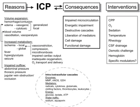
The most feared complication following severe trau-matic brain injury (TBI) is an increase in intracranial pressure (ICP) which may also become therapy-refrac-tory making ensuing death inevitable. Elevated ICP as measured continuously is a number which requires thor-ough and differentiated analysis of various possible rea-sons (Figure 1). In this context, the time point of intra-cranial hypertension is crucial: during the early phase (minutes/hours to initial 2–3 days), new onset or signifi-cant progression of hemorrhages and edema are the most common reasons [1]. Developing intracranial hy-pertension at later time points which is not observed under standard experimental conditions can be caused by progressing perifocal and generalized edema forma-tion and disturbed vascular responsiveness. Overall, compression and edema form a vicious circle with vari-ous potential therapeutic targets (Figure 2). In this con-text, the heterogeneity of TBI with its different lesions might require lesion-dependent interventions. The easi-est treatment step is the surgical removal of space-occu-pying lesions compressing the brain (subdural and epi-dural hematoma [SDH, EDH]) providing their speed of development has not surpassed the time point of sal-vageable iatrogenic intervention. More complicated to
1 Department of Surgery, Division of Surgical Intensive Care Medicine, University Hospital Zurich, Switzerland. Received: May 22, 2005; revision accepted: June 27, 2005.
treat and correct are different chang-es developing in parallel and sequen-tially which can induce or aggravate underlying damage over time. De-spite the intense research and the promising results obtained by spe-cifically modulating identified cellu-lar targets in “non-intensive care unit (ICU) rodents” [2], the equally successful implementation of these laboratory results in ICU patients with severe TBI has failed. Just re-cently, the Dexanabinol trial which had been envisioned to provide an effective therapy by inhibiting cellu-lar damage mediated by reactive oxygen species, glutamate and tu-mor necrosis factor was announced a failure. Consequently, we are left with our basic treatment possibili-ties.
In this review, the authors have tried to address the old issues and questions on how to treat intracra-nial hypertension, focusing on find-ings published in the past 5 years.
Identification of Secondary Injury and Deterioration
Both, clinical and experimental re-search strongly support the percep-tion that reliable and easy-to-per-form monitoring techniques and methods are indispensable to un-mask further deterioration and to also initiate and guide specific ther-apeutic and pharmacological inter-ventions. For this, measuring chang-es in ICP is the most efficient basic monitoring method to reveal patho-logic alterations within the intracra-nial compartment which is
comple-mented by additional monitoring approaches. ICP values as such are nonspecific as they cannot differenti-ate the actual cause for the observed increase in ICP (Figure 1). Nevertheless, ICP monitoring is crucial in prompting diagnostic steps and guiding therapeutic in-terventions.
Overall, secondary injury is characterized by its dy-namic development initiated by the primary injury. In this context, speed, duration, and extent of injury pro-gression are variable in time and location and appear to be different for structural and functional damage. Pro-gression and new onset of structural lesions occur
pre-Reasons
Reasons
ICP
ICP
Consequences
Consequences
Interventions
Interventions
Volume expansion: hemorrhage/contusion edema - vasogenic - cytotoxic blood volume vasodilation, vasoparalysis Increased metabolism: ischemia - local - global fever hyperglycolysis seizure Impaired outflow: abdominal pressure thoracic pressure jugular vein obstruction/ occlusion generalized focal Impaired microcirculation Energetic impairment Destructive cascades Liberation of mediators Cell damage Functional damage Intra-/extracellular cascades Enzymes: MMP, nNOS, GDH Mediators:
radicals, cytokines, glutamate, clotting factors, thrombocytes, leukocytes Energy:
glucose, lactate, ATP Water traffic: sodium, aquaporin CPP O2 Sedation Temperature Rheology CSF drainage Osmotic challenge Hemoglobin Specific modulators? vasoconstriction, compression, microthrombosis impaired cardiac output inadequate oxygenation, O2transport and delivery
Figure 1. Schematic drawing showing possible pathophysiological circuits which contribute to
intracranial hypertension following severe TBI which, in turn, can activate or aggravate under-lying pathologic changes. Corrective interventions are limited.
Craniectomy HemostasisSurgery
CSF drainage Furosemide Sedation Head elevation Hyperventilation Osmotherapy Barbiturates Hypothermia Ergotamine Vintracranial= Vbrain+VCSF+Vblood+Vmass lesion
Vintracranial= Vbrain+ VCSF+ Vblood+ Vmass lesion
Vasoregulation Edema Hemorrhage Hemorrhage Outflow Pathology Therapy Osmotherapy
Figure 2. The intracranial volume is influenced by different compartimentalized components
which, however, influence each other. To a certain extent these components are individual therapeutic targets. However, it is important to understand that aggressive treatment of one component might induce adverse and deleterious effects in a different part owing to the tight functional dependencies.
dominantly within the first 24–72 h. Edema formation develops in parallel but persists longer than hemorrhag-es which show signs of rhemorrhag-esolution within 5 days. Conse-quently, the initial hours to days are the most crucial phase requiring intensive monitoring to identify second-ary deterioration. Nevertheless, the subsequent days to weeks are overshadowed by potential secondary insults related to focal or diffuse ischemia, persistingly open blood-brain barrier, the inflammatory response, infec-tions, and other organ complications which have been shown to influence morbidity and mortality [3].
Since revelation of functional deterioration is the key in improving our current therapy, the following sec-tion summarizes the different monitoring techniques which are used to assess development, extent, and re-gression of various causes of elevated ICP.
Brain Imaging
While the readily available CT (computed tomography) analysis is an integral and widely employed part in eval-uating presence, severity, and development of structural intracranial pathologies, it does not reflect new onset or progression of functional deterioration. Other imaging techniques known to disguise functional changes (PET [positron emission tomography], SPECT [single-photon emission computed tomography], H-MRS [proton mag-netic resonance spectroscopy], MRI [magmag-netic reso-nance imaging]) are not readily available, difficult to conduct in cardiopulmonary and hemodynamically un-stable patients, can only be performed discontinuously, are still academic in nature, and have not been integrat-ed in the actual clinical management. Assessment of functional disturbances is crucial to induce and adapt therapeutic interventions. In the classic ICU setting with sedated and mechanically ventilated patients “functional disturbance” refers to changes on the cellu-lar level which involves not only neurons but also astro-cytes and endothelial cells. In this context, it is impor-tant to comprehend that actual cell-based alterations are highly dynamic processes which influence each oth-er in a hetoth-erogeneous spatial and temporal mannoth-er.
Present conclusions. In daily routine CT analysis is
the mainstay within the diagnostic and therapeutic deci-sion-making. During the acute phase only MRI studies with their logistic and patient-dependent difficulties are justified to determine brain stem lesion which otherwise are not visible on CT scans. Assessing functional altera-tions by MRI and PET requires an active and
coopera-tive patient and, thus, is of inferior assistance in coma-tose and sedated patients.
Intracranial Pressure (ICP) and Compliance
The hallmark of posttraumatic structural and functional disturbance is an elevation in ICP due to increased in-tracranial volume related to expanding hemorrhages (EDH, SDH, contusions) and/or accumulation of water within the extracellular and intracellular compartment, known as vasogenic and cytotoxic edema. Furthermore, insufficient as well as sustained cerebral perfusion (hy-peremia) due to ischemia and increased intracranial blood volume, respectively, can induce intracranial hy-pertension [4, 5]. For this, introduction of ICP monitor-ing devices within the subdural space, brain parenchyma and ventricular system are integrated in the routine treatment of these patients. As evaluated within the guidelines brought forward by the American Associa-tion of Neurological Surgeons (AANS;
www2.brain- trauma.org/guidelines/downloads/btf_guidelines_man-agement.pdf) subarachnoid, subdural, and epidural
monitors are less accurate compared to parenchymal and ventricular ICP probes. In this context, it is impor-tant to comprehend that the suggested ICP threshold of 15 mmHg is based on supratentorial measurements, as patients can also herniate with normal values, if the ex-panding lesion is located in close proximity of the brain stem. In general, ICP values > 15 mmHg reflect underly-ing pathology and any increase exceedunderly-ing 25 mmHg should prompt further diagnostic evaluation (see AANS guidelines).
Determination of intracranial compliance as a mea-sure of increased stiffness of the brain by infusing fixed amounts of fluid in the ventricular system is thought to unmask a pathologic pressure-volume relationship and to also predict forthcoming hypertension when small in-creases in volume induce a larger than normal increase in pressure under conditions of exploited compensatory mechanisms as seen in cases of space-occupying lesions. As a function of elevated ICP the tolerable amount of injectable volume into the ventricular system decreases significantly, as ICP exceeds 20 mmHg, irrespective of age [6]. However, given a low incidence of episodes identified to adequately predict pathologically elevated ICP, i.e., 16% of 225 episodes determined in ten patients suffering from severe TBI, this technique requires fur-ther modification before it can be introduced in the clinical routine as an early warning system for ensuing deterioration [6].
Present conclusions. Measuring ICP is the crucial
and central monitoring parameter for any physician treating patients with TBI. Changes in ICP need to prompt more in-depth diagnosis to determine the cause for intracranial hypertension, thereby allowing more specific treatment, and to prevent uncontrollable de-compensation. Assessing ICP automatically provides information about global cerebral perfusion, as the ce-rebral perfusion pressure (CPP) results from ICP and mean arterial blood pressure (MABP). Translation of bedside analysis of disturbed intracranial compliance to daily routine has not been completed due to technical limitations.
Electrophysiological Investigations (EEG, EPs)
Disturbances in neuronal activity and axonal conduc-tance can be assessed by electroencephalography (EEG) and evoked potentials (EPs) which can determine the site of functional impairment and even allow prognostic statements [7]. Cortical responsiveness to exteroceptive tactile, acoustic, and noxious stimuli requires intact transmission from the periphery to central areas (sen-sory stimulation) with adequate processing in the gray matter. On a cellular level, excitatory and inhibitory in-puts require viable and metabolically active neurons and astrocytes to generate and modulate electrogenic potentials arising from cortical and subcortical struc-tures. Any disturbance due to structural lesions within the white or gray matter of brain and spine or functional impairment related to hypothermia and administered sedative drugs can hamper the reliability of these inves-tigations. Nevertheless, these techniques allow insight into pathologic alterations which in conjunction with metabolic analysis might unmask severity of injury and pharmacological reversibility as suggested by the find-ings of Vespa et al. who were able to reveal nonconvul-sive status epilepticus which otherwise had gone unde-tected [8]. Consequently, specialists recommend continuous EEG monitoring during the initial 48 h [9].
Present conclusions. EEG analysis remains a
con-troversial area due to its physiological, pathophysiologi-cal and technipathophysiologi-cal complexity, since we can neither deter-mine the actual anatomic depth at which changes are induced nor assess the extent of neuronal and glial im-balance. More refined and simplified approaches as e.g., the BIS (bispectral index) EEG might reveal neuronal stability or instability continuously at bedside, possibly helping to disguise neuronal from glial activity,
when-ever surrogate markers as e.g., decreased jugular venous oxygen saturation (SjvO2) occur in face of decreased EEG (neuronal) activity. EPs are helpful in assessing brain stem lesions, thereby aiding the complex decision process.
Microdialysis
Recent introduction of microdialysis in the clinical rou-tine has opened the possibility to unmask otherwise oc-cult pathologic metabolic alterations based on routine analysis of glutamate, glucose, lactate, pyruvate, and glycerol [10], and calculated indices. In addition, phar-macokinetic studies can be performed [11]. A large number of reports have convincingly demonstrated its applicability and usefulness in assessing cellular impair-ment following TBI, providing certain limitations are considered. The low spatial and temporal resolution dictated by the position and speed of dialysis, respec-tively [12], do not allow to transfer these local altera-tions to distant areas and might not be able to reveal ensuing changes early enough to allow preemptive in-terventions. In addition, recovery rate is not routinely determined under in vivo conditions by injecting a met-abolically inert substance via a second microdialysis catheter inserted in close proximity [13]. Furthermore, we cannot correct for concentration or dilution effects due to edema-related shrinkage or expansion of the ex-tracellular space and we are not able to exclude diffu-sion handicap caused by activated glia surrounding the catheter over time.
Present conclusions. Cerebral microdialysis
disguis-es local pathologic procdisguis-essdisguis-es as well as pharmacody-namic effects. Whether the latency of these changes might be overcome by a higher sampling rate (1- to 5-min intervals vs. routine 30-min intervals) which in-creases the logistical and personal workload remains unclear. In this context, a sophisticated automated arti-fact recognition tool becomes indispensable. The asso-ciated costs limit its general integration.
Cerebrospinal Fluid (CSF) Analysis
In patients with noncompressed lateral ventricles, ven-tricular catheters are inserted to lower ICP by draining CSF. This approach can also be used to determine meta-bolic and immunologic changes [14] in CSF to unmask underlying pathologic changes. This technique, howev-er, is compromised by certain methodological pitfalls. For one, drained CSF reflects global changes and
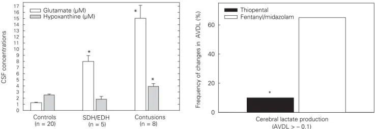
mea-sured solutes are strongly influenced by intracisternal blood, the bulk flow toward the ventricular system washing these substances out of the brain, and the speed and amount of produced and resorbed CSF which might change over time and is not quantified under clinical conditions. Furthermore, there is no clear consensus whether single or repetitive measurements are required or if CSF needs to be collected continuously. The fact that ventricular catheters cannot be inserted in patients with narrow or compressed lateral ventricles or CSF cannot be drained due to progressive edema-related compression of the ventricular system implies that only patients with brain injuries of a presumably lesser ex-tent or a more complicated secondary development are investigated. This, in turn, does not allow to extrapolate the determined changes to all patients. Nevertheless, this technique allows to assess the impact of underlying type of brain lesions and can also be used for pharma-codynamic and pharmacokinetic studies. In this con-text, highest glutamate and hypoxanthine concentra-tions were determined in patients with contusions and mixed lesions compared to control patients (lumbar CSF) and patients with isolated SDH or EDH (Figure 3). Inducing a burst suppression pattern by the admin-istration of thiopental resulted in a significant decrease in CSF glutamate, lactate and hypoxanthine concen-trations [15].
Present conclusions. CSF analysis remains a simple
monitoring approach which, however, is limited to
pa-tients with less or more severe injuries, depending on the size of the ventricular system. The fear for ventricu-lostomy-related infections might restrain this ap-proach.
Jugular Bulb Catheter
Retrograde insertion of a special catheter in the internal jugular vein allows to continuously determine changes in SjvO2 [16] and discontinuously measure arterio-jugu-lar venous differences of humoral and celluarterio-jugu-lar compo-nents leaving or entering the brain [17–20]. This moni-toring approach reflects global alterations and is routinely used to guide and control hyperventilation to reduce elevated ICP. This allows to detemine hyperven-tilation-induced vasoconstriction and prevent injury-ag-gravating ischemia [21]. As any attempt to study meta-bolic and thus functional alterations, concomitant drug administration needs to be considered, as pharmacody-namic influences may supervene. In this context, we have observed a significant reduction in lactate pro-duction reflected by the lower frequency of negative arterio-jugular venous lactate differences in patients subjected to high-dose barbiturate coma compared to patients receiving benzodiazepines and opioids (Fig-ure 4).
Different SjvO2 values were identified to have a therapy-guiding potential: while SjvO2 values < 50% re-flect ischemia, levels between 50 and 65% could reveal impending ischemia, and SjvO2 between 65 and 75% are considered normal; levels > 76% reflect hyperemia or
CSF concentrations 0 1 2 3 4 5 6 7 8 9 10 11 12 13 14 15 16 17 Glutamate (µM) Hypoxanthine (µM) SDH/EDH (n = 5) Contusions (n = 8) Controls (n = 20) * * *
Frequency of changes in AVDL (%)
0 20 40 60 Thiopental Fentanyl/midazolam
Cerebral lactate production (AVDL > – 0.1) *
Figure 3. Influence of different traumatic brain lesions on CSF
gluta-mate and hypoxanthine concentrations, reflecting underlying excito-toxicity and energetic impairment. Highest levels were observed in patients presenting with contusions (*p < 0.05 vs. controls).
Figure 4. In patients with therapy-refractory intracranial
hyperten-sion, administration of high-dose thiopental significantly reduced the frequency of metabolic compromising events reflected by a decreased number of pathologically elevated jugular venous lactate (negative arterio-jugular venous difference) compared to patients treated with fentanyl and midazolam (*p < 0.001).
vast depression of metabolism characteristic of severe injuries. Most frequent causes for pathologically de-creased SjvO2 values are hyperventilation, hypovole-mia, and anemia which can be prevented or corrected, thereby reducing incidence of intracranial hypertension and improving outcome [1].
Present conclusions. Retrograde cannulation of the
internal jugular bulb to monitor cerebral alterations is technically easy but is limited by the frequency of analy-sis and the declining data quality within several days fol-lowing insertion of the fiber-optic catheter. Measuring arterio-jugular venous differences might complement multimodal monitoring especially in regard to systemic influences related to diseases known to develop over time in critically ill patients (e.g., organ dysfunction, in-fections, sepsis, etc.). With this approach more patients could be monitored especially if penetrating probes (microdialysis, tissue probes) cannot be introduced.
Brain Tissue Oxygenation
Following TBI, the injured brain is in a state of distress characterized by sustained vulnerability owing, among other reasons, to its limited energetic reserves in face of sustained and dysregulated excitation. Consequent-ly, the brain requires sufficient oxygen to prevent ad-ditional cell damage from impaired oxidative metabo-lism necessary to meet activation-induced increases in energy demands. Changes in tissue pO2 (ptiO2) reli-ably reveal evolving tissue perturbation. Detrimental ptiO2 threshold below which ischemic damage devel-ops and which is associated with pathologic neuro-chemical alterations [22] and a worse outcome is 8–10 mmHg [23, 24]. Changes in ptiO2 can also serve to dis-criminate anaerobic from nonoxidative metabolism, as an increase in extracellular lactate can also be caused by sustained activity and adapted lactate utilization in face of adequate perfusion and supply with oxygen and nutrients. Similar to the SjvO2, local ptiO2 changes can be used to assess limits of hyperventilation and thus guide this therapeutic intervention [25]. This technique with its monitoring confined to a small area of interest at the cortical-subcortical junction within the frontal cortex of the uninjured or lesser injured hemisphere faces similar methodological limitations as the bio-chemical monitoring via microdialysis. This cannot be overcome by its high temporal resolution with a sam-pling rate of 1–10 recordings/min.
Present conclusions. As seen with microdialysis,
ptiO2 recordings require insertion of penetrating probes which might not be feasible in patients with bifrontal lesions. Due to the high frequency of data recording a sophisticated artifact recognition software is indispens-able to guarantee reliindispens-able interpretation of the collected data. The long “calibration” of up to 6 h limits its useful-ness during the early period following probe place-ment.
Cerebral Perfusion
Ever since its initial description by Graham et al. in 1978 [26] reporting ischemic damage in 91% of pa-tients succumbing to their nonmissile head injuries, diagnosis, avoidance, and treatment of impaired per-fusion following severe TBI have prompted a multi-tude of clinical and experimental studies. The easiest but also most crude assessment of cerebral perfusion is to calculate the difference between MABP and ICP. This number, however, does not reflect regional alterations which are known to be extremely hetero-geneous. Assessing cerebral perfusion can be per-formed invasively and noninvasively. As recently and convincingly shown by Vajkoczy et al. [27], insertion of a special thermodilution probe allows to continu-ously and reliably determine changes in cerebral per-fusion as evaluated by simultaneous stable xenon-enhanced computerized tomography scanning (sXe-rCBF) which contrary to the laser Doppler methods reveals absolute flow values [ml/100 g/min]. In a small series of patients, Jaeger et al. [28] could show that this technique allows to detect impaired perfusion judged by the changes in ptiO2. Unfortu-nately, as other catheters used to aid neurointensive care, this probe “only” reveals local alterations. As-sessing global changes in cerebral blood flow (CBF) at bedside using the transcranial thermo-dye-dilution technique, however, has yielded uncertain results compared to xenon-CT measurements [29].
Noninvasive bedside transcranial Doppler evalua-tion does not only reveal changes in blood flow velocity of the extra- and intracranial vessels but may also reflect microcirculatory impairment following TBI, as the low flow velocity which was reported to be most prominent ipsilateral to a focal pathology is associated with de-creased ptiO2, especially within the first days following injury [30]. This technique is performed discontinuously but with a higher frequency compared to imaging stud-ies.
Imaging studies with their high spatial resolution are limited by the fact that these discontinuously per-formed and logistically challenging studies can only pro-vide “snapshot” views. Perfusion CT [31], SPECT [32], PET [21], perfusion-weighted MRI [33], and xenon-CT [29] can reveal localization and extent of underlying im-paired perfusion and ischemia and even unmask basal ganglia hypoperfusion.
Apart from identifying ischemia, assessing changes in perfusion is essential to determine if increased CBF exceeding the metabolic demand reflects hyperemia. This could result from sustained brain damage or an in-creased pressure load due to impaired autoregulation which could be reduced by lowering CPP.
Present conclusions. Although decreased as well as
increased cerebral perfusion are the most crucial patho-physiological changes accounting for secondary dam-age, routine surveillance of disturbed perfusion is pre-dominantly based on surrogate markers (ptiO2, SjvO2, metabolic changes). More widespread application of bedside continuous approaches combining regional and global perfusion is important to improve both under-standing and therapeutic guidance. This will automati-cally facilitate interpretation of other monitoring pa-rameters, and thereby either initiate or stop therapeutic interventions.
Proteomics
Due to its recent commercial availability, analysis of various proteins determined in microdialysis or CSF samples can be used to investigate changes of extracel-lular and even intracelextracel-lular proteins following their re-lease or liberation. This approach might allow to iden-tify proteins which might be useful predictors for disease characteristics following TBI as seen after stroke [34].
Present conclusions. More data is needed before a
meaningful conclusion may be drawn.
Cerebral Monitoring: Is it Local Versus Global or a Combination of Both?
Successful development and feasible transfer from bench to bedside have facilitated the integration of vari-ous monitoring techniques in the daily routine support and care of patients with severe TBI. Nevertheless, as with any – especially novel – “experimental” diagnostic approach certain limitations inherent to the applied techniques and methods can make the clinical decision
process more difficult, as differentiated interpretation of collected data might not reveal pathologic alterations at the time point of evaluation but may only become obvious following post hoc analysis. Insertion of special catheters despite their small diameters (ICP probe: 1 mm; ptiO2: 0.8 mm; microdialysis: 0.8 mm; thermodilu-tion probe: 1 mm) carries a risk of addithermodilu-tional damage, mainly hemorrhage especially in preinjured regions. Thus, these catheters should only be inserted within the frontal cortex to avoid any damage to the primary and secondary sensory and motor fields located more poste-rior.
While these catheters are generally assumed to only reflect local changes confined to a small volume sur-rounding these probes – used to argue against their use-fulness –, this must be challenged by the fact that this assumption might only hold true at low ICP levels and maybe also in craniectomized patients. Under experi-mental [35] and clinical [36] conditions the rigid skull will translate increased pressure caused by a focal or hemispheric lesion to the contralateral hemisphere and also to other regions within the ipsilateral hemisphere over time as the existing pathology extends. Conse-quently, monitoring the contralateral or lesser injured hemisphere will reveal serious and relevant alterations reflecting severity and extent of underlying disturbance. This, in turn, implies that whenever pathologic altera-tions occur in the contralateral hemisphere, functional integrity of the entire brain needs to be considered at stake. The initial conception that metabolic abnormali-ties are restricted to superficial regions has been recent-ly questioned, as a significant reduction in metabolic oxygen rate was also observed in subcortical white mat-ter remote from focal hemorrhagic lesions, suggesting more diffuse injuries [37]. However, the studied patients were heterogeneous in terms of injury severity, consid-ered time points, and administconsid-ered drugs.
The strong need to closely monitor patients with se-vere TBI and to start as early as possible to avoid miss-ing preventable insults which increase morbidity and mortality has become self-understood. This is strength-ened by retrospective reports showing a significant reduction in mortality, increase in functional out -come, shortened hospitalization, and reduction in costs [38–40]. Nevertheless, critics still question the need for intensified monitoring, as the efficacy of ICP and multi-modal monitoring has never been studied in a prospective, randomized clinical trial [41]. Overall, there is a growing body of evidence showing that these
monitoring techniques and methods integrated in the neurointensive care setting to date can detect deteriora-tion, guide therapeutic and pharma-cological interventions, and thereby contribute to the improvement of these patients. Thus, designing a randomized trial assigning patients to a nonmonitoring arm would be unethical and therefore inapplica-ble. However, the extent and fre-quency of monitoring could be sub-jected to a randomized trial. Furthermore, the length of monitor-ing required to guarantee a more fa-vorable outcome still remains under debate. While some centers discon-tinue monitoring and sedation after a few days, other centers maintain monitoring and sedation until ICP remains stable without exceeding 20 mmHg. Based on our own experi-ence, ICP can steadily increase
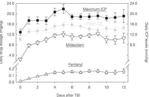
dur-ing the first week, surpass 15 mmHg durdur-ing the second week and approach 20 mmHg by the end of the second week (Figure 5).
Present conclusions. Further technical development
and more widespread application of various monitoring techniques in primary and tertiary hospitals will gener-ate more data and substantigener-ate our knowledge which, in turn, will improve the overall treatment. It remains to be determined which parameters must be monitored and which are facultative to disguise evolving deteriora-tion and improvement. In general, various parameters need to be combined to guarantee a holistic surveillance of local and systemic as well as cell-dependent changes. In addition, monitoring needs to be adapted to the tem-poral profile characterized by different influences and requirements.
Treatment of Secondary Injury and Deterioration
In principal, dictated by the progressive nature of evolv-ing structural and functional deterioration our duty is to find ways and means to attenuate (= realistic goal) or even stop (= ideal situation) this development without endangering the patient. What is oversimplified by the dichotomy of e.g., ICP- versus CPP-targeted therapy is, in reality, much more complex. In fact, we might have to
combine the AANS guidelines and the treatment strate-gies advocated by Grände et al. known as the “Lund concept” [42] in the same patient at different time points. The following part summarizes new knowledge and novel developments for the different treatment steps considered essential in the treatment of intracra-nial hypertension.
CPP- or ICP-Targeted Therapy – the All-Time Controversial Expanded by the Volume-Targeted Therapy (“Lund Concept”)
Based on a multitude of publications using different methods to determine cerebral perfusion (PET, sXe-CT) posttraumatic brain ischemia strongly influences neuro-logic outcome. Consequently, maintenance of adequate cerebral perfusion is essential to avoid additional cell damage possibly aggravating underlying injury. In this context, adequacy of perfusion is difficult to define as it is subject to heterogeneous local and temporal changes with a strong interindividual variability [43], ranging from impaired perfusion [44] due to clotted or vasocon-stricted or compressed vessels to increased perfusion (hyperemia) [43] caused by vasodilated or paralyzed vessels. If adequacy of perfusion is defined as sufficient metabolic support of neurons and glia, then metabo-lism-related parameters in addition to cerebral
perfu-Days after TBI
0 2 4 6 8 10 12
Daily drug dosage (mg/kg)
0.0 0.1 0.2 4.0 8.0 12.0 16.0 20.0 24.0 0.0 0.1 0.2 4.0 8.0 12.0 16.0 20.0 24.0
Daily ICP levels (mmHg)
Fentanyl Midazolam
Maximum ICP
mean ICP
Figure 5. Temporal profile of changes in maximum and mean ICP determined in 16
representa-tive patients suffering from severe TBI. ICP was significantly increased on the first posttrau-matic day and after 1 week, remaining elevated thereafter. Within 4 days, fentanyl and mid-azolam were administered in their highest possible daily amount.
Under clinical conditions, infusion of fentanyl and midazolam followed the increases in ICP. Thus, it remains to be determined if a higher starting dose might attenuate subsequent intra-cranial hypertension.
sion must be continuously determined at bedside. This will allow us to respond to changes over time influenced by progression and resolution of hemorrhages and ede-ma forede-mation and also enable us to adequately react to iatrogenic manipulations and therapeutic interventions. Thus, aggressive therapeutic measures employed to in-fluence cerebral perfusion without proper control might result in blood volume overload (hyperemia or flow-me-tabolism uncoupling) under conditions of endogeneous-ly or pharmacologicalendogeneous-ly depressed metabolic demand [43]. This, in turn, could induce aggravating elevated ICP. The initial CPP value of 70 mmHg set as the thera-peutic goal by the AANS in 2000 was recently ques-tioned and revised by the AANS in 2003 based on the observed volume- and pressor-induced increase in inci-dence of adult respiratory distress syndrome (ARDS) in patients who had been randomized to the CBF-targeted therapy [45]. As recently concluded by Juul et al., a CPP > 60 mmHg does not improve outcome providing ICP can be maintained < 20 mmHg [46]. Then again, others request to maintain CPP > 70 mmHg [47].
As clearly stated in the initial guideline, the AANS repeats that we are still faced with “insufficient data to
support treatment standards” and suggests: “Cerebral perfusion pressure (CPP) should be maintained at a min-imum of 60 mmHg. In the absence of cerebral ischemia, aggressive attempts to maintain CPP above 70 mmHg with fluids and pressors should be avoided because of the risk of adult respiratory distress syndrome.” The crucial
detail in guiding adequate cerebral perfusion is exten-sive monitoring. For this, we need to combine different approaches simultaneously, including focal and global assessment of metabolism and perfusion.
Contrary to the initial perception of a static ICP-CPP relationship, novel data show that the spatial and tem-poral heterogeneity in impaired perfusion, disturbed metabolism, and metabolism-flow mismatch reflect a more dynamic relationship which appears to be differ-ent for various lesions and could change over time with-in the same patient. Consequently, the optimal thresh-olds for ICP as well as CPP might not be isolated setpoints but could require more iatrogenic flexibility to adapt and guide therapeutic interventions by monitor-ing cerebral metabolism, allowmonitor-ing higher ICP values (> 20 mmHg) as long as an adequate perfusion thresh-old is maintained [48, 49]. Based on the calculated dy-namic threshold levels for CPP and ICP, Chambers et al. could show that determined minimal CPP and maxi-mal ICP values differ depending on extent/severity of
underlying lesions [5]. Interestingly, minimum required CPP values were highest in patients with nonevacuated mass lesions (94 mmHg), possibly related to the con-comitant compression of the microcirculation, requiring an increase in pressure gradient. An increase in CPP from approximately 70 to 90 mmHg appears necessary to reduce the ischemic brain volume, improve flow-me-tabolism coupling, decrease oxygen consumption, and increase ptiO2 [50, 51].
As suggested by Cremer et al. in a small series of patients, daily pharmacological manipulations of CPP are suggested to determine the dynamic changes in CPP requirements [52]. Unfortunately, this small series of patients was heterogeneous in terms of underlying inju-ries and concomitant therapeutic interventions.
Present conclusions. Despite the beneficial effects
reported upon rigid dichotomy of CPP- versus ICP-tar-geted therapy, the dynamic pathologic processes with their strong temporal and lesion dependency might re-quire a more flexible approach allowing a combination of the CPP- versus ICP-targeted treatment strategies.
Hyperventilation
Initially envisioned as an elegant way to control ICP by simply increasing ventilation frequency or tidal volumes in mechanically ventilated patients, the therapeutic hy-perventilation has been subject to controversial dis-cussions, as hypocapnia can induce adverse effects and contribute to a worse outcome. In this context, vasocon-striction with ensuing or impending ischemia as evi-denced by decreases in cerebral perfusion [21, 53], re-duced ptiO2 and SjvO2 values [25], and increased extracellular glutamate and lactate concentrations [53] have been reported. As shown by Carmona Suazo et al., daily short hyperventilation trials of 15 min reveal an increasing risk for hyperventilation-induced compro-mise of cerebral oxygenation during the first 5 days as reflected by an elevated ptiO2/paCO2 reactivity ratio [54]. As suggested by Coles et al., threshold value of paCO2 ranges from 34 to 38 mmHg (= 4.5–5.0 kPa), im-plying that even small changes in paCO2 during nor-moventilation with normocapnic values can induce un-favorable alterations [21]. Other groups, however, did not report any adverse effects and highlight a consistent decrease in ICP [55]. Brief episodes of hyperventilation which reduced posttraumatic cerebral perfusion were not associated with energy failure, even in regions where ischemic perfusion values were reached. Thus, Diringer
et al. concluded that underlying suppressed metabolism protects from hyperventilation-induced impaired per-fusion [56].
Due to extensive experience gained within the past 20 years, the official AANS guidelines suggest that chronic hyperventilation should definitely not be per-formed during the first 5 days following TBI. In particu-lar, hyperventilation should definitely be avoided within the first 24 h, as this time period is characterized by im-paired cerebral perfusion and disturbed metabolic and energetic homeostasis. Hyperventilation decreases the already impaired perfusion even further, limiting nutri-tional supply and oxygenation to damaged tissue, and may also induce lesion growth by disturbing cerebral va-soreactivity and autoregulation. This may be of utmost importance in patients with more severe injuries as e.g., SDH and multiple contusions in whom vascular respon-siveness to CO2 is reduced [57, 58]. In addition, hypo-capnia can also impair cardiac, pulmonary and cardio-vascular function [59].
Present conclusions. As concluded by a Cochrane
review, randomized controlled trials are required to as-sess the effectiveness of hyperventilation [60]. Hyper-ventilation might only be an option if detailed assess-ment of individual perfusion and metabolism pattern is available. Specific bedside monitoring is definitely in-dispensable, since even small changes in paCO2 within its normal range can already induce adverse effects.
Osmotherapy: Mannitol and Small-Volume Resuscitation
Shifting of electrolytes and changes in osmotic gradients across the blood-brain barrier contribute to brain ede-ma and brain swelling. Thus, ede-manipulation of this os-motic gradient appears to be a feasible therapeutic ap-proach. In this context, mannitol and hypertonic saline solution with and without dextran have been investi-gated under clinical conditions. Despite extensive expe-rience with mannitol it still lacks evidence of efficacy, as concluded by a recent Cochrane review [61]. Mannitol with its strong osmotic potential expands plasma, there-by reducing the hematocrit and blood viscosity, and in-creases CBF and oxygen delivery within several minutes following its administration. These effects are predomi-nant in patients with CPP values < 70 mmHg and re-duced serum osmolarity [62]. Ultra-early administration of high-dose mannitol (1.4 g/kg) in the emergency room appears superior to low-dose mannitol (0.7 g/kg) to
re-verse clinical signs of impending brain death and im-prove long-term neurologic outcome, as studied in se-verely injured patients with a Glasgow Coma Scale score of 3 and bilateral abnormal pupillary widening [63]. Continuous infusion should be avoided, as mannitol may damage the blood-brain barrier or accumulate within the extracellular space which, in turn, could raise brain osmolality, thereby exacerbating ICP and increas-ing brain swellincreas-ing.
An alternative to mannitol are hypertonic solutions, referred to as small-volume resuscitation, as a small dose, usually 4 ml/kg body weight (approximately 250 ml) of 7.2–7.5% NaCl/colloid solution is infused in ap-proximately 20 min. In addition to its primary resuscita-tive potential from trauma and hypotensive shock, this approach might also gain access to perioperative and intensive care treatment of patients with elevated ICP as suggested by Kreimeier & Messmer [64]. The high tonicity and the fact that hypertonic saline does not pen-etrate the blood-brain barrier makes it a promising os-motherapeutic agent. The decrease in ICP is thought to result from reduced water content in noninjured brain areas and the cerebellum without significantly changing global cerebral perfusion [65]. The transient reduction in ICP might be prolonged by continuous infusion but requires careful control to avoid adverse effects as e.g., electrolyte abnormalities, cardiac failure, bleeding dia-thesis, phlebitis, central pontine myelinolysis, and re-bound intracranial hypertension following uncontrolled infusion.
As recently published, administration of hyperton-ic saline solution results in superior effects compared to mannitol by decreasing daily episodes of intracra-nial hypertension with a reduced rate in therapy-re-fractory increases in ICP [66]. However, morbidity and mortality were similar in both groups and the differ-ence in osmotic strength (350 mOsmol/dose vs. 175 mOsmol/dose) limits the power of this study. As sug-gested by Horn et al., hypertonic saline solution might be an effective measure to decrease ICP which is oth-erwise refractory to standard therapeutic approaches, i.e., mannitol and barbiturates [67]. When adminis-tered during the prehospital phase, hypertonic saline solution did not result in improved mortality or mor-bidity, as seen in a double-blind, randomized con-trolled trial enrolling 229 comatose and hypotensive TBI patients [68].
In a recently published pilot study, equimolar doses of hypertonic saline and dextran solution (HSD,
Res-cueflow) were compared to 20% mannitol solution. HSD caused a significantly greater decrease in ICP than mannitol and had a longer duration of effect than man-nitol [69].
Present conclusions. As concluded in two recently
published Cochrane database reviews, further detailed prospective, randomized, and blinded large-scale clini-cal studies are required to determine if the wide confi-dence intervals observed in the present investigations will reveal if hypertonic solutions are of significant im-portance [70] and if mannitol is indicated to manage in-tracranial hypertension in patients without an operable intracranial hematoma [61]. In this context, this treat-ment modality needs to be investigated before admis-sion, at various time points following admission to the hospital in dependence of underlying lesion, and in re-gard to intracranial hypertension.
Choice of Volume to Increase CPP
To date, it is well accepted that volume infusion to cor-rect hemodynamic stability should precede administra-tion of vasopressors. However, the type and amount of fluids are still under debate. As recently shown, normo-volemia with a fluid balance ranging from –594 to 4,859 ml was associated with the least incidence of poor out-come in hypothermic as well as normothermic patients compared to patients with lower or higher fluid balance [47]. Unfortunately, data of volume balance in patients treated according to the “Lund concept” are not avail-able which might allow to more efficiently define re-quired normovolemia in patients with severe TBI. Due to its plasma-expanding effects, colloid solutions are su-perior to crystalloid solutions which need to be adminis-tered in higher amounts to induce similar hemodynamic stability. As recently published, high amounts of hy-droxyethyl starch administered repeatedly in severe TBI patients are safe [71]. As reported by the Saline versus Albumin Evaluation study, 4% albumin was not associated with an overall benefit compared to saline. However, patients with severe TBI within the albu-min-treated group revealed a trend toward increased mortality [72].
Present conclusions. The ideal fluid balance remains
to be determined in patients suffering from severe TBI. Providing a lower CPP can be adequately controlled and justified by specific monitoring, a timely reduction in volume administration might attenuate side effects,
as e.g., ARDS known to increase morbidity in these pa-tients.
Barbiturate Coma
Barbiturates can decrease elevated ICP in patients pre-senting with otherwise therapy-refractory intracranial hypertension by reducing cerebral metabolism and oxy-gen consumption, thereby decreasing vascular tone, in-hibiting excitatory input and energetic impairment. However, adverse systemic side effects which can lead to serious complications related to hemodynamic insta-bility and infections due to impaired immunocompe-tence limit the administration to most extreme situa-tions. The fact that patients can suffer from further episodes of increased ICP despite complete suppression of neuronal activity as evidenced by continuous EEG recording and an initial responsiveness, suggests that barbiturate coma might not be the ultimate step in our therapeutic hierarchy but might require additional spe-cific treatment approaches. In this context, we must comprehend that EEG monitoring employed to guide barbiturate administration will only reflect changes in cortical neuronal activity. Alterations occurring in deep structures or involving astrocytes which encompass nearly 75% of the brain remain disguised. As observed under experimental conditions, barbiturates dose-de-pendently decrease astrocytic clearance of glutamate [73], possibly resulting in additional functional and structural cell damage, as glutamate cannot be used to fuel intracellular energetic processes or aid in synthesiz-ing the antioxidant glutathione. Under clinical condi-tions, this, in turn, could result in increased energetic impairment as reflected by sustained cerebral oxygen extraction, progressing edema formation, and further increase in ICP. This increase in ICP during barbiturate coma could also stem from hyperemia, if CPP is not ad-justed to the lowered metabolic demand during barbitu-rate coma. The statement published within the official AANS guidelines that prophylactic administration of barbiturates is not indicated following severe TBI is questioned by the “Lund concept” and the fact that many features within the overall intensive care treat-ment of these patients have changed due to extensive pathophysiological characterization of the different fea-tures of TBI and development of specific treatment op-tions. Controlling energetic impairment and deteriora-tion by suppressing cerebral metabolism through co-infusion of low-dose thiopental could prevent pro-gression of structural and functional cell damage and
reduce the risk of CPP-dependent increases in cerebral blood volume, as a lower CPP can be tolerated [74]. This hypothesis needs to be prospectively investigated at dif-ferent centers.
Present conclusions. As concluded in a Cochrane
database review published in 2000, barbiturate coma does not improve outcome in patients with severe TBI owing to its hypotensive effect [75]. Prophylactic barbi-turate administration discouraged by the AANS, how-ever, is challenged by the “Lund concept” and awaits detailed investigation. More detailed investigations us-ing refined monitorus-ing techniques and modern knowl-edge are required to determine the benefit of initial low-dose barbiturate administration, at which ICP high-dose barbiturate coma is to be initiated, for how long barbiturate coma (low and high dose) needs to be maintained, and what level of neuronal suppression as determined by continuous EEG recording and meta-bolic/neurochemical supervision is adequate.
Sedation and Analgesia
Following severe TBI, the injured brain is highly vulner-able to any potentially adverse influence. In this con-text, coughing and straining exert a certain mechanical stress due to an increase in ICP caused by decreased ve-nous outflow [76]. This, in turn, carries the risk of caus-ing additional hemorrhages and aggravatcaus-ing existcaus-ing space-occupying lesions, thereby adding to the underly-ing pathologic vicious circle of compression and edema formation. In addition, a concomitant increase in oxy-gen consumption reflected by decreased SjvO2 in face of reduced CPP may occur during coughing while endotra-cheal suctioning inadequately sedated TBI patients [76].
Sustained cellular activity is an energetically com-promising condition of stress which adds to a depletion of energetic reserves, thereby reducing the threshold for additional, otherwise tolerable compromising events. To date, neurons with their different excitatory and in-hibitory circuit receptors remain the primary direct pharmacological target although astrocytes account for a large portion of energy-consuming and -producing ef-fects, owing to their plethora of different functions with-in the neuronal-glial-endothelial syncytium [77].
A common manner to counteract these stress-relat-ed changes and thus prevent additional injury is to in-duce and maintain adequate analgesia and sedation. As outlined by Citerio & Cormio [78], many
pharmacologi-cal agents with characteristic differences in their phar-macokinetic and pharmacodynamic profile are avail-able to specifically control ICP and cerebral metabolism and reduce seizure activity in addition to the more gen-eral aims of controlling pain, anxiety, and agitation, lim-iting the stress response, facilitating care, and managing ventilatory support. For brain-injured patients it is im-portant to maintain adequate supply with nutrients and oxygen to guarantee sufficient energy production. Be-sides increasing oxygen delivery by optimized hemody-namics, ventilation, and oxygen carriers, oxygen con-sumption and demand are reduced by decreasing cerebral metabolism. Providing an intact metabo-lism-flow coupling, a reduction in cerebral metabolism will decrease CBF, thereby reducing cerebral blood vol-ume and ICP. In this context, it is important to remem-ber that metabolism is suppressed dose-dependently until the EEG becomes isoelectric. Thereafter, minimal basal energy consumption cannot be suppressed any further. Thus, reduction of intracranial blood volume is also restricted.
In general, benzodiazepines (e.g., midazolam, lo-razepam) or propofol are combined with opioids (e.g., morphine, fentanyl). As recently shown, disturbances in metabolism and metabolic suppression reactivity quan-tified by transient propofol-induced burst suppression pattern are heterogeneous and influenced by underly-ing brain lesions [79].
On a cellular level opioids activate specific µ-, δ-, κ-, and σ-receptors, of which µ-receptors mediate analgesia by inhibiting presynaptic release of excitatory transmit-ters via decreased availability of calcium and stabilizing postsynaptic membranes by increasing permeability for potassium. While activation of µ-receptors reduces lib-eration of norepinephrine and substance P, δ-receptors attenuate release of acetylcholine, and κ-receptors de-crease liberation of dopamine.
Benzodiazepines require the endogenous inhibitory transmitter GABA (γ-aminobutyric acid) to increase the opening time of the chloride channel by allosteric modulation of the GABAA receptor. This results in a stabilization of pre- and postsynaptic membranes, by which excitatory inputs are blunted.
Propofol has several targets, as it directly activates
GABAA receptors, inhibits the NMDA
(N-meth-yl-D-aspartate) receptor and modulates calcium influx through slow calcium ion channels [80].
Overall, the choice of agent, dosage, and duration of administration show a strong variability between
coun-tries and hospitals which might have contributed to the failure of various pharmacological trials aimed at modu-lating/inhibiting transmitter-mediated influences [81]. Some favor propofol to treat intracranial hypertension, as it dose-dependently reduces EEG activity. Others prefer benzodiazepines despite their “ceiling effect” in reducing cerebral metabolism and blood flow in fear of propofol-induced increase in hemodynamic instability, lipid load, and the potentially fatal “propofol infusion syndrome” characterized by severe metabolic acidosis, cardiac and peripheral muscle damage (necrosis), and circulatory collapse. This syndrome can be provoked by elevated catecholamine, glucocorticoid and cytokine levels [82] encountered in patients with severe TBI and is thought to be related to disturbed structural and func-tional integrity of mitochondria [80].
The beneficial effects of these drugs in terms of re-ducing cerebral metabolism and oxygen consumption are confronted with certain side effects. The most com-mon problem is receptor desensitization or downregu-lation developing during chronic continuous adminis-tration of opioids and benzodiazepines which accounts for the diminishing effectiveness of these drugs as evi-denced by coughing, straining and eye opening despite continuously infusing these drugs at their maximally recommended dose. In clinical practice, dosage of opi-oids and benzodiazepines is increased following changes in ICP (Figure 5), suggesting that starting with higher or submaximal dose or adding a more potent sedating agent as e.g., low-dose thiopental as advocated by the “Lund concept” [42] might prevent further cellular de-terioration known to contribute to subsequent intracra-nial hypertension. Contrary to benzodiazepines, barbi-turates activate GABAA receptors independently of GABA (at high dosage). Since CSF glutamate and hy-poxanthine concentrations remained unchanged and even showed a trend to elevated levels during continu-ous administration of midazolam and fentanyl (Figure 5) and since thiopental significantly reduced CSF gluta-mate and hypoxanthine in patients who developed ther-apy-refractory intracranial hypertension during admin-istration of fentanyl and midazolam [15], a combination of these drugs might be a more promising approach in conveying neuroprotection.
A further complication is drug-induced reduction in bowel function leading to impaired transport and up-take of nutrients which compromises nutritional supply of energetic compounds. In addition, disturbed bowel function may even progress to paralytic ileus. Serious
constipation might add to intracranial hypertension by elevating intraabdominal pressure known to reduce ce-rebral venous outflow. Furthermore, prolonged admin-istration of opioids may suppress pituitary function, thereby disturbing the hypothalamic-pituitary-adrenal axis, causing hormonal dysregulation, and suppression of the immune system [83].
While inducing and maintaining sedation in patients with severe TBI is a rather straightforward approach, the awakening phase might be underestimated in its pathophysiological importance. In this context, continu-ous administration of opioids and benzodiazepines in-duces tolerance and dependency due to altered receptor function and expression. Clinically, patients are ex-tremely stressed upon reduction or discontinuation of drugs. The observed psychovegetative dysregulation re-sulting in hyperdynamic circulation, fever, and physical agitation might again endanger cerebral viability. Usu-ally, neuromonitoring has already been discontinued, leaving us without any data for this serious phase. In general, administration of the α2-adrenergic agonist clonidine is used to attenuate these alterations.
Present conclusions. To date, choice, dosage, and
length of administered analgetics and sedatives are at the discretion of individual centers. A standardized pro-tocol might improve comparability of patients in multi-center studies. Since level of sedation assessed by EEG activity is judged as incomplete upon administration of benzodiazepines, specific receptor modulation might regain importance when aiming at metabolic stability to reduce intracranial hypertension and increase survival rate rather than anticipating complete restoration of higher cognitive functions. More detailed investigations are required to determine potential adverse effects dur-ing the arousal phase overshadowed by withdrawal symptoms which might destroy previous successful pro-tection.
Fever, Therapeutic Hypothermia, and Temperature Control
Based on experimental and clinical data, increases in rectal temperature > 38.5 °C corresponding to brain temperature levels of 40–41 °C are associated with ad-verse biochemical alterations leading to functional and structural deterioration, resulting in increased posttrau-matic morbidity and mortality [84]. Consequently, con-trolled normothermia of body and brain as well as pre-vention of fever are considered important basic
treatment modalities in modern therapy of patients with severe TBI [84]. The next logical step was to induce hy-pothermia by which reduced cerebral metabolism might convey protective effects. However, therapeutic hypo-thermia following severe TBI could not reproduce the beneficial effects observed in patients with cardiac ar-rest who had been cooled to 32–33 °C for 24 h [85]. In a recently published carefully conducted clinical study, Tokutomi et al. [86] concluded that maintaining rectal temperature between 35 and 35.5 °C results in an opti-mal relationship between decreased ICP, metabolic suppression, adequate cerebral perfusion, and least sys-temic side effects known to offset anticipated protection once body temperature drops to < 35 °C [87].
As observed with the administration of sedatives and analgetics, induction and maintenance of hypothermia and rewarming are at the discretion of different centers and reveal strong variations [88]. In this context, begin-ning, degree and length of hypothermia, means to induce hypothermia, and speed of rewarming need to be rede-fined. Some favor normothermia (37–38 °C) which, how-ever, is associated with elevated ICP [86], while others maintain body temperature between 35 and 37 °C. Other groups will only initiate deep hypothermia (32–34 °C) in patients requiring barbiturate coma due to refractory in-tracranial hypertension [89]. Rewarming ranges from 1 °C/d to 1 °C/h [88]. Furthermore, the technique of mea-suring temperature shows a strong variability between centers. In this context, brain temperature is accepted to exceed body core temperature determined in the jugular bulb, pulmonary artery, esophagus, tympanic membrane, and bladder by 2 °C, which means that we might be un-derestimating intracerebral changes.
Fever or temperature control also differs strongly between institutions. While some centers propagate early administration of the antipyretic drugs acetamino-phen and ibuprofen directly upon admission, other cen-ters are more reluctant to give these antipyretic drugs to avoid possible adverse side effects in ICU patients.
The least expensive way to reduce body temperature is to use surface cooling blankets. However, this me -thod results in episodes of difficult-to-control decreases and increases in body temperature, sometimes > 2 °C within a few hours. In addition, body temperature usually increases abruptly within a few hours to levels > 38 °C. In the meantime, more sophisticated techniques have been developed, ranging from hydrogel-coated wa-ter-circulating energy transfer pads applied directly to the trunk and thighs [90] to special cooling helmets [91]
and endovascular cooling employing heat exchange cath-eters [92] which are superior in terms of controlling tem-perature during hypothermia and rewarming.
Present conclusions. The need to prevent fever is
unanimously accepted. However, therapeutic hypother-mia remains a topic fueling controversial discussions. To date, degree of hypothermia, means and time point of inducing as well as maintaining hypothermia are at the discretion of individual centers, requiring a stan-dardized protocol, also referring to use of antipyretic drugs. It remains to be determined if quantitative or qualitative changes in body temperature during main-tained hypothermia are harmful, which speed of re-warming is allowed without inducing additional injuries, and if neuromonitoring needs to be extended to the post-rewarming phase.
Elevated Intraabdominal Pressure
In recent years, clinical awareness concerning deleteri-ous effects of intraabdominal hypertension on nearly all organ systems has prompted different means of treat-ment, ranging from curarization to application of exter-nal negative abdomiexter-nal pressure and surgical decom-pression [93] which have been shown to reduce ICP and decrease mortality [94]. Following TBI, abdominal hy-pertension has been shown to elevate ICP and decrease CPP closely following the dynamics and kinetics of the intraabdominal pressure which is of high concern in pa-tients presenting with already increased ICP [95].
Dysregulated bowel movements may also contrib-ute to abdominal hypertension. In this context, our own investigations suggest that some patients might also profit from enemas, as elevated ICP was significantly decreased from 22 ± 9 to 13 ± 7 mmHg for a median duration of 4 h (1–23 h) in 38% of prospectively investi-gated 67 patients with severe TBI. These results reveal that exploited intracranial compensatory mechanisms to counteract elevated ICP strongly depend on mea-sures which can decrease pressure load transmitted from lower body cavities by improving cerebral venous outflow.
As recently shown, the composition of the enema used might also directly influence the cerebral compart-ment as cerebral extracellular glycerol was significantly increased [96]. Whether the absorbed glycerol is able to equally reduce elevated ICP and improve CPP as ob-served following intravenous infusion remains to be de-termined.
Present conclusions. Mechanistically,
intraabdomi-nal hypertension following fluid overload or dysregu-lated bowel movements contributes to intracranial hy-pertension. Prospective, randomized studies are needed to determine if prophylactic measures are required and which interventions need to follow a standardized pro-tocol.
Steroids
Ever since its identification to inhibit the activity of
phospholipase A2, thus preventing downstream
gen-eration of various destructive prostaglandins and leu-kotrienes, steroids were praised for their antiedema-tous potency. This beneficial action as documented for brain tumors, however, has been subject to controver-sial discussions for TBI patients. As shown by the re-cently published results of the international multi-center, prospective, randomized, and double-blind study CRASH, this issue has finally been resolved. In this study, 10,008 patients with mild and severe TBI were randomized to receive methylprednisolone (load-ing dose 2 g/first hour, followed by 0.4 g/h for the fol-lowing 48 h) or placebo. With 5,007 patients in the verum and 5,001 in the placebo group, mortality was significantly increased during the initial 2 weeks fol-lowing injury with a trend to a higher incidence in sei-zure activity, gastrointestinal hemorrhages, wound in-fections, and pneumonia in patients receiving the steroid [97]. Given the plethora of different side effects which are possibly potentiated by other drugs, as e.g., catecholamines and certain pathologic alterations which are characteristic of severely injured critical care patients, as e.g., peripheral insulin resistance, increased energy expenditure with hormonal, inflammatory, and immunologic dysfunction, steroids should no longer be considered for acute cerebral (and spinal) injuries. In this context, glucocorticoids inhibit the cellular and humoral immune response, decrease insulin respon-siveness of muscle and fat tissue, increase blood glu-cose levels, decrease muscle mass, may promote myo- and neuropathy, disturb plasma electrolytes via their mineralocorticoid effects, and can hamper the func-tion of the hypothalamic-pituitary-adrenal axis which can already be disturbed by TBI itself. Under experi-mental conditions, glucocorticoids have been shown to aggravate underlying brain damage by increasing met-abolic vulnerability in neurons due to inhibition of glu-cose uptake, exacerbating glutamate-induced excito-toxicity, disrupting mobilization of neurotrophins,
inhibiting the local inflammatory response [98], and reducing cerebral antioxidative capacity [99].
Present conclusions. Contrary to the hypothesized
improved outcome following TBI, the large multicenter, placebo-controlled, double-blind study conducted by the MRC CRASH trial has negated its implementation within the standard treatment regimen of patients with severe TBI.
Hormonal Dysregulation
TBI is associated with structural and functional distur-bances of the hypothalamic-pituitary-adrenal axis re-sulting in diminished blood levels of various hormones originating from the anterior and posterior pituitary. These disturbances which depend on the severity of un-derlying TBI [100] occur early after TBI [101] and have been shown to persist up to 5 years [100, 102, 103]. Due to prolonged persistence of these endocrinological al-terations and the fact that these disorders contributing to morbidity following TBI are treatable, more detailed investigations to determine need and efficacy of correc-tive treatment paradigms starting during the intensive care phase must be conducted [100, 102].
Structural disturbances can result from hemorrhag-es and infarctions of the pituitary or hypothalamus as well as traumatic stalk resections. Functional distur-bances can result from sustained or impaired feedback inhibition or continuous activation by routinely admin-istered drugs (e.g., opioids, catecholamines). In this context, exogenously administered dopamine can in-duce hypopituitarism reflected by suppression of circu-lating concentrations of all anterior pituitary-dependent hormones [104] and noradrenaline might contribute to the observed adrenal insufficiency [105]. Disturbances of the anterior and posterior pituitary can result in sig-nificant changes in blood electrolyte levels which, in turn, can induce edema formation and aggravate preex-isting edema due concomitant osmotic shifts (hypo- and hypernatremia), upregulation of glial aquaporin (hypo-natremia), and changes in volume-regulatory osmolytes (taurine, glutamate, aspartate, glutamine) leading to sustained intracellular water accumulation. Blunted or sustained release of the antidiuretic hormone (ADH) from the posterior pituitary gland results in increased and decreased plasma sodium levels, respectively. Apart from TBI-induced alterations, barbiturates and opioids are also known to stimulate release of ADH. Sustained TBI-induced stimulation of the anterior pituitary gland