Investigations into the cytotoxicity of titania ceramics surfaces
Blum J., Eckert K.-L., Schroeder A., Petitmermet M., Ha S.-W., Wintermantel E.
Chair of Biocompatible Materials Science and Engineering, Department of Materials, ΕΤΗ Z rich, CH-8952 Schlieren, Switzerland
INTRODUCTION:
Titanium dioxide ceramics are potentially useful s porous cell carrier materials whose properties, such s good permeability, serve to enhance cell vitality. In porous materials, the exchange area is very high, which necessitates high chemical resistance in biological media. This condition can be met by the use of titanium dioxide materials. Furthermore, it was demonstrated that the biocompatibility of titanium and its alloys is associated with the formation of a chemically stable oxide film on thesurface [1,2].
Titanium dioxide is commercially available s fine-grained powders with crystallite sizes below l μπι. As this does not favour the development of porous ceramics, a process was established to increase the grain sizes to bet\veen 5 and 15 μπι [3]. The effect of tantalum and tungsten oxide on grain growth was investigated. Earlier studies revealed that tantalum oxide additions support the formation of porous titanium dioxide ceramics [4]. Tungsten oxide was chosen s an alternative dopant s it acts s a sintering aid.
The cytocompatibility of these materials was assessed by culturing cells on flat samples. As most ceramic studies have concentrated on bone cells, the osteogenic cell line MC3T3-E1 was used. This paper describes the effect of dififerent titanium dioxide materials on cell growth and distribution.
MATERIALS AND METHODS: Ceramics:
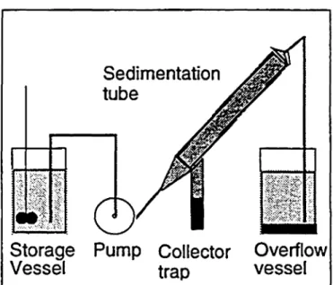
Commercial titanium dioxide powder (1171, Kronos) was suspended in water (50 wt%). 6.52 % tantalum oxide or 2.86 % tungsten oxide (l Mol %) respectively, were added, ball milled, spray dried, and heated to 1300 °C (3 K/min) for 20 min. Deagglomeration was achieved by dry milling for l h. Small Grains (< 3μπι) and large agglomerates were eliminated by a Sedimentation process (Fig. 1).
10 g titanium dioxide powder was mixed with 0.6 g graphite powder (KS 6, Lonza) and 2.8 g paraffin (MP 64 °C, Fluka) into a thermoplastic mixture. A composition with reduced porosity was made by mixing 10 g of undoped powder with 4 g of as-delivered, untreated powder and 1.5 g paraffin. The mixture was heated to 100 °C andpressed into round discs of 15 mm diameter. After a low-temperature firing cycle for polymer bumout at 300 °C the samples were sintered at 1350 °C for 20 min. Finishing was done by ultrasonic cleaning for 15 s and flushed with deionized water. Open porosity was measured by cold water uptake and the material structure was assessed by SEM (Hitachi S 2500 C).
Sedimentation tube s«
$m
*·:··}&&··$ X4;^-<V'At
Storage Pump Collector Overflow Vessel trap vessel
Figure 1: Sedimentation devicefor Separation of grain fractions. The Suspension is pumped into the Sedimentation tube where the coarse fraction settles in the collector trap. Fine material is drawn into the outlet vessel. The grain size of the fractions can be varied by alteration ofthe pump rate.
Cytocompatibility Assays:
Cytocompatibility testing was performed using the osteogenic cell line MC3T3-E1 (Dept. Pathophysiology, Uni. Bern, Switzerland) cultured in oc-MEM medium containing 5% fetal calf serum and antibiotics (all from Gibco) at 37°C and 5% CO2. Test ceramic discs were
individually placed in wells of 24 well tissue culture plates (Nunc), seeded at 5000 cells/well in l ml medium and incubated with medium change every 3 days. Cells in wells without ceramics were used s positive controls. All determinations were carried out in triplicate.
Figure 2: undoped, x5000. Large grains with elongated mor-phology prevai. l Figure 3: Ta2O5-doped, xSOOO. Small grain size and open structure areformed. Figure 4: WOz-doped xSOOO. Grains have rounded morphology.
Proliferation of cells on ceramic samples was evaluated with the MTT test [5]. At intervals, medium from sample wells was replaced with 100 μΐ (5 mg/ml) MTT (Sigma) in phosphate buffered saline (PBS) and incubated for 1.5 h. Reduction of MTT into a colored tetraformazan
product was determined spectroscopically (SLT multiwell reader) at 560 nm after ethanol elution.
Cell viability and distribution on ceramic surfaces were monitored by double fluorescent labeling using a kit (CPAK CVO 01, Millipore) containing calcein-AM (excitation 485 nm, emission 530 nm) and ethidium homodimer (excitation 485 nm, emission 620 nm). At intervals, medium from sample wells was replaced by fluorescent reagents in l ml PBS. Cells were examined after l h by fluorescence microscopy (Zeiss).
RESULTS: Ceramics:
The investigated materials showed open porosities of 57-60 % by volume. Due to the high porosity of these compositions the surface of the samples was sensitive to erosion during handling and testing. This behaviour was especially marked in tantalum oxide-doped material. The compositions with reduced porosity had a pore content of 33.2 %. It was of markedly higher strength. Undoped material has the highest grain size of the investigated mixtures (Fig. 2). Tantalum-doped material is ι characterized by its small crystallite size (Fig. 3). i Crystallite size in tungsten-doped material is smaller man : in undoped material (Fig. 4). Tungsten doping has a i noticeable effect on grain shape: the grains are rounded in |moφhology, whereas elongated grain shapes are more ; frequent in undoped and tantalum-doped materials.
ΙΙΛΓ: οΓΐ-ϊ yu~ /S *V 80 7Λ —/U 60" »· Λ 50 Af\~ 40 on oU"· PO-10" di "·'"·*·;' v :·'· ·' ·;,····.·· .X i;' 'ί \ ί ΐ: >: . .::·., ·.-..·< ···'>·;·· • ^->.:-i'^ ' ::··;· :..* ''': '$?&' •'ΐ,ΐϊγ. ···'·.' ">:·. • . ;·. ' • > · f \ , •·%.\'&·'·~' •.'•i'.v.·':'·«*·^;': ·< ^ .'•fr':*'·· ^.^ W£i '••:?^'^:'v':-y% : .,-ί^.?^; 1
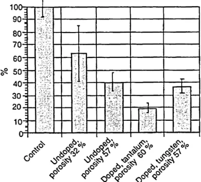
3Φ->Γ'3:>\·/..^ V.: _ -'··'·. ':i/:^· . •^'rf:·;· ..':··' V Λ»;';···;. < W o<T# ^ \% f&Figure 5: Cell viability assessed by MTT lest after 10 d in culture. Results are given s percentages ofcontrol Cell Assays:
The MTT assay indicated that the cytocompatibility of control cells was approached most closely by the titanium dioxide samples with reduced porosity, followed by undoped samples and those doped with tungsten oxide, both showing similar cell growth and viabilities. Tantalum oxide-doped samples demonstrated the lowest cell vitality (Fig. 5). Fluorescent labeling confirmed these results and in addition, showed the cell distribution on the ceramic samples to be patchy and uneven in comparison to controls. Furthermore, cells on tungsten-doped ceramic appeared to be smaller and rounder than those on undoped samples (Figs. 6-8).
DISCUSSION:
The difierence in cell viabilities which was observed between undoped titanium dioxide samples of high and reduced porosities indicates that material structure plays an important role in cytocompatibility. Considering the low strength of the samples and consequent particle release, it is unclear whether the low viabilities measured for tantalum oxide-doped material were due to the chemical composition. This was obvious in the case of tantalum-doped material where the Inhibition of sintering caused by doping lead to low strength. With undoped and tungsten-doped ceramics, it may also be possible that the advantages of high porosities, such s good permeability, might be counteracted by the deleterious effects of wear particle release on cells. This question may be clarifled by setting up a correlation between porosity and biological response. This study indicates that the porosity of the ceramics should be optimized to levels where release of wear particles does not occur during use.
Figure 6: Distribution of viable cells on undoped titanium dioxide ceramic visualized by calcein-AM fluorescent labe-ling. Magn-i catMagn-ion χ 100. Figure 7: Distribution of viable cells on tungsten oxide-doped samples. Magni cation xlOO. Figure 8: Distribution of viable cells on tantalum oxide-doped samples. Magni ca on xlOO. REFERENCES:
1. A. Wisbey et al.; Biomaterials 12, 470-473 (1991) 2. J.E. Ellingsen; Biomaterials 12, 593-596 (1991) 3. K.-L. Eckert et al.; Bioceramics 8, 447-452 (1995) 4. R. Ebert; PhD-thesis University Erlangen-N rnberg,
(1983)
5. T. Mosmann; J. Immunol. Methods 65, 55-63 (1983)