Genet. Res., Camb. (1991), 57, pp. 279-282 With 2 text-figures Printed in Great Britain 279
Muscle opacity (mo), a new mutant gene in Xenopus laevis,
linked to the rusty locus
ANNE DROIN
Station de Zoologie expe'rimentale, Universite de Geneve, 154 route de Malagnou, CH-1224 Chene-Bougeries (Switzerland) (Received 29 August 1990 and in revised form 15 October 1990)
Summary
A new developmental mutant is described in Xenopus laevis (Amphibia, Anura): muscle opacity
{mo). Homozygotes die at larval stage 48. The underlying defect, visible at stage 47, is a
degeneration of cephalic musculature. The mo gene is linked to the rusty locus, with a map distance of 6-1 %.
1. Introduction
Linkage data are still relatively scarce in amphibian genetics. In Urodela, numerous mutant genes have been discovered in the axolotl (Armstrong, 1985); several of them have been tested for linkage but only the genes / (fluid imbalance) and g (gill lethal) have been confirmed to be linked (Humphrey, 1959); for others, linkage was suggested but not confirmed, or independent assortment was shown (Armstrong, 1984). In Pleurodeles waltl, a peptidase enzyme locus and two genes affecting larval and postmetamorphic skin pigmentation are linked to the sex chromosomes (Collenot et al. 1989).
In Anura, the first case of linkage was presented by Browder (1972) in Rana pipiens: a dominant Subvital gene (Sbv) is linked to the Burnsi locus. Since then, eight linkage groups, mainly of enzyme loci, have been established in this species and several others have been identified in other species (Wright & Richards, 1987). In Xenopus laevis linkage between mutant genes affecting development has not been systematically searched for, most of the genes being recessive and lethal. However, genes have been mapped to the centromere by means of diploid gynogenesis. Within the last ten years, the localization of several genes has been established (Kobel, 1981; Colombelli et al. 1984; Thiebaud et al. 1984; Reinschmidt et al. 1985). Moreover, with the use of isozyme markers and RFLP (restriction fragment length polymorphism), linkage groups of enzyme loci have also been mapped in this species (Graf, 1989 a).
We present here a new recessive lethal mutant gene,
muscle opacity (mo), which is linked to the
pig-mentation gene rusty (ry) (Uehlinger & Droin, 1969). The main characteristic of the mo syndrome consists
of a degenerative process affecting the muscles of the head and finally leading to the death of the tadpoles. In addition to a description of the mo phenotype, linkage data are reported.
2. Material and Methods
Laboratory-bred as well as imported adult Xenopus
laevis laevis were used in the crosses. Egg laying,
fertilization, and rearing of tadpoles were according to standard methods used in our laboratory (Droin & Chavane, 1976). Developmental stages (st.) are num-bered according to Nieuwkoop and Faber (1956). For histolpgical analysis, tadpoles were fixed in Zenker's or Bouin's fluids, serially cut at 6 /im and stained with haemalum-eosin.
3. Results and Discussion
(i) Phenotype
Mutant embryos and tadpoles develop normally up to the 9th day (st. 47). At this stage, mutant tadpoles can be distinguished from normal sibs by a characteristic opacity of the ventral muscles of the head. This anomaly seems more conspicuous in tadpoles, homo-zygous for the rusty mutant gene; presumably this is an effect of the reddish tinge of their skin, apt to improve visibility under incident light. The tinge is due to the persistence of egg pigment into larval stage. No other abnormality has been found in rusty (Uehlinger et al. 1971).
Muscle opacity in mo affects particularly the large interhyoideus muscle extending transversely through-out the ventral part of the head; it is more pronounced in the anterior part of the muscle. Two other muscles
GRH 57
https:/www.cambridge.org/core/terms. https://doi.org/10.1017/S0016672300029438
Anne Droin 280
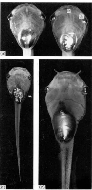
Fig. 1. (a) Ventral view of a mo ry mutant (left) and of a normal ry tadpole (right) aged 10 days (st. 48). The opacity of the rostral part of the interhyoideus muscle (ih) and of the orbito-hyoideus muscles (oh) of the mo tadpole is clearly visible, x 10-5. (b) A mo tadpole aged 20 days arrested at st. 48. (c) A normal tadpole of the same age and at the same magnification. Note the small size of the mutant and the marbled aspect of the iridophore layer of the belly (arrow), x 7-2.
of the hyoid arch are also opaque, the thick lateral symmetrical muscles situated ventrally to the eyes, m. orbito-hyoideus or levator hyoideus (Fig. 1 a). In more advanced stages, opacity can affect three other muscles, namely the two other symmetric ones of the hyoid arch, m. quadrato-hyoangularis or depressor mandibulae and the small transverse muscle situated rostrally to the heart, m. transversus ventralis. Sections through these muscles reveal that the opaqueness is due to degeneration of the muscles. The periphery of their bundles is disorganized, with loose cytoplasm and blood infiltration, becoming more pronounced as time proceeds (Fig. 2). This deterioration affects also the fibres themselves which lose their striation. By contrast, the entire musculature of the rump and tail
Fig. 2. Longitudinal sections of the orbito-hyoideus muscles of two mutants. At 11 days (a), the bundle structure is still very similar to that of a normal muscle, while at 23 days (b), blood infiltration (arrow) and progressive degeneration of the fibres are observed, x 320.
regions seems hardly affected, and heart muscle is normal.
Evolution of the syndrome is slow; tadpoles feed normally. At fifteen days, other abnormalities typical of an arrest of growth are observed, probably due to the fact that the muscles are no longer functional and prevent the tadpoles from feeding. When normal tadpoles have reached st. 49, the mutants are blocked at st. 48; their head and body are smaller and narrower, the gut slender with a marbled aspect of the iridophore layer (Fig. 1 b, c), and the thymus and limb buds less developed. The heart, blood vessels and pronephros become red due to a transient accumu-lation of blood. In the following days, the phenotype presents the typical aspect of general degeneration, the tadpoles become blind, the gut very thin with a protruding gall bladder and distended pro- and mesonephros tubuli. The mutant tadpoles more and more resemble the various lethal dwarf mutants (Droin, 1974, 1988). They begin to die around the 20th day.
This mutation is the third one found in our laboratory involved with the development of muscles of young tadpoles of Xenopus. The mutant phenotype
folded jaw was found to be due to a morphological
deficiency of the m. quadrato-hyoangularis, which is not long enough to be inserted on the cartilage of the mandibular arch (Droin et al. 1968). In the immobile mutant, a defect in the function of the muscles prevents them from contracting (Droin & Beau-chemin, 1975). In the mo mutant, the muscles are well developed and functioning, but then degenerate. Such
https:/www.cambridge.org/core/terms. https://doi.org/10.1017/S0016672300029438
Linkage in Xenopus laevis 281
ry mo ry mo
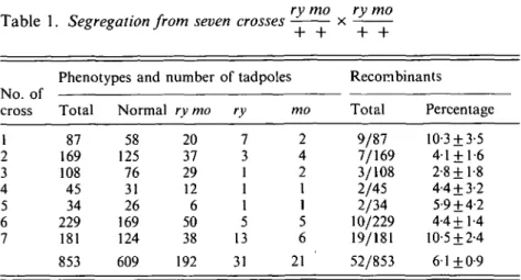
Table 1. Segregation from seven crosses x
Phenotypes and number of tadpoles Recombinants No. of
cross Total Normal ry mo ry mo Total Percentage
1 2 3 4 5 6 7 87 169 108 45 34 229 181 853 58 125 76 31 26 169 124 609 20 37 29 12 6 50 38 192 7 3 1 1 1 5 13 31 2 4 2 1 1 5 6 21 9/87 7/169 3/108 2/45 2/34 10/229 19/181 52/853 10-3 + 3-5 4-1 + 1-6 2-8+1-8 4-4 + 3-2 5-9 + 4-2 4-4+1-4 10-5 ±2-4 6-1+0-9
mutations illustrate the complex mechanisms of gene action intervening at different levels of muscle differ-entiation. It should be possible, by chimaeric combina-tions, to determine whether mo selectively acts upon head musculature in particular, or whether postcranial musculature appears unaffected only because of differential maturation along the body axis.
(ii) Genetics
The rusty mutation, which is inherited as a recessive mendelian factor with a constant expression, was found in the offspring of two families, one parent of which came from a nuclear transplantation egg. Its origin was not determined (Uehlinger & Droin, 1969). Since then the rusty mutation, which can be useful as a marker in embryological experimentation, has been maintained through several generations in hetero- and homozygous stocks. The mo mutation was found in the F2 of a family created by a mating between a homozygous rusty female (? 30) and a <$ (S 34) descended also from an adult <$ issued from a nuclear transfer. As the mutant tadpoles were identified only after the death of their parents and grandparents, backcrosses were impossible; the origin of this mutation cannot be determined either.
When mutant tadpoles of the first F2 mating were observed (obtained from a cross between two Fx individuals), it was obvious that nearly all the rusty tadpoles presented the abnormal phenotype while only a few non-rusty tadpoles showed the same abnormalities. The data of the different matings are summarized in Table 1. They show that segregation deviates from independence in a two-factor cross (9:3:3:1), thus demonstrating linkage between mo and rusty. In these matings between heterozygotes for the two mutations (ry/+ and mo/ + ), in addition to the double-recessive mutants (192/853) obtained, there was a small number of either rusty or mo single-recessive mutants. The number of recombinants - 52 tadpoles out of 853 examined in 7 different matings -corresponds to 6 1 % of recombination. From these data we conclude that the two mutations were linked in coupling phase.
Since the terminal phenotype of the mo mutant is similar to that of various dwarfmutants, matings were performed between individuals, heterozygous for mo and the four dwarf genes of our mutant collection. No mutant segregant was found, showing that mo and the different dwarf mutations are not allelic.
Recently, two developmental mutations have been attributed to different linkage groups, periodic al-binism (ap) to group I (Graf, 1989 a) and polydactyly (pd) to group HI (Graf, 1989 ft). Analysis by
inter-subspecies hybridization involving isozyme markers might allow us to assign mo and ry to one of the . known linkage groups.
I am grateful to Professor H. Gloor for critically reading the manuscript. I thank Mrs M. Quentin for technical as-sistance, MM. A. Solaro and A. Portianucha for the photo-graphs and Mrs Y. Develey for typing assistance. This work was supported by the University of Geneva.
References
Armstrong, J. B. (1984). Genetic mapping in the Mexican axolotl, Ambystoma mexicanum. Canadian Journal of
Genetics and Cytology 26, 1-6.
Armstrong, J. B. (1985). The axolotl mutants. Developmental
Genetics 6, 1-25.
Browder, L. W. (1972). A subvital gene in Rana pipiens linked to the Burnsi locus. Genetical Research 20,263-268. Collenot, A., Dournon, C. & Lauthier, M. (1989). Genetic evidence for linkage with the Z and W sex chromosomes of two distinct couples of alleles controlling larval and post-metamorphic skin pigmentation in salamander.
Biology of the Cell 67, 1-7.
Colombelli, B., Thiebaud, Ch. H. & Muller, W. P. (1984). Production of WW super females by diploid gynogenesis in Xenopus laevis. Molecular and General Genetics 194, 57-59.
Droin, A. (1974). Trois mutations recessives letales, dwarf I (dw-l), dwarf-ll (dw-ll) et precocious oedema (p.oe) affectant les tetards de Xenopus laevis. Annales
dEmbry-ologie et de Morphogenese 7, 141-150.
Droin, A. (1988). Three new analogous mutations in
Xenopus laevis. Ada Embryologiae et Morphologiae experimentalis. (New series) 9, 115-127.
Droin, A. & Beauchemin, M. L. (1975). Immobile (im), a recessive lethal mutation of Xenopus laevis tadpoles.
Journal of Embryology and Experimental Morphology 34,
435-449.
https:/www.cambridge.org/core/terms. https://doi.org/10.1017/S0016672300029438
Anne Droin 282 Droin, A. & Chavane, M. C. (1976). A recessive semi-lethal
mutation, distended lungs (dl) affecting the tadpoles of
Xenopus laevis. Ada Embryologiae Experimentalis 3,
273-279.
Droin, A., Reynaud, J. & Uehlinger, V. (1968). Folded jaw
(fj), une mutation letale recessive affectant le
developpe-ment de la machoire chez Xenopus laevis. Revue Suisse de
Zoologie 75, 531-538.
Graf, J. D. (1989a). Genetic mapping in Xenopus laevis: eight linkage groups established. Genetics 123, 389-398. Graf, J. D. (1989a). Genetic mapping of a mutation causing
polydactyly (pd) in Xenopus laevis. Experientia 45, A64
(Abstract).
Humphrey, R. R. (1959). A linked gene determining the lethality usually accompanying a hereditary fluid im-balance in the Mexican axolotl. The Journal of Heredity 50, 279-286.
Kobel, H. R. (1981). Gene mapping in Xenopus (Anura, Pipidae). Monitore zoologico italiano 17, 109-118. Nieuwkoop, P. D. & Faber, J. (1956). Normal Table of
Xenopus laevis (Daudin) Amsterdam: North-Holland.
Reinschmidt, D., Friedman, J., Hauth, J., Ratner, E., Cohen, M., Miller, M., Krotoski, D. & Tompkins, R. (1985). Gene-centromere mapping in Xenopus laevis. The
Journal of Heredity 76, 345-347.
Thiebaud, Ch. H., Colombelli, B. & Miiller, W. P. (1984). Diploid gynogenesis in Xenopus laevis and the localization with respect to the centromere of the gene for periodic albinism ap. Journal of Embryology and Experimental
Morphology 83, 33-42.
Uehlinger, V., Beauchemin, M.-L. & Droin, A. (1971). The behaviour of the egg pigment in wild-type and rusty tadpoles of Xenopus laevis. Journal of Embryology and
Experimental Morphology 26, 571—585.
Uehlinger, V. & Droin, A. (1969). Origine et heredite de la mutation rusty (ry) du batracien anoure Xenopus laevis (Daudin). Archiv der Julius Klaus-Stiftung 43/44, 48-54. Wright, D. A. & Richards, C. M. (1987). Linkage groups in
the leopard frog, Rana pipiens. In Genetic Maps (ed. S. J. O'Brien), vol. 4, pp. 417-421. Cold Spring Harbor Laboratory.
https:/www.cambridge.org/core/terms. https://doi.org/10.1017/S0016672300029438