The Tyr (albino) locus of the laboratory mouse
Friedrich Beermann,
1Seth J. Orlow,
2M. Lynn Lamoreux
31ISREC (Swiss Institute for Experimental Cancer Research), National Center of Competence in Research (NCCR)
Molecular Oncology, Chemin des Boveresses 155, 1066 Epalinges, Switzerland
2The Ronald O. Perelman Department of Dermatology & The Department of Cell Biology, New York University School of Medicine,
New York, New York 10016, USA
38255 Sandy Point Road, Bryan, Texas 77807, USA
Received: 26 July 2002 / Accepted: 24 May 2004
Abstract
The albino mouse was already known in ancient times and was apparently selectively bred in Egypt, China, and Japan. Thus, it is not surprising that the c or albino locus (now the Tyr locus) was among the first used to demonstrate Mendelian inheritance in mammals at the dawn of the past century. This locus is now known to encode tyrosinase, the rate-limiting enzyme in the production of melanin pig-ment, and the molecular basis of the albino (Tyrc) mutation is known. Here we describe the congenic series of Tyr-locus alleles, from wild type to null (albino). We compare eye and skin pigmentation phenotypes and the genetic lesions that cause each. We suggest that this panel of congenic mutants contains rich, untapped resources for the study of many questions of basic cell biological interest.
The albino mouse was already known in ancient times and, over a century ago, was used to first demonstrate Mendelian inheritance of a genetic trait in mammals (Castle and Allen 1903). Very early on it was suggested that the albino gene locus was responsible for a ‘‘factor’’ that is necessary for mel-anin pigment to form in the melanocytes. This ‘‘factor’’ has been identified as tyrosinase, the rate-limiting enzyme for melanogenesis (Coleman 1982). Tyrosinase is encoded at the albino (Tyr) locus of the mouse on Chromosome 7 (Kwon et al. 1989b), where multiple natural mutations and manmade muta-tions (Fig. 1) have helped to define the funcmuta-tions and
interactions of this enzyme with other proteins that together effect normal pigmentation. In human, the defect in tyrosinase is called oculocutaneous albi-nism type 1 (OCA1) and is often specified as OCA1A or OCA1B to distinguish between no pigment or less pigment, respectively. The genetic defect in the tyrosinase gene affects the quantity of pigment pro-duced within the melanosome; melanin is absent or reduced, but melanocytes are present in the skin and hair follicles and they contain melanosomes. In addition to effects on pigmentation, albino mice have defects in the visual projections at the optic chiasm (Jeffery et al. 1994), decreased numbers of rod photoreceptors (Donatien and Jeffery 2002; Rachel et al. 2002a), and spatiotemporal defects in neuronal production (Rachel et al. 2002a). Furthermore, a role for tyrosinase in the occurrence of glaucoma by a mechanism apparently unlinked to melanin pro-duction has recently been suggested (Libby et al. 2003).
Melanin pigment is produced primarily in two different cell types, the neural crest-derived mela-nocytes found in skin, hair follicle, and the choroid, ciliary body, and iris of the eye, and the retinal pig-ment epithelium, a cell layer of the retina that is derived from the optic cup. In the pigment cells, melanin is synthesized and deposited within endo-lysosome-like organelles, termed ‘‘melanosomes,’’ by a series of enzymatic reactions beginning with tyrosine as substrate, and involving the copper–gly-coenzyme tyrosinase (Garcia–Borron and Solano 2002). The melanin product is deposited within the melanosome as eumelanic (brown or black) or pheomelanic (yellow/red) pigment. It had long been believed that the first two reactions in the melano-genic pathway—the hydroxylation of tyrosine to dopa (3,4-dihydroxyphenylalanine) and the oxidation of dopa to dopaquinone—are catalyzed by the en-zyme tyrosinase (del Marmol and Beermann 1996).
Correspondence to: Friedrich Beermann; E-mail: [email protected]
More recent chemical analyses have suggested, however, that tyrosinase-catalyzed oxidation of tyrosine leads directly to dopaquinone, which then can lead to eumelanin formation via spontaneous formation of dopachrome (Wakamatsu and Ito 2002). Accordingly, dopa itself can act as a cofactor in tyrosine oxidation and is not derived by tyrosinase enzyme activity, but indirectly by reduction of dop-aquinone (Riley 1999). The pheomelanic pathway is thought to be initiated by a reaction between cyste-ine and dopaquinone, thus leading to cysteinyldopa, and further to benzothiazoles. Accordingly, the bal-ance between pheomelanin and eumelanin may be determined by the availability of cysteine as pre-cursor (Land and Riley 2000).
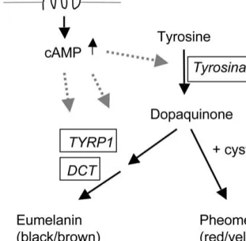
Besides tyrosinase, two other enzymes function in melanogenesis, dopachrome tautomerase (DCT) and tyrosinase-related protein 1 (TYRP1). DCT is encoded at the slaty (now Dct) locus of the mouse, and TYRP1 is encoded at the black/brown (now Tyrp1) locus. When all three of these enzymes function normally, eumelanin pigment is deposited within the melanosome (Fig. 2). Analysis of mice that are mutant at the Tyr or Tyrp1 locus has shown that mutation in one may affect the phenotype associated with the other. Accordingly, TYRP1 may affect stability of tyrosinase (Manga et al. 2000), and both proteins are transported together from the endoplasmic reticulum to the melanosome
(Toyof-uku et al. 2001). Mice lacking Tyrp1 [Tyrp1 deletions (Rinchik et al. 1994)] or Dct [Dct knockout (Gu-yonneau et al. 2004)] are only slightly affected in pigmentation and show a rather brown (Tyrp1 defi-ciency) or dark gray (Dct defidefi-ciency) coat color. Several other gene loci function within the mela-nosome and are necessary for normal pigmentation but have not been shown to be so intimately inter-active with tyrosinase protein (Bennett and Lamo-reux 2003). These loci include pink-eyed dilution (p, mutation causing OCA2 in human) (Chiu et al. 1993), underwhite (uw, now Matp, mutation causing OCA4 in human) (Newton et al. 2001; Costin et al. 2003), and MITF (Mitf) that modulates the expres-sion of a number of melanocyte-specific genes, including Tyr and Tyrp1, at the transcriptional level and influences the eumelanin/pheomelanin switch (Goding 2000; Widlund and Fisher 2003).
Eumelanin (which is black or brown depending upon the genotype at the Tyrp1 locus) is produced in the melanosome as a result of the normal activity of the MSH (melanocyte stimulating hormone) recep-tor, which regulates levels of cyclic AMP (cAMP) within the cell and is present in melanocyte cell membranes (Fig. 2). The MSH receptor (MC1R) is encoded at the melanocortin-1 receptor (Mc1r) locus [formerly extension (e) locus] in the mouse and is
Fig. 1. The mouse tyrosinase gene and location of identi-fied mutations. (a) The Tyr gene locus, showing relative size of introns (Ruppert et al. 1988) and location of the enhancer/dominant control region at )15 kb (black arrowhead). The transcription start site is indicated as +1. (b) Exon/intron structure of the tyrosinase gene and loca-tion of mutaloca-tions which were identified within coding sequence. Note that Tyrc-em is listed twice since it ap-peared on a Tyrc-mbackground.
Fig. 2. Scheme of melanin synthesis. Activation of mela-nocortin receptor 1 (MC1R) increases cAMP levels, thus enhancing tyrosinase activity and favoring eumelanin synthesis (del Marmol and Beermann 1996; Barsh 2003). Tyrosinase-related protein 1 (TYRP1) and dopachrome tautomerase (DCT) are enzymes of eumelanin synthesis only.
responsive to the environment of the hair follicle in which the melanocyte resides. It is thus capable of switching from an active state that raises cAMP levels and results in the production of eumelanin within the melanosomes of the cell to an inactive state, when pheomelanin is produced (Barsh 2003). In the eumelanic state, elevated cAMP levels are followed by an increase in activity of tyrosinase, DCT, and TYRP1. In the pheomelanic state, the melanosomes produce yellow-colored melanin, cAMP levels are reduced, tyrosinase activity is lower, and DCT and TYRP1 activities are absent (Lamoreux et al. 1995). Mutation at Mc1r can result in melanocortin receptors that are not responsive to the environment and are constitutively active, resulting in production of only eumelanin, as in the sombre(Mc1rE-so) mutant, or constitutively inactive, resulting primarily in the production of pheomela-nin as in the hair follicles of the yellow mutant (Mc1re) mouse. Tyrosinase is required for both types of pigment, but activity is reduced in pheomelanic melanocytes. Wild-type MC1R is active unless blocked by the protein encoded at the agouti locus, thus switching pigment synthesis from the eumela-nin pathway toward the pheomelaeumela-nin pathway (Barsh 2003). Thus, mice that are yellow because of mutation at the agouti locus continue to produce the agouti-locus protein inappropriately and do not switch back and forth from the production of eu-melanin to the production of pheoeu-melanin as is normal in wild-type agouti mice. In addition, several other pigment loci encode proteins that interact with the Mc1r/agouti protein switch mechanism. These include mahogany (Atrn) and mahoganoid, both of which result in a reduction of pheomelanin, or rather an increase in eumelanin, in the hair coats of mutant mice (He et al. 2003). Interestingly, the Tyr-locus mutants (except for platinum) preferen-tially reduce the amount of pheomelanin compared with the reduction in eumelanin. Hence, the impact of Tyr-locus mutations is greater in pheomelanic mice than in eumelanic mice (Lamoreux and Pend-ergast 1987; Lamoreux et al. 2001) and also in pheomelanic locations on a mouse as for example the belly.
Availability of multiple mouse Tyr (albino) locus alleles with various sorts of genetic lesions provides an opportunity to evaluate the dynamic interactions in the processes that intervene between the tran-scription of the tyrosinase gene and the resulting phenotype of the animal. These include transcrip-tion, translatranscrip-tion, post-translational processing, and transport mechanisms, as well as interactions with the products of other loci. Moreover, many other mutations causing albinism act via tyrosinase by
affecting tyrosinase processing [e.g., pink-eyed dilu-tion (Chen et al. 2002)] or tyrosinase trafficking [e.g., forms of Hermansky–Pudlak syndrome (Huizing et al. 2002)]. In this review, we report on the current state of knowledge regarding the molecular bases and phenotypic consequences of mutations at the mouse Tyr (albino) gene locus (Fig. 3, Table 1).
The allelic series
In the absence of mutations at other loci, mice that are wild type at the Tyr locus are fully pigmented and are black (Tyr+/Tyr+, a/a). Wild type is dominant to all other alleles at the locus, though one semidomi-nant mutant (albino-strong, Tyrc-s) is reported at the JAX web site (http://www.informatics.jax.org/sear-ches/mlc.cgi?14347). Mice that are lacking the Tyr locus as a result of overlapping deletions (Tyrc-6H/
Tyrc-14CoS) are unpigmented, although melanosomes
are present, confirming the requirement of the Tyr locus and a functional tyrosinase for pigment pro-duction (Russell et al. 1982; Rinchik et al. 1993). Similarly, two natural mutations have been identi-fied that result in an albino phenotype associated with lack of tyrosinase activity, Tyrcand Tyrc-2J. The classic mouse albino mutation, Tyrc, which is pres-ent in common albino mouse strains such as BALB/c or FVB, is characterized by the complete absence of pigmentation in both skin and eyes and by aberrant decussation of the optic nerve at the level of the chiasm (Guillery 1974; LaVail et al. 1978). Even though tissue homogenates of BALB/c mice might retain a slight amount of tyrosinase-dependent mel-anin synthesis in vitro (Hearing 1973), Tyrc is
con-sidered a null mutation since the mutated protein is not active in vivo and is retained in the ER (Halaban et al. 2000; Toyofuku et al. 2001). The molecular change in Tyrcis a G-to-C mutation at position +387, which results in the substitution of a cysteine for a serine residue at position 103 (Kwon et al. 1989b; Shibahara et al. 1990; Yokoyama et al. 1990). The Tyrc-2J mutation arose spontaneously in C57BL/6J mice, and homozygous Tyrc-2J/Tyrc-2J mice are phe-notypically identical to Tyrc/Tyrc mice. The muta-tion was identified as a G-to-T change at posimuta-tion +309, resulting in an arginine-to-leucine substitution at codon 77 and furthermore led to increased alter-native splicing within exon 1 (Lefur et al. 1996, 1997). Enzymatic activity of tyrosinase is absent in normal melanocytes of these mice, but melanomas occurring on this Tyrc-2Jgenetic background can be pigmented and are tyrosinase positive (Cohen–Solal et al. 2002), in contrast to melanomas appearing on BALB/c or FVB mice (Tyrc) (Cohen–Solal et al. 2001). Thus, it is
conceivable that Tyrc-2Jis not a true null mutation
and is able to produce an unstable but enzymatically active protein in melanoma cells.
Tyr locus alleles that fall between the two ex-tremes of black and albino can be classified into several groups by phenotype. First, there are alleles affecting ocular and cutaneous pigmentation simi-larly. The chinchilla allele was procured in 1922 by Feldman from a fancier (Feldman 1922). C57BL/6J– Tyrc-ch/Tyrc-ch (chinchilla) mice are phenotypically very similar to mice that are wild type at the Tyr locus, with black eyes and very dark gray, almost black, hair coat, though the tyrosinase activities of their skin or eyes is approximately one third that of wild-type mice. Moyer (1966) reported that mela-nosomes look normal in size and number, at least in the retina. In eumelanic brown (Tyrp1b) mice, the
effect of chinchilla is not evident, in either the intensity of pigmentation or reduced tyrosinase activity or change of melanosome structure (Russell 1948; Gru¨neberg 1952; Lamoreux et al. 2001). Interestingly, in pheomelanic chinchilla (Tyrc-ch/ Tyrc-ch) mice, pigmentation is much reduced com-pared with that of pheomelanic mice that are wild type at the Tyr locus. This dichotomy is typical of the phenotypes of most of the Tyr-locus mutations
(Silvers 1979; Lamoreux and Pendergast 1987), with the exception of platinum. Pheomelanic chinchilla melanocytes exhibit a greatly reduced number of melanosomes compared with normal pheomelanic melanocytes. Northern blot data and RT-PCR failed to reveal any difference in expression between Tyr
c-ch and wild type (Halaban et al. 1988; Ganss et al.
1994), and it was suggested that chinchilla tyrosi-nase enzyme is less stable than the wild-type tyrosinase enzyme (Halaban et al. 1988). Sequence analysis of the entire coding region revealed a G-to-A point mutation at nucleotide +1523, resulting in an amino acid substitution of alanine to threonine at position +482, close to the transmembrane region (Beermann et al. 1990).
Platinumoccurred as a spontaneous mutation in DBA/2 (Dickie 1966). Homozygous platinum (C57BL/6J–Tyrc-p/Tyrc-p) mice are very pale with pink eyes, yet their tyrosinase activity is higher than that of chinchilla mice (Tyrc-ch/Tyrc-ch). In skin
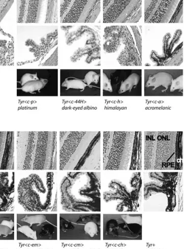
ex-Fig. 3. Phenotype of alleles at the Tyr gene locus. Shown are (top row) sections of the retina of mice mutant at the Tyr locus, (middle row) sections through the uvea and ciliary body, and (bottom row) pictures of the mutant mice (RPE, retinal pigment epithelium; ch, choroid; ONL, outer nu-clear layer; INL, inner nunu-clear layer). First and last columns contain no mouse photograph because albino (Tyrc-2J/ Tyr c-2J) and wild-type (C57BL/6J–Tyr+/Tyr+) mice are used as negative and positive controls in several other columns. Each column is labeled at the bottom with the name of the mutant illustrated as follows: Albino, C57BL/6J–Tyrc-2J/ Tyrc-2J. Platinum, C57BL/6J–Tyrc-p/Tyrc-p: mouse in the foreground is platinum, just slightly more pigmented than the albino mutant mouse in the background. Dark-eyed albinoC57BL/6J–Tyrc-44H/Tyrc-44H. Himalayan, C57BL/6J– Tyrc-h/Tyrc-h: the himalayan mutant mouse, on the right, is more darkly pigmented on the extremities than in the warmer areas of its body. Control mice in this picture are albinoon the left and C57BL/6J in the center. Acromelanic, C57BL/6J–Tyrc-a/Tyrc-a: the acromelanic mutant mouse on the left is somewhat darker in body pigmentation than the himalayanmutant mouse (previous column) but shares the characteristically darker extremities. Extreme dilution, C57BL/6J–Tyrc-e/Tyrc-e: the extreme dilution mutant mouse, in the foreground, is compared with wild type and albinocontrols. Pigmentation in hair coat of this mutant is uniformly distributed. Extreme dilution mottled, C57BL/ 6J–Tyrc-em/Tyrc-em: in the foreground are the control mice, albino(Tyrc-2J), and wild type at the Tyr locus. The mouse in the background has intermingled areas of beige, and paler and darker hair. Chinchilla mottled, C57BL/6J– Tyrc-m/Tyrc-m: the control mice are on the left and in the foreground. The chinchilla mottled mutant mouse has intermingled areas of dark gray and very dark gray hair. Chinchilla, C57BL/6J–Tyrc-ch/Tyrc-ch: the bright area on the chinchillamutant mouse, on the left, is an artifact of the reflected light. In fact, chinchilla mutant mice that are black are superficially difficult to distinguish from wild-type black mice. Wild wild-type, C57BL/6J–Tyr+/Tyr+.
tracts of platinum mice, a large proportion of tyros-inase is present in soluble form (Townsend et al. 1981). Furthermore, phenotypic differences in intensity of pigmentation between pheomelanic and eumelanic platinum mice are not evident, and both appear equally pale. These differences between platinum and chinchilla mice suggested that tyros-inase is functional in platinum mice, as the tyrosi-nase activities of skin and eyes are higher than those of chinchilla mice, which have lower tyrosinase activity but much more intense pigmentation. In addition, the effect of pheomelanogenesis on tyrosi-nase activity is sidestepped in the case of platinum mice. Analysis of the tyrosinase protein suggested a mutation at the carboxy terminal part of the protein (Orlow et al. 1993), and electron microscope studies demonstrated tyrosinase activity in the trans-Golgi network and in nearby vesicles, but missing activity in melanosomes (Beermann et al. 1995). Instead, tyrosinase was found at the cell surface. Since melanogenesis is confined essentially to the mela-nosome, it is thus reasonable not to detect major differences between pheo- and eumelanogenesis in this specific mutant allele. The mutation in plati-num is a G-to-A change at +1523 (Beermann et al. 1995), inferring a replacement of a lysine residue in the cytoplasmic tail by a termination codon. The lack of the cytoplasmic tail, which contains the essential di-leucine sorting motif, is causing misro-uting of platinum tyrosinase to the cell surface (Beermann et al. 1995; Simmen et al. 1999).
Second, there are alleles with more effect on coat pigmentation, e.g., extreme dilution (Tyrc-e). The
original Tyrc-e-mutation was found in the wild (Det-lefsen 1921). C57BL/6J mice that are homozygous at
this locus can be characterized as ‘‘midgray’’ in phe-notype, more or less midway in intensity between wild type and albino but with eyes that are nearer to black. Both tyrosinase activity and amount of eu-melanin are greatly reduced in C57BL/6J–Tyrc-e/Tyr
c-emice; melanosomes of Tyrc-e/Tyrc-eare reduced in
both number and size (Markert and Silvers 1956; Moyer 1966). Northern blot analysis of newborns’ skins (not shown) showed no reduction in abundance of tyrosinase mRNA in Tyrc-e/Tyrc-e mice that are eumelanic. Sequencing the complete coding region demonstrated a deviation from wild type in exon 5, leading to exchange of an alanine by a threonine, the very same mutation (A482T) that had been identified in the chinchilla (Tyrc-ch) mutation (Beermann et al.
1990). A482T is the only mutation found in the coding sequence of the chinchilla (Tyrc-ch) tyrosinase
gene; it affects tyrosinase in vivo (Halaban et al. 1988) and following transfections (unpublished data). Therefore, it is rather unlikely that A482T is a polymorphism, with two unidentifed mutations still existing for both chinchilla and extreme dilution. It is more likely that the Tyrc-e mutation may have occurred on a Tyrc-chbackground and might contain a yet unidentified second mutation, for example, in the regulatory region.
Himalayan is a spontaneous mutation which occurred in offspring of a cross between DBA/2 and AKR/J (Green 1961). Mice homozygous for the himalayan mutation (Tyrc-h), similar to himalayan
cats or rabbits, over time develop more intense pigmentation at the extremities where the body is cooler. Their body color is beige, with darker-beige extremities, and eyes are dark ruby. The mutation is an A-to-G change at nucleotide 1338 that alters
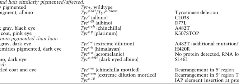
Table 1. Phenotype and molecular lesion of selected Tyr locus alleles
Pigment phenotype Allele Molecular lesion
Eyes and hair similarly pigmented/affected:
Fully pigmented Tyr+, wildtype
No pigment, albino Tyrc-6H/Tyrc-14cos Tyrosinase deletion
Tyrc(albino) C103S
Tyrc-2J(albino) R77L
Dark gray, black eye Tyrc-ch(chinchilla) A482T
Pale coat, pink eye Tyrc-p(platinum) K507STOP
Eyes more pigmented than hair:
Mid gray, dark eye Tyrc-e(extreme dilution) A482T (additional mutation?)
Extremities pigmented, dark eye Tyrc-h(himalayan) H420R
Tyrc-a(acromelanic) No protein detected, RNA low
Albino, dark eye Tyrc-44H(dark eyed albino) S146I
Mottled:
Mottled coat and eye Tyrc-m(chinchilla mottled) Rearrangement in 5¢ region
Tyrc-em(extreme dilution mottled) Rearrangement in 5¢ region T373I
Tyrc-1R IAP element insertion at promoter
See text for references and further details. Numbering of nucleotides and amino acids varied in the past, depending upon the definition of the start site and upon whether the signal peptide was included. In this article we refer to the published transcriptional start site (+1) (Bentley et al. 1994). For the amino acid sequence, the start codon (ATG) is counted as +1.
a histidine residue to an arginine residue at amino acid 420 (Kwon et al. 1989a). The activity of tyrosinase isolated from skins of Tyrc-h/Tyrc-h mice is heat labile (Coleman 1962), but the protein itself has been reported not to be heat-sensitive (Town-send et al. 1985), and it has been stated that the himalayan tyrosinase binds an inhibitor differen-tially at different temperatures (Kidson and Fabian 1981). A similar mutation in human (Giebel et al. 1991), which is located only two codons away, re-sults in a thermosensitive protein upon transfec-tion into HeLa cells. Thus, the himalayan (Tyrc-h/
Tyrc-h) mouse would seem to be an excellent model
for the condition in man and deserves further study to understand the cause of this thermosen-sitivity.
Acromelanic (Tyrc-a) is a spontaneous mutation which occurred on the C3H/HeJ strain (Sweet 1987). Acromelanic mice are beige in coat color (similar to himalayan) but have dark eyes and pig-ment appearing on tail, ears, and extremities. We sequenced the complete coding sequence, including about 270 bp upstream of the transcription start site, and no change to the wild-type tyrosinase se-quence was detected. This result is in accordance with failure to detect protein and message on Western blots (not shown) and Northern blots (not shown). The message was nevertheless detectable by RT-PCR (not shown), thus suggesting a defect in transcriptional regulation or RNA stability. No major rearrangements were detected by Southern blot analysis covering about 15 kb of tyrosinase 5¢ sequence (not shown). Since the mutation affects RNA levels, three unsequenced areas remain to be tested for the presence of mutation: (1) the en-hancer region (Ganss et al. 1994; Porter and Meyer 1994), (2) the 3¢ noncoding sequences which might be involved in tyrosinase regulation and mRNA stability (Takeuchi et al. 2000), and (3) the exon/ intron boundaries, which might affect the correct splicing (Ruppert et al. 1988; Lefur et al. 1997). A defect in the enhancer region might explain the different effect in skin melanocytes versus RPE pigmentation by affecting regulation preferentially in either cell type (Porter et al. 1999; Camacho– Hu¨bner and Beermann 2001). On the other hand, the choroidal layer, which equally consists of neu-ral crest-derived melanocytes, is pigmented in ac-romelanic mice. Thus, it might rather be the steady accumulation of low levels of melanin within the RPE (and the choroidal layer) that makes the eye pigmented but keeps the skin and hair rather un-pigmented. In addition, the presence of pigment at the extremities might point to a certain tempera-ture-sensitive effect. How this is explained without
an obvious mutation in the cDNA remains to be discovered.
In homozygous dark-eyed albino (Tyrc-44H) mice, overall pigment production is greatly reduced and is obvious only in the eyes (Cattanach and Rasberry 1988). Dark-eyed-albino mice are born white with ruby-colored eyes, which darken to become almost black by 3–4 months of age. The hair coat, by con-trast, remains essentially unpigmented. Enzymatic activity of tyrosinase and melanin levels in the ret-ina of Tyrc-44H/Tyrc-44Hnewborn mice reached levels
of only 2.6% (tyrosinase activity) and 11.8% (mela-nin) of wild type (Rachel et al. 2002b). By Southern blot, Northern blot, and RT-PCR analyses, it was demonstrated that the basis of the phenotype resides in the coding sequences, with a point mutation (G-to-T) in exon 1, at position +515, inferring a substi-tution of the amino acid serine by isoleucine (posi-tion +146) (Schmidt and Beermann 1994).
Third, alleles exist that depict a mottled or var-iegated coat color (Tyrc-m, Tyrc-1R, Tyrc-em). Mice carrying the chinchilla-mottled mutation (Tyrc-m) were found in the offspring of a neutron-irradiated male (Phillips 1970), and Tyrc-1Rarose spontaneously in 1988 in the Oak Ridge National Laboratory in a C3Hf/RI strain (Wu et al. 1997). Northern blot analyses and RT-PCR data showed that expression of tyrosinase is significantly diminished in homozy-gous Tyrc-1R mutant mice when compared with wild-type controls (Wu et al. 1997). Both Tyrc-mand Tyrc-1R cause a phenotype of mottled pigmentation resembling a chimerism of chinchilla color and a paler shade in homozygous mice. In Tyrc-m, which exhibit dark and light gray stripes on the coat, the mottled pigmentation is due to differential tyrosi-nase gene expression and changed chromatin struc-ture of the Tyr gene locus in melanocytes within a stripe (Porter et al. 1991). This inherited mottling, as seen also in some tyrosinase-transgenic mice, results from the formation of phenotypically different but genetically identical developmental clones among cells of the same type (Bradl et al. 1991). Eyes of Tyr c-m/Tyrc-m mice appear dark, and older findings
indi-cate that they are chimeric, with patches of darker and lighter pigmented cells (Deol and Truslove 1980). Molecular analysis of Tyrc-m/Tyrc-m DNA demonstrated a normal coding region but a major rearrangement involving 30 kb of 5¢ upstream tyrosinase regulatory sequences, including the locus control region (Porter et al. 1991; Porter and Meyer 1994; Lavado Judez and Montoliu 2002). Molecular analysis of Tyrc-1R revealed insertion of a 5.4-kb intracisternal A particle (IAP) element at )225 bp upstream of the tyrosinase promoter (Wu et al. 1997). Thus, this IAP element isolates the promoter of the
tyrosinase gene from the upstream tyrosinase locus control region, thereby either increasing the distance between this enhancer and the promoter or directly negatively affecting tyrosinase gene expression. The tyrosinase locus control region, which equally exists in human tyrosinase (Fryer et al. 2003; Regales et al. 2003), has recently been shown to have boundary activity, protecting the tyrosinase gene regulation from negative effects of neighboring chromatin (Giraldo et al. 2003). Thus, in the case of the mottled mutations, it is feasible that (1) the boundary activ-ity cannot be exerted, (2) the new introduced se-quences result in novel ‘‘negative’’ influences as hypermethylation, and (3) interaction of the en-hancer sequences with promoter sequences such as the MITF binding site is affected. A third mottled mutation, extreme-dilution mottled (Tyrc-em), arose spontaneously in Harwell (UK) in breeding chin-chilla mottled mice (Tyrc-m). Homozygotes for this allele possess black eyes and light gray fur that is variegated. The molecular basis of this mutation has been identified, on top of the rearrangements inher-ent in the mottled stock (Tyrc-m, see above), as a point mutation (C to T) in exon 3 of tyrosinase at position +1197 (+1220 according to the numbering of the authors), implying a substitution of the amino acid threonine by isoleucine (position +373) (Lavado Judez and Montoliu 2002).
Conclusion
We have reviewed and described alleles at the Tyr locus in the mouse and have added some new information. Most mice congenic with C57BL/6J should soon be available and thus offer a unique re-source for the study of genic action and interactions. Regarding pigmentation of the albino series, it is striking that effects on eye and fur pigmentation seem to differ. This might be due to transfer of melanosomes from neural crest-derived melanocytes in skin and hair follicles, whereas they are retained in the retinal pigment epithelial cells and the cho-roidal melanocytes. This is exemplified by recent analyses on the Tyrc-44H (dark-eyed albino) allele, where tyrosinase activities in the retina of homo-zygotes at birth were much more reduced (2.6%) compared with the melanin levels (11.8% of wild type) (Rachel et al. 2002b). Alternatively, there might exist differences in tyrosinase gene expression between the two cell types. Initial experiments by Porter and Meyer (1994) had indicated that the en-hancer region (dominant control region) of the mouse tyrosinase gene could be a candidate for such a differential regulation. The presence of the en-hancer increased melanin deposition primarily in
the neural crest cells (e.g., iris) but not to the same degree in cells of the retinal pigment epithelium (Porter and Meyer 1994). This observation was con-firmed later using transgenic mice and transfection experiments (Porter et al. 1999; Camacho–Hu¨bner and Beermann 2001), suggesting that there might be a differential regulation between optic cup-derived and neural crest-derived pigment cells.
Several of the mutants at the Tyr locus indirectly affect phenotypes associated with other loci. For example, brown (TYRP1) protein and tyrosinase protein interact rather closely to produce the pig-ment phenotype. A chinchilla mutant (Tyrc-ch)
mouse that is black (Tyrp1+) exhibits a slight but visible reduction in pigment intensity. In contrast, the difference between a brown (Tyrp1b) mouse and a brown chinchillamutant (Tyrp1b, Tyrc-ch) mouse is not obvious. It has been shown that tyrosinase-neg-ative albinism, at least in some instances, is an ER-retention disease, with tyrosinase retained in the ER, which also affects localization of TYRP1 (Toyofuku et al. 2001). The availability of multiple alleles at this Tyr gene locus which is essential for pigmen-tation and retinal development but dispensable for survival, and which contains various genetic lesions, provides an opportunity to evaluate the dynamic interactions in the processes that intervene between the transcription of the tyrosinase gene and the resulting phenotype of the animal. This rich source of mutations has allowed and will allow studies to address various cellular mechanisms ranging from defects in transcriptional regulation to protein mi-slocalization and retinal development/organization. Acknowledgments
Thanks are due to Andrea Schmidt for help with the DNA and RNA analyses of Tyrc-eand Tyrc-amutant mice. Work in the laboratory of FB was supported by grants from the Swiss Cancer League, by grant 3100-066796.01 from the Swiss National Science Foun-dation, and by the National Center of Competence in Research (NCCR) Molecular Oncology, a research instrument of the Swiss National Science Founda-tion, and in the laboratory of SJO by PHS grants EY10223 and AR41880.
References
1. Barsh G (2003) What controls variation in human skin color? PLoS Biol 1, 019–022
2. Beermann F, Ruppert S, Hummler E, Bosch FX, Mu¨ller G, et al. (1990) Rescue of the albino pheno-type by introduction of a functional tyrosinase gene into mice. EMBO J 9, 2819–2826
3. Beermann F, Orlow SJ, Boissy RE, Schmidt A, Boissy YL, et al. (1995) Misrouting of tyrosinase with a trun-cated cytoplasmic tail as a result of the murine plati-num (cp) mutation. Exp Eye Res 61, 599–607
4. Bennett D, Lamoreux M (2003) The color loci of mice —a genetic century. Pigment Cell Res 16, 333–344 5. Bentley NJ, Eisen T, Goding CR (1994)
Melanocyte-specific expression of the human tyrosinase promoter: activation by the microphthalmia gene product and role of the initiator. Mol Cell Biol 14, 7996–8006 6. Bradl M, Larue L, Mintz B (1991) Clonal coat color
variation due to a transforming gene expressed in melanocytes of transgenic mice. Proc Natl Acad Sci USA 88, 6447–6451
7. Camacho–Hu¨bner A, Beermann F (2001) Increased transgene expresssion by the mouse tyrosinase en-hancer is restricted to neural crest-derived pigment cells. Genesis 29, 180–187
8. Castle W, Allen G (1903) The heredity of albinism. Proc Am Acad Arts Sci 38, 603–621
9. Cattanach B, Rasberry C (1988) Dark-eyed albinism. Mouse News Lett 81, 64
10. Chen K, Manga P, Orlow S (2002) Pink-eyed dilution protein controls the processing of tyrosinase. Mol Biol Cell 13, 1953–1964
11. Chiu E, Lamoreux M, Orlow S (1993) Postnatal ocular expression of tyrosinase and related proteins: disrup-tion by the Pink-eyed Unstable (pun) mutadisrup-tion. Exp Eye Res 57, 301–305
12. Cohen–Solal K, Reuhl K, Ryan K, Roberts K, Chen S (2001) Development of cutaneous amelanotic mela-noma in the absence of a functional tyrosinase. Pig-ment Cell Res 14, 466–474
13. Cohen–Solal K, Crespo–Carbone S, Namkoong J, Mackason K, Roberts K, et al. (2002) Progressive appearance of pigmentation in amelanotic melanoma lesions. Pigment Cell Res 15, 282–289
14. Coleman D (1962) Effect of genic substitution on the incorporation of tyrosine into the melanin of the mouse skin. Arch Biochem Biophys 96, 562–568 15. Costin G, Valencia J, Vieira W, Lamoreux M, Hearing V
(2003) Tyrosinase processing and intracellular traffick-ing is disrupted in mouse primary melanocytes carrytraffick-ing the underwhite (uw) mutation. A model for oculocuta-neous albinism (OCA) type 4. J Cell Sci 116, 3203–3212 16. del Marmol V, Beermann F (1996) Tyrosinase and re-lated proteins in mammalian pigmentation. FEBS Lett 381, 165–168
17. Deol M, Truslove G (1980) Nonrandom distribution of unpigmented melanocytes in the retina of chinchilla-mottled mice and its significance. Proc XIth Int Pig-ment Cell Conf, Sendai, Japan, pp 153–157
18. Detlefsen J (1921) A new mutation in the house mouse. Am Naturalist 55, 469–473
19. Dickie M (1966) Platinum. Mouse News Lett 34, 30 20. Donatien P, Jeffery G (2002) Correlation between rod
photoreceptor numbers and levels of ocular pigmen-tation. Invest Ophthalmol Vis Sci 43, 1198–1203 21. Feldman H (1922) A fourth allelomorph in the albino
series of mice. Am Naturalist 56, 573–574
22. Fryer J, Oetting W, King R (2003) Identification and characterization of a DNase hypersensitive region of the human tyrosinase gene. Pigment Cell Res 16, 679– 684
23. Ganss R, Montoliu L, Monaghan A, Schu¨tz G (1994) A cell-specific enhancer far upstream of the mouse tyrosinase gene confers high level and copy number-related expression in transgenic mice. EMBO J 13, 3083–3093
24. Garcia–Borron J, Solano F (2002) Molecular anatomy of tyrosinase and its related proteins: beyond the histi-dine-bound metal catalytic center. Pigment Cell Res 15, 162–173
25. Giebel LB, Tripathi RK, King RA, Spritz RA (1991) A tyrosinase gene missense mutation in temperature-sensitive type I oculocutaneous albinism. A human homologue to the Siamese cat and the Himalayan mouse. J Clin Invest 87, 1119–1122
26. Giraldo P, Martinez A, Regales L, Lavado A, Garcia– Diaz A, et al. (2003) Functional dissection of the mouse tyrosinase locus control region identifies a new putative boundary activity. Nucleic Acids Res 31, 6290–6305
27. Goding C (2000) Mitf from neural crest to melanoma: signal transduction and transcription in the melano-cyte lineage. Genes Dev 14, 1712–1728
28. Green M (1961) Himalayan, a new allele of albino in the mouse. J Hered 52, 73–75
29. Gru¨neberg H (1952) Genetics of the Mouse (The Hague: Nijhoff)
30. Guillery R (1974) Visual pathways in albinos. Sci Am 230, 44–54
31. Guyonneau L, Murisier F, Rossier A, Moulin A, Beer-mann F (2004) Melanocytes and pigmentation are af-fected in Dopachrome tautomerase knockout mice. Mol Cell Biol 24, 3396–3403
32. Halaban R, Moellmann G, Tamura A, Kwon B, Ku-klinska E, et al. (1988) Tyrosinases of murine mela-nocytes with mutations at the albino locus. Proc Natl Acad Sci USA 85, 7241–7245
33. Halaban R, Svedine S, Cheng E, Smicun Y, Aron R, et al. (2000) Endoplasmic reticulum retention is a common defect associated with tyrosinase-negative albinism. Proc Natl Acad Sci USA 97, 5889–5894 34. He L, Eldridge A, Jackson P, Gunn T, Barsh G (2003)
Accessory proteins for melanocortin signaling: attrac-tin and mahogunin. Ann NY Acad Sci 994, 288–298 35. Hearing V (1973) Tyrosinase activity in subcellular
fractions of black and albino mice. Nat New Biol 245, 81–83
36. Huizing M, Boissy R, Gahl W (2000) Hermansky– Pudlak syndrome: vesicle formation from yeast to man. Pigment Cell Res 15, 405–419
37. Jeffery G, Schu¨tz G, Montoliu L (1994) Correction of abnormal retinal pathways found with albinism by introduction of a functional tyrosinase gene in trans-genic mice. Dev Biol 166, 460–464
38. Kidson S, Fabian B (1981) The effect of temperature on tyrosinase activity in himalayan mouse skin. J Exp Zool 215, 91–97
39. Kwon B, Halaban R, Chintamaneni C (1989a) Molec-ular basis of mouse himalayan mutation. Biochem Biophys Res Commun 161, 252–260
40. Kwon B, Haq A, Wakulchik M, Kestler D, Barton D, et al. (1989b) Isolation, chromosomal mapping and expression of the mouse tyrosinase gene. J Invest Dermatol 93, 589–594
41. Lamoreux M, Pendergast P (1987) Genetic controls over melanocyte differentiation: interaction of agouti-locus and albino-agouti-locus genetic defects. J Exp Zool 243, 71–79
42. Lamoreux M, Zhou B, Rosemblat S, Orlow S (1995) The pinkeyed-dilution protein and the eumelanin/ pheomelanin switch: in support of a unifying hypoth-esis. Pigment Cell Res 8, 263–270
43. Lamoreux M, Wakamatsu K, Ito S (2001) Interaction of major coat color gene functions in mice as studied by chemical analysis of eumelanin and pheomelanin. Pigment Cell Res 14, 23–31
44. Land E, Riley P (2000) Spontaneous redox reactions of dopaquinone and the balance between the eumelanic and phaeomelanic pathway. Pigment Cell Res 13, 273– 277
45. Lavado Judez A, Montoliu L (2002) Histological, enzymatic and molecular analysis of chinchilla-mot-tled (Tyrc-m) and extreme dilution mottled (Tyrc-em) mouse mutant tyrosinase alleles. Pigment Cell Res 15 Suppl 9, 63(abstract)
46. La Vail J, Nixon R, Sidman R (1978) Genetic control of retinal ganglion cell projections. J Comp Neurol 182, 399–421
47. Lefur N, Kelsall SR, Mintz B (1996) Base substitution at different alternative splice donor sites of the tyros-inase gene in murine albinism. Genomics 37, 245–248 48. Lefur N, Kelsall SR, Silvers WK, Mintz B (1997) Selective increase in specific alternative splice vari-ants of tyrosinase in murine melanomas — a projected basis for immunotherapy. Proc Natl Acad Sci USA 94, 5332–5337
49. Libby R, Smith R, Savinova O, Zabaleta A, Martin J, et al. (2003) Modification of ocular defects in mouse developmental glaucoma models by tyrosinase. Sci-ence 299, 1578–1581
50. Manga P, Sato K, Ye L, Beermann F, Lamoreux M, et al. (2000) Mutational analysis of the modulation of tyrosinase by tyrosinase-related proteins 1 and 2 in vitro. Pigment Cell Res 13, 364–374
51. Markert C, Silvers W (1956) The effects of genotype and cell environment on melanoblast differentiation in the house mouse. Genetics 41, 429–450
52. Moyer F (1966) Genetic variations in the fine structure and ontogeny of mouse melanin granules. Am Zool 6, 43–66
53. Newton J, Cohen–Barak O, Hagiwara N, Gardner J, Davisson M, et al. (2001) Mutations in the human orthologue of the mouse underwhite gene (uw) underlie a new form of oculocutaneous albinism, OCA4. Am J Hum Genet 69, 981–988
54. Orlow SJ, Boissy RE, Moran DJ, Pifko–Hirst S (1993) Subcellular distribution of tyrosinase and
tyrosinase-related protein-1: implications for melanosomal bio-genesis. J Invest Dermatol 100, 55–64
55. Phillips R (1970) Chinchilla-mottled. Mouse News Lett 42, 26
56. Porter SD, Meyer CJ (1994) A distal tyrosinase up-stream element stimulates gene expression in neural-crest-derived melanocytes of transgenic mice: posi-tion-independent and mosaic expression. Develop-ment 120, 2103–2111
57. Porter S, Larue L, Mintz B (1991) Mosaicism of tyros-inase-locus transcription and chromatin structure in dark vs. light melanocyte clones of homozygous chinchilla-mottledmice. Dev Genet 12, 393–402 58. Porter S, Hu J, Gilks C (1999) Distal upstream
tyrosi-nase S/MAR-containing sequence has regulatory properties specific to subsets of melanocytes. Dev Genet 25, 40–48
59. Rachel R, Dolen G, Hayes N, Lu A, Erskine L, et al. (2002a) Spatiotemporal features of early neurogenesis differ in wild-type and albino mouse retina. J Neurosci 22, 4249–4263
60. Rachel R, Mason C, Beermann F (2002b) Influence of tyrosinase levels on pigment accumulation in the retinal pigment epithelium and on the uncrossed ret-inal projection. Pigment Cell Res 15, 273–281 61. Regales L, Giraldo P, Garcia–Diaz A, Lavado A,
Mon-toliu L (2003) Identification and functional validation of a 5¢ upstream regulatory sequence in the human tyrosinase gene homologous to the locus control re-gion of the mouse tyrosinase gene. Pigment Cell Res 16, 685–692
62. Riley PA (1999) The great DOPA mystery: The source and significance of DOPA in phase I melanogenesis. Cell Mol Biol 45, 951–960
63. Rinchik EM, Stoye JP, Frankel WN, Coffin J, Kwon BS, et al. (1993) Molecular analysis of viable spontaneous and radiation-induced albino (c)-locus mutations in the mouse. Mutat Res 286, 199–207
64. Rinchik E, Bell J, Hunsicker P, Friedman J, Jackson I, et al. (1994) Molecular genetics of the brown (b)-locus region of mouse chromosome 4. I. Origin and molec-ular mapping of radiation-and chemical-induced lethal brown deletions. Genetics 137, 845–854
65. Ruppert S, Mu¨ller G, Kwon B, Schu¨tz G (1988) Mul-tiple transcripts of the mouse tyrosinase gene are generated by alternative splicing. EMBO J 7, 2715– 2722
66. Russell E (1948) A quantitative histological study of the pigment found in the coat color mutants of the house mouse. 2. Estimates of the total volume of pig-ment. Genetics 33, 228–236
67. Russell L, Montgomery C, Raymer G (1982) Analysis of the albino-locus region of the mouse: IV. Charac-terization of 34 deficiencies. Genetics 100, 427–453 68. Schmidt A, Beermann F (1994) Molecular basis of
dark-eyed albinism in the mouse. Proc Natl Acad Sci USA 91, 4756–4760
69. Shibahara S, Okinaga S, Tomita Y, Takeda A, Ya-mamoto H, et al. (1990) A point mutation in the tyrosinase gene of BALB/c albino mouse causing the
cysteine–serine substitution at position 85. Eur J Bio-chem 189, 455–461
70. Silvers WK (1979) The coat colors of mice—a model for mammalian gene action and interaction(New York: Springer)
71. Simmen T, Schmidt A, Hunziker W, Beermann F (1999) The tyrosinase tail mediates sorting to the lysosomal compartment in MDCK cells via a di-leu-cine and a tyrosine-based signal. J Cell Sci 112, 45–53 72. Sweet H (1987) Acromelanic (ca). Mouse News Lett 78,
56
73. Takeuchi S, Takeuchi T, Yamamoto H (2000) A pos-sible mechanism for feedback regulation of the mouse tyrosinase gene by its 3¢ non-coding RNA fragments. Pigment Cell Res 13, 109–115
74. Townsend D, Witkop CJ, Mattson J (1981) Tyrosinase subcellular distribution and kinetic parameters in wild type and C-locus mutant C57BL/6J mice. J Exp Zool 216, 113–119
75. Townsend D, Guillery P, King R (1985) Himalayan tyrosinase does not demonstrate temperature sensi-tivity. In: Bagnara J, Klaus S, Paul E, Schartel M. eds Biological, Molecular and Clinical Aspects of
Pig-mentation(Tokyo: University of Tokyo Press) 76. Toyofuku K, Wada I, Valencia J, Kushimoto T, Ferrans
V, et al. (2001) Oculocutaneous albinism types 1 and 3 are ER retention diseases: mutation of tyrosinase or Tyrp1 can affect the processing of both mutant and wild-type proteins. FASEB J 15, 2149–2161
77. Wakamatsu K, Ito S (2002) Advanced chemical meth-ods in melanin determination. Pigment Cell Res 15, 174–183
78. Widlund H, Fisher D (2003) Microphthalmia-associated transcription factor: a critical regulator of pigment cell development and survival. Oncogene 22, 3035– 3041
79. Wu M, Rinchik EM, Wilkinson E, Johnson DK (1997) Inherited somatic mosaicism caused by an intracisternal particle insertion in the mouse tyrosinase gene. Proc Natl Acad Sci USA 94, 890– 894
80. Yokoyama T, Silversides DW, Waymire KG, Kwon BS, Takeuchi T, et al. (1990) Conserved cysteine to serine mutation in tyrosinase is responsible for the classical albino mutation in laboratory mice. Nucleic Acids Res 18, 7293–7298