Early and locally advanced non-small-cell lung cancer
(NSCLC): ESMO Clinical Practice Guidelines for
diagnosis, treatment and follow-up
†
J. Vansteenkiste
1, D. De Ruysscher
2, W. E. E. Eberhardt
3, E. Lim
4, S. Senan
5, E. Felip
6& S. Peters
7,
on behalf of the ESMO Guidelines Working Group*
1
Respiratory Oncology (Pulmonology), University Hospitals Leuven/KU Leuven, Leuven;2
Radiation Oncology, University Hospitals Leuven/KU Leuven, Leuven, Belgium;
3
Department of Medical Oncology, West German Cancer Centre, University Hospital, University Duisburg-Essen, Essen, Germany;4
Imperial College and the Academic Division of Thoracic Surgery, Royal Brompton Hospital, London, UK;5Radiation Oncology, VU University Medical Centre, Amsterdam, The Netherlands;6Medical Oncology, Vall d’Hebron University Hospital, Barcelona, Spain;7
Département d’ Oncologie, Centre Hospitalier Universitaire Vaudois (CHUV), Lausanne, Switzerland;
These Clinical Practice Guidelines are endorsed by the Japanese Society of Medical Oncology (JSMO)
incidence/epidemiology
Lung cancer is the leading cause of cancer mortality worldwide, accounting for∼1.3 million deaths each year. Overall incidence and epidemiology data are summarised in the ESMO Clinical Practice Guidelines (CPG) on‘metastatic non-small-cell lung cancer (NSCLC)’ [1].
screening for lung cancer
A high proportion of NSCLC patients present with symptoms in advanced disease stage. The aim of lung cancer screening is to reduce lung cancer-related mortality by detecting more patients in earlier—potentially curable—stages. Historical randomised, controlled trials (RCTs) on the use of periodical chest X-ray and/or sputum cytology were negative, and screening based on these techniques is therefore not recommended [I, C]. Low-dose computed tomography (LDCT)—more sensitive for the detection of small nodules—was studied in more recent RCTs. The National Lung Screening Trial (NLST) compared LDCT with chest X-ray in 53 454 current or former heavy smokers (≥30 pack-years or ≤15 years since smoking cessation) aged between 55 and 74 years. In this high-risk group, a 20% reduction in lung cancer related deaths, from 425 to 326 (P = 0.004), was reported. All-cause mortality was reduced by 6.7% [2]. LDCT screening thus reduces lung cancer-related mortality [I, A]. It is, however, not ready for large-scale
population-based implementation because of remaining questions surrounding the definition of the at-risk population, timing, interval and method of computed tomography (CT) (particularly 2D versus 3D nodule interpretation [3]), how to handle (false)
positivefindings and, especially, cost-effectiveness when compared with, e.g. smoking cessation alone [4]. Further analyses and results of several ongoing European trials are eagerly awaited.
Other potential methods of screening, such as exhaled breath or blood biomarkers lack validation and are therefore not recommended in clinical practice [II, C].
diagnosis
Bronchoscopy is the appropriate test for centrally located tumours, where a pathological diagnosis will be obtained in ∼90% of cases by means of forceps biopsy, bronchial brushing or washing [III, A]. For the pathological classification, we refer to the diagnostic paragraph in the CPG‘metastatic non-small-cell lung cancer (NSCLC)’ [1].
the solitary pulmonary nodule
Peripheral lesions, especially solitary pulmonary nodules, are a diagnostic challenge. Typical calcification patterns usually point to a benign aetiology. Likewise, a solid nodule that has been stable for at least 2 years upon comparison with prior imaging results is very likely to be benign. Factors such as age, smoking history, size, borders or density can be of help in risk
assessment, but do not replace evaluation by an experienced multidisciplinary team. Teams are strongly encouraged to follow existing guidelines of pulmonary or imaging societies for initial solitary pulmonary nodule management [III, A]. Examples are the recently revised American College of Chest Physicians (ACCP) guidelines [5], or the Fleishner Society
recommendations for small solid nodules [6] or for sub-solid nodules [7].
In nodules considered suspect, a pre-treatment pathological proof, preferably obtained by biopsy, is recommended [III, A]. Appropriate techniques are peripheral sampling by
bronchoscopy [directed by either radial endobronchial ultrasound (EBUS) or navigational CT], tru-cut biopsy under †Approved by the ESMO Guidelines Working Group: March 2010, last update May 2013.
This publication supersedes the previously published version—Ann Oncol 2010; 21 (Suppl. 5): v103–v115.
*Correspondence to: ESMO Guidelines Working Group, ESMO Head Office, Via L. Taddei 4, CH-6962 Viganello-Lugano, Switzerland;
E-mail: clinicalguidelines@esmo.org
clinical
pr
a
ctice
guidelines
© The Author 2013. Published by Oxford University Press on behalf of the European Society for Medical Oncology. All rights reserved. For permissions, please email: journals.permissions@oup.com.
CT guidance or video-assisted thoracoscopy (VATS) biopsy. However, in some patients with clinical stage I/II lesions, diagnostic samples may not be accessible and resection or stereotactic ablative radiotherapy (SABR) may be selected if there is a high likelihood of malignancy based on assessment of clinical and imagingfindings by an experienced
multidisciplinary group [III, A].
staging and risk assessment
The overall staging and risk assessment is summarised in the CPG‘metastatic non-small cell lung cancer (NSCLC)’ [1].
locoregional staging
After distant metastases have been ruled out, a more detailed locoregional staging is mandatory to distinguish early stages (I/II) from potentially resectable stage IIIA and from unresectable stage III, according to the seventh TNM staging system [8] [III, A]. This is a multidisciplinary process involving imaging, endoscopic and surgical techniques [9]. Accuracy is vital to avoid erroneous interpretations leading to a false stage III or stage IV diagnosis in early-stage patients and vice versa. Absence of suspect mediastinal lymph node (LN) metastasis on both CT and positron emission tomography (PET) images has a high negative predictive value, and these patients can in general proceed to surgery, except in the case of a centrally located tumour or hilar LNs
[I, A]. In these cases the probability of mediastinal LN disease is increased and may also be obscured on imaging [10].
In patients with mediastinal LNs on imaging, The ACCP makes the distinction between discrete (non-bulky) LNs (i.e. discrete nodes seen on CT, well enough defined to measure their size) and extensive (bulky) LN infiltration (i.e. nodes can no longer be distinguished or measured, multiple nodes are matted together and the boundary between them is obscured,
mediastinal vessels may be encircled) [10]. Except for those with bulky LN disease, nodal tissue confirmation is recommended [11] [I, A]. For that purpose, endoscopic techniques such as endobronchial (EBUS) or oesophageal ultrasound (EUS)-guided sampling by expert teams are preferred as afirst step to confirm LN disease, as they are less invasive and less costly than mediastinoscopy [I, A]. For patients with suspect LNs on imaging and negative EBUS/EUS results, an additional mediastinoscopy is recommended [12]. Figure1provides a practical schematic for rational locoregional staging.
preoperative risk assessment
In patients scheduled for radical treatment (surgery alone, surgical or non-surgical combined modality treatment), risk assessment is an integral aspect to allow joint decision-making with the patient, even if data supporting its relevance in decision-making are limited [III, A].
If pulmonary function tests point to a forced expiratory volume in 1 s (FEV1) and diffusing capacity for carbon monoxide (DLCO) >80%, patients can proceed to surgery without further pulmonary evaluation [13] [III, A]. When either FEV1 or DLCO is <80%, additional ergospirometry helps to assess the risk for postoperative complications or mortality.
Patients with a maximal oxygen consumption (VO2max) >20 ml/kg/min may proceed to surgery as far as pneumonectomy, while those with a VO2max <10 ml/kg/min have a very high postoperative risk [13] [III, A]. For the others, more formal lung function testing—including segment counting used to estimate postoperative lung function—is needed, as the risk of
postoperative shortness of breath is important to both the patient and clinician. As a guide, a predicted postoperative FEV1 and DLCO >40% is acceptable [14].
The risk of in-hospital death can also be also estimated using a validated scoring method such as the Thoracoscore [15] [III, A]. There have been a number of risk models to predict adverse cardiac outcomes, but they do not seem to discriminate well in patients undergoing thoracic surgery [16].
It is best practice to evaluate and optimise any co-morbidities [chronic obstructive pulmonary disease (COPD), heart failure, ischaemic heart disease, diabetes, renal insufficiency,
coagulation disorders] before planned surgery [14] [III, A].
treatment of early stages I and II
surgery
Surgery remains the cornerstone of treatment of early-stage NSCLC for patients willing to accept the procedure-related risks [III, A]. Those not willing to accept these risks should be informed about other radical options such as SABR. Current randomised trials comparing surgery with stereotactic body radiotherapy in patients with stage I disease will be relevant for future guideline and policy recommendations.
Based on data from the LSCG 821 trial, lobectomy is the current treatment of choice (given sufficient lung function) [I, A]. More limited resections (segmentectomy and wedge resection) are associated with increased local recurrence [17]. The optimum extent of resection for small lesions (<2 cm), adenocarcinoma in situ and minimally invasive
adenocarcinoma are the subject of investigations in randomised trials and cohort studies, respectively.
In terms of LN management, randomised trials have not solved the controversial issue of LN sampling versus systematic nodal dissection. These two options do not differ in terms of complication rates, but data are conflicting with respect to survival outcome [18,19]. The extent of LN dissection is therefore nowadays mainly dictated by staging requirements for guaranteed‘R0 resection’ status. This is stipulated by the International Association for the Study of Lung Cancer (IASLC) as a minimum of six nodes/stations, three of which should be mediastinal including the sub-carinal station, with negativity of the highest resected node [20] [III, A].
Minimal access surgery (VATS lobectomy) is increasingly carried out. A systematic review reported improved 5-year survival and reduced systemic recurrences in patients who received VATS lobectomy [21], and another meta-analysis reported lower in-hospital complications [22]. It is important to appreciate, however, that most of the comparative studies included were non-randomised; and hence, there is a possibility of selection bias influencing good outcomes for VATS
lobectomy. To date there have not been any RCTs sufficiently powered to detect important differences between the two
techniques in either overall or disease-free survival. Either open thoracotomy or VATS access can thus be utilised as appropriate to the expertise of the surgeon [III, A].
chemotherapy
There is a broad evidence base supporting the use of adjuvant cisplatin-based doublet chemotherapy after surgery for stages II and III NSCLC, originating from 23 randomised trials
published between 1992 and 2005 [I, A]. Five ensuing
meta-analyses have been undertaken, summarising the beneficial effects as a hazard ratio ranging between 0.74 and 0.87 [23]. Individual patient data meta-analysis affirmed the beneficial effects of cisplatin-based chemotherapy per subgroup of patients with lung cancer, and it is recommended for stages II–III [24] [I, A]. Most studies used a two-drug combination with cisplatin and an attempted cumulative cisplatin dose of at least 300 mg/ m², delivered in three to four cycles. The most frequently used regimen across trials is cisplatin–vinorelbine. Efficacy in stage IB remains controversial since the results are inconsistent in this subgroup [II, B]. Data suggest that there is no overall survival (OS) benefit, except for patients with tumours >4 cm in the CALGB 9633 study [25], or patients with tumours >5 cm in the JBR.10 study [26]. Currently, adjuvant chemotherapy is not recommended in stage IA, with reports of potential harm, although the number of patients in this subgroup was small [II, B].
As lung cancer patients often have smoking-induced cardiopulmonary co-morbidity, the indication of adjuvant chemotherapy after thoracic surgery needs to be assessed individually according to the risk-benefit ratio. Age per se is not a contra-indication for adjuvant chemotherapy [27]. Patients must be well recovered following surgery.
In contrast, data regarding neo-adjuvant chemotherapy are scarce. With 10 trials—some small-sized—addressing this
Figure 1. Suggested algorithm for locoregional lymph node staging in patients with non-metastatic non-small-cell lung cancer (adapted from De Leyn P, Lardinois D, Van Schil PE, et al. ESTS guidelines for preoperative lymph node staging for non-small cell lung cancer. Eur J Cardiothorac Surg 2007; 32(1): 1–8. By permission of Oxford University Press for the European Association for Cardio-Thoracic Surgery).
Table 1. Comparison of 5-year survival rates with surgery alone, surgery followed by adjuvant chemotherapy or neo-adjuvant chemotherapy followed by surgery. Data adapted from Lim et al. [23].
Stage 5-year survival Postoperative chemotherapy Preoperative chemotherapy
Reported Expected 95% CI Expected 95% CI
IA 73 78.4 76.4–80.3 78.1 73.7–81.8
IB 54 63.2 59.8–66.4 62.7 55.2–69.0
IIA 48 58.5 54.6–62.0 57.9 49.4–64.9
IIB 38 50.5 45.9–54.7 49.8 39.7–58.2
IIIA 25 40.1 34.5–45.3 39.3 27.0–49.4
Reproduced with permission from Lim et al. [23].
question, the most recent published in 2011 [28], results are largely dominated by the MRC LU22 study which reported a favourable hazard ratio of 0.88 (P = 0.07) [29]. When the results of adjuvant versus neo-adjuvant chemotherapy were compared using indirect comparison meta-analysis [23] (Table1), or directly as in the NATCH trial [30], no clinically important differences in OS were discerned. Induction chemotherapy can be given with the intent of tumour down-staging allowing complete resection, but this has not been evaluated in a formal randomised setting [III, B]. However, indirect data from the MRC LU22 study suggest that 31% of patients are down-staged as a result of preoperative chemotherapy, even though, in this study, all tumours were considered suitable for resection at baseline [29].
Several predictive factors for the use of adjuvant cisplatin-based chemotherapy have been described, such as
immunohistochemistry staining for the DNA repair component Excision Repair Cross Complementation Group 1 (ERCC1) [31]. They should guide neither the indication of adjuvant therapy nor the choice of therapy [IV, B], since ERCC1findings could not be validated in recent analyses [32], and none of these markers has been prospectively validated in large cohorts. The same holds true for molecular analyses for, for example, EGFR, KRAS or ALK mutations. Actually, randomised study data suggested a worse survival with postoperative use of the EGFR-tyrosine kinase inhibitor gefitinib compared with placebo in the adjuvant setting, even in the subgroup with EGFR-mutated tumours [33]. Hence, at present, targeted agents should not be used in the adjuvant setting [II, A].
primary radiotherapy
In patients unfit for surgery, SABR is the treatment of choice for peripherally located stage I NSCLC (if SABR is not available, a hypofractionated radiotherapy schedule with a high biologically equivalent dose is advised) [III, A].
SABR has led to improved population-based survival in elderly patients [34], and the convenience of this outpatient therapy over three to eight visits has also led to a reduction in the proportion of untreated patients. The SABR dose should be to a biologically equivalent tumour dose of≥100 Gy, prescribed to the encompassing isodose.
A systematic review comparing outcomes of SABR and surgery in patients with severe COPD revealed a higher 30-day mortality following surgery but similar OS at 1 and 3 years [35]. Analysis of SABR outcomes in 676 patients found a median OS of 40.7 months, and actuarial 5-year rates of initial local, regional and distant recurrence of 10.5%, 12.7% and 19.9%, respectively [36]. A systematic review of SABR in centrally located tumours found local control rates of >85% with biologically equivalent doses≥100 Gy [37]. The risk of high-grade toxic effect was <9% when the biologically equivalent normal tissue dose was≤210 Gy. Prospective trials of SABR versus primary resection are now underway.
For tumours with a size >5 cm and/or central location, far less data are available for SABR. These patients are preferentially treated with radical radiotherapy using more conventional daily or accelerated schedules [38] [III, A].
Adding chemotherapy to radiotherapy for patients with stage II-N1 disease may be considered [V, C]. Although this was not
properly assessed in clinical studies, there may be a similar benefit as for resected patients with stage II-N1 disease.
postoperative radiotherapy
There is no indication that postoperative radiotherapy (PORT) improves outcome in patients with completely resected N0 or N1 disease, with a meta-analysis in fact demonstrating a detrimental effect on survival in these cases [39]. For patients in whom unsuspected mediastinal nodal metastases are discovered during surgery, PORT has not been shown to improve OS in prospective randomised studies. PORT in completely resected early-stage NSCLC is therefore not recommended [I, A]. It can however be indicated after incomplete surgery [III, B].
treatment of locally advanced stage III
chemotherapy
Chemotherapy is an integral part in the treatment of locally advanced NSCLC (LA-NSCLC) as it improves survival in all subgroups of patients, whether treated with surgery or radiotherapy, as shown in meta-analyses based on individual patient data [40,41] [I, A].
The optimal chemotherapy regimen has not been investigated in randomised studies which were specifically designed for this purpose. In the absence of such trials, no strict recommendation can be given as to the best combination. Of the six randomised studies in the meta-analyses,five used cisplatin-based schedules and only one carboplatin. In the carboplatin study, a weekly infusion was used, but this relates to only 91 patients out of a total of 1205 in the meta-analysis. Cisplatin-based schedules are therefore recommended [II, A]. Moreover, in a recent meta-analysis on risk factors for symptomatic radiation pneumonitis, elderly patients who received carboplatin–paclitaxel
chemotherapy were at highest risk [42].
Of thefive phase III studies that used cisplatin concurrently with radiotherapy, two used only two cycles of chemotherapy during radiotherapy, one trial used two cycles concomitantly during radiotherapy followed by two cycles of consolidation chemotherapy and two studies used daily cisplatin. There was no significant heterogeneity in the benefit between these schedules, although the daily administration of cisplatin has not been adopted widely because of practical issues. Therefore, two to four cycles of cisplatin-based doublet chemotherapy are recommended [III, B]. The most commonly used drugs together with cisplatin are etoposide (at full systemic dose 100–120 mg/m²) and vinorelbine (at 60% of systemic dose, 15 mg/m²). On average, a cisplatin dose of 80 mg/m2per cycle was administered.
In a phase III study, patients were randomised between carboplatin–paclitaxel induction chemotherapy followed by either concurrent radiotherapy with the same chemotherapy or concurrent chemoradiotherapy alone [43]. No differences in any end point were observed with the exception of more
haematological toxic effect in the induction arm. Carboplatin-based induction chemotherapy before concurrent chemo-radiotherapy is therefore not recommended [I, C].
Consolidation treatment after concurrent chemoradiotherapy either with docetaxel [44,45] or with gefitinib [46] did not result in improved survival in phase III studies—with even a
detrimental effect regarding gefitinib—and is therefore not recommended [I, A].
In sequential approaches, i.e. in the induction setting either before surgery or radiotherapy, the same cisplatin-based doublet regimens as in other NSCLC settings are recommended, with the number of cycles ranging from two to four [I, A].
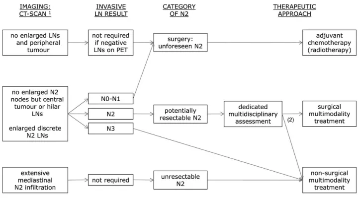
resectable LA-NSCLC
Patients are defined as having potentially resectable LA-NSCLC when a dedicated multidisciplinary assessment—including an experienced thoracic surgeon—judges a complete resection (R0) may be feasible after induction treatment. The treatment of LA-NSCLC remains a matter of debate [47] (Figure2). Aside from many dedicated prospective studies on surgical multimodality treatment in potentially resectable stage IIIA-N2 patients— including more than 2000 patients—there is only one
randomised trial in this setting [48]. The Lung Intergroup Trial 0139 randomised patients with resectable N2 disease (75% had single nodal station N2 disease) between either concurrent radiotherapy to a dose of 45 Gy and cisplatin–etoposide followed by surgery or definitive concurrent chemoradiotherapy to a dose of 61 Gy. No significant difference in OS was observed (P = 0.24), but progression-free survival was significantly better in the trimodality arm (P = 0.017). This points to the possibly higher early toxic death rate in the surgery arm as an explanation for the non-significant improvement in OS in the surgical arm. This was indeed the case for patients having
pneumonectomy. In an exploratory unplanned, matched subgroup analysis, patients treated with a lobectomy after induction concurrent chemoradiotherapy had a significantly better survival than those treated non-surgically or with a pneumonectomy.
Following the non-significant difference in the primary end point result in the only randomised trial, the choice of treatment varies across countries and centres. The optimal treatment plan is to be discussed in a multidisciplinary tumour board, taking into account the local treatment expertise. Both definitive chemoradiotherapy and induction therapy followed by surgery are options [II, A]. Surgery is preferably considered in patients in whom a complete resection by lobectomy is expected [II, B]. More complex surgical resections after induction treatment should be carried out in experienced centres [III, B]. Down-staging of the mediastinal nodes in the resection specimen is associated with better prognosis.
A randomised phase III study compared induction chemo-radiotherapy to a dose of 45 Gy followed by surgery to
induction chemotherapy followed by surgery and postoperative radiotherapy [49]. No differences were observed.
unresectable LA-NSCLC
Patients are defined as having unresectable LA-NSCLC when a dedicated multidisciplinary assessment—including an
experienced thoracic surgeon—judges a complete resection (R0) to be unlikely, even after induction treatment.
Figure 2. Suggested algorithm for treatment in patients with locoregional non-small-cell lung cancer, based on imaging, invasive lymph node staging tests and multidisciplinary assessment.
Table 2. Summary of recommendations Incidence/Epidemiology
• Screening with low-dose CT reduces lung cancer-related mortality [I, A]. It is not yet ready for large-scale implementation, because of unanswered questions regarding the definition of the at-risk population, timing, interval and method of CT (particularly 2D versus 3D evaluation), how to handle (false) positive findings, and, especially, cost-effectiveness in relation to smoking cessation.
• Screening with low-dose CT should not be offered on an individual basis. It can, however, be considered for current or former heavy smokers (≥30 pack-years or≤15 years since smoking cessation) aged 55–74 years, who are well informed about potential benefits and risks, and who are referred to a dedicated screening programme in an experienced multidisciplinary team [I, B].
• Other screening methods, such as chest X-ray, sputum analysis or biomarkers are not recommended for clinical use [I, C]. Diagnosis
• Bronchoscopy is the recommended method of obtaining a pathological diagnosis of centrally located tumours [III, A]. • The diagnostic approach to non-calcified pulmonary nodules should be based on existing guidelines [III, A].
• A pre-treatment pathological diagnosis is recommended. In some patients with clinical stage I/II lesions, this is not feasible, and a high likelihood of malignancy based on assessment of clinical and imagingfindings in an experienced multidisciplinary group may be considered sufficient [III, A]. Staging and risk assessment
• In non-metastatic NSCLC, detailed locoregional staging, according to the seventh TNM staging system, and the cardiopulmonaryfitness of the patient determine the choice of treatment [III, A].
• For patients with a non-centrally located resectable tumour and absence of nodal metastasis on both CT and PET images, surgical resection is recommended [I, A].
• For patients with suspect mediastinal lymph node metastasis on CT or PET images (unless bulky) pathological confirmation of nodal disease is recommended [I, A].
• The preferredfirst technique for pathological confirmation of suspect nodes is needle aspiration under EBUS and/or EUS guidance. Mediastinoscopy is the test with the highest negative predictive value to rule out mediastinal lymph node disease [I, A].
• The risk of postoperative morbidity and mortality should be estimated using validated risk-specific models [III, A].
• Formal lung function testing should be undertaken to estimate postoperative lung function. For patients with FEV1 and DLCO >80% in their pulmonary function tests and no other major co-morbidities, surgical resection is recommended. For others, additional ergospirometry, echocardiography, coronary tests etc. may be warranted [III, A].
• Co-morbidities should be evaluated and optimised before surgery [III, A]. Treatment of early stages I and II
• Surgery should be offered to patients with stage I or II NSCLC who are willing to accept procedure-related risks [III, A]. • Anatomical resection (lobectomy) is preferred over lesser resections such as wedge or segment resection [I, A]. • Lymph node dissection should conform to IASLC specifications for staging [III, A].
• Either open thoracotomy or VATS access can be utilised as appropriate to the expertise of the surgeon [III, A].
• Adjuvant chemotherapy should be offered to patients with resected stage II or III NSCLC [I, A] and can be considered in patients with resected stage IB disease and a primary tumour >4 cm [II, B]. However, pre-existing co-morbidity and postoperative recovery need to be taken into account in this decision. • For adjuvant chemotherapy, a two-drug combination with cisplatin is preferable [I, A]. In randomised studies, the attempted cumulative cisplatin dose was
up to 300 mg/m², delivered in three to four cycles. The most frequently studied regimen is cisplatin–vinorelbine.
• Given the current state of knowledge, the choice of adjuvant therapy should not be guided by molecular analyses such as ERCC1 or mutation testing [IV, B]. • Given the current state of knowledge, targeted agents should not be used in the adjuvant setting [II, A].
• In view of the equivalence of neo-adjuvant and adjuvant chemotherapy for overall survival, the consistent results and broad evidence base support adjuvant chemotherapy as the timing of choice [I, A].
• The non-surgical treatment of choice for stage I NSCLC is stereotactic ablative radiotherapy (SABR). The dose should be to a biologically equivalent tumour dose of≥100 Gy, prescribed to the encompassing isodose [III, A].
• SABR for early-stage peripheral lung tumours is associated with low toxic effect in patients with COPD and the elderly [III, A].
• For tumours with a size >5 cm and/or central location, radical radiotherapy using more conventional daily or accelerated schedules is recommended [III, A]. • Postoperative radiotherapy in completely resected early-stage NSCLC is not recommended [I, A]. It can however be indicated after incomplete surgery [III, B]. Treatment of locally advanced stage III
• Chemotherapy should be offered to all patients with LA-NSCLC who can tolerate it [I, A].
• Cisplatin-based regimens (e.g. cisplatin–etoposide or cisplatin–vinorelbine) delivered concurrently with radiotherapy have been studied most extensively, and are therefore recommended [II, A]. Studies using carboplatin–paclitaxel or other carboplatin-based combinations generally showed inferior outcomes, but may be chosen individually based on co-morbidity issues. The number of cycles ranges from two to four, and the cisplatin dose per cycle was in the range of 80 mg/m2[III, B].
Continued
In the EORTC (European Organisation for Research and Treatment of Cancer) trial, patients with unresectable N2 disease who showed at least a minimal tumour response after three cycles of very heterogeneous induction chemotherapy protocols were randomised between radiotherapy (60 Gy in 30 fractions in 6 weeks) and surgery [50]. No survival differences were observed. The preferred treatment of unresectable LA-NSCLC is definitive concurrent chemotherapy and radiotherapy with a dose no less than the biological equivalent of 60 Gy in 2.0 Gy fractions [I, A].
Induction chemotherapy followed by radiotherapy (mostly to a dose of 60–66 Gy in 30–33 fractions in 6–7 weeks), so-called sequential chemoradiotherapy, was compared with concurrent chemoradiotherapy to the same dose in many phase III trials and in a meta-analysis [51]. Concurrent chemotherapy and radiotherapy lead to higher 5-year survival rates at the cost of a higher rate of reversible oesophagitis. Infit patients, this is the standard treatment. Accurate locoregional staging is
recommended in analogy with resectable LA tumours [III, B]. In elderly patients or in those with clinically relevant co-morbidities [13], sequential chemotherapy and radiotherapy is a reasonable therapy choice [I, A]. A randomised trial in elderly, frail patients reported better median survival when low-dose daily carboplatin was added to radiotherapy alone [52]. In a meta-analysis based on individual patient data from phase III trials, accelerated radiotherapy schedules (i.e. delivered in shorter overall treatment times) led to higher 5-year OS rates at the expense of transient acute oesophagitis in patients treated with non-concurrent schedules [38]. Accelerated radiotherapy schedules are therefore recommended [I, A], e.g. 66 Gy in 24 fractions.
High-dose radiotherapy should be delivered according to quality standards such as those of the EORTC [53].
postoperative radiotherapy
PORT may be considered forfit patients with completely resected NSCLC with N2 nodal involvement, preferably after completion of adjuvant chemotherapy. This may reduce local recurrences, although no survival benefit has been demonstrated [54] [III, B]. A randomised clinical trial to assess the effect on survival is ongoing (LUNGART, NCT00410683). PORT can be indicated in case of a R1 or R2 resection, although survival in these patients remains poor [54] [III, B].
personalised medicine
In this disease setting, more research is needed to identify molecular markers which could lead to advances in personalised medicine.
follow-up
NSCLC patients treated with radical intent should be followed for treatment-related complications, detection of treatable relapse or occurrence of second primary lung cancer [III, A].
Except for one small underpowered study, there are no prospective comparative trials on the question of what is the most effective follow-up for patients with non-metastatic NSCLC [55]. How often and by which methods surveillance is indicated is guided by knowledge about relapse patterns, not by evidence that earlier detection and treatment of recurrence leads to a better outcome.
At least two-thirds of the relapses occur in the initial 2–3 years after treatment [56]. Hence, a follow-up visit every 3–6 months is recommended during 2–3 years, annually thereafter
Table 2. Continued
• In sequential approaches, a platinum-based two-drug combination is the preferred choice, with the number of cycles ranging from two to four [I, A]. • Carboplatin-based induction chemotherapy before concurrent chemoradiotherapy can generally not be recommended [I, C]. Data on cisplatin-based
induction chemotherapy are few and individual patients may benefit from this approach [III, B].
• Consolidation treatment with, e.g. docetaxel or an EGFR-tyrosine kinase inhibitor after concurrent chemoradiotherapy is not recommended [I, A]. • The preferred treatment of unresectable LA-NSCLC is definitive concurrent chemotherapy and radiotherapy [I, A].
• Definitive thoracic radiotherapy should be no less than the biological equivalent of 60 Gy in 2.0 Gy fractions [I, A].
• In patients who are unfit to receive concurrent chemotherapy and radiotherapy, the sequential approach should be offered as an alternative treatment with curative intent [I, A].
• In non-concurrent schedules, radiotherapy delivered in a short overall treatment time is recommended [I, A].
• For resectable LA-NSCLC, especially single nodal stage N2 disease, both definitive chemoradiotherapy and induction therapy followed by surgery are options [II, A].
• Surgery is preferably considered in patients in whom a complete resection by lobectomy is expected [II, B]. More complex surgical resections after induction treatment should be carried out in experienced centres [III, B].
• Routine use of PORT is, as yet, unproven but it may be considered in N2 patients after resection. In these cases, radiotherapy was delivered after chemotherapy in randomised studies. PORT is indicated after incomplete surgery [III, B].
Follow-up
• NSCLC patients treated with radical intent should be followed for treatment-related complications, detection of treatable relapse or occurrence of second primary lung cancer [III, A].
• A follow-up visit every 3–6 months is recommended during 2–3 years, less often—e.g. annually—thereafter [III, B]. • For follow-up, history and physical examination, chest CT and, to a lesser extent, chest X-ray, are appropriate tools [III, B].
• NSCLC patients should be offered smoking cessation, as this leads to superior treatment outcomes. Combining behaviour techniques with pharmacotherapy is the preferred approach [I, A].
[III, B]. New abnormalities deserve discussion in a multidisciplinary team with experience in both treatment complications, and in the distinction between recurrence or metachronous second primary tumour, which occurs in 5–10% of the patients.
History and physical examination, chest X-ray and annual CT are appropriate tests, CT in particular for earlier detection of a second primary tumour.
smoking cessation
Smoking is the main cause of lung cancer, responsible for 80% of cases. Smoking cessation is of major value for NSCLC patients—especially those with early and locally advanced stages and a potential for cure—as it is associated with significantly decreased risks of mortality, development of a second primary tumour lung cancer or recurrence [57]. Combining behaviour techniques with pharmacotherapy is the best approach, with success rates up to 25% [58] [I, A].
note
A summary of recommendations is given in Table2. Levels of evidence and grades of recommendation have been applied using the system shown in Table3. Statements without grading were considered justified standard clinical practice by the experts and the ESMO faculty.
con
flict of interest
Prof. Vansteenkiste is holder of the Eli Lillly Chair in Respiratory Oncology at the Leuven University (research funding). Prof. Vansteenkiste is holder of the AstraZeneca Chair in Personalised Lung Cancer Care at the Leuven University (research funding). Dr Eberhardt has reported: Advisory board: GlaxoSmithKline, Amgen, Novartis, Merck, Teva, Roche, AstraZeneca, Eli Lilly, Boehringer Ingelheim, Pfizer, Bristol-Myers Squibb; Speakers’ bureau: Roche, AstraZeneca, Eli Lilly,
Boehringer Ingelheim, Pfizer, GlaxoSmithKline, Amgen, Novartis, Hexal, Merck; Research grants: Eli Lilly. Mr. Lim has reported: Research support from SCreenCell and PointHope; Speakers’ bureau member for Roche and Imedex; Advisory board member for Strategen, Abbott Molecular and
GlaxoSmithKline; Patent pending with Clearbridge BioMedics; Holding stock in Pfizer. Prof. Senan has reported: Research grants and honoraria: Varian Medical Systems; Member of a trial management group in a phase III trial conducted by Lilly Oncology. Dr Felip has reported: Consultancy/honoraria: Eli Lilly, GlaxoSmithKline, Pfizer, Roche and Boehringer Ingelheim. Dr Peters has reported: Consultancy/honoraria: Roche, Eli Lilly, AstraZeneca, Pfizer, Boehringer Ingelheim, Bristol-Myers Squibb, Merck Serono, Daiichi Sankyo, Tesaro. Prof. De Ruysscher has reported no potential conflicts of interest.
references
1. Peters S, Adjei AA, Gridelli C et al. Metastatic non-small-cell lung cancer (NSCLC): ESMO Clinical Practice Guidelines for diagnosis, treatment and follow-up. Ann Oncol 2012; 23(Suppl 7): vii56–vii64.
2. Aberle DR, Adams AM, Berg CD et al. Reduced lung-cancer mortality with low-dose computed tomographic screening. N Engl J Med 2011; 365: 395–409. 3. Van Klaveren RJ, Oudkerk M, Prokop M et al. Management of lung nodules
detected by volume CT scanning. N Engl J Med 2009; 361: 2221–2229. 4. Aberle DR, Abtin F, Brown K. Computed tomography screening for lung cancer:
Has itfinally arrived? Implications of the national lung screening trial. J Clin Oncol 2013; 31: 1002–1008.
5. Gould MK, Donington J, Lynch WR et al. Evaluation of individuals with pulmonary nodules: When is it lung cancer?: Diagnosis and management of lung cancer, 3rd ed: American College of Chest Physicians evidence-based clinical practice guidelines. Chest 2013; 143(Suppl 5): e93S–e120S.
6. Macmahon H, Austin JH, Gamsu G et al. Guidelines for management of small pulmonary nodules detected on CT scans: a statement from the Fleischner Society. Radiology 2005; 237: 395–400.
7. Naidich DP, Bankier AA, MacMahon H et al. Recommendations for the management of subsolid pulmonary nodules detected at CT: a statement from the Fleischner Society. Radiology 2013; 266: 304–317.
Table 3. Levels of evidence and grades of recommendation (adapted from the Infectious Diseases Society of America-United States Public Health Service Grading Systema)
Levels of evidence
I Evidence from at least one large randomised, controlled trial of good methodological quality (low potential for bias) or meta-analyses of well-conducted randomised trials without heterogeneity
II Small randomised trials or large randomised trials with a suspicion of bias (lower methodological quality) or meta-analyses of such trials or of trials with demonstrated heterogeneity
III Prospective cohort studies
IV Retrospective cohort studies or case–control studies V Studies without control group, case reports, experts opinions Grades of recommendation
A Strong evidence for efficacy with a substantial clinical benefit, strongly recommended
B Strong or moderate evidence for efficacy but with a limited clinical benefit, generally recommended
C Insufficient evidence for efficacy or benefit does not outweigh the risk or the disadvantages (adverse events, costs, ...), optional D Moderate evidence against efficacy or for adverse outcome, generally not recommended
E Strong evidence against efficacy or for adverse outcome, never recommended
aDykewicz CA. Summary of the guidelines for preventing opportunistic infections among hematopoietic stem cell transplant recipients. Clin Infect Dis 2001;
33: 139–144. By permission of the Infectious Diseases Society of America.
8. Goldstraw P, Crowley J, Chansky K et al. The IASLC Lung Cancer Staging Project: proposals for the revision of the TNM stage groupings in the forthcoming (seventh) edition of the TNM Classification of malignant tumours. J Thorac Oncol 2007; 2: 706–714.
9. Vansteenkiste J, Dooms C, De Leyn P. Early stage non-small-cell lung cancer: challenges in staging and adjuvant treatment: evidence-based staging. Ann Oncol 2010; 21(Suppl 7): vii189–vii195.
10. Silvestri GA, Gonzalez AV, Jantz MA et al. Methods for staging non-small cell lung cancer: diagnosis and Management of Lung Cancer, 3rd ed: American College of Chest Physicians Evidence-Based Clinical Practice Guidelines. Chest 2013; 143 (Suppl 5): e211S–e250S.
11. De Leyn P, Lardinois D, Van Schil PE et al. ESTS guidelines for preoperative lymph node staging for non-small cell lung cancer. Eur J Cardiothorac Surg 2007; 32: 1–8. 12. Annema JT, van Meerbeeck JP, Rintoul RC et al. Mediastinoscopy vs.
endosonography for mediastinal nodal staging of lung cancer: A randomized trial. JAMA 2010; 304: 2245–2252.
13. Brunelli A, Charloux A, Bolliger CT et al. ERS/ESTS clinical guidelines onfitness for radical therapy in lung cancer patients (surgery and chemo-radiotherapy). Eur Respir J 2009; 34: 17–41.
14. Lim E, Baldwin D, Beckles M et al. Guidelines on the radical management of patients with lung cancer. Thorax 2010; 65(Suppl 3): iii1–iii27.
15. Falcoz PE, Conti M, Brouchet L et al. The Thoracic Surgery Scoring System (Thoracoscore): risk model for in-hospital death in 15,183 patients requiring thoracic surgery. J Thorac Cardiovasc Surg 2007; 133: 325–332.
16. Brunelli A, Cassivi SD, Fibla J et al. External validation of the recalibrated thoracic revised cardiac risk index for predicting the risk of major cardiac complications after lung resection. Ann Thorac Surg 2011; 92: 445–448.
17. Ginsberg RJ, Rubinstein LV. Randomized trial of lobectomy versus limited resection for T1 N0 non-small cell lung cancer. Lung Cancer Study Group. Ann Thorac Surg 1995; 60: 615–622.
18. Wu YI, Huang ZF, Wang SY et al. A randomized trial of systematic nodal dissection in resectable non-small cell lung cancer. Lung Cancer 2002; 36: 1–6. 19. Darling GE, Allen MS, Decker PA et al. Randomized trial of mediastinal lymph node
sampling versus complete lymphadenectomy during pulmonary resection in the patient with N0 or N1 (less than hilar) non-small cell carcinoma: results of the American College of Surgery Oncology Group Z0030 Trial. J Thorac Cardiovasc Surg 2011; 141: 662–670.
20. Goldstraw P. International Association for the Study of Lung Cancer Staging Manual in Thoracic Oncology. Florida: Editorial Rx Press 2009.
21. Yan TD, Black D, Bannon PG, McCaughan BC. Systematic review and meta-analysis of randomized and non-randomized trials on safety and efficacy of video-assisted thoracic surgery lobectomy for early-stage non-small cell lung cancer. J Clin Oncol 2009; 27: 2553–2562.
22. Cao C, Manganas C, Ang SC et al. Video-assisted thoracic surgery versus open thoracotomy for non-small cell lung cancer: A meta-analysis of propensity score-matched patients. Interact Cardiovasc Thorac Surg 2013; 16: 244–249.
23. Lim E, Harris G, Patel A et al. Preoperative versus postoperative chemotherapy in patients with resectable non-small cell lung cancer: systematic review and indirect comparison meta-analysis of randomized trials. J Thorac Oncol 2009; 4: 1380–1388.
24. Pignon JP, Tribodet H, Scagliotti GV et al. Lung adjuvant cisplatin evaluation: a pooled analysis by the LACE Collaborative Group. J Clin Oncol 2008; 26: 3552–3559.
25. Strauss GM, Herndon JE, 2nd, Maddaus MA et al. Adjuvant paclitaxel plus carboplatin compared with observation in stage IB non-small-cell lung cancer: CALGB 9633 with the Cancer and Leukemia Group B, Radiation Therapy Oncology Group, and North Central Cancer Treatment Group Study Groups. J Clin Oncol 2008; 26: 5043–5051.
26. Winton T, Livingston R, Johnson D et al. Vinorelbine plus cisplatin vs. observation in resected non-small cell lung cancer. N Engl J Med 2005; 352: 2589–2597. 27. Pepe C, Hasan B, Winton TL et al. Adjuvant vinorelbine and cisplatin in elderly
patients: National Cancer Institute of Canada and Intergroup Study JBR.10. J Clin Oncol 2007; 25: 1553–1561.
28. Scagliotti GV, Pastorino U, Vansteenkiste JF et al. Randomized phase III study of surgery alone or surgery plus preoperative cisplatin and gemcitabine in stages IB to IIIA non-small cell lung cancer. J Clin Oncol 2012; 30: 172–178. 29. Gilligan D, Nicolson M, Smith I et al. Preoperative chemotherapy in patients with
resectable non-small cell lung cancer: results of the MRC LU22/NVALT 2/EORTC 08012 multicentre randomised trial and update of systematic review. Lancet 2007; 369: 1929–1937.
30. Felip E, Rosell R, Maestre JA et al. Preoperative chemotherapy plus surgery versus surgery plus adjuvant chemotherapy versus surgery alone in early-stage non-small cell lung cancer. J Clin Oncol 2010; 28: 3138–3145.
31. Olaussen KA, Dunant A, Fouret P et al. DNA repair by ERCC1 in non-small cell lung cancer and cisplatin-based adjuvant chemotherapy. N Engl J Med 2006; 355: 983–991.
32. Friboulet L, Olaussen KA, Pignon JP et al. ERCC1 isoform expression and DNA repair in non-small cell lung cancer. N Engl J Med 2013; 368: 1101–1110.
33. Goss GD, Lorimer I, Tsao MS et al. A phase III randomized, double-blind, placebo-controlled trial of the epidermal growth factor receptor inhibitor gefitinb in completely resected stage IB-IIIA non-small cell lung cancer (NSCLC): NCIC CTG BR.19. J Clin Oncol 2010; 28: 18s (Suppl: LBA7005).
34. Haasbeek CJ, Palma D, Visser O et al. Early-stage lung cancer in elderly patients: a population-based study of changes in treatment patterns and survival in the Netherlands. Ann Oncol 2012; 23: 2743–2747.
35. Palma D, Lagerwaard F, Rodrigues G et al. Curative treatment of stage I non-small-cell lung cancer in patients with severe COPD: Stereotactic radiotherapy outcomes and systematic review. Int J Radiat Oncol Biol Phys 2012; 82: 1149–1156.
36. Senthi S, Lagerwaard FJ, Haasbeek CJ et al. Patterns of disease recurrence after stereotactic ablative radiotherapy for early stage non-small-cell lung cancer: a retrospective analysis. Lancet Oncol 2012; 13: 802–809.
37. Senthi S, Haasbeek CJ, Slotman BJ, Senan S. Outcomes of stereotactic ablative radiotherapy for central lung tumours: a systematic review. Radiother Oncol 2013; 106: 276–282.
38. Mauguen A, Le Péchoux C, Saunders MI et al. Hyperfractionated or accelerated radiotherapy in lung cancer: an individual patient data meta-analysis. J Clin Oncol 2012; 30: 2788–2797.
39. PORT meta-analysis trialists group. Postoperative radiotherapy for non-small cell lung cancer. Cochrane Database Syst Rev 2005; CD002142.
40. Arriagada R, Auperin A, Burdett S et al. Adjuvant chemotherapy, with or without postoperative radiotherapy, in operable non-small-cell lung cancer: two meta-analyses of individual patient data. Lancet 2010; 375: 1267–1277.
41. Pignon JP, Stewart LA. Randomized trials of radiotherapy alone versus combined chemotherapy and radiotherapy in stages IIIa and IIIb non-small cell lung cancer: a meta-analysis. Cancer 1996; 77: 2413–2414.
42. Palma DA, Senan S, Tsujino K et al. Predicting radiation pneumonitis after chemoradiation therapy for lung cancer: an international individual patient data meta-analysis. Int J Radiat Oncol Biol Phys 2013; 85: 444–450.
43. Vokes EE, Herndon JE, Kelley MJ et al. Induction chemotherapy followed by chemoradiotherapy compared with chemoradiotherapy alone for regionally advanced unresectable stage III non-small cell lung cancer: Cancer and Leukemia Group B. J Clin Oncol 2007; 25: 1698–1704.
44. Hanna N, Neubauer M, Yiannoutsos C et al. Phase III study of cisplatin, etoposide, and concurrent chest radiation with or without consolidation docetaxel in patients with inoperable stage III non-small cell lung cancer: The Hoosier Oncology Group and U.S. Oncology. J Clin Oncol 2008; 26: 5755–5760.
45. Jalal SI, Riggs HD, Melnyk A et al. Updated survival and outcomes for older adults with inoperable stage III non-small-cell lung cancer treated with cisplatin, etoposide, and concurrent chest radiation with or without consolidation docetaxel: analysis of a phase III trial from the Hoosier Oncology Group (HOG) and US Oncology. Ann Oncol 2012; 23: 1730–1738.
46. Kelly K, Chansky K, Gaspar LE et al. Phase III trial of maintenance gefitinib or placebo after concurrent chemoradiotherapy and docetaxel consolidation in inoperable stage III non-small cell lung cancer: SWOG S0023. J Clin Oncol 2008; 26: 2450–2456.
47. Vansteenkiste J, Van Damme V, Dooms C. Generalized or personalized treatment for stage IIIA-N2 non-small cell lung cancer? Expert Opin Pharmacother 2010; 11: 1605–1609.
48. Albain KS, Swann RS, Rusch VW et al. Radiotherapy plus chemotherapy with or without surgical resection for stage III non-small cell lung cancer: a phase III randomised controlled trial. Lancet 2009; 374: 379–386.
49. Thomas M, Rübe C, Hoffknecht P et al. Effect of preoperative chemoradiation in addition to preoperative chemotherapy: a randomised trial in stage III non-small cell lung cancer. Lancet Oncol 2008; 9: 636–648.
50. Van Meerbeeck JP, Kramer GW, Van Schil PE et al. Randomized controlled trial of resection versus radiotherapy after induction chemotherapy in stage IIIA-N2 non-small cell lung cancer. J Natl Cancer Inst 2007; 99: 442–450.
51. Auperin A, Le Péchoux C, Rolland E et al. Meta-analysis of concomitant versus sequential radiochemotherapy in locally advanced non-small cell lung cancer. J Clin Oncol 2010; 28: 2181–2190.
52. Atagi S, Kawahara M, Yokoyama A et al. Thoracic radiotherapy with or without daily low-dose carboplatin in elderly patients with non-small cell lung cancer: a randomised, controlled, phase 3 trial by the Japan Clinical Oncology Group (JCOG0301). Lancet Oncol 2012; 13: 671–678.
53. De Ruysscher D, Faivre-Finn C, Nestle U et al. European Organization for Research and Treatment of Cancer recomendations for planning and delivery of high-dose, high-precision radiotherapy for lung cancer. J Clin Oncol 2010; 28: 5301–5310.
54. Le Péchoux C. Role of postoperative radiotherapy in resected non-small cell lung cancer: a reassessment based on new data. Oncologist 2011; 16: 672–681.
55. Schmidt-Hansen M, Baldwin DR, Hasler E. What is the most effective follow-up model for lung cancer patients? A systematic review. J Thorac Oncol 2012; 7: 821–824.
56. Demicheli R, Fornili M, Ambrogi F et al. Recurrence dynamics for non-small-cell lung cancer: effect of surgery on the development of metastases. J Thorac Oncol 2012; 7: 723–730.
57. Parsons A, Daley A, Begh R, Aveyard P. Influence of smoking cessation after diagnosis of early stage lung cancer on prognosis: systematic review of observational studies with meta-analysis. BMJ 2010; 340: b5569.
58. Stead LF, Lancaster T. Behavioural interventions as adjuncts to pharmacotherapy for smoking cessation. Cochrane Database Syst Rev 2012; 12: CD009670.