Studies of Copper Complexes Displaying N
3S Coordination as Models for Cu
BCenter of
Dopamine
β-Hydroxylase and Peptidylglycine r-Hydroxylating Monooxygenase
Fre´de´ric Champloy, Nourredine Benali-Che´rif, Pascale Bruno, Ingrid Blain, Marcel Pierrot, and Marius Re´glier*
Chimie, Biologie et Radicaux Libres, UMR CNRS 6517, Universite´s d’Aix-Marseille 1 et 3,
Faculte´ des Sciences et Techniques de Saint Je´roˆme, case 432, avenue Escadrille Normandie-Niemen, 13397 Marseille Cedex 20, France
Alain Michalowicz
Laboratoire pour l’Utilisation du Rayonnement Electromagne´tique, Baˆtiment 209D, Centre Universitaire de Paris-Sud, 91405 Orsay Cedex, France
ReceiVed July 24, 1997
We describe the studies of new copper complexes [MeSPY2]CuPF6, 2, and [MeSPY2]Cu(ClO4)2‚CH3CN, 3, as models for the CuBcenter of dopamineβ-hydroxylase and peptidylglycine R-hydroxylating monooxygenase. The structure of [MeSPY2]Cu(ClO4)2‚CH3CN, 3, has been established by X-ray crystallography. The copper coordination exhibits a square pyramidal geometry where the equatorial plane is occupied by the SCH3group and three nitrogen atoms (tertiary amine, one pyridine, and acetonitrile solvent), whereas the axial position binds the second pyridine. Using FEFF calculations and multiscattering interaction, EXAFS refinements show that the SMe group lies in the coordination sphere of copper complexes [MeSPY2]CuPF6, 2, and [MeSPY2]Cu(ClO4)2‚CH3CN, 3. While [MeSPY2]CuPF6, 2, reacts with dioxygen in dichloromethane without oxidation of the ligand, we observed an oxidation of the sulfide ligand when [MeSPY2]Cu(ClO4)2‚CH3CN, 3, reacts with hydrogen peroxide in methanol. Considering results, we propose that Met314, crucial for DBH and PHM activity, could be the site of the H
2O2(or ascorbate) inactivation by oxidation to the sulfoxide group.
Introduction
Dopamineβ-hydroxylase (DBH; EC 1.14.17.1),1,2a copper-containing monooxygenase present in a variety of mammalian tissues, catalyzes the benzylic hydroxylation of dopamine into norepinephrine. All known results from EPR,3 ESEEM,4 EXAFS,5and biochemical studies6suggest that DBH contains two inequivalent copper atoms per active site. A CuAsite is proposed to be the ascorbate binding site, where this reductor delivers one electron at a time. At a distance greater than 4 Å, a CuB center involved in dioxygen fixation is responsible for the hydroxylation of dopamine.7 Whereas it is well established
that oxidized DBH has a CuA(His)3(H2O)‚‚‚CuB(His)2X(H2O) type of configuration, the structure of the reduced form is not really clear. EXAFS data of reduced DBH show the loss of two water molecules with the appearance of one-half of a sulfur ligand per copper(I) center. While this S ligand, assigned to a Met residue, was first proposed to be coordinated to the CuA center,4a recent preparation of one-half apo reduced DBH form seems to demonstrate that the MetS ligand was coordinated to the CuB center.8 This MetS ligand which is not detected by EXAFS in the oxidized DBH form could be also present but as a weakly bound axial ligand. In addition, DBH presents some structural and functional similarities with another copper-containing monooxygenase, peptidylglycine R-hydroxylating monooxygenase (EC 1.14.17.3, PHM) which catalyses the R-amidation of several bioactive peptides.2,9,10 The amino acid sequences of rat PHM and rat DBH exhibit 32% identity over a 291-amino acid region, and some residues in PHM are conserved in DBH. Mutation of the Met residues common to PHM and DBH to Ile have identified Met314as being crucial for catalytic activity. Therefore, we decided to focus our (1) Villafranca, J. J. Metal Ions in Biology; Spiro, T. G., Ed.; John Wiley
and Sons: New York, 1981; Vol 3, pp 263-290. Stewart, L. C.; Klinman, J. P. Annu. ReV. Biochem. 1988, 57, 551.
(2) Klinman, J. P. Chem. ReV. 1996, 96, 2541.
(3) Blackburn, N. J.; Collison, D.; Sutton, J.; Mabbs, F. E. Biochem. J. 1984, 220, 447. Blackburn, N. J.; Concannon, M.; Khosrow Shahiyan, S.; Mabbs, F. E.; Collison, D. Biochemistry 1988, 27, 6001. (4) McCracken, J.; Desai, P. R.; Papadopoulous, N. J.; Villafranca, J. J.
Biochem. 1988, 27, 4133.
(5) Hasnain, S. S.; Diakun, G. P.; Knowles, P. F.; Binsted, N.; Garner, C. D.; Blackburn, N. J. Biochem. 1988, 221, 545. Scott, R. A.; Sullivan, R. J.; De Wolf, W. E., Jr.; Dolle, R. E.; Kruse, L. I. Biochemistry 1988, 27, 5411. Blumberg, W. E.; Desai, P. R.; Powers, L.; Freedman, J. H.; Villafranca, J. J. J. Biol. Chem. 1989, 264, 6029. Pettingill, T. M.; Stange, R. W.; Blackburn, N. J. J. Biol. Chem. 1991, 266, 16996. Blackburn, N. J.; Hasnain, S. S.; Pettingill, T. M.; Stange, R. W. J.
Biol. Chem. 1991, 266, 23120.
(6) Stewart, L. C.; Klinman, J. P. Biochemistry 1987, 26, 5302. Brenner, M. C.; Murray, C. J.; Klinman, J. P. Biochemistry 1989, 28, 4656. (7) Anh, N.; Klinman, J. P. Biochemistry 1983, 22, 3090. Miller, S. M.;
Klinman, J. P. Biochemistry 1985, 24, 2114.
(8) Blackburn, N. J. In Bioinorganic Chemistry of Copper; Karlin, K. D., Tyeclar, Z., Eds.; Chapman & Hall: New York, 1993; pp 164-183. Reedy, B. J.; Blackburn, N. J. J. Am. Chem. Soc. 1994, 116, 1924. (9) Boswell, J. S.; Reedy, B. J.; Kulathila, R.; Merkler, D.; Blackburn,
N. J. Biochemistry 1996, 35, 12241.
(10) During the writing of this article, the X-ray structure of the oxidized form of PAM was solved by Prigge, S. T.; Kolhekar, A. S.; Eipper, B. A.; Mains, R. E.; Amzel, L. M. Science 1997, 278, 1300. This work confirms the presence of a Met residue as a weakly bound ligand in the coordination sphere of the CuBcenter (dCu-SMet) 2.68 Å).
S0020-1669(97)00928-2 CCC: $15.00 © 1998 American Chemical Society Published on Web 07/08/1998
attention on the chemistry and the X-ray spectroscopy of copper ions with N3S-type ligand. We report here the synthesis, the characterization by X-ray spectroscopies, and the reactivity toward dioxygen and hydrogen peroxide of copper complexes coordinated to MeSPY2 ligand 1 (Scheme 1).
Experimental Section
General Methods. Solvents were freshly distilled under nitrogen: MeOH from magnesium, diethyl ether from sodium benzophenone ketyl, and acetonitrile from calcium hydride. Deoxygenation of solvents and solutions was carried out by three vacuum/purge cycles. Com-mercial starting materials were used without further purification, except for 2-vinylpyridine which was distilled prior to use. Cu(CH3CN)4PF6
was obtained by reaction of Cu2O with HPF6 in deoxygenated
acetonitrile.11 Preparations and handling of air-sensitive compounds
were carried out by using standard Schlenk techniques. 1H and13C
NMR spectra were recorded on a Bruker AC-200 spectrometer. Chemical shifts are reported asδ values in ppm downfield from TMS as internal standard. Elemental analyses were obtained on a CHN Technicon microanalyzer.
N-2-(Methylthio)ethyl-N-bis-2-(2-pyridyl)ethylamine (MeSPY2,
1). Freshly distilled 2-vinylpyridine (35 g, 0.33 mol), 2-methylthio-ethylamine (3 g, 33 mmol), and acetic acid (10 g, 0.17 mol) in MeOH (100 mL) were refluxed for 2 days. Then, MeOH was evaporated under vacuum, and 100 mL of 15% NaOH solution was added. After the mixture was extracted with CH2Cl2(3× 50 mL), the organic layers
were dried over Na2SO4. Evaporation of CH2Cl2under reduced pressure
(18 mmHg) gave a crude product which still contained 2-vinylpyridine, which was eliminated by vacuum distillation (25°C, 0.01 mmHg). Flash chromatography (silica gel, CH2Cl2/MeOH 95:5) of crude product
afforded 6.5 g (65%) of amine 1. Anal. Calcd (found) for C17H23N3S: C, 67.74 (67.80); H, 7.69 (7.74); N, 13.94 (14.01). 1H NMR (200 MHz, CD3CN)δ: 8.46 (dt, J 4.6, 1 Hz; 2 H), 7.60 (td, J 7.5, 1.6 Hz; 2 H), 7.23-7.03 (m; 4H), 2.89 (bs; 8 H), 2.74 (m; 2 H); 2.47 (m; 2 H); 2.03 (s; 3 H). 13C NMR (50 MHz, CD 3CN)δ: 161.46 (2 C), 149.72 (2 CH), 137.10 (2 CH), 124.12 (2 CH), 121.93 (2 CH), 54.34 (2 CH2), 51.46 (CH2), 36.72 (CH2), 35.15 (CH2), 15.60 (CH3).
[MeSPY2]CuPF6, 2. Amine 1 (2 g, 6.6 mmol) and Cu(CH3CN)4PF6
(2.5 g, 6.7 mmol) in deoxygenated acetonitrile (10 mL) were stirred under Ar for 1 h. Copper(I) complex 2 was precipitated by addition of anhydrous diethyl ether. Filtration under Ar and drying under high
vacuum (0.01 mmHg) afforded 2.5 g (73%) of the very air-sensitive yellow copper(I) complex 2. Anal. Calcd (found) for C17H23N3
-SCuPF6: C, 40.04 (40.07); H, 4.55 (4.58); N, 8.24 (8.30). 1H NMR (200 MHz, CD3CN)δ: 8.56 (bd, J 4 Hz; 2 H); 7.80 (bt, J 7 Hz; 2 H); 7.50-7.20 (m; 4 H); 2.77 (bs; 12 H); 2.21 (s; 3 H). 13C NMR (50 MHz, CD3CN)δ: 150.99 (2 CH); 138.89 (2 CH); 126.46 (2 CH); 123.81 (2 CH); 54.34 (2 CH2); 51.46 (CH2); 36.72 (2 CH2); 35.15 (CH2); 18.77 (CH3).
[MeSPY2]Cu(ClO4)2‚CH3CN, 3. Amine 1 (2 g, 6.6 mmol) and Cu(ClO4)2‚6H2O (2.5 g, 6.7 mmol) in acetonitrile (10 mL) were stirred
for 1 h. Copper(II) complex 3 was precipitated by addition of diethyl ether. Filtration and drying under vacuum (0.01 mmHg) afforded 3.6 g (90%) of blue copper(II) complex 3. Anal. Calcd (found) for [C17H23N3S]Cu(ClO4)2‚CH3CN: C, 37.72 (37.99); H, 4.33 (4.36); N,
9.26 (9.55).
Crystal Structure Determination. Crystal data for compound 3 with details of the X-ray diffraction experiment are reported in Table 1. Cell constants and the orientation matrix for data collection were obtained from least-squares refinement, using setting angles of 25 reflections in the range 20°< 2θ < 28°. Two intense reflections were recorded throughout the data every hour and showed no decay during the exposure time. The data were corrected for Lpbut not for absorption
and secondary extinction, resulting in the unique set of 3496 reflections with I > 3σ(I) used in the structure determination. All non-H atoms were found by direct methods,12 and the structure was refined
successfully in space group P21/c by a full-matrix least-squares
procedure using anisotropic thermal parameters for all non-H atoms. The weighting scheme used is
Full-matrix least-squares refinement based on F converged at R ) 0.0568, Rw) 0.0657; S ) 1.35; (∆/σ)max) 0.3 for all atoms; residual
( de ) +0.78, -0.60 e Å-3in a final DF synthesis, atomic scattering factors from International Tables for X-ray Crystallography.13
Positional parameters and selected bond distances and angles with ORTEP perspective view are given in Table 2 and Figure 1, respectively. Perchlorate anions showed high thermal parameters due to disordered positions as indicated in DF maps. Oxygen O4 attached
(11) Kubas G. J. Inorg. Synth. 1979, 19, 90.
(12) Frenz, B. A. The Enraf-Nonius CAD-4 SDP-A Real Time System for Concurrent X-ray Data Collection and Crystal Structure Solution. In
Computing in Crystallography; Schenk, H., Olthof-Hazekamp, R., Van
Konigsveld, H., Bassi, G. C., Eds.; Delft University Press: The Netherlands, 1978.
(13) International Tables for X-ray Crystallography; Ibers, J., Hamilton, W. C., Eds.; Kynoch Press: Birmingham, 1974; Vol. IV. (Present distributor Kluwer Academic Publishers: Dordecht, The Netherlands.)
Scheme 1. Blackburn Model for the Oxidized and Reduced DBH and PHM Active Sites (X Could Be either a His or an O-donor ligand)
Table 1. Experimental Details of the Crystal Structure Determination of Copper(II) Complex 3
formula C19H26Cl2CuN4O8S formula weight 604.95 space group P21/c a, Å 14.131(3) b, Å 14.782(4) c, Å 12.280(3) β, deg 97.02(5) V, Å3 2545.9(5) Z 4 dcalc, g/cm3 1.58 µ(Mo KR), cm-1 11.98
total no. of unique data 4157
no. with F2> 3σ(F2) 3496
Ra 0.0568
Rwb 0.0657
goodness of fitc 1.35
largest shift, esd in final cycle 0.3
largest peak, e/Å3 +0.78, -0.60
aR )∑||F o| - |Fc||/∑|Fo.bRw) [∑w(|Fo| - |Fc|)2/∑w|Fo|2]1/2; w ) 1/σ2|F c|.cw ) (|Fo| - |Fc|)2/∑w|Fo|2]1/2. w ) [σ(Iobs) 2+ (0.04I obs) 2 ]-1 (1) B
to Cl1 was shared into two different positions of equal multiplicity (O4 and O-4).
X-ray Absorption. X-ray absorption measurements were made at LURE-Orsay on XAS II beamline. The storage ring delivered a current of 320 mA at 1.85 GeV. The monochromator was Si(111). The X-ray spectra were recorded in transmission mode using standard He-filled ionization chambers. Energies were calibrated by means of a Cu-foil internal standard, the first inflection point on the Cu edge was assigned as 8978 eV.
EXAFS Data Collection. The spectra were recorded in the transmission mode, the amount of copper complexes in each sample was calculated in order to obtain an edge jump∆µ(x) equal to 1, completed with cellulose powder and compressed as homogeneous pellets (1 mm thick) supported in an Al spacer with 63.5-mm Mylar tape windows. For each sample three XANES (8950 to 9130 eV, 0.35 eV step, 512 points, 2 s/points) and six EXAFS (8900 to 9900 eV, 1.95 eV step, 512 points, 2 s/points) scans were recorded at ambient temperature (295 K). Data reduction was accomplished with the software package “EXAFS pour le Mac”.14 Before being averaged,
each spectrum corresponding to one sample is treated separately using standard procedures.15 Pre-edge background is removed with a linear
fit up to the edge and extrapolated using the Lengeler-Eisenberger method.16 Atomic background µ
0 is removed with a fifth-order
polynomial fit in the range 9050-9900 eV, followed with a 6 knots spleen fit in the same range. The EXAFS signal is extracted with the standard formula
in the range 9006-9900 eV.
The energy origin E0was chosen in the edge jump at the maximum
of the first-order derivative (8985 eV for 3 and 8990 eV for 2). These E0values will be allowed to vary during the fitting procedure. After
extraction of the EXAFS signals on each recording, the average signals χ(k) and their standard deviations are calculated. These standard deviations are used in the estimation of standard error of the fitted parameters.17 The systematic errors in the distances between the metal
atom and its neighbors are typically estimated as ca. 0.01 and 0.02 Å, respectively, for short and long distances (and 20% for the number of atoms per shell); these values are used in the text. The EXAFS signals were used between 3 and 13 Å-1, and the Fourier transforms of the averaged EXAFS signal k3χ(k) multiplied by the Kaiser-Bessel
apodiza-tion window (τ ) 2.5) were calculated in this range. Fourier filtering procedure is used for two purposes (i) to remove the noise (filtering window, 0-13 Å) and (ii) to separate each component of the EXAFS signal when it is possible without spoiling of the data.
Oxidation of [MeSPY2]Cu(ClO4)2‚CH3CN, 3, with H2O2 in MeOH. In a Shlenck tube placed under argon and containing 20 mL of deoxygenated MeOH, 100 mg (0.15 mmol) of copper(II) complex 2, and 150µL (1.5 mmol) of 30% H2O2were introduced. After the
mixture stood for 2 h at room temperature and after vacuum evaporation of MeOH and dissolution of the residues in 50 mL of CH2Cl2, the
decomplexation with 35% NH4OH solution gave 40 mg (84%) of
oxidized ligand 4 (MeSOPY2). 1H NMR (CDCl
3)δ: 2.40 (s; 3 H), 2.70 (m; 2 H), 2.90 (m; 10 H), 7.05 (m; 4 H), 7.50 (td, J 1.7, 7.6 Hz; 2 H), 8.49 (d, J 4.6 Hz; 2 H). 13C NMR (CDCl 3)δ: 35.85 (2 CH2), 38.73 (CH3), 46.92 (CH2), 53.39 (CH2), 53.85 (2 CH2), 121.22 (CH), 123.87 (CH), 136.33 (CH), 149.12 (CH), 160.4 (C). Results and Discussion
Synthesis. The Michael-type addition of
2-methylthioethyl-amine to freshly distilled 2-vinylpyridine in a MeOH-acetic (14) Michalowicz, A. EXAFS pour le Mac; Ed. Socie´te´ Franc¸aise de
chimie: Paris, 1991, p 101.
(15) Teo, B. K. EXAFS: Basic principles and data analysis; Springer-Verlag: Berlin, 1986. Koningsberger, D. C., Prins, R., Eds. X-ray
Absorption Principles, Techniques, Applications of EXAFS, XANES, SEXAFS; Chemical Analysis; Wiley: New York, 1988; Vol. 92.
(16) Lengeler, P.; Eisenberger, X. X. Phys. ReV. 1980, 21, 4507. (17) Lytle, F. W.; Stern, E. A.; Sayers D. E. Physica 1989, B158, 701.
Michalowicz, A. The`se University of Paris-Val de Marne, 1990. Table 2. Positional Parameters and Their Estimated Standard
Deviation in Copper(II) Complex 3
atoms x y z B (Å2)a Cu 0.78215(4) 0.02430(4) 0.23409(4) 2.93(1) C1 0.6654(4) -0.1315(4) 0.2785(5) 5.0(1) C2 0.6648(5) -0.0865(4) 0.3887(5) 5.5(1) C3 0.7269(5) 0.0757(5) 0.4863(5) 6.7(2) C4 0.6809(4) -0.1174(4) 0.0859(4) 4.3(1) C5 0.7841(4) -0.1362(4) 0.0711(5) 4.6(1) C6 0.8282(4) -0.0544(4) 0.0260(4) 3.8(1) C7 0.8647(4) -0.0556(5) -0.0733(5) 5.6(1) C8 0.8997(5) 0.0243(5) -0.1120(5) 5.9(1) C9 0.8971(4) 0.1024(4) -0.0532(4) 5.1(1) C10 0.8606(4) 0.0989(4) 0.0459(4) 3.9(1) C11 0.5812(3) -0.0118(4) 0.1714(4) 4.2(1) C12 0.5826(4) 0.0685(4) 0.0928(5) 4.5(1) C13 0.6200(3) 0.1549(4) 0.1452(4) 3.8(1) C14 0.5725(4) 0.2361(4) 0.1219(6) 5.6(1) C15 0.6067(4) 0.3127(4) 0.1725(6) 6.3(2) C16 0.6876(4) 0.3092(4) 0.2459(6) 5.9(2) C17 0.7324(4) 0.2277(4) 0.2644(5) 4.9(1) C18 0.9782(4) 0.1077(4) 0.3232(4) 3.7(1) C19 1.0756(4) 0.1357(5) 0.3609(5) 5.1(1) Cl1 0.6225(1) -0.1123(1) 0.7061(1) 6.15(4) Cl2 1.0001(1) 0.32928(9) 0.1470(1) 4.52(3) N1 0.6718(3) -0.0633(3) 0.1881(3) 3.39(8) N2 0.8266(3) 0.0228(3) 0.0847(3) 3.33(8) N3 0.6997(3) 0.1507(3) 0.2152(3) 3.63(9) N4 0.9034(3) 0.0855(3) 0.2921(3) 3.77(9) O1 0.9402(4) 0.2913(4) 0.2155(5) 12.2(2) O2 1.0725(5) 0.2791(5) 0.1212(6) 16.9(2) O3 0.9740(6) 0.4075(5) 0.0898(8) 15.0(3) O4 1.0608(7) 0.3755(7) 0.2274(9) 8.7(3) O-4 0.9462(9) 0.2994(9) 0.0490(9) 10.6(4) O5 0.7011(4) -0.1424(5) 0.6627(5) 10.8(2) O6 0.6236(5) -0.1377(6) 0.8130(4) 12.8(2) O7 0.5970(7) -0.0311(5) 0.678(1) 28.1(3) O8 0.5437(5) -0.1573(8) 0.6560(7) 16.6(3) S 0.7700(1) -0.0183(1) 0.4150(1) 4.57(3)
aAnisotropically refined atoms are given in the form of the isotropic equivalent displacement parameter defined as4/
3[a2B11+ b2B22+ c2B33
+ ab(cos γ)B12+ ac(cos β)B13+ bc(cos R)B23].
Figure 1. ORTEP perspective view of copper(II) complex 3 displaying the numbering scheme and selected bond distances and angles. Bond distances are in Å: Cu-S, 2.335(1); Cu-N1, 2.052(4); Cu-N2, 2.011(4); Cu-N3, 2.199(4); Cu-N4, 1.990(4). Bond angle are in deg: S-Cu-N, 187.1(1); S-Cu-N2, 158.8(1); S-Cu-N3, 103.3(1); S-Cu-N4, 86.7(1); Cu-N2, 93.1(2); Cu-N3, 97.6(1); N1-Cu-N4, 167.7(2); N2-Cu-N3, 97.7(2); N2-N1-Cu-N4, 88.9(2); N3-Cu-N4, 94.2(2). Numbers in parentheses are the estimated standard deviation in the least significant digits.
χ(E) )[µ(E) - µ0(E)] µ0(E)
acid mixture leads to ligand 1 in good yields.18 Interaction of 1 under Ar with 1 equiv of Cu(CH3CN)4PF6in deoxygenated acetonitrile affords the very sensitive pale-yellow copper(I) complex 2 which was isolated by precipitation with anhydrous diethyl ether. Copper(II) complex 3 was obtained in good yields by treatment of 1 equiv of Cu(ClO4)2‚6H2O with ligand 1 in acetonitrile. Crystallization of complex 3 (Scheme 2) from a saturated acetonitrile solution using the diethyl ether vapor diffusion technique gave blue crystals which were suitable for X-ray diffraction analysis (Table 1 and Figure 1).
X-ray Structure Description of Copper(II) Complex 3.
The copper coordination exhibits a square pyramidal geometry as is usual for this type of copper complex (Figure 1).19 The SCH3 group is perhaps not expected in the axial but in the equatorial position at a distance Cu-S of 2.335(2) Å. Three nitrogen atoms complete the equatorial plane, the tripodal nitrogen atom N1, the nitrogen pyridine atom N2, and the nitrogen acetonitrile atom N4 with bond distances equal to 2.056(4), 2.011(4), and 1.993(4) Å, respectively. These dis-tances are similar to those found in the literature for the Cu-N and the Cu-S bonds.20 The pyridine nitrogen atom N3 in the axial position is at 2.209(4) Å, and this type of long Cu-N bond has also been observed in other copper complexes.21 The central copper atom is out of the plane (N1, N2, N4, S) toward the fifth axial ligand N3, and the distance of the copper to the mean square plane is 0.301(4) Å. The dihedral angle between the two pyridine rings is 99.4(2)°.
EXAFS Studies of Copper Complexes 2 and 3. The
XANES spectra at the Cu K edge and their first derivatives are reported in Figure 2. The spectrum of the copper(I) complex 2 is different from that of the copper(II) complex 3. The first derivative Cu K edge spectra exhibits maxima at 8980 and 8986 eV, respectively, for complexes 2 and 3, corresponding to the
energy edge. These results are similar to those described in the literature for copper(I) and copper(II) complexes.22
The analysis of EXAFS spectra was performed by means of FEFF 6.01 code23for the ab initio EXAFS calculations using the curved-wave multiple-scattering and X-Fine24 for the optimization of the FEFF input data. The calculations included 201 and 245 unique paths, respectively, for compounds 2 and
3 with effective length <5.1 Å and up to six legs.
Details of the EXAFS refinement are given in Table 3. For both compounds, the varied parameters were the threshold energy E0, the scale factor S02, and the Debye-Waller factors σ2 for each path. Positional parameters are included in the refinement of compound 2 only. For the EXAFS refinement of 3, a model comprising the non-H atoms was constructed using the known geometry. Calculations with initial values for S02 (1.0) and〈σ2〉(7× 10⁻³) correspond to an R factor of 37.3% but readily converge with R ) 6.44%. However, several steps were necessary for the refinement of compound 2:
(i) A starting model was constructed on the base of a regular tetrahedral coordination of Cu(I) with three Cu-N distances (18) Karlin K. D.; Hayes J. C.; Gultneh Y.; Cruse R. W.; McKown J. W.;
Hutchinson J. P.; Zubieta J. J. Am. Chem. Soc. 1984, 106, 2121. (19) Alilou, E. H.; Amadei, E.; Giorgi, M.; Pierrot, M.; Re´glier, M. J. Chem.
Soc., Dalton Trans. 1993, 549.
(20) Orpen, A. G.; Brammer, L.; Allen, F. H.; Kennard, O.; Watson, D. G.; Taylor, R. J. Chem. Soc., Dalton Trans. 1989, 12, S1. Baker, E. N.; Garrick, P. M. J. Chem. Soc., Dalton Trans. 1978, 416. (21) Kitajima, N.; Moro-oka, Y. J. Chem. Soc., Dalton Trans. 1993, 2665.
(22) Kau, L. S.; Spira-Solomon, D. J.; Penner-Hahn, J. E.; Hodgson, K. O.; Solomon, E. I. J. Am. Chem. Soc. 1987, 109, 6433.
(23) Rehr, J. J.; Mustre de Leon, J.; Zabinsky, S. I.; Albers, R. C. J. Am.
Chem. Soc. 1991, 113, 5135.
(24) Champloy, F.; Giorgi, M.; Michalowicz, A.; Pierrot, M. J. Synchroton
Radiat. 1997, 4, 36. Scheme 2. Synthesis of Copper(I) and Copper(II)
Complexes 2 and 3
Figure 2. (a) Cu K edge spectra and (b) its first derivative for copper(I) complex 2 (dashed line) and copper(II) complex 3 (solid line). B
of 2.0 Å and a Cu-S distance of 2.3 Å (values obtained after a single scattering analysis). Calculations with initial values for S02and〈σ2〉give R ) 48%.
(ii) The scale factor and the global Debye-Waller factor for the multiple-scattering paths were optimized using a polynomial fitting. Final values are S02) 1.1,〈σ2〉) 9.5 × 10⁻³, and R ) 27%.
(iii) The atomic model was then minimized. The four atoms linked to the copper were expressed in spherical coordinates,
and the minimization was performed by a simplex method with the X-Fine routine. At the end of this step, R is equal to 12%, Cu-S ) 2.14 Å, Cu-Nt) 1.835 Å and Cu-Npyr) 2.0 Å.
(iv) Constraints in the above model were relaxed by molecular modeling using prosimulate software.25 Ten carbon atoms were included in the second shell, and the last refinement cycle was performed with individual Debye-Waller factors for each multiple-scattering path and converged with R ) 7.01%, on the two first shells (0.91-3 Å).
Final values of the EXAFS refinement as well as bond distances andσ2values found for complexes 2 and 3 are given in Table 3. The observed and the calculated EXAFS spectra and the radial distribution functions are given in Figures 3-6. Different situations are illustrated in order to point out the following two important features:
(i) Multiple scattering interactions can often contribute to the outer shell data, and these can arise either from the presence of rigid ligand groups such as imidazoles26,27or in the case of a pathway involving an Fe-O-C unit.28 This study offers the opportunity to draw attention to the effects of a linear ligand
(25) Oxford Molecular S. A., X.-Poˆle Ecole Polytechnique, F-91128 Palaiseau Cedex.
(26) Co, M. S.; Scott, R. A.; Hodgson, K. O. J. Am. Chem. Soc. 1981,
103, 986.
(27) Hasnain, S. S., Ed. Synchrotron Radiation and Biophysics; Ellis Horwood Ltd.: Chichester, U.K. 1990; Chapters 3 and 4. (28) Westre, T. E.; Di Cicco, A.; Filipponi, A.; Natoli, C. R.; Hedman, B.;
Solomon, E. I.; Hodgson, K. O. J. Am. Chem. Soc. 1995, 117, 1566. Table 3. EXAFS Analysis of Copper Complexes 2 and 3
3 2 E0(eV) 8978.42 8985.78 So2 1.1029 0.9513 R factora 3.13(37.3) 7.01(48) Fourier transform atoms distancesb (Å) σ2 (×10-3) distancesb (Å) σ2 (×10-3) Npyr 2.01 5.01 2.00 6.43 Nt 2.06 5.66 1.84 6.76 Npyr(axial) 2.21 29.77 2.00 6.67 S 2.34 6.20 2.16 6.24 N(CH3CN) 1.99 7.08 aR )∑(k χexp 3 (k) - k χth 3 (k))2/∑(k χexp 3 (k))×100. Refinement starting
values in parentheses.bCrystallographic values. Filter window 0-12 Å (whole signal).cFilter window 0.91-3 Å (first and second shells).
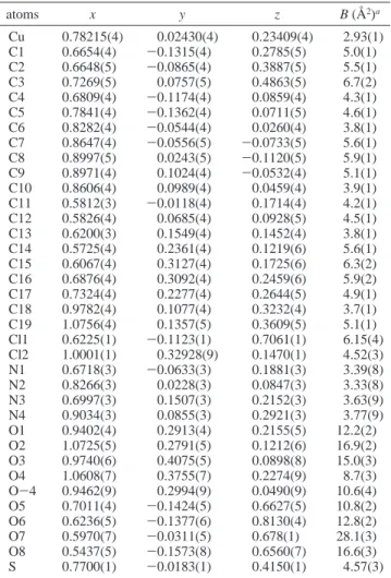
Figure 3. EXAFS data and simulations with CH3CN for copper(II) complex 3: (a) k3χ(k) and (b) radial distribution function (bold solid line, experimental signal; dashed line, simple scattering; straight solid line, multiple scattering).
Figure 4. EXAFS data and simulations without CH3CN for copper(II) complex 3: (a) k3χ(k) and (b) radial distribution function (bold solid line, experimental signal; dashed line, simple scattering; straight solid line, multiple scattering).
like CH3CN (Figures 3 and 4). When multiple scattering is neglected, the experimental curve can be fitted; however the corresponding Cu-S distance is not acceptable, 2.40 Å vs 2.335(1) Å for the crystallographic value. A shift of the S-peak is also observed when the CH3CN contribution is not taken into account in the FEFF calculations, and the experimental curve cannot be fitted, even giving Cu-S the crystallographic value. Therefore, it can be said that CH3CN is the major source of the multiple scattering effects on the first and second shells in compound 3.
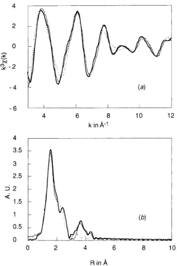
(ii) The presence of the S atom in the copper coordination sphere of compound 2 is clearly demonstrated in Figure 6.29 For this compound, the second peak of the radial distribution curve is not adequately modeled, and it is likely that there are multiple-scattering contributions from the aromatic groups. However, in the absence of CH3CN, the multiple scattering is less important, and the results are reliable.
Structures of complexes 2 and 3 are marked by some features which have been identified by this combined crystallographic
and XAS study. The change in the coordination geometry from a square-plane pyramid to a distorted tetrahedron is also completed by a shortening of the copper(I)-to-ligand distances. The most important variations are observed for the Cu-S distance, 2.34 Å in 3 and 2.14 Å in 2, and for the Cu-Nt distance, 2.06 Å in 3 and 1.84 Å in 2. According to the Cambridge Data Bank, such a short Cu(I)-N distance has been found in more than 20 crystallographic structures in which the nitrogen atoms have an sp2(enamine or aromatic) and an sp3 (five- or six-membered metallo-ring) hybridization (Scheme 3).
Reactivity with O2and H2O2. We did not detect any ligand oxidation with complexes derived from MeSPY2, 1, ligand, contrary to those complexes [RPY2]CuPF6for which oxygen atom transfers to the ligand (OATL) were observed upon dioxygen exposure.30,31 The clear yellow CH
2Cl2solution (1 mM) of [MeSPY2]CuPF6, 2, complex rapidly turned into a green solution upon exposure to an O2atmosphere. The stoichiometry of Cu/O2measured by manometry was 4:1. Precipitation with diethyl ether afforded a green powder corresponding to a copper(II) complex which released, in quantitative yield, unchanged ligand 1 after demetalation with a 35% NH4OH solution. The same result was obtained by reaction of the (29) The presence of the MeS group in the copper coordination sphere of
compound 2 is confirmed both by1H and13C NMR spectra where
large downfield effects are observed on MeS resonances (∆δ2- ∆δ1
) 2.21 - 2.03 ) 0.18 ppm for1H and∆δ
2- ∆δ1) 18.77 - 15.60
) 3.20 ppm for13C). See: Karlin, K. D.; Haka, M. S.; Cruse, R. W.;
Meyer, G. J.; Farooq, A.; Gultneh, Y.; Hayes, M. S.; Zubieta, J. J.
Am. Chem. Soc. 1988, 110, 1196. Gagne, R. R.; Kreh, R. P.; Dodge,
J. A.; Marsh, R. E.; McCool, M. Inorg. Chem. 1982, 21, 254. Nelson, S. M.; Lavery, A. Drew, M. G. B. J. Chem. Soc., Dalton Trans. 1986, 911.
(30) Amade´i, E.; Alilou, E. H.; Eydoux, F.; Pierrot, M.; Re´glier, M.; Waegell, B. J. Chem. Soc., Chem. Commun. 1992, 1782.
(31) Itho, S.; Kondo, T.; Komatsu, M.; Ohshiro, Y.; Li, C.; Kanehisa, N.; Kai, Y.; Fukuzumi, S. J. Am. Chem. Soc. 1995, 117, 4714. Figure 5. EXAFS data and simulations on first and second shells for
copper(II) complex 3: (a) k3χ(k) and (b) radial distribution function (bold solid line, experimental signal; dashed line, simulation without
S; straight solid line, simulation with S).
Figure 6. EXAFS data and simulations on whole signal for copper(I) complex 2: (a) k3χ(k) and (b) radial distribution function (bold solid line, experimental signal; dashed line, simulation without S; straight solid line, simulation with S).
copper(II) complex [MeSPY2]Cu(ClO4)2, 3, with dioxygen in the presence of reductant (benzoin/NEt329or ascorbate/phosphate buffer pH 7.4).32
Copper/dioxygen chemistry is well documented.33 The structures of copper(II) peroxo species formed upon dioxygen exposure depend on the ligand. Indeed, with tridentate ligands (RPY2,34HB(3,5-iPr
2pz)3,35and iPr3tacn36), copper(I) species are known to lead to side-on (µ-η2:η2-peroxo)dicopper(II) species upon reaction with O2. It was shown that side-on peroxo species which possess nonbasic/electrophilic properties37have oxidizing capability toward cyclohexene38and PPh
3.39 In some cases, this side-on peroxo species have the capability to insert an oxygen atom into a C-H bond of the ligand.40 Recently, Tolman et al. have reported that (µ-η2:η2-peroxo)dicopper(II) species can exist in equilibrium with a bis(µ-oxo)dicopper(III) species.41 These authors proposed that{Cu
2(µ-η2:η2-O2)}2+ and {Cu2(µ-O)2}2+ cores are isomers residing at the same
overall oxidation level and, therefore, each can be considered equally competent as potential oxidants in copper/dioxygen chemistry. Such {Cu2(µ-η2:η2-O2)}2+ and {Cu2(µ-O)2}2+ intermediates could be responsible for observed OATL in [RPY2]CuPF6chemistry (Scheme 4).30,31 Tripodal tetradentate ligand such as TMPA42and quinolyl-substituted43and binucle-ating analogues44give copper(I) complexes which bind O
2to (32) Uchida, K.; Kawakishi, S. Biochim. Biophys. Acta 1990, 347, 1034.
Uchida, K.; Kawakishi, S. Arch. Biochem. Biophys. 1990, 283, 20. (33) Karlin, K. D.; Kaderli, S.; Zuberbu¨hler, A. D. Acc. Chem. Res. 1997,
30, 139.
(34) Sanyal, I.; Mahroof-Tahir, M.; Nasir, S.; Ghosh, P.; Cohen, B. I.; Gultneh, Y.; Cruse, R.; Farooq, A.; Karlin, D. D.; Liu, S.; Zubieta, J.
Inorg. Chem. 1992, 31, 4322. Blackburn, N. J.; Strange, R. W.; Farooq,
A.; Haka, M. S.; Karlin, K. D. J. Am. Chem. Soc. 1988, 110, 4263. (35) Kitajima, N.; Fujisawa, K.; Moro-Oka, Y. J. Am. Chem. Soc. 1989,
111, 8975. Kitajima, N.; Moro-Oka, Y. Chem. ReV. 1994, 94, 737.
(36) Mahapatra, S.; Halfen, J. A.; Wilkinson, E. C.; Que Jr., L.; Tolman, W. B. J. Am. Chem. Soc. 1994, 116, 9785.
(37) Paul, P. P.; Tyeklar, Z.; Jacobson, R. R.; Karlin, K. D. J. Am. Chem.
Soc. 1991, 113, 5322.
(38) Kitajima, N.; Koda, T.; Iwata, Y.; Moro-Oka, Y. J. Am. Chem. Soc. 1990, 112, 8823.
(39) Karlin et al. have observed PPh3 oxidation with spectroscopically
characterized side-on (µ-η2:η2-peroxo)dicopper(II) species derived
from RPY2 ligands.35However, with X-ray characterized (µ-η2:η2
-peroxo)dicopper(II) species derived from HB(3, 5-iPr2pz)3 ligand,
Kitajima et al. observed the formation of Cu(I)-PPh3complexes with
concomitant liberation of O2and no PPh3oxidation.36
(40) Karlin, K. D.; Hayes, J. C.; Gultneh, Y.; Cruse, R. W.; McKown, J. W.; Hutchinson, J. P.; Zubieta, J. J. Am. Chem. Soc. 1984, 106, 2121. Mahapatra, S.; Halfen, J. A.; Wilkinson, E. C.; Que, L., Jr.; Tolman, W. B J. Am. Chem. Soc. 1994, 116, 9785. Mahapatra, S.; Halfen, J. A.; Wilkinson, E. C.; Pan, G.; Cramer, C. J.; Que, L., Jr.; Tolman, W. B J. Am. Chem. Soc. 1995, 117, 8865. Halfen, J. A.; Young, V. G., Jr.; Tolman, W. B J. Am. Chem. Soc. 1996, 118, 10920. Allen, W. E.; Sorrell, T. N. Inorg. Chem. 1997, 36, 1732.
(41) Halfen, J. A.; Mahapatra, S.; Wilkinson, E. C.; Kaderli, S.; Young, V. C., Jr.; Que, L., Jr.; Zuberbu¨hler, A. D.; Tolman, W. B. Science 1996, 271, 1397. Mahapatra, S.; Halfen, J. A.; Wilkinson, E. C.; Pan, G.; Xuedong, W.; Young, V. C., Jr.; Cramer, C. J.; Que, L., Jr.; Tolman, W. B. J. Am. Chem. Soc. 1996, 118, 11555. Mahapatra, S.; Halfen, J. A.; Tolman, W. B. J. Am. Chem. Soc. 1996, 118, 11575. (42) Jacobson, R. R.; Tyeklar, Z.; Farooq, A.; Karlin, K. D.; Liu, S.; Zubieta, J. J. Am. Chem. Soc. 1988, 110, 3690. Tyeklar, Z.; Jacobson, R. R.; Wei, N.; Murthy, N. N.; Zubieta, J.; Karlin, K. D. J. Am. Chem. Soc. 1993, 115, 2677.
(43) Karlin, K. D.; Wei, N.; Jung, B.; Kaderli, S.; Niklaus, P.; Zuberbu¨ler, A. D. J. Am. Chem. Soc. 1993, 115, 9506.
(44) Lee, D.-H., Wei, N.; Murthy, N. N.; Tyeklar, Z.; Karlin, K. D.; Kaderli, S.; Jung. B.; Zuberbu¨hler, A. D. J. Am. Chem. Soc. 1995, 117, 12498.
give end-on trans-(µ-1,2-peroxo)dicopper(II) products. Contrary to the side-on species, the end-on peroxide moieties are basic/ nucleophilic35and react with PPh
3to give Cu(I)-PPh3 com-plexes with concomitant liberation of O2, and no ligand oxidations are observed. Although it was not possible to detect any peroxo species by UV-vis spectroscopy (even at low temperature in CH2Cl2, -80°C), MeSPY2, 1, is a tetradentate ligand, and it is reasonable to propose the formation of a trans-(µ-1,2-peroxo) species upon reaction of [MeSPY2]CuPF6, 2, with O2. This intermediate which is basic/nucleophilic reacts with the electrophilic copper(I) complex 2 with O-O bond cleavage leading to a µ-oxo-Cu(II) complex (O2 4 e- re-duction).
More interesting is the reaction of the copper(II) complex [MeSPY2]Cu(ClO4)2, 3, with hydrogen peroxide. In the pres-ence of an excess of H2O2 (10 equiv in MeOH at room temperature) copper(II) complex 3, after demetalation (35% NH4OH solution), gave 84% of a new compound 4. A thorough analysis of the spectral data compared with those of an authentic sample obtained by oxidation of ligand 1 with meta-chloro-perbenzoic acid indicated that the sulfoxide 4 is the oxidation product of the ligand sulfide group.
As mentioned in the Introduction, the mechanism of DBH catalysis has been a subject of many studies6,7from which it is well established that a Cu(II)-OOH species formed by oxygen activation is responsible for the hydroxylation of dopamine. May et al. have reported that DBH catalyzes the conversion of phenyl-2-aminoethyl sulfides to the corresponding sulfoxides at a rate which is considerably higher than the hydroxylation of the corresponding carbon analogues.45 On the other hand, recent studies of Karlin’s group on copper-dioxygen chemistry have shown that Cu(II)-OOH species are able to transfer an oxygen atom to tetrahydrothiophene, giving the corresponding sulfoxide.46 Therefore, taking into account these results, we can reasonably propose that the first step of the reaction is the formation of a [MeSPY2]Cu(II)-OOH species. Then, the
oxygen atom transfer to the sulfide group could occur by a concerted mechanism; however, a single-electron transfer of the sulfide group leading to MeS•+and Cu(II)-O•radical species may be also considered. The strong tendency of the MeS•+ toward sulfoxidation produces the sulfoxide by rebounding with Cu(II)-O•radical species (Scheme 5).
Conclusion and Biological Relevance
The CuB(His)2X(H2O) four-coordinated geometry (without strong SMe binding) known for DBH and PHM is quite distinct from the square pyramidal CuN4S geometry (with strong equatorial SMe binding) of the copper complex 3 described in this paper. Our work has the merit to clarify some observations reported for the inactivation of DBH and PHM by hydrogen peroxide, and thus 3 can be considered as a good functional model.47 Indeed, some years ago Skotland and Ljones reported irreversible inactivation of the water soluble form of bovine adrenal DBH by hydrogen peroxide (or ascorbate) in the absence of any substrate.48 More recently, the same irreversible inactivation was reported by Merkler et al. for PHM.49 Considering that the H2O2(or ascorbate)-inactivated DBH binds copper more weakly than the native form, it was proposed that
(45) May, S. W.; Phillips, R. S. J. Am. Chem. Soc. 1980, 102, 5981. (46) Karlin, K. D.; Ghosh, P.; Cruse, R. W.; Farooq, A.; Gultneh, Y.;
Jacobson, R. R.; Blackburn, N. J.; Strange, R. W.; Zubieta, J. J. Am.
Chem. Soc. 1988, 110, 6769. Mahroof-Tahir, M.; Murthy, N. N.;
Karlin, K. D.; Blackburn, N. J.; Shaikh, S. N.; Zubieta, J. Inorg. Chem. 1992, 31, 3001. Karlin, K. D.; Cruse, R. W.; Gultneh, Y. J. Chem.
Soc., Chem. Commun. 1987, 599.
(47) Further modeling is needed to design the best copper ligand to achieve hydroxylation as in DBH and PHM. The approach used here needs to be improved to attain the goal of a copper complex with catalytic hydroxylation properties toward exogenous ligands. Thus, the problem to achieve association of the substrate into the copper coordination sphere still remains to be solved.
(48) Skotland, T.; Ljones, T. Arch. Biochem. Biophys. 1979, 201, 81. (49) Merkler, D. J.; Kulathila, R.; Tamburini, P. P.; Young, S. D. Arch.
Biochem. Biophys. 1992, 294, 594.
Scheme 5 Scheme 6. Mechanisms Proposed for Hydrogen Peroxide
Inactivation of DBH and PHM B
the enzyme-bound copper catalyzes the partial destruction of its own binding site by hydroxylation of one histidine residue. This hypothesis was supported by Uchida and Kawakishi who observed histidine-residue hydroxylation of angiotensin by copper(II) salts in the presence of ascorbate and dioxygen.32 However, in the light of the recent XAS studies of DBH and PHM active sites which show the presence of Met314and taking into account our present findings, we suggest that Met314could be the site of the inactivation by oxidation into a sulfoxide group (Scheme 6). Confirmation of these proposals and characteriza-tion of the modified protein will certainly provide valuable
information about the active site of DBH whose crystal structure is still lacking. This work is in progress in our group.
Acknowledgment. We acknowledge the use of the
synchro-tron radiation facilities and the help given by personnel at LURE and, more particularly, Dr. Isabella Ascone.
Supporting Information Available: Tables of positional and thermal parameters, interatomic distances and angles, and general displacement parameter expressions (B’s) for compound 3 (10 pages). Ordering information is given on any current masthead page. IC9709281