Caged phosphopeptides and phosphoproteins:
Probes to dissect the role of phosphorylation in complex signaling pathways
by
Elizabeth Maura Vogel
B.A. Chemistry Hamilton College, 2001
Submitted to the Department of Chemistry in Partial Fulfillment of the Requirements for the
Degree of Doctor of Philosophy
at the
Massachusetts Institute of Technology
February 2007
© 2007 Massachusetts Institute of Technology All rights reserved
Signature of Author: ,
-Department of Chemistry December 19, 2006
Certified by:.
Barbara Imperiali Class of 1922 Professor of Chemistry and Professor of Biology Thesis Supervisor
Accepted by:.
Robert W. Field Haslam and Dewey Professor of Chemistry Chairman, Departmental Committee on Graduate Students
ARCHIVES
MASSACHUSETTS INSTITUTEi OF TECHNOLOGYMAR
0 3 2007
LIBRARIES
lPThis doctoral thesis has been examined by a committee of the Department of Chemistry as follows:
Professor Timothy F. Jamison
Chairman
(I
Professor Barbara Imperiali Thesis Supervisor
Caged phosphopeptides and phosphoproteins:
Probes to dissect the role of phosphorylation in complex signaling pathways by
Elizabeth Maura Vogel
Submitted to the Department of Chemistry
on December 19, 2006 in partial fulfillment of the requirements of the Degree of Doctor of Philosophy in Organic Chemistry
ABSTRACT
Protein phosphorylation is a central regulatory mechanism in signal transduction pathways and cellular migration. Current genetic strategies for the study of phosphorylation, including gene knockout and point mutation, are limited in providing temporal information. As a complement to these techniques, the synthesis and semisynthesis of probes that enable researchers to observe the downstream effects of kinase-mediated phosphorylation in "real time" are presented in this thesis.
The release of a physiologically-relevant concentration of a phosphopeptide with temporal and spatial control is accomplished by the photolysis of a photolabile precursor, a caged phosphopeptide. The synthesis and application of NI-Fmoc-protected 1-(2-nitrophenyl) ethyl (NPE) caged phosphothreonine, serine, and tyrosine building blocks facilitate the straightforward assembly of any caged phosphopeptide through Fmoc-based solid phase peptide synthesis. Removal of the NPE caging group by irradiation with long-wavelength UV light generates a concentration burst of the corresponding phosphopeptide.
In addition, the installation of a caged phosphoamino acid into a full-length, multi-domain protein, the cellular migration protein paxillin, is described. A strategy, which is applicable to any expressible protein target, is detailed for the semisynthesis of a paxillin variant with a caged phosphorylated tyrosine at residue 31 of the 557-residue protein using native chemical ligation. Paxillin is a 61-kDa protein known to orchestrate the interaction of signaling proteins involved in cell migration by acting as a molecular adaptor, with the creation of specific binding sites dependent on paxillin phosphorylation. Therefore, the semisynthetic probe comprises the entire paxillin macromolecule, including all other binding and localization domains, which are essential for creating a native-like system to probe the effect of phosphorylation. The comprehensive biochemical characterization of the paxillin probe and quantification of uncaging
following irradiation with long-wavelength UV light are also described.
Additionally, the strategy developed for the paxillin semisynthesis was applied to incorporate a different unnatural amino acid, the fluorescent chemosensing residue Sox, into a protein-domain sensor for ERK2 kinase activity. The protein domain sensor demonstrated significantly improved sensitivity for ERK2 phosphorylation over the corresponding peptide probe.
Thesis Supervisor: Barbara Imperiali
Acknowledgements
First, I thank my advisor, Professor Barbara Imperiali, for her creativity, her wealth of ideas, and her support. She has created a lab environment that has been a joy to work in for the past five years. In addition, I have learned much from the breadth of her scientific knowledge and interests, and through her inspiring skills at teaching, both within the group and in the classroom. It has been a true privilege to be a part of the Imperiali lab.
Next, I thank all the past and present members of the Imperiali group, who have made day-to-day research particularly fun, and who have provided so many ideas, patient instruction, and great friendships. Debbie Rothman and Melissa Shults have been both appreciated scientific advisors and dear friends. I thank Debbie for taking me under her wing when I first joined the lab, and for her great energy and enthusiasm for all she does. To Melissa Shults, for inspiring me with her dedication and work ethic and for her thoughtful and sincere attention to others. Many thanks to Jebrell Glover for his expertise and patience in teaching me molecular biology. To Elvedin Lukovic, Dora Carrico Moniz, Mary O'Reilly, Bianca Sculimbrene, Anne Reynolds, Galen Loving, Langdon Martin and Eranthie Weerepana, I thank them for their trusted scientific advice and their valued friendships. I would also like to thank and specially acknowledge the Imperiali-Red Sox alliance of Debbie, Melissa, Bianca, Langdon, Anne, and Seungib Chou. A special thanks to Brenda, Dora, Anne, Alex, and Elvedin for each tackling a chapter of my thesis with their brilliant editing skills. Thank you and best of luck to Matthieu Sainlos, Andreas Aemissegger, Nelson Olivier, Mark Chen, and our great second years, Brenda Goguen, Meredith Hartley, Wendy Iskenderian, and Angelyn Larkin.
I would also like to thank Professor Tim Jamison for his guidance as my thesis chair. An enormous thank you to Elizabeth Fong and Susan Brighton for all that they do to make the lab and department run smoothly.
I have many people I would like to acknowledge outside of MIT. I thank Professor Ian Rosenstein for first sparking my enthusiasm for organic chemistry and for being and incredible teacher and mentor. To Chris Steed and John Doench, for being like brothers to me, and for their support and friendship. I especially thank my amazing girlfriends, Janet Damaske, Meg Hem Steed, my roommate Eliz Guancial, Lynne Myrth, Amy Lawrence, and Catherine Wolf.
I thank Alex Taylor for being my most trusted editor, for his fantastic ideas, and for all the happiness he brings me.
And most importantly, I thank my family (ALL of you), and particularly Alex, Mom, Dad, Sara, Kev, Ty, Jilly, and Auntie. You make my life in every way joyful. I dedicate this thesis to you.
Table of Contents Abstract... ... ... 3 Acknowledgements ... ... 4 Table of Contents ... .. 5 List of Figures ... 7 List of Schemes ... ... List of Abbreviations... 10
Chapter 1 Introduction to native chemical ligation: semisynthesis of post-translationally modified proteins and biological probes 1-1 Introduction ... 12
1-2 Engineering design considerations for NCL ... 13
1-3 Thioester generation... 17
1-4 N-terminal Cys fragments ... ... 22
1-5 Extensions of NCL... 23
1-6 Applications ... ...24
Chapter 2 Synthesis and characterization of caged phosphopeptides Introduction ... 53
Results and Discussion ... 55
2-1 Interassembly approach ... 56
2-2 Building block approach ... 58
2-3 Quantum yield calculation... 60
Conclusion...61
M ethods ... 61
Acknowledgements...70
References ... 70
Chapter 3 Semisynthesis of caged phosphoTyr31 paxillin Introduction ... .. 73
Results and Discussion ... 77
3-1 Overview of paxillin semisynthesis... 77
3-2 Synthesis of paxillin peptide thioesters ... 78
3-3 Expression of the C-terminal precursor for NCL ... 80
3-4 Protease selection...85
3-5 Native chemical ligation ... 89
C onclusion ... 92
Acknowledgements ... 98
R eferences... ... 98
Chapter 4 In vitro characterization of semisynthetic paxillin analogs and preliminary cellular studies on adhesion dynamics Introduction ... . ... 101
Results and Discussion ... ... 104
4-1 Paxillin binding assays ... 104
4-2 In vitro paxillin phosphorylation ... 107
4-3 Uncaging of cpY31 paxillin ... 110
4-4 Phosphorylation dependent Crk binding ... 112
4-5 Focal adhesion turnover assays ... 113
C onclusion ... .. 117
M ethods ... .. 118
Acknowledgements ... 126
R eferences ... .. 126
Chapter 5 Semisynthesis of a Sox-based chemosensor for ERK2 phosphorylation Introduction ... 129
Results and Discussion ... 132
5-1 Design of the Sox-based ERK2 phosphorylation sensor ... 132
5-2 Preparation of the C-terminal sensor fragment ... 134
5-3 Synthesis of peptide thioesters... 134
5-4 Test ligation and product isolation with Cys-PNT(46-138)-His 6... 137
5-5 NCL of the first generation ERK2 kinase sensor...139
C onclusion ... .. 140
Acknowledgements ... 141
M ethods ... .. 147
R eferences ... .. 147
List of Figures Chapter 1
1.1 Native chemical ligation of unprotected polypeptide fragments...13
1.2 Mechanism for NCL with thiol catalysis... 16
1.3 Fmoc-based SPPS methods for a-thioester synthesis...19
1.4 M echanism of protein splicing... ...21
1.5 Recombinant methods for generating N-terminal Cys-containing fragments...23
1.6 Sequential NCL ... . ... 25
1.7 Semisynthesis of phosphorylated and glycosylated proteins ... 29
1.8 Semisynthesis of proteins with post-translational lipidation ... 32
1.9 Fluorescent labeling of AANAT and GST...34
1.10 Unnatural amino acids incorporated into semisynthetic proteins ... 36
1.11 Applications of unnatural amino acids introduced into proteins by NCL...40
1.12 Immobilization of proteins onto microarrays ... 43
Chapter 2 2.1 A 2-nitrophenylethyl-caged phosphopeptide ... 54
2.2 Caged phosphopeptide cpChk2, Ac-MARHFD(cpT)YLIRR-NH 2 . . . ..57. .
2.3 Possible oxidation of methionine and tryptophan residues ... 58
2.4 Structure of caged phosphoamino acid building blocks for Fmoc-bases SPPS....59
Chapter 3 3.1 Cellular migration... ....... 73
3.2 Caged phophoTyr31 paxillin... ... .... 74
3.3 Paxillin structural domains and selected paxillin-interacting proteins ... 75
3.4 Semisynthesis of paxillin analogs ... .. 78
3.5 Representative paxillin expression in the BL21 cell line ... 81
3.6 Complete isolation of GST-PCS-Cys-Pax(38-557)-FLAG ... 83
3.7 Purified GST-paxillin fusion protein following expression in codon-enhanced cells and purification via the N and C-terminal tags ... 84
3.8 Treatment of GST-IEGR-Cys-Pax(38-557)-FLAG with Factor Xa ... 87
3.9 TEV protease cleavage of GST-ENLYFQ-Cys-Pax(38-557)-FLAG...89
3.10 The reaction of an N-terminal cysteine residue with glyceraldehydes ... 90
3.11 Semisynthesis of caged phosphorylated paxillin ... ...93
Chapter 4 4.1 Semisynthetic paxillin analogs ... 101
4.2 Paxillin with selected binding partners and upstream kinases ... 102
4.3 Cartoon of a migrating cell ... ... 103
4.4 Elutions from GST-affinity purifications of FAK, GIT, PTP-PEST, and GST... 105
4.5 GST and GST fusion proteins following 1 week of storage at 4 C ... 105
4.7 Paxillin phosphorylation by Src, ERK, and JNK kinases ... 109 4.8 Chemiluminescent detection of ligated paxillin (21a) ... 110 4.9 Uncaging of cpY31Pax (21c) ... 111 4.10 Analysis of paxillin-CrkSH2 binding prior to and following uncaging of 21c...113 4.11 Assay protocol to probe the effect of Tyr-31 paxillin phosphorylation on focal
adhesion dynamics ... ... .... 114 4.12 Snapshots from focal adhesion movies of MEF cells ... ... 1...15
Chapter 5
5.1 Sox-based fluorescent chemosensors for kinase activity ... 130 5.2 Structure of the ERK2 kinase and of Ets- 1 residues 1-138 ... 131 5.3 ERK2 Sox-sensor design ... 133 5.4 Expression and TEV proteolysis of GST-ENLYFQC-PNT(46-138)-His 6 (24)..135
5.5 PNTD peptide thioesters for ligation to Cys-PNT(46-138)-His6 (24)...1..36 5.6 Test ligation and purification of Ac-FLAG-Cys-PNT(46-138)-His 6 (27)...138 5.7 Analysis of PNT domain stability following NCL ... 139 5.8 NCL to generate the ERK2 kinase activity sensor 22a...140 5.9 Direct comparison of ERK2 activity sensing of the Sox protein 22a versus the
List of Schemes Chapter 2
2.1 Photolysis of molecules masked by o-nitrobenzyl-derived caging groups...55 2.2 Synthesis of phosphitylating agent 1 ... 56 2.3 Synthesis of a caged phosphothreonine peptide via the interassembly approach..57 2.4 Synthesis of N-a-Fmoc-phospho(1-nitro-phenylethyl-2-cyanoethyl)-L-theonine,
10, for Fmoc-based SPPS ... 60
Chapter 3
List of Abbriviations
Standard one-letter and three-letter codes are used for the 20 natural amino acids.
Ac Acm ATP BAL BME BSA calcd CBD CHEF Cit CNBr C-terminus DCM DDI DIPEA DMF DMSO DTT ECM EDT EDTA ENLYFQC EPL EPR ERK ESI-MS FAK Fmoc FHA FRET GFP GIT GPI GST IMPACT HATU HBTU Hepes HOBt HPCL acetyl acetamidomethyl adenosine-5'-triphosphate backbone amide linker beta-mercaptoethanol bovine serum albumin calculated
chitin binding domain
chelation enhanced fluorescence citrulline
cyanogen bromide carboxy-terminus dichloromethane
DNA directed immobilization N,N-diisopropylethylamine N,N-dimethylformamide dimethylsulfoxide dithiothreitol extracellular matrix ethanedithiol ethylenediaminetetraacetic acid
TEV protease cleavage sequence (Glu-Asn-Leu-Tyr-Phe-Gln-Cys) expressed protein ligation
electron paramagnetic resonance extracellular signal regulated kinase electrospray ionization mass spectrometry quantum yield of uncaging
focal adhesion kinase
N-cz-fluorenylmethoxylcarbonyl fork head associated
fluorescence resonance energy transfer green fluorescent protein
GRK interactor 1
glycosyl phosphatidylinositol glutathione S-transferase
intein-mediated purification with affinity chitin binding tag O-(7-azabenzotriazol-1-yl)-1,1,3,3-tetramethyluronium hexafluorophosphate 2-(1H-benzotriazole-l -yl)-1,1,3,3-tetramethyluronium Hexafluorophosphate N-(2-hydroxyethyl)piperazine-N'-ethanesulfonic acid N-hydroxylbenzotriazole
IEGR IPTG MALDI MS MAPK MBP mCPBA MeCN MEF MESNA MPPA N-terminus Nle NCL NPE OD PBS PBS PCS PDE PNT domain Pfa PKL Pma Pmp PNA PTP pSer pThr pTyr PyBOP RBD SDS-PAGE SARA SeM SH2 SPPS tl/2 TBS TEV TFA THF TIRF TMS TNBS UV
FLAG protease cleavage sequence (Ile-Glu-Gly-Arg) isopropyl-3-D-thiogalactopyranoside
matrix assisted laser desorption ionization mass spectrometry mitogen-activated protein kinase
myelin basic protein
meta-chloroperbenzoic acid acetonitrile
murine embryonic fibroblast 2-mercaptoethanesulfonic acid (4-carboxymethyl)thiophenol amino-terminus
norleucine
native chemical ligation 2-nitrophenylethyl optical density
phosphate buffered saline paxillin binding subdomain protease cleavage sequence phosphodiesterase
pointed domain
phosphono-difluoro-methylenealanine paxillin kinase linker
phosphonomethylenealanine
phosphonomethylene phenylalanine polyamide nucleic acid
protein tyrosine phosphatase phosphoserine
phosphothreonine phosphotyrosine
benzotriazole- 1-yl-oxy-tris-pyrrolidino-phosphonium hexafluorophosphate
Ras binding domain
sodium dodecyl-sufate polyacrylamide gel electrophoresis Smad anchor for receptor activation
selenomethionine Src homology 2
solid phase peptide synthesis half life
tris buffered saline tobacco etch virus trifluoroacetic acid tetrahydrofuran
total internal reflection fluorescence trimethylsilane
trinitrobenzene sulfonic acid ultraviolet
Chapter 1. Introduction
NCL: Semisynthesis of post-translationally modified proteins and biological probes
A version of this chapter has been submitted for publication in Protein Engineering. Copyright © 2006 Springer.
1-1. Introduction to native chemical ligation
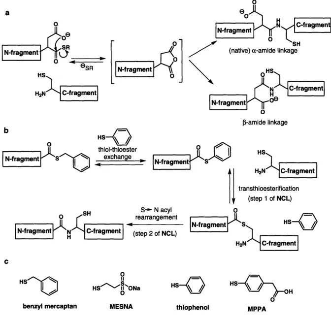
Methods for the modification of proteins to introduce novel chemical functionalities and for the preparation of homogenous samples of post-translationally-modified proteins are critical for probing protein structure/function relationships and protein/protein interactions. Native chemical ligation is a chemoselective reaction that joins synthetic or biologically-expressed protein fragments via a native amide bond, enabling the construction of modified proteins in multi-milligram quantities sufficient for biophysical and biochemical studies.' Native chemical ligation can be carried out with fully deprotected peptide and protein fragments in neutral aqueous media, enabling modified peptides to be incorporated into target peptides or proteins. Required for the reaction are an N-terminal fragment containing a C-terminal (c-thioester, and a C-terminal fragment with an N-terminal cysteine residue. The general reaction is shown in Fig. 1.1 a. The transformation begins with a reversible transthioesterification to associate the two fragments via a thioester bond involving the cysteine residue of the C-terminal fragment. An energetically favorable acyl rearrangement involving a 5-membered ring transition state then occurs to form a stable amide bond. Since the thioester-linked intermediates have never been observed, it is presumed that the rate-limiting step of NCL is transthioesterification. Typically, the reaction is run in the presence of thiol additives, which both suppress cysteine oxidation and catalyze the reaction by generating more reactive thioesters.2 Importantly, NCL is compatible with the presence of fully-deprotected internal cysteine residues in either protein fragment (Fig. 1.1b). Thioester exchange is fully reversible, and it is only upon reaction with the terminal cysteine that the S - N acyl rearrangement can occur, resulting in the thermodynamically stable
NCL was first introduced in 1994 with the synthesis of a 72-residue polypeptide comprising entirely native peptide bonds, thus overcoming the practical limits of SPPS through the ligation of two fully deprotected peptide fragments.' While NCL was originally limited to ligations involving a-thioesters generated by chemical synthesis, the scope of the reaction was advantageously extended to proteins of any size through the application of 'expressed protein ligation' (EPL), in which a-thioester or N-terminal Cys-containing fragments that are generated via recombinant methods are used for the semisynthesis of protein domains and full-length proteins.3-5 EPL was originally defined specifically as the extension of NCL that involves recombinant a-thioester peptides, but the term is now used more generally to designate NCL processes involving any recombinant fragments, either thioester or N-terminal Cys-containing.6
0 transthioesterification thiol, neutral pH N-terminal
(N4
polypeptide C-terminal H2N polypeptide S--N acyl rearrangement b 0O N-terminal(4
polypeptide eSR HS H2N C-terminal 2N polypeptide transthioesterification IN-termmalI polypeptide H21 H! e 0 S N-terminal N C-terminal polypeptide H polypeptideFigure 1.1. a) Native chemical ligation (NCL) of unprotected polypeptide fragments. b) Reversible transthioesterification with internal cysteine residues.
1.2 Engineering design considerations for NCL
An important consideration in the design of semisynthetic proteins is the designation of a ligation junction, Xaa-Cys. Since the practical limit of solid phase
peptide synthesis (SPPS) is between 40 and 60 residues, junction sites for ligations involving a synthetic peptide must fall within those distances from the N- or C-terminus of the protein target. In some cases naturally-occurring cysteine residues are present in the protein of interest at a suitable position for the ligation. However, in the absence of a suitably placed cysteine, one may be introduced in the place of a non-essential residue. Appropriate residues may be selected based on structural knowledge or previous experiments that demonstrate that the residues are not involved in protein function. The ease of site-directed mutagenesis allows for further confirmation that the cysteine mutation does not significantly alter protein structure or function. In general, ideal ligation sites are in segments of a protein that lack secondary structure, such as in terminal regions, loop regions or linker regions between two domains. If the reaction is carried out under non-denaturing conditions, and if the protein will not be subjected to refolding, it is particularly important that the folded state of the protein is not disturbed by the ligation site, regardless of whether a naturally-occurring or an introduced cysteine residue is used as the Xaa-Cys junction.
While standard NCL dictates that a cysteine residue be present at the N-terminus of the C-terminal fragment, it is also important to consider the effect of the C-terminal residue of the thioester fragment, -XaSR, on the ligation. It has been reported that a-thioester peptides containing any of the 20 encoded amino acid residues at the terminal position are compatible with NCL, but the kinetics of ligation differ dramatically depending on the properties of that residue.7 In general, the reaction proceeds most rapidly when the C-terminal position is occupied by the sterically-unhindered glycine residue, or by histidine or cysteine residues, which are hypothesized to increase the rate of thioester exchange by catalysis with the imidazole or thiol side-chain functionalities. Other terminal residues shown to result in rapid product formation are phenylalanine, methionine, tyrosine, alanine, and tryptophan. Ligation has been found to be prohibitively slow only with two of the n-branched amino acids (valine and isoleucine) and proline, although it may be possible to overcome these residue limitations using highly-activated thioesters.
In another study, ligations involving aspartic or glutamic acid residues in the C-terminal position of an a-thioester were shown to result in significant side product
formation.8 In this case, the proximity of the carboxyl functionality in the amino acid side chains to the activated thioester can result in the formation of backbone isomers, in which a 3- or y- amide bond forms between the Cys-containing peptide and the aspartate or glutamate residues respectively (Fig. 1.2a). Side product formation can be avoided by orthogonal protection of the carboxyl side chains. However, when possible, selecting an Xaa-Cys ligation site that avoids sluggish ligations or side reactions is beneficial to ensure maximal product formation.
Another factor affecting the efficiency of NCL is the presence of thiol additives,2 which potentially combat difficult ligation reactions. The alkylthioesters commonly used in NCL react slowly because of the poor leaving group properties of the corresponding alkyl thiols, requiring thiol-thioester exchange with the thiol additive to promote ligation with the Cys-containing fragment. (To minimize confusion, this exogenous thiol replacement will be referred to throughout the chapter as thiol-thioester exchange to distinguish it from the attack of a Cys residue in the first step of NCL, which will be referred to as transthioesterification.) Rapid and complete ligation is best facilitated by thiols that are both good nucleophiles, to promote the in situ formation of a more reactive thioester, and good leaving groups, to favor the transthioesterification (Fig. 1.2b). For NCL involving peptide fragments, a benzylmercaptan/thiophenol mixture is commonly employed in an aqueous/organic buffer (Fig. 1.2c). Thiophenol promotes rapid thiol-thioester exchange and serves as a good leaving group, but has poor aqueous solubility and therefore can only be used at very low concentrations for ligations involving proteins that cannot tolerate the addition of organic solvent. MESNA (2-mercaptoethanesulfonic acid) has excellent solubility in water and is a popular choice for ligations using recombinant protein fragments. However, a recent investigation of a number of thiol additives revealed that MESNA, an alkanethiol with pKa of 9.2, shows rapid thiol-thioester exchange, but has poor leaving group properties.9 In the case of MESNA, the rate limiting step for NCL is transthioesterification, whereas with certain other thiols the thiol-thioester exchange is rate limiting. In general alkylthioesters, such as those generated by MESNA or benzyl mercaptan, were found to be less reactive than phenylthioesters. The investigators found (4-carboxymethyl)thiophenol (MPPA) to be a superior catalyst with peptide-based test ligations and recommend use of MPPA for
protein ligations as well, predicting that it may be more effective than MESNA for EPL. Since this report was published within one month of writing this chapter, there have been no other reports to date that apply MPPA, and it will be interesting to see if MESNA is eventually replaced as the predominantly used thiol for EPL by MPPA or another aryl thiol catalyst. Another option for rapid ligation is the use of preformed aryl thioesters,1'1'(", which eliminate the thiol-thioester exchange step that is rate limiting with certain thiol additives.
o
a 0
O N C-fragment
SSN-fragmentH
'S
FI 0 SHN-fragment (native) a-amide linkage
So
9 e .k~"
l~"-'-',t
0 Pj-amide linkageb
0 thiol-thioester ON-fragment ••
N-fr••agment1s'o
< > -fra ment Sl
HS H2N transthioesterification (step 1 of NCL) S-- N acyl 0 SH rearrangement -fragment S HSN-ragment N C-fragm (step 2 of NCL)
H2N C-fragment
HSbenzyl
mercaptan
benzyl mercaptan HSO ' ONa 0 MESNAHS
-
o thiophenol HS OH MPPAFigure 1.2. a) Generation of a 3-amide-linked side product for NCL with an Asp-Cys ligation junction. b) Mechanism for NCL with thiol catalysis. An ideal thiol additive results in rapid thiol-thioeser exchange of the thioester fragment and provides a good leaving group for transthioesterification with the Cys-fragment. c) Thiols used for NCL.
1.3 Thioester generation
1 Synthetic thioester peptides
The synthesis of a-thioester peptides for NCL can be accomplished using a variety of SPPS-based methods. The modular nature of SPPS allows facile replacement of any residue of the target peptide thioester with non-encoded amino acids, as well as the
site-specific incorporation of tags and probes. Initially most investigators employed acid-labile Boc-protection strategies for SPPS,1"12 since the thioester linkage is not stable to the
basic deprotection treatment with piperidine used in Fmoc-based SPPS. However, a number of approaches have been developed to circumvent the sulfur-carbonyl cleavage associated with Fmoc deprotection, allowing the generation of thioester peptides without the harsh cleavage conditions employed in Boc-based SPPS. This use of milder conditions is particularly important for the synthesis of glycopeptide and phosphopeptide thioesters, since glycoside and phosphoryl linkages are labile to the anhydrous HF required for peptide cleavage in Boc-based SPPS.
In one Fmoc-based approach, deblocking agents were developed as alternatives to piperidine to effectively remove the Fmoc protecting group without nucleophilic cleavage of the thioester linkage.13,14 In an alternative strategy, a backbone amide linkage (BAL) is used to anchor the penultimate residue, which contains an orthogonally protected C-terminus, to the solid support.15 The peptide can be elaborated using standard Fmoc-based SPPS protocols, and subsequently deprotected at the C-terminus prior to being coupled to a thioester amino acid and finally fully deprotected and cleaved from the resin (Fig. 1.3a). Additional BAL and side-chain linkage-based approaches have been reported,16 including an example that masks the terminal glycine thioester as a trithioortho-ester and releases the deprotected thioester by acid treatment.17
A more commonly applied strategy utilizes an alkanesulfonamide "safety-catch" linker, in which a peptide is assembled on solid support linked via the C-terminus by a sulfonamide bond. This sulfonamide linkage is stable to the repeated piperidine deprotection treatments in Fmoc-based SPPS. Following peptide synthesis, the sulfonamide is activated by N-alkylation and subsequently cleaved from solid support by nucleophilic attack of a thiol additive. The thioester bond is stable in TFA, and peptide can therefore be deprotected with a standard TFA cleavage cocktail (Fig. 1.3b).'8 19 A
similar method employs an aryl hydrazide linker that can be activated following peptide synthesis by mild oxidation.2 0
Arguably the most straightforward Fmoc-based SPPS approach for generating thioesters involves peptide synthesis on highly acid-labile resin, such as commercially available 2-chlorotrityl or TGT resin, followed by cleavage of the fully-protected peptide from solid support with mild acid treatment. The acid releases the peptide as a
C-terminal carboxylic acid, but does not affect the Fmoc-compatible side-chain protecting groups. The protected peptide is then derivatized to a C-terminal thioester in situ by treatment with the desired thiol and standard peptide-coupling activating agents.2 1'22 Side-chain deprotection affords the corresponding free thioester (Fig. 1.3c). An evaluation of activating agents for this strategy has identified conditions, specifically phosphonium-salt based activating agents, that result in high yields and low levels of epimerization (<1.4%).10
While still in a preliminary stage of development, a final method of o-thioester formation is worth mentioning both for its creative approach and its similarity to the protein splicing mechanism that is exploited for the generation of recombinant protein a-thioesters (Sect. 1.3.2). This method involves the generation of a thioester-linked peptide by utilizing an N-S acyl shift facilitated by a protected thiol-containing auxiliary attached to the peptide backbone (Fig. 1.3d).23 Following Fmoc-based SPPS of a peptide linked to resin by an amide bond, deprotection of the peptide side chains with a TFA cleavage cocktail concurrently deprotects the thiol moiety of the auxiliary, initiating an acyl shift that results in a thioester bond-linkage of the peptide to solid support. Subsequent thiolysis releases the corresponding peptide thioester.
A recent review of methods for thioester synthesis can provide more information on these and other approaches.24
2. Recombinant thioester fragments
While direct synthesis is suitable for peptide fragments under approximately 60 residues in length, the scope of NCL was initially limited by the lack of recombinant methods to generate longer peptide or protein fragments containing C-terminal a-thioesters. This limitation was overcome in 1998 with the introduction of EPL, which
a
t-Bu BocI t-Bu Boc
peptide 1 I I
pNideN
1) Allyl deprotection peptide 1 \ deprotection ca peptide N SRAllylO R 2) coupling of terminal Ol HN a R'
amino acid thioester R . 0 R'
0 RS) T H •' "R
R' O
t-Bu Boc t-Bu Boc
t-Bu Boc
Speptide eptdeNi peptide ý SR SR' peptide SR'
' O'O 0i activationn 01 0a thiolysis o deprotection
O o O deprotection
C
t-Bu Boc
t-Bu Boc
t-Bu Boc
peptide peptide OH peptide SR peptide SR
O Ph cleavage 0 thioester- ification deprotection
o o
d
t-R.
Ron Sdeprotection/ N-S acyl shift TrtS -OMe OMe 0 thiolysis Fder SRFigure 1.3. Fmoc SPPS-based methods for peptide a-thioester synthesis include the a) BAL strategy, b) "safety catch" linker strategy, c) in situ thioesterification of a protected peptide, and d) thiolysis following a thiol-auxiliary mediated N - S acyl shift.
applies a modified protein splicing mechanism to generate thioesters suitable for ligation.3'4 To place the technology developed for recombinant protein thioesters in context, a brief explanation of protein splicing follows.
Protein splicing Protein splicing is a protein processing event that results in the extrusion of an internal protein segment, termed an intein, and the concomitant joining of two flanking regions, termed N- and C-exteins, through an amide bond.25 Inteins, which
catalyze the intramolecular protein rearrangement that mediates their excision (Fig. 1.4a). A number of conserved or key residues in inteins contribute to the splicing event through structural or electronic influences.26 In general, inteins contain a cysteine or serine residue at the N-terminal position (termed the 1 position), a conserved asparagine residue at the C-terminal position, and a cysteine, serine, or threonine residue at the first position of their flanking C-extein (termed the +1 position). In the initial step of standard protein splicing, an N-S (or 0) acyl shift occurs, involving the cysteine (or serine) at position 1, transferring the N-extein from the backbone to the side chain of residue 1 of the intein. While this step appears thermodynamically unfavorable, it is favored by the conformation of the intein, which is thought to distort the scissile amide bond into a higher energy conformation. In the next step, transesterification involving the cysteine (or serine or threonine) at position +1 joins the two exteins through a thioester bond. In the third step, the asparagine residue cyclizes, resulting in cleavage at the C-terminal splice junction to liberate the intein as a C-terminal succinimide. In the final step, which is the sole reaction that does not require catalysis by the intein, a spontaneous acyl rearrangement produces the spliced exteins with an amide linkage.6
Mutant (Asn to Ala) inteins to generate recombinant thioesters Protein splicing
has been exploited with great success for the generation of recombinant thioesters. Inteins have been engineered with an asparagine to alanine mutation, which permits the initial step of the protein splicing mechanism involving an N - S acyl shift to produce a thioester linkage, but prevents subsequent succinimide formation.27 Addition of exogenous thiols results in release of the N-extein as the corresponding C-terminal thioester. The isolation of recombinant thioesters from a resin-bound intein system has been commercialized by New England Biolabs as the IMPACTTM (intein-mediated purification with an affinity chitin binding tag) -system. In this system, a target gene, functioning as the N-extein, is cloned immediately N-terminal to a genetically modified (Asn to Ala) intein gene. A chitin binding domain (CBD) is cloned C-terminal to the intein, functioning as the C-extein and facilitating immobilization of the resulting protein construct on chitin beads. Thus, the expressed three-segment (target protein-intein-CBD)
construct can be isolated from all other cellular proteins by immobilization and washing, and the target protein thioester can be released by subsequent thiolysis (Fig. 1.4b).28
Expressed protein ligation can be performed with the eluted thioester or directly on the chitin beads.3'4'29 For solid phase ligation, the thiol and Cys-containing peptide/protein fragment can be simultaneously incubated with the resin-bound protein fusion, enabling concurrent thiolysis and ligation.
position 1 position +1 b a Cys Cvs N-extein ste N to S ac acyl transfer t~rn~t i
(
I o
O modifiedo
I _ H2N intein +k;,-,h,=;=) k·, 4·r exogenous thiol I-succinimide formation step 4: S to N acyl transfer HS V 0H2N m dif iedintein
01
SH
HS
N-extein N C-extein H2N intein 0
NH H20
Figure 1.4. a) Mechanism of protein splicing. Splicing results in the joining of two exteins through a native peptide bond and the extrusion of the intein segment. b) Recombinant generation of an a-thioester using a modified intein system. The target protein is N-terminal to the intein, and a chitin binding domain (CBD) is C-terminal to the intein, immobilizing the construct on solid support. An Asn to Ala mutation prevents succinimide formation. Thiolysis results in release of the a-thioester target protein.
i E
1-4. N-terminal Cys fragments
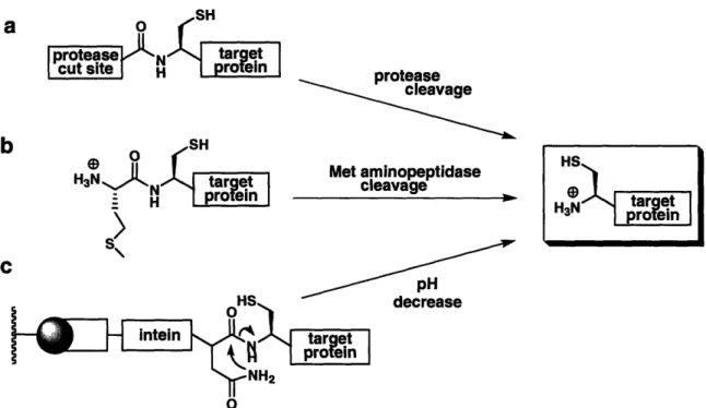
The synthesis of peptides containing an N-terminal cysteine residue is straightforward using SPPS and requires no additional manipulations. For longer fragments, there are several approaches for accessing biochemically-expressed proteins with an N-terminal cysteine. The most commonly applied methods involve the generation of a precursor protein designed with a cysteine residue immediately C-terminal to a protease cleavage site (Fig. 1.5a). The first example of affinity cleavage for NCL used factor Xa, a protease that cuts C-terminal to its Ile-Glu-Gly-Arg recognition sequence.5 Other terminal cleaving proteases include enterokinase, ubiquitin C-terminal hydrolase, and furin. The single disadvantage with employing proteases is the possibility for undesired cleavage at secondary sites. For instance, factor Xa can cleave after Gly-Arg pairs or other basic residues in a target protein.
Recently tobacco etch virus (TEV) protease, a highly specific cysteine protease with a seven-residue recognition sequence, was applied for the generation of N-terminal Cys fragments.3 0 TEV typically recognizes a Ser or Gly residue in the Pl' site, but will
also tolerate a Cys in that position. TEV demonstrates high sequence selectivity and overexpresses well on a large scale, making it an ideal protease for EPL applications.
Endogenous Met aminopeptidases have also been utilized to access N-terminal Cys proteins from the corresponding Met-Cys containing precursor proteins (Fig. 1.5b). The resulting Cys-polypeptides can be isolated from cell lysate using aldehyde-functionalized resin,3 1 or reacted directly for in vivo NCL.32 For exogenous cleavage of a Met-Cys junction, cyanogen bromide (CNBr) was successfully applied to access an N-terminal Cys in a recombinant glycoprotein that was insoluble in buffers compatible with the commonly employed proteases.33
An intein-based strategy can also be applied to generate N-terminal Cys proteins (Fig. 1.5c). Several commercially available expression vectors contain genetically modified inteins that lack the conserved cysteine (or serine) residue at the N-terminus (1 position) of the intein for this purpose. Cleavage of the intein by succinimide formation, induced by lowering the pH and increasing temperature, simultaneously releases the N-terminal Cys protein.28 The limitation to this method is the possibility for spontaneous
SH
a
o
protease N target
cut site H protein
cleavage
L Ou
H)N N H p-ropa target Met aminopeptidase cleavage H "
S >;
C oHS decrease intein t NH2 0Figure 1.5. Recombinant methods for generating N-terminal Cys-containing protein fragments using a) exogenous protease, b) endogenous methionine aminopeptidase, and c) a genetically modified intein system.
1-5. Extensions of NCL 1. Sequential NCL
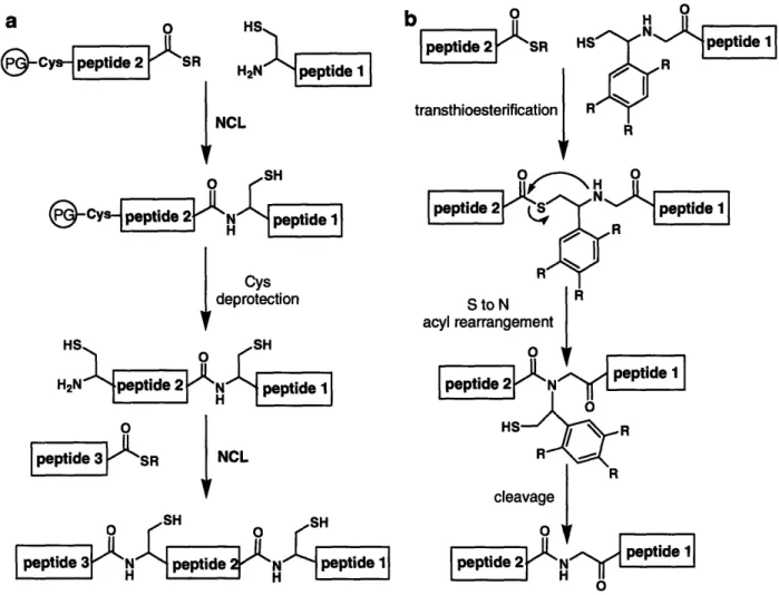
Although many NCL applications require the ligation of only two fragments, the technique is not limited to a single ligation step. Multiple modifications throughout a protein or modification in the middle of a large protein can be accomplished by sequential ligations (Fig. 1.6a).34 In sequential NCL, an initial ligation is carried out between an N-terminal Cys-containing fragment and a second fragment containing a thioester moiety and a protected N-terminal Cys residue, masked to prevent the thioester fragment from undergoing intra- or intermolecular ligations. Strategies used to mask the cysteine residue include the incorporation of the factor Xa pro-sequence immediately N-terminal to the cysteine residue,34 acetamidomethyl (Acm) protection of the side-chain thiol,3 5 thiazolidine cysteine protection,36 and photolabile protection of the thiol or amino group.37 Following the initial ligation, the cysteine is deprotected and a second ligation is carried out. Theoretically there is no limit to the number of fragments that can be ligated
in this fashion. Segmental ligation has been performed on solid support, in a strategy analogous to SPPS,38 as well as in situ, either in "one pot" reactions 37,39 or with HPLC or
affinity-tag purification of the ligated intermediates.39
2. Accessing Xaa-Ala and Xaa-Gly ligation junctions
The only major limitation of NCL is the general requirement for a cysteine residue at the ligation site. For specific applications, the requisite N-terminal Cys residue can be replaced by another nucleophilic residue, such as selenocysteine.40 Alternatively, NCL can be combined with post-ligation desulfurization with palladium or H2/Raney nickel to
convert the resultant cysteine to an alanine, and thereby access peptides or proteins with an Xaa-Ala ligation junction.41 This method has been applied to several proteins that lack cysteine residues,42'43 but cannot be used for proteins that contain a cysteine anywhere in the protein sequence, since all thiols will be reduced by the desulfurization step.
In a more generally applicable strategy, NCL can be performed with a removable Cys-mimic to ultimately produce an Xaa-Gly ligation junction (Fig. 1.6b).44 In this approach the C-terminal ligation fragment is linked via the N-terminal amine to a thiol-containing auxiliary that facilitates thioester exchange with the Qo-thioester fragment. Following transthioesterification, an S - N acyl shift results, and subsequent cleavage of
the N-linked auxiliary yields a native amide bond at the ligation site. Rapid ligation is best promoted by auxiliaries that proceed through a five-membered ring acyl rearrangement intermediate, and most of these utilize a 2-aryl mercaptoethyl group as the Cys-mimic. Depending on the aryl substituents, removal of the auxiliary is accomplished with strong acid treatment,45 milder TFA treatment,46 or photolysis.47
1.6 Applications
The strongest testament to the utility and generality of NCL is the impressive number of applications that have been reported for the study of complex biological
a
0 HSb
peptide 2 Cy peptide SR H2N peptide 1 t tietransthioesterification NCL
SH
Cys peptide N peptide 1
Cys deprotection
HS
SH
H2N peptide 2 N peptide 1 0 peptide 3 SR NCL cleavage SH SHpeptide 3 Nb peptide N peptide 1 peptide 2 HN peptide 1
Figure 1.6. a) Sequential NCL. PG = Cys protecting group. b) NCL with a 2-aryl mercaptoethyl ligation auxiliary to produce an Xaa-Gly ligation junction.
systems using ligation methodology to access homogenous samples of native or modified proteins or protein analogs. Due to the vast number of applications reported in the twelve years since NCL was first introduced, only a fraction of these can be covered with adequate detail in this chapter. Since a number of excellent reviews are available,'626,48 particular focus is on reports from the past three years.
1. Post-translational modifications
Post- (and co-) translational modification of proteins increases the diversity of the proteome by more than an order of magnitude beyond that programmed by the genetic code.4 9 Direct characterization of the impact of post-translational modifications,
including phosphorylation, glycosylation, lipidation, and acetylation, on the structure and function of proteins can be prohibitively complex due to the difficulty of obtaining homogenously modified protein in high yields. The nature of this difficulty stems from the lack of genetic encoding for these modifications, the heterogeneity of biological samples, and the inherent non-specificity in the enzymes that catalyze post-translational modifications. NCL and EPL allow the generation of modified proteins in milligram quantities by the reaction of a biologically-expressed non-modified fragment with a synthetic fragment containing the desired modification. Many post-translational modifications occur within the N- or C-terminus of proteins, making the corresponding modified proteins ideal targets for NCL. However, segmental ligation can be used to incorporate modifications within the central regions of a protein as well.
Phosphorylation Phosphorylation is the most prevalent post-translational modification, affecting an estimated one third of human proteins.49 Protein kinases, enzymes which catalyze transfer of the y-phosphoryl from ATP to a serine, threonine, or tyrosine side chain within a peptide or protein, can have tens or hundreds or substrates, and may modify multiple sites in a single protein, making the production of discretely phosphorylated proteins challenging. NCL has facilitated the preparation of homogeneous samples of phosphoproteins, enabling investigators to isolate the specific roles of phosphorylation on protein function in vitro.
In one example, synthetic variants of a Cys2His2 zinc finger protein were prepared
by NCL in various phosphorylated forms and used to study the effects of linker phosphorylation on DNA binding.50 Cys
2His2 zinc finger proteins are a class of
transcription factors comprising zinc-binding domains joined by highly conserved linker regions, which each include a single threonine or serine residue. These linker regions are known to be phosphorylated, but previous studies on the effects of phosphorylation were limited to using partially-purified protein isolated from cell lysates, prohibiting detailed quantitative evaluation. NCL enabled the preparation of pure phosphorylated variants of a representative 86-residue protein containing three zinc finger domains joined by two linker regions. The proteins were synthesized by sequential ligation of peptide fragments to access variants with either a single phosphothreonine residue in the N-terminal or
C-terminal linker, phosphothreonine residues in both linkers, or no linker phosphorylation (Fig. 1.7a). Direct comparison of the DNA-binding affinity of the three phosphorylated variants and the non-phosphorylated protein using a fluorescence anisotropy-based assay indicated that phosphorylation of either linker resulted in a - 40-fold decrease in DNA binding affinity, and that phosphorylation of both linkers resulted in a 130-fold loss. These quantitative measurements, made possible by pure preparations of phosphorylated variants, provide strong support for a model the authors set out to evaluate, in which coordinated regulation of zinc finger transcription factors is achieved by cellular phosphorylation of the linker regions.
In another study, singly- and dually-phosphorylated variants of a signaling protein, Smad2, were prepared using EPL, allowing investigators to deconvolute the impact of each phosphoryl group on protein oligomerization and on further Smad2 phosphorylation by an upstream kinase.51 Phosphorylation of receptor-activated Smad (R-Smad) proteins, such as Smad2, occurs on the final two serine residues of a C-terminal phosphorylation sequence. Phosphorylation of this C-terminus results in the dissociation of the R-Smad from the membrane-bound receptor complex, and the formation of a new heteromeric complex between the R-Smad and a related co-Smad protein, which subsequently translocates to the nucleus and regulates gene expression. Phosphorylated R-Smads can also form homotrimers in vitro. With the serine residues under investigation located at the extreme C-terminus, Smad2 and the three possible phosphorylated variants were ideal candidates for EPL and were prepared by ligation of an expressed ox-thioester corresponding to Smad2 (residues 241-462) with one of four synthetic pentapeptides containing a phosphoserine residue at either position 465 or 467, both 465 and 467, or at neither position (Fig. 1.7b). Biophysical studies on the oligomerization state of the variants, including analytical ultracentrifugation, revealed that stable Smad2 oligomer formation requires phosphorylation at both serine residues but that phosphorylation of Ser465 provides the driving force for oligomerization. In addition, phosphorylation of Smad2 at Ser467 proceeds more rapidly when the protein is already phosphorylated at Ser465, while the rate of phosphorylation of Smad2 at Ser465 does not significantly increase by prephosphorylation at Ser467. Since Smad2 is enzymatically phosphorylated on both sites by the same kinase, a semisynthetic strategy
was critical to enable access to pure samples of singly phosphorylated Smad2 for these studies. Interestingly, the receptor kinase that phosphorylates Smad2, TPfRI, is itself activated by phosphorylation and a tetraphosphorylated variant of this kinase was among the first applications of NCL to produce phosphoproteins52,53 and was utilized in the Smad2 phosphorylation assay.
Glycosylation Homogenous samples of glycosylated proteins are particularly
challenging to access because cellular glycoproteins exist as complex mixtures of glycoforms, which are difficult to purify or even characterize.54 Similar to phosphorylation, enzymatic glycosylation results in heterogeneous samples due to modification of multiple residues, and glycoprotein mixtures may be further complicated by the diversity of oligosaccharides and possible sugar linkages. Tremendous effort has been devoted to the chemical and chemoenzymatic synthesis of glycopeptides,5 5 and these advances have been applied to the synthesis and semisynthesis of homogenous glycoproteins by NCL.19,5 6-5 9 For the preparation of glycopeptide a-thioesters,
Fmoc-based SPPS strategies are exclusively applied, since glycosidic linkages are not stable to the repetitive acid treatments required in Boc-based SPPS.
In an impressive synthetic undertaking, a 132-residue mucin-like glycoprotein, GlyCAM-1, was semisynthesized in three distinct glycoforms containing up to 13
glycans for future investigations on the effect of glycosylation on GlyCAM-1 structure and function.35 GlyCAM1 consists of a central unglycosylated domain flanked by two mucin domains, characterized by dense groups of a-O-linked N-acetylgalactosamine (GalNAc) on serine and threonine residues. All glycans were introduced as the mono-saccharide GalNAc derivative, which, in principle, can be elaborated enzymatically to produce fully active glycoprotein. The semisynthetic glycoforms generated in this study include GlyCAM-1 variants glycosylated exclusively in either the N-terminal or C-terminal mucin domain and a variant glycosylated in both mucin domains. The authors applied three different NCL approaches to achieve all the glycoforms, including a sequential strategy using two ligations and involving a recombinant a-thioester with a masked N-terminal cysteine to generate the GlyCAM-1 variant glycosylated on both mucin domains (Fig. 1.7c).
synthetic synthetic
HS0 HS
Fmoc-Cys airS R H2N s6 Doain
(residues 38-64) !
(resdues1) NCL (residues 66-86)
2) Fmoc deprotection HS
H2N-Cys Domain Z C - Domain
synthetic
Domain _
(residues 1-36)
Domain Cys Domain Domai n
2 S recombinant 0 Smad2 O MH2 domain SR (residues 241-462) synthetic H2N-Cys-Ser-Ser-Met-Ser (residues 463-467) NCL
IMH2 domairl-: ys-Ser-Ser-Met-Ser
recombinant synthetic
HS 0 HS
cy ýjesi ýd mucin omain 2
SFactor Xa site S (42-7) ~H" R 2N-Cys75 lmui• 1) NCL 2) Factor Xa protease cleavage HS
S• r residues mucin domain 2
synthetic H2N-Cysy--l )YS (79-132)
sa i L7E1
0
domun 1 SR
2LSR
we.
mucin residues | | mucin domain 2
domain 1 ys (42-77)Cys (79-132) synthetic
O
HS )recombinant H2N-Cys-- 3 NCL L CysFigure 1.7. Semisynthesis of a) a dually-phosphorylated zinc finger protein, b) Smad2 phosphorylated on residues 465 and 467 of the C-terminal tail, c) GlyCAM- 1 with 13 GalNAc modifications on two mucin-like domains, and d) an N-linked glycoprotein variant of Im7.
NCL has also been applied to the semisynthesis of an N-linked glycoprotein variant of a well-studied bacterial protein, Im7, to investigate the influence of glycosylation on protein folding.6 0 N-linked glycosylation is a co-translational event that
is thought to assist in the correct folding of expressed proteins. The four-helix Im7 protein, which is not naturally glycosylated, served as a tractable model for folding studies due to the significant quantity of data available on the kinetics and thermodynamics of its three-state folding mechanism. The glycoprotein analogue was prepared with an Asn-linked chitobiose building block at residue 13 on helix I by the
O O-0- 0
ILl
HO Ac H 0 >11 OH OH NHAc NHAc " Iligation of a glycosylated a-thioester to an expressed C-terminal fragment comprising Im7 residues 29-87 (Fig. 1.7d). Biophysical analysis of the glycosylated and non-glycosylated semisynthetic Im7 variants revealed that glycosylation at position 13 had minimal effect on protein folding. Investigations of other Im7 glycovariants are currently underway to probe the effect of glycosylation at other sites.
Lipidation The post-translational lipidation of proteins is involved in regulating
function by targeting modified proteins to specific membranes. The covalent addition of lipid anchors can be divided into four main classes: N-terminal myristoylation, C-terminal addition of a glycosyl phosphatidylinositol (GPI), acetylation, and prenylation.4 9 As with other post-translationally modified proteins, lipoproteins are challenging to access by genetic or enzymatic methods and have become exciting targets for NCL.
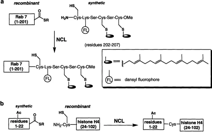
Prenylation involves the addition of a farnesyl or geranylgeranyl group to one or two cysteines at the C-terminus of a protein. The C-terminal location of the modified cysteine residues renders prenylated proteins well suited for semisynthesis by EPL. A successful application has been the semisynthesis of fluorescently-labeled mono- and diprenylated variants of Rab7, a Rab guanosine triphosphatase (GTPase) of the Ras-GTPase superfamily.61 An expressed a-thioester corresponding to Rab7 (residues 1-201)
was reacted with fluorescently-labeled and prenylated hexapeptides to generate Rab7 variants with geranylgeranyl modifications on either Cys205 or Cys 207, or on both Cys205 and Cys207 of the C-terminus (Fig. 1.8a). Semisynthesis involving the hydrophobic lipopeptides required several modifications to standard EPL protocol, including the addition of specific detergents and an organic extraction of unreacted peptide, which binds to the protein non-covalently, from precipitated protein. In addition, a Rab chaperone protein was necessary to stabilize the denatured Rab7 variants during refolding. A novel fluorescence-based prenylation assay, utilizing the environment-sensitive properties of the dansyl fluorophore, was developed to probe the mechanism of diprenylation. The results of the prenylation assay support a proposed random sequential mechanism of prenylation. The straightforward assay, facilitated by the creative application of a fluorescent tag, was possible because the researchers were able to generate homogenous monoprenylated protein.
As with glycosylation, a primary challenge in the EPL of prenylated proteins is the synthesis of modified peptides. Comparison of a number of solution-phase and solid-phase methods for synthesizing prenylated peptides revealed the strength of a hydrazide linker-based solid-phase approach that was used to incorporate several prenylated and fluorescently labeled peptides onto the oz-thioester fragment of Rab7.62 The extensive semisynthetic work with Rab7 was recently extended to other Ras-type GTPases: those in the Ras subfamily.63 Prenylated Ras proteins pose additional semisynthetic challenges
because they are not known to interact with a chaperone, the use of which was essential for the stabilization and purification of Rab7. This challenge was successfully addressed by the use of polybasic prenylated peptides, which eliminated non-specific peptide/protein aggregation and enabled ligation and purification in non-denaturing conditions.
Progress has also been made in the preparation of proteins with GPI anchors. GPI proteins are modified via a C-terminal amide linkage with a lipo-pentasaccharide anchor. Using EPL, lipidated analogs of green fluorescent protein (GFP) were created with a simplified GPI anchor, a phospholipid without glycans, to demonstrate a flexible strategy for generating proteins lipidated at the C-terminus.64 The lipidated GFP variants were shown to incorporate stably into supported membranes, and quantification of their lateral fluidity was achieved by fluorescence imaging. This strategy can therefore be applied to the semisynthesis of naturally lipidated proteins and their study in lipid bilayers.
Acetylation Reversible acetylation involves modification of proteins, notably
histones and transcription factors, on the E-amino group of lysine residues. In core histones, which comprise the octomeric protein core of nucleosomes, post-translational modification of the N-terminus alters histone-DNA interactions and is implicated in regulating gene transcription.49 NCL has been employed to generate pure samples of modified histones, acetylated or methylated on the N-terminal tail.42,65 In a recent example, a homogenous monoacetylated variant of histone H4 was prepared by NCL to characterize the structural and functional effects of acetylation of Lysl6.66 Toward this end, a synthesized peptide thioester acetylated at Lysl6 and corresponding to residues 1-22 of histone H4 was ligated to a recombinant C-terminal fragment (residues 23 to 102)
(Fig. 1.8b). The H4 variant was incorporated into nucleosomal arrays and found to inhibit the formation of higher order chromatin structures and prevent the functional interaction of histones with a specific chromatin-associated protein. This characterization of a selectively acetylated variant complements previous peptide competition studies and provides direct evidence not accessible with truncated or randomly hyperacetylated histone derivatives.
a recombinant HS synthetic
O
Rab 7 H2N-Cys-Lys-Ser-C -Ser-C s-OMe
(1
-2
0
1
)
S
S
6
s
S
NCL (residues 202-207) HSRab 7 HS
(1-201) Cys-L -Ser-C s-Ser-C s-OMe
FS S = dansyl fluorophore
synthetic recombinant
Ac HS Ac
residues SR histone H4 NCL residues Cys histone H4
1-22 NH2-C s-- (24-102)I 1-22 ys (24-102)
Figure 1.8. Semisynthesis of a) fluorescently-labeled and diprenylated Rab7, and b) monoacetylated histone H4.
2. Fluorescent probes
Genetically-encoded fluorophores are widely used for imaging proteins in live cells. Green fluorescent protein (GFP) and the ever increasing number of GFP variants with improved properties and a spectrum of excitation and emission wavelengths are invaluable probes for imaging protein localization and examining intramolecular and intermolecular protein interactions. One powerful application of fluorophore-labeled
proteins is fluorescence resonance energy transfer (FRET). FRET results when two fluorophores are within close proximity and the emission spectrum of the "donor fluorophore" overlaps with the excitation spectrum of the "acceptor fluorophore." The proximity and spectral overlap enable a transfer of energy and a corresponding increase in the intensity of the acceptor fluorophore emission, allowing for quantification of the distance between the two fluorophores. The major drawback of using genetically-encoded fluorophores for FRET and other fluorescence-based imaging is the significant size of the fluorophores (27 kDa for GFP) appended onto the protein of interest, potentially altering native interactions and localization. Organic fluorophores are significantly smaller (< 1 kDa) and often possess superior photophysical properties, such as higher extinction coefficients and greater resistance to photobleaching, but can be challenging to incorporate into proteins in a chemoselective manner.
NCL has been used to chemoselectively install a donor-accepter pair of organic fluorophores into proteins to study both intramolecular conformational changes38 and intermolecular interactions6 7 using FRET. In an example of the latter, variants of
serotonin N-acetyltransferase (AANAT), a circadian rhythm enzyme, were constructed
via EPL with a fluorescein or rhodamine-containing peptide at the C-terminus.67 Since standard methods for determining oligomerization state, such as size-exclusion chromatography and dynamic light scattering, proved inconclusive with AANAT, FRET was employed to probe for homo-oligomerization (Fig. 1.9a). Fluorescence studies showed a significant increase in FRET upon incubation of the donor (fluorescein) and acceptor (rhodamine)-containing AANAT variants, indicating a preference for AANAT to oligomerize.
Along with FRET, there have been numerous applications reported for fluorescent probes incorporated into proteins by NCL, some of which are discussed in the context of other sections in this chapter. Examples include the C-terminal labeling of proteins for anisotropy-based binding studies,68 the domain-specific replacement of tryptophan residues with a red-shifted and environment-sensitive tryptophan analog to probe domain function in the context of a full-length protein,6 9 and the site-specific incorporation of an
environment-sensitive fluorophore into an effecter domain to monitor protein-domain interactions."M Fluorescent labeling of proteins has also been accomplished in vivo.7 1 A
cell permeable fluorescent thioester was introduced into cells expressing a target protein with an N-terminal cysteine, generated by intein-mediated splicing, resulting in NCL to generate a protein variant labeled at the C-terminus (Fig. 1.9b).
k
a i
AANAT FL
AANATRh
monomers - no FRET oligomers - increase in FRET
b
I LnI overexpressed /" GST HS in vivc H2N-Cys GST NCL I I ST I TMR-containing thioesterFigure 1.9. a) AANAT labeled with fluorescein (FL) and rhodamine (Rh). Expected FRET outcome for AANAT as a monomer (no FRET) and oligomer (FRET). b) In vivo labeling of GST (containing an N-terminal Cys residue) with the membrane permeable TMR thioester.
3. Unnatural amino acids
The power of NCL is even more pronounced in its application to the semisynthesis of protein domains or full-length proteins containing unnatural amino acids. Amino acid analogs chemoselectively introduced by NCL can be used to probe specific aspects of amino acid or protein function. This approach can involve introducing residues that differ from the natural amino acid at a position of interest in a single aspect, such as side-chain geometry, steric effects, or electronic effects. In one example, variants