Characterization of a Perfused 3D Liver Bioreactor
by
Jennifer Mitchel
Submitted to the Department of Mechanical Engineering
in partial fulfillment of the requirements for the degree of
Bachelor of Science in Engineering
as recommended by the Department of Mechanical Engineering
at the
MASSACHUSETTS INSTITUTE OF TECHNOLOGY
June 2007
@ Massachusetts Institute of Technology 2007. All rights reserved.
Is7
Author ...
7.
.* . ./,. . V .. .. . . . . ...Department of Mechanical Engineering
May 11, 2007
Certified by
Linda G. Griffith
SoE Professor of Teaching Innovation, Department of Biological Engineering
Thesis Supervisor
Accepted by...
...
Lenhard V
John H. Lienhard V
Professor, Department of Mechanical Engineering
Ch.airm.n_ ndprgr.ladinat.p Thpqsi.s Cnmmitpp
ASSACHUSETTS INSTiTE OF TECHNOLOGY
iJUN
2 2007
LIBRARIES
ARCHIVES
M ... &..oCharacterization of a Perfused 3D Liver Bioreactor
by
Jennifer Mitchel
Submitted to the Department of Mechanical Engineering on May 11, 2007, in partial fulfillment of the
requirements for the degree of Bachelor of Science in Engineering as recommended by the Department of Mechanical Engineering
Abstract
The liver is the most important site of drug and nutrient metabolism in the body, and we desire an accurate in vitro model that allows us to perform long term drug and metabolism studies. To this end of developing an assaying tool, I used an existing multi-well bioreactor that allows for formation of perfused, three dimensional tissue structures, and began charac-terization of tissue behavior over time. One issue in the multi-well bioreactor is the unknown profile of cell retention over time, which is an important specification for normalizing data from drug metabolism studies. Number of cells can be indirectly assessed by measuring total protein or RNA levels when direct counting is problematic. To the end of compar-ing these methods, an additional goal of this thesis was to develop a protocol to measure both protein and RNA levels from a single sample using the commercially available reagent
RNAlater. RNAlater was shown, however, to be incompatible with certain existing protocols
for isolating both protein and RNA. Thesis Supervisor: Linda G. Griffith
1
Introduction
It is desirable to perform long term studies on liver parenchymal cells (hepatocytes) in an in
vitro environment, but they have classically been difficult to maintain in culture, with much
of their liver-specific functionality lost after only one day in conventional cultures. Though research has been conducted to determine the optimal method of preserving hepatospecific morphology and function, both reliable long term study and cultivation of complex tissue structures remain as embryonic technologies. A microfluidic bioreactor designed to foster the development of three dimensional (3D) liver tissue-like structures when seeded with hepatocytes was developed by the Griffith Lab[l]. In this bioreactor, spheroidal aggregates have been shown to form qualitatively better tissue than single hepatocytes [2], though recently protocols were developed to facilitate single cell seeding. The device uses pneumatic pressure to move fluid through the tissue-containing channels, subjecting the hepatocytes to shear stress as they would experience in vivo and supplying the tissue with more oxygen than it would receive in static culture where diffusion is solely responsible for oxygen transport.
The availability of oxygen is a huge factor in the viability of the cells in this reactor, and was a concern directing the experiments of this thesis. One parameter determined in part by the oxygen limitations is the initial number of cells to add to the scaffold. When we consider seeding the reactor with the ideal number of hepatocytes, we see that the need for a large number of cells in order to get accurate tissue structures and assay responses must be balanced with the fact that a larger number of cells means more oxygen is consumed per area and the available oxygen in a given well is finite. Scaffolds of the size used here have previously been seeded with between 3 x 10⁵ and 1.2 x 10⁶ hepatocytes; it was determined here that the optimal number of cells for initial seeding is 8 x 10⁵ per reactor well.
After determining the optimal number of hepatocytes to seed in each reactor well, it was desired to find a method by which both total protein and RNA could be isolated from a single reactor well. Currently experiments are being conducted which look at the drug metabolism of cultured liver tissue in the mult-well bioreactor, generally after 4 or 7 days in culture, in comparison with freshly isolated cells. Metabolism studies, which measure the abilities of the hepatocytes to perform their liver specific functions and the expression levels of liver specific genes aim to show that hepatocellular functionality remains relatively constant or well behaved, indicating that the multi-well bioreactor is suitable for long term studies. In order for these studies to yield accurate results, it is important to have a method of accurately determining the number of cells inside the bioreactor, so that data can be normalized to per cell rates. Two methods for analyzing cell numbers are using total protein and total RNA isolated from samples.
The method for counting cells based on total RNA relies on the assumption that to-tal RNA is relatively constant across a cell type. Though it is possible that RNA levels may change as cells are cultured in the bioreactor, compared to freshly isolated cells, it is believed that this metric is reliable enough to be used. Total RNA can also be used for drug metabolism studies in that it represents the current expression patterns of the hepato-cytes. It is possible, therefore, to measure the relative expression levels of genes known to play crucial roles in liver specific drug metabolism, and compare these expression levels to those found in freshly isolated and traditionally cultured hepatocytes. Phenol-based RNA extraction methods, such as use of the TRIzoL reagent, are common, and allow for isola-tion of protein and RNA as well. However, the TRIZOL protocol is time consuming, and does not allow for processing many samples in parallel, especially when compared to protein assays. In addition, the most time consuming steps of the protocol are required regardless whether protein or RNA is being isolated. Together, these factors make it desirable to find an alternative to TRIZOL as a sample preservative. Additionally, it would be beneficial to preserve samples such that one could choose at the time of analysis, rather than at the time of preservation, whether RNA, protein, or both are required. To this end, we looked at the commercially available reagent RNAlater as a sample preservative. Obtaining protein data from samples stored in RNAlater simply requires removal of the reagent, and obtaining RNA data requires fewer steps than if samples were preserved in TRIzoL.
Lastly, it was desired to know what the cell loss profile over time looks like, because though it is definitely known that many (approximately two thirds to three quarters) cells are lost between day 0 and the end of the experiment (day 4 or 7), it is not known when and at what rates these cells are lost. This is another application where RNAlater was desired to be used to obtain protein and RNA data from a single sample, in order to develop a correlation between RNA and protein profiles over time, in addition to comparing these methods for cell counting. Because of the many complications with using RNAlater, however, the final profile discussed was obtained using TRIZOL.
2
Background
2.1
The Multiwell Bioreactor
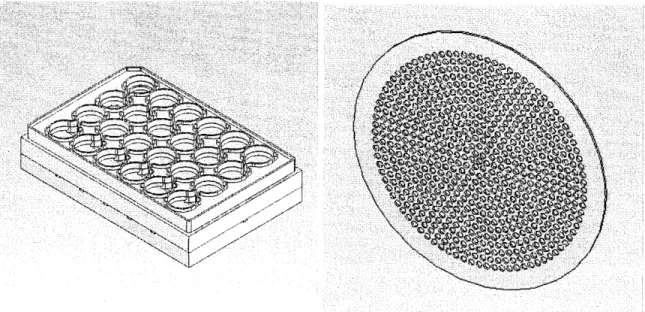
For this thesis I worked with the Generation F multiwell bioreactor, shown in Figures 1(a) and 1(c)1 and described previously[4]. This is a high throughput system that allows for up to twelve experiments to be done in parallel under virtually identical experimental
(b) Model of an 860 channel, silicon scaffold
(c) Multiwell Bioreactor and Control System
Figure 1: The MNultiwell bioreactor and a Scaffold. (a) CAD model of bioreactor. The inner two rows of wells are the reactor wells, and contain scaffolds and are seeded with cells, while the outer two rows are the resovoir wells which contain media that is being replenised with oxygen. (b) CAD Model of a typical scaffold with 860 channels, ~1 cm in diameter. This scaffold would likely be seeded with between .5 and 1 million hepatocytes. (c) Photograph of the bioreactor and its controller and air lines.
tions (temperature, humidity, user variability, etc) which will generally allows variation of two parameters in parallel, with adequate control and repetition of conditions. The mul-tiwell bioreactor also lends itself well to time course studies in which samples have to be permanantly removed to obtain data. The reactor wells, which are in the two inner rows, are supplied with media that is continually oxygenated, from the resovoir wells, which are the two outer rows. The reactor wells hold scaffolds which can be seeded with freshly isolated cells, where they can form 3D tissue structures attached to the walls of the scaffolds. The scaffolds are 150 microns thick, a length designed to minimize the oxygen limitations of the system. These scaffolds sit on top of a porous filter (Millipore) which is meant to even out the flow, and the filter sits on top of a support scaffold which allows medium to flow through from either direction. The resovoir wells have porous filters as well. The scaffolds, shown in Figure 1(b), are coated with Collagen I, an extracellular matrix protein, to encourage adhesion to the scaffold walls.
2.2
TRIZOL
TRIzoL(Invitrogen) is a commercially available reagent meant for the isolation of total RNA from biological samples. The reagent is a mono-phasic solution of phenol and guanidine isothiocyanate, and preserves the RNA present in a sample, while being highly effective dur-ing homogenization because it helps to dissolve and denature cells and cellular components
(without degrading proteins or DNA).
2.3
RNAlater
RNAlater (Ambion) is a reagent advertised to preserve RNA in cells and tissues, eliminating the need to analyze samples immediately due to RNA's high susceptibility to degradation[5],[6]. According to the official protocol guide, RNAlater should be able to preserve RNA at room temperature for 1 week without compromising RNA quality, or at 20"C or 80'C indefinitely. Both the Ambion and Qiagen protocol notes indicate that RNAlater preserved cells should be compatible with standard RNA isolation procedures, such as the Qiagen RNeasy Mini Kit, indicating that TRIzoL may not be a necessary reagent to isolate RNA. This reagent was thought to be the answer to the question of how to easily and effectively preserve RNA in a way such that it would be simple to obtain protein from the same sample, because both the RNA isolation and the protein assay have the same first step of sample homogenization. The ideal protocol would involve this common homogenization step followed by a choice to be made by the researcher as to which information is desired; such freedom and versatility is helpful to build into protocols, because of the reality in labwork that experimental needs
change dynamically.
Use of RNAlater in the literature has had variable results. Some studies using this reagent with whole tissue report that prognostic and diagnostic qualities of the tissue are not compromised [7],[8], though they do not report a comparison between RNAlater and TRIZOL and do not consider the potential relative loss of total RNA. RNAlater is often compared snap-freezing (immersion in liquid nitrogen), which is a third standard method of RNA preservation, with conflicting conclusions. Some studies report that RNAlater is a preservation method preferable to snap-freezing [7],[9], and others report that the two methods are equally effective [8], [10], [11], indicating that RNAlater is the preferred choice for RNA preservation because of its ease of use. One study does report however that snap-freezing is the optimal method of preservation when compared with RNAlater [12].
In addition to this conflicting information about the usefulness of RNAlater as an RNA preservative, it is unclear how well it functions during the homogenization step of standard RNA isolation protocols. It is indicated in the manufacturer's notes that RNAlater stabilizes cells against lysis, and we conclude that it must therefore be removed from samples before the necessary step of homogenization can take place. Some studies which report that RNAlater can be used to stabilize RNA also used TRIzoL during the isolation steps, even though this reagent was not used for initial preservation [13],[14].
One study found TRIZOL to be highly superior to RNAlater as a preservative[16], show-ing that total RNA yields were approximately tenfold higher and significantly more pure for the former reagent. Another study concludes that RNA extraction can be optimized by the use of TRIZOL, and that RNAlater is not suitible for RNA expression studies in dissected biopsy material[17].
3
Materials and Methods
3.1
Storing Cells in RNAlater
Samples were preserved by adding 5 to 10 times their volume of RNAlater reagent at room temperature, and storing for up to 1 month at 4VC, as directed by the manufacturer's note. In order to isolate cells from the RNAlater suspension, cells were either spun down at '5000g for -5 minutes, or first resuspended in an equal volume of PBS and then spun down.
3.2
RNA Isolation and Measurement
A great deal of care was taken to ensure that the samples remained RNAase free, so as to minimize degradation. All surfaces and tools were treated before starting the isolation with
RNaseZap (Ambion), as were the gloves of the researcher handling samples. In addition, RNase free tubes, pipette tips, and reagents were used. A maximum of 8 samples were treated at once, to further minimize RNA degradation due to the instability of RNA at room temperature. Samples were kept on ice whenever possible.
The RNA isolation protocol used is a modified version of the Qiagen isolation protocol; all reagents come from the Qiagen RNAeasy Kit. To each sample 350 or 700pL of Buffer RLT, the lysis buffer available with the Qiagen RNeasy Kit, was added. This solution was passed approximately 10 times through a 25G syringe tip to homogenize the sample. An equal volume of ethanol was added, the sample was inverted to mix, and up to 7001 L of this
solution was added to a Qiagen Spin Column. The column was centrifuged for 30 seconds at ,40.2 rpm in a table-top centrifuge. The eluent was passed a second time through the column to ensure maximal retention of RNA. Eluent was discarded and the process repeated for any remaining sample until all sample was processed.
The sample was washed in succession with Buffer RW1 (700pL, 30 seconds) and Buffer RPE (500pL, 30 seconds, followed by a second wash for 2 minutes) to removed contaminating cellular components. The spin column was transfered to a new collection tube, and the sample .was spun down at full speed for 1 minute, to ensure the removal of any traces of ethanol. The spin column was then transfered to an RNase-free collection tube, 50•L of 550C DPEC treated water was added to the center of the collection membrane, sample was
allowed to incubate for approximately 1 minute at room temperature, and then the spin column in the collection tube was spun down for 1 minute -10.2 rpm.
The eluent was measured for RNA content by the Nanodrop ND-1000 Spectrophotometer (NanoDrop Technologies, Wilmington DE). The sample is measured for absorbance over a broad spectrum and the NanoDrop software calculates the concentration of RNA. The 280/230 and 260/230 ratios are checked to be over 1.8 to ensure purity of the samples. A low 280/230 ratio implies contamination by protein, salts, or solvents. Data points with low purity ratios were not used in analysis.
For samples stored in TRIZOL, the above protocol was followed with the addition of the following preceeding steps and modifications: Samples were stored in 1 mL TRIzoL at -80oC for a minimum of 15 minutes (until frozen). When the samples were ready for processing they were allowed to thaw on ice. The thawed sample was passed approximately 10 times through a 25G syringe tip to homogenize the sample. 200 pL of chloroform was added to the sample, which was then shaken vigorously and allowed to incubate at room temperature
for 2-3 minutes. This step, followed by a 15 minute centrifugation at 12,000 RPM at 4VC, yields a biphasic mixture of aqueous and organic phases. RNA remains exclusively in the aqueous phase, which is removed and placed into a 1.5 mL tube, where the above protocol
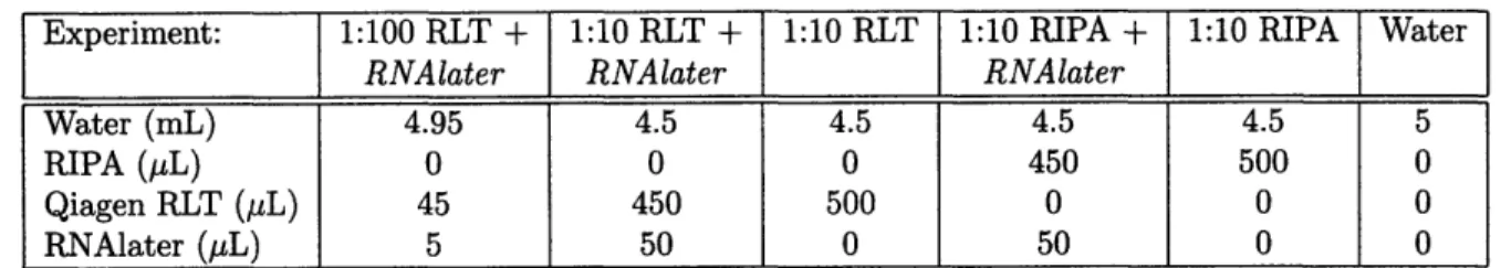
Experiment: 1:100 RLT + 1:10 RLT + 1:10 RLT 1:10 RIPA + 1:10 RIPA Water
RNAlater RNAlater RNAlater
Water (mL) 4.95 4.5 4.5 4.5 4.5 5
RIPA (AL) 0 0 0 450 500 0
Qiagen RLT (pL) 45 450 500 0 0 0
RNAlater (pL) 5 50 0 50 0 0
Table 1: Buffer Concentrations for BCA Assay Experiment to Determine Compatibility of RNAlater with Quantitation of Total Protein.
can be followed, beginning with the addition of ethanol to precipitate the DNA.
3.3 Protein Isolation and Measurement
The second method aimed at quantifying the number of cells in a scaffold is based on measuring protein levels. This assay is performed using the BCA Protein Assay Reagent Kit (Prod# 23227, Pierce, Rockford IL). Prior to use in the protein assay, samples were lysed with 0.7-1 mL either RIPA Lysis Buffer (TEKnova) or Qiagen RLT Lysis Buffer. 1% PMSF and 10% PIC (v/v) were added to the lysis buffer prior to lysis. Samples were either sonicated for 10 minutes or snap-frozen to aid in lysis. The lysed samples were then diluted, usually 1:100,. in order to be within the working range of the assay (2-40pg/mL). BSA standards were prepared at the following concentrations: 0, 5, 10, 15, 20, 25, 30, 35 (pg/mL). These standards were run with each assay plate. 100 or 150 pL of BSA standard or sample was added to an equal volume of Working Reagent from the Pierce kit. Plates were mixed on a plate shaker for 30 seconds, and then incubated for approximately two hours at 370C. Plates were cooled and then read by a SpectraMax platereader at 562 nm. The BSA standards allow the absorbance values for the samples to be correlated with a protein concentration.
4 Results
4.1
Protein Assay Results Depend on Buffer Composition
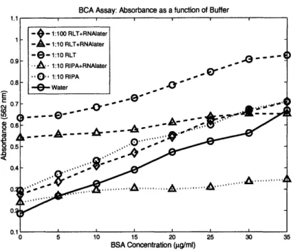
In order to determine the feasibility of using the BCA Protein Assay on samples preserved in RNAlater, the compatibility of the BCA reagents was tested for each of the following conditions: Qiagen RLT Lysis Buffer, RIPA Lysis Buffer, Qiagen RLT Lysis Buffer plus a 1:10 Volume of RNAlater, and RIPA Lysis Buffer plus a 1:10 Volume of RNAlater. All conditions were then diluted 1:10 in water, with the RLT plu RNAlater condition also diluted 1:100. The relative levels of water and lysis buffer in each sample were determined based on expected dilutions of RNAlater and lysis buffer, and are documented in Table 1. It was
BCA Assay: Absorbance as a function of Buffer E o4 CD <ca 0 BSA Concentration (gpg/ml)
Figure 2: BCA Assay Experiment to Determine Compatibility of RNAlater with Quantitation
of Total Protein. The lysis buffers increase background signal levels, while RNAlater appears to reduce
the slope of the curve.
predicted that up to a one tenth volume of RNAlater may remain after samples were stored in the preservative and required resuspension in the lysis buffer.
The results of this assay are shown in Figure 2. Both RIPA and RLT raise the signal background, RLT significantly moreso. This high signal background implies that care must be taken that samples are sufficiently dilute such that the signal is not saturated for more concentrated samples. This background due to lysis buffer is normally accounted for in the protein assay by subtracting a blank from all values obtained. RNAlater, on the other hand, has the effect of reducing the signal level and flattening out the curve. For example, when looking at the dashed RLT curves in Figure 2, it is clear that the highest signal is present when the BSA is diluted in RLT only, and the signal is reduced for the 1:10 dilution of
RNAlater. The same trend appears in the RIPA curve. High levels of RNAlater appear to
be incompatible with the BCA Protein Assay, as the absorbance values at 562 nm span a range approximately an order of magnitude less than for low or zero levels of RNAlater.
The curve most similar to that obtained from the control, BSA in water, is the 1:100 dilution of RLT and RNAlater, indicating that the protein isolation protocol (described in Section 3.3) can be followed with the addition of RNAlater to samples without detriment, as long as the residual RNAlater occupies less than one thousanth part of the final lysate. Care must therefore be taken to remove as much RNAlater as possible before the addition of lysis buffer.
4.2 Determination of Optimal Cell Number
A simple qualitative experiment was performed to compare the two mostly commonly used seeding densities, 500 x 103 and 800 x 103 cells per reactor well. Two reactors were seeded by different users, each reactor having 6 wells of each seeding density, and were allowed to culture for 4 days. At the end of the incubation period, it was clear that the higher seeding density was preferable. Individual sections of tissue appeared morphologically similar between the two seeding densities, but for the lower density there was a prevalence of empty patches on the scaffold, while for the higher density the scaffold appeared much more uniform.
4.3 RNA and Protein Level Standard Curves
The protocol outlined in Section 3.2 for isolating RNA was repeated over five times with cells preserved in RNAlater, over a range of concentrations with results shown in Figure 3(a), where RNA levels are plotted as a function of cell number. Cells were either freshly isolated and incubated in RNAlater at room temperature for less than an hour, or preserved at 4VC overnight up to two weeks. There is no obvious correlation between storage methods and quality of the standard curve, however, though the literature indicates that incubation overnight is a requirement when using RNAlater. Because each of these five curves was so radically different, it is apparent that with current protocols RNAlater cannot be used to establish cell numbers with any accuracy or precision.
In one instance, data not shown, the Qiagen RLT lysis buffer was prepared as indicated in Section , with PIC and PMSF added, and the curve appeared to radically shift downward,
with the no cell control giving large negative values. This condition was important to test, as it would reflect the lysis buffer being used if a portion of the lysate was to be removed for protein analysis (while the rest of the lysate would be analyzed for RNA content). In a different experiment, however, these additions to the lysis buffer did not appear to have an effect on the RNA levels detected; if this system is used in the future, this potential effect should be futher investigated.
Figure 3(b) shows a TRIzoL standard curve, in which RNA levels are plotted as a function of cell number. The curve is highly linear and well behaved, and indicates that each cell contains approximately 50 pico grams of RNA. This curve is sufficiently similar to others found previously by various researchers that it was not repeated additional of times.
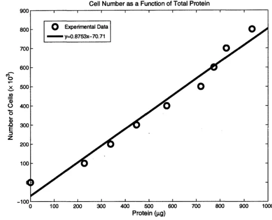
Last, a standard curve in which cell number is plotted as a function of total protein, is shown in Figure 42. Some researchers force a y-intercept of zero, reasoning that logically once background is subtracted, zero protein should correspond to zero cells; however, this
RNA Standard Curves, 3/5-4/10
3/5 Stored at 40C
0- 3/15 Fresh Cells
'
3/20 Stored at 40C
II1 4/3 Fresh Cells
4/10 Stored at 40C
"4O
r 100 200 300 400 500 Number of Cells (x 103) (a) RNAlater 600 700 800RNA Standard Curve, Using Trizol
Cell Number (x 103 ) (b) TRIzol Figure 3: Standard Curves to Measure RNA Levels as treated with RNAlater. These curves were not obtained with variation between each curve. (b) Cells stored in TRIZOL
a Function of Cell Number. (a) Cells a satisfactory protocol, as shown by the
25 20 S15 J z cc M I I I I I I I I I I I I I II
Cell Number as a Function of Total Protein '0 0 CO ox o a) .0 E z 0 100 200 300 400 500 600 Protein (gpg)
Figure 4: Total Protein Standard Curve. Cell number is This curve can be used directly to determine how many cells are measurement.
700 800 900 1000
shown as a function of protein levels. present based on a given total protein
imposition on the data changes the values obtained when the curve is used to calculate the number of cells corresponding to a given value of total protein, and was not implemented here. However, a data point of (0,0) was included to reflect this phenomenon of zero protein corresponding to zero cells, though the curve obtained is not identical to if the y-intercept was forced to be at zero. This may account for a slight difference in the numbers presented in Section 4.4 from those appearing elsewhere.
4.4
Cell Loss Over Time in the Multiwell Bioreactor
An experiment to determine the cell loss profile over time was performed using RNAlater to preserve RNA and protein, though no direct data was obtained. Two reactors were seeded with 800 x 103 cells per reactor well, with row A containing silicon scaffolds and row B containing PEEK scaffolds. Three reactor wells were taken out of the reactor on days 1, 3, 5, and 7, and stored in 1 mL RNAlater. The samples were stored for approximately 6 weeks at 4VC, which is longer than recommended by the manufacturer's note, due to complications with protocol development. When samples were ready to be analyzed, the
RNAlater was aspirated and RLT added but it was very difficult to remove tissue from the
scaffolds. RNAlater stabilizes tissue, in this case so well that the tissue remained attached to the scaffolds even after additional sonication and snap freezing. In addition, once the
RNAlater is removed the RNA starts to degrade at room temperature; this combination of
effects led us to discard the samples.
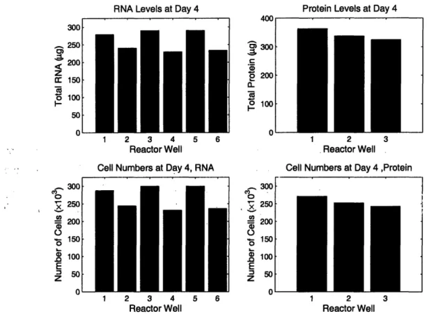
A second experiment was performed in which reactor wells were seeded with 800 x 10 and scaffolds were removed at day 4. Three scaffolds were analyzed for total protein3 and
six were stored in TRIzoL and analyzed for total RNA. Results are shown in Figure 5 and are very promising, in that cell numbers obtained by either method are very similar. The day 4 cells population determined by total RNA was 267±33 (x 103), while the day 4 cell population determined by total protein was 299±17 (x103).
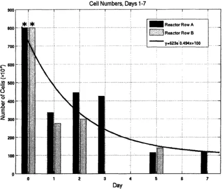
Last, an experiment was performed with reasonable success to determine the cell loss profile over time, the results of which are shown in Figure 6. On Day 0, 800x 103 cells were seeded in each reactor well (PEEK scaffolds coated with collagen I). Two scaffolds were removed on each of days 1, 2, 3, 5, and 74. These scaffolds were placed in 1 mL of TRIZOL but were not frozen immediately because of potential damage to the scaffolds. The TRIZOL was passed through the scaffold using a micropipetter until the tissue was removed from the scaffold. There appeard to be about 10% of the sample remaining on the scaffold, which
3Data obtained by Michal Bokovza 4
Only 10 out of 12 vreactor wells were used because two wells had pumping problems after the start of the experiment.
Protein Levels at Day 4
U
300 200 100 1 2 3 4 5 6 Reactor Well Cell Numbers at Day 4, RNA2 3 4
Reactor Well
5 6
2 3
Reactor Well
Cell Numbers at Day 4 ,Protein
2
Reactor Well
Figure 5: Number of Cells on Day 4 as measured by total protein and total RNA. Each measurement was taken from a different well in the same reactor. The day 4 cells population determined by total RNA was 267+33 (x 103), while the day 4 cell population determined by total protein was 299±17
(x
10
3) 300 250 200 M 150 B 100 I--50 0 o 2, 0x12
E zRNA Levels at Day 4
!
i m
I
Cell Numbers, Days 1-7 A-o 0 x " 0 0 E z S 1 2 3 4 5 7 Day
Figure 6: Cell Loss Profile Over 7 Days. Cell numbers were analyzed using TRIZoL Reagent to isolate total RNA. A curve was fitted based on the initial seeding density of 800x 103 cells per reactor well. Day 0
data (**) was not measured directly but was included as a useful comparison and default time point.
could not be removed. It is unclear if snap freeing would have reduced this.
When the calculated cell numbers were plotted as a function of time, along with the day 0 initial data point, it was possible to fit an exponential curve to the data. This curve qualitatively describes the trend we have seen in many reactors, in which a great number
of cells are lost between day 0 and day 1, and are presumed to simply never attach to the scaffold; following this initial rapid loss, cell numbers reduce more slowly, until a steady state value is reached.
5
Discussion
RNAlater was not shown to be a viable solution to the problem of easily extracting RNA and protein data from a single sample, but could be further investigated. If RNAlater is used in the future, care should be taken that all samples incubate in RNAlater overnight at 40C, so that the reagent has time to adequetly penetrate the cells. Though this requirement was not described in the manufacturer notes and did not appear to make a difference in the standard curves developed here, it was a common trend in the literature and is worth pursuing as potential partial remedy to current state of RNAlater protocols.
References
[1] Sivaraman, A. et al. A Microscale in vitro Physiological Model of the Liver: Predictive Screens for Drug Metabolism and Enzyme Induction. Curr. Drug Meta. 2005, 6, 569-591. [2] Powers MJ, Domansky K, Kaazempur-Mofrad MR, Kalezi A, Capitano A, Upadhyaya Ar, Kurazawski P, Wack KE, Stolz DB, Kamm R, Griffith LG. A Microfabricated Array
Bioreactor for Perfused 3D Liver Culture. 2002. Biotechnol Bioeng 78:257-269.
[3] Domansky K, Inman W, Serdy J, Griffith L. Perfused microreactors for liver tissue engi-neering. Conf Proc IEEE Eng Med Biol Soc. 2005;7:7490-2.
[4] Poster: Domansky K, Inman W, Serdy J, Owens B, Wittemore M, Vineyard L, Griffith LG. Multiwell tissue Culture Plate as a Platform for 3D Perfused Cell-Based Screening Assays. 2006.
[5] RNAlater Tissue Collection: RNA Stabilization Solution, manufacturer's protocol (Am-bion). Available at http://www. ambion. com/techlib/resources/rnalater/.
[6] RNAlater Handbook, manufacturer's protocol (Qiagen). Available at http://www1.
qiagen. com/literature/protocols/RNAlater. aspx.
[7] Florell SR, Coffin CM, Holden JA, Zimmermann JW, Gerwels JW, Summers BK, Jones DA, Leachman SA. Preservation of RNA for functional genomic studies: a multidisci-plinary tumor bank protocol. 2001. Mod Pathol. 14(2):116-28.
[8] Stemmer K, Ellinger-Ziegelbauer H, Lotz K, Ahr HJ, Dietrich DR. Establishment of a protocol for the gene expression analysis of laser microdissected rat kidney samples with affymetrix genechips. 2006. Toxicol Appl Pharmacol. 217(1):134-42.
[9] Chowdary D, Lathrop J, Skelton J, Curtin K, Briggs T, Zhang Y, Yu J, Wang Y, Mazumder A. Prognostic Gene Expression Signatures Can Be Measured in Tissues Col-lected in RNAlater Preservative. 2006. J Mol Diagn. 8(1):31-9.
[10] Grotzer MA, Patti R, Geoerger B, Eggert A, Chou TT, Phillips PC. Biological stability of RNA isolated from RNAlater-treated brain tumor and neuroblastoma xenografts. 2000. Med Pediatr Oncol. 34(6):438-42.
[11] Mutter GL, Zahrieh D, Liu C, Neuberg D, Finkelstein D, Baker HE, Warrington JA. Comparison of frozen and RNALater solid tissue storage methods for use in RNA expres-sion microarrays. 2004. BMC Genomics 5(1):88.
[12] Wang SS, Sherman ME, Rader JS, Carreon J, Schiffman M, Baker CC. Cervical tissue collection methods for RNA preservation: comparison of snap-frozen, ethanol-fixed, and RNAlater-fixation. 2006. Diagn Mol Pathol. 15(3):144-8.
[13] Malik KJ, Chen CD, Olsen TW. Stability of RNA from the retina and retinal pigment epithelium in a porcine model simulating human eye bank conditions. Invest Ophthalmol Vis Sci. 2003 Jun;44(6):2730-5.
[14] Rodrigo MC, Martin DS, Redetzke RA, Eyster KM. A method for the extraction of high-quality RNA and protein from single small samples of arteries and veins preserved in RNAlater. J Pharmacol Toxicol Methods. 2002 Mar-Apr;47(2):87-92.
[15] Kasahara T, Miyazaki T, Nitta H, Ono A, Miyagishima T, Nagao T, Urushidani T. Evaluation of Methods for Duration of Preservation of RNA Quality in Rat Liver Used for Transcriptome Analysis. 2006. J of Tox Sci 31(5):509-519.
[16] Morrison C, Palatini J, Riggenbach J, Radmacher M, Porcu P. Fine-Needle Aspira-tion Biopsy of Non-Hodgkin Lymphoma for Use in Expression Microarray Analysis. 2006. Cancer 108(5'):311-18.
[17] Roos-van Groningen MC, Eikmans M, Baelde HI, de Heer E, Bruijn JA. Improvement of Extraction and Processing of RNA from Renal Biopsies. 2004. Kidney Int. 65(1):97-105.