HAL Id: pasteur-03093327
https://hal-pasteur.archives-ouvertes.fr/pasteur-03093327
Preprint submitted on 3 Jan 2021
HAL is a multi-disciplinary open access archive for the deposit and dissemination of sci-entific research documents, whether they are pub-lished or not. The documents may come from teaching and research institutions in France or abroad, or from public or private research centers.
L’archive ouverte pluridisciplinaire HAL, est destinée au dépôt et à la diffusion de documents scientifiques de niveau recherche, publiés ou non, émanant des établissements d’enseignement et de recherche français ou étrangers, des laboratoires publics ou privés.
Anna Spier, Martin Sachse, To Nam Tham, Mariette Matondo, Pascale
Cossart, Fabrizia Stavru
To cite this version:
Anna Spier, Martin Sachse, To Nam Tham, Mariette Matondo, Pascale Cossart, et al.. Bacterial FtsZ induces mitochondrial fission in human cells. 2021. �pasteur-03093327�
1 2 Bacterial FtsZ induces mitochondrial fission in human cells 3 4 5 6
Anna Spier 1,2,3,4,$, Martin Sachse 5, Nam To Tham 1,2,3, $, Mariette Matondo 6,7, Pascale
7 Cossart 1,2,3, and Fabrizia Stavru 1,2,3,8,$,#,* 8 9 10 1 Unité des Interactions Bactéries-Cellules, Institut Pasteur, Paris, France 11 2 Institut National de la Santé et de la Recherche Médicale (INSERM), U604, Paris, France 12 3 Institut National de la Recherche Agronomique (INRA), USC2020, Paris, France 13 4 Université Paris Diderot, Sorbonne Paris Cité, Paris, France 14 5 Unité Technologie et service BioImagerie Ultrastructurale, Institut Pasteur, Paris, 15 France 16 6 Plateforme Protéomique, Unité de Spectrometrie de Masse pour Biologie (UTechS 17 MSBio), Institut Pasteur, Paris, France 18 7 Centre National de la Recherche Scientifique (CNRS), USR 2000, Paris, France 19 8 CNRS SNC5101, Paris, France 20 $ present address : Unité de Biologie Evolutive de la Cellule Microbienne, Institut 21 Pasteur, Paris, France 22 # CNRS ERL6002, Paris, France 23 24 25 26 * Correspondence and requests for materials should be addressed to 27 fabrizia.stavru@pasteur.fr, lead contact 28 29 Keywords: mitochondrial division, bacterial division, Drp1, mtDNA, inner mitochondrial 30 membrane 31
Abstract 32
Mitochondria are key eukaryotic organelles that evolved from an intracellular bacterium, 33
in a process involving bacterial genome rearrangement and streamlining. As 34
mitochondria cannot form de novo, their biogenesis relies on growth and division. In 35 human cells, mitochondrial division plays an important role in processes as diverse as 36 mtDNA distribution, mitochondrial transport and quality control. Consequently, defects 37 in mitochondrial division have been associated with a wide range of human pathologies. 38 While several protists have retained key components of the bacterial division machinery, 39
none have been detected in human mitochondria, where the dynamin-related protein 40
Drp1, a cytosolic GTPase is recruited to the mitochondrial outer membrane, forming 41 helical oligomers that constrict and divide mitochondria. Here, we created a human codon 42 optimized version of FtsZ, the central component of the bacterial division machinery, and 43 fused it to a mitochondrial targeting sequence. Upon expression in human cells, mt-FtsZ 44 was imported into the mitochondrial matrix, specifically localizing at fission sites prior to 45
Drp1 and significantly increasing mitochondrial fission levels. Our data suggests that 46
human mitochondria have an internal, matrix-localized fission machinery, whose 47
structure is sufficiently conserved as to accommodate bacterial FtsZ. We identified 48 interaction partners of mt-FtsZ, and show that expression of PGAM5, FAM210, SFXN3 and 49 MTCH1 induced mitochondrial fission. Our results thus represent an innovative approach 50 for the discovery of novel critical mitochondrial fission components. 51 52 53
Introduction 54
Mitochondria are key eukaryotic organelles, which have retained their own genome and 55
are delimited by two membranes. The bacterial origin of mitochondria originally 56
proposed by Lynn Margulis (then Lynn Sagan1) is now largely accepted, and even complex
57
mitochondrial features, such as the inner mitochondrial membrane invaginations termed 58
cristae, have recently been shown to be evolutionarily conserved in specific bacterial 59
lineages2. However, one conundrum is the apparent lack of evolutionary conservation of
60
the division machinery between bacteria and mitochondria of fungi and metazoa. 61
Division is an essential process for both bacteria and mitochondria. In the vast 62
majority of bacteria, cell division is performed by a multiprotein complex, at the heart of 63
which lies the tubulin homologue and evolutionarily conserved protein FtsZ3–5. In
64
bacteria, FtsZ assembles early at prospective fission sites, forming the Z-ring and 65 recruiting several additional proteins to mediate cell division (reviewed in 6,7). 66 Mitochondrial fission is necessary for proper distribution of the organelle during 67 mitosis and in highly polarized cells8 and the dynamic equilibrium between fission and 68 fusion is tightly connected to mitochondrial function in both human and yeast cells9. At 69
the molecular level, important differences exist between organisms. While several 70
protists have retained key components of the bacterial division machinery, none have 71
been detected in fungi and metazoa10, where mitochondrial fission is thought to be
72
governed by a cytosolic machinery11. An intermediate situation has been described in the
73
red alga Cyanoschizon merolae, where assembly of an intramitochondrial FtsZ-based 74
fission machinery appears coordinated with the assembly of a cytosolic Dynamin-based 75
fission machinery12,13. In human cells a series of events involving the ER, the actin and
76
septin cytoskeleton and receptors on the mitochondrial outer membrane (OMM) 77 culminates in the assembly of a dynamin-related protein (Drp1) on the OMM8; Drp1 then 78 constricts mitochondria, leading to division, with potential synergistic action of Dyn214– 79 16. Fission ensues after constriction beyond a critical threshold17. During mitochondrial 80
fission the two membranes that delimit mitochondria represent a challenge, as fusion 81
between the outer and inner mitochondrial membranes has to be prevented to avoid 82
leakage of mitochondrial content (Fig 1A). One possible scenario is that the inner 83
membranes reach the necessary curvature and fusogenic distance earlier than the outer 84
membrane, spontaneously fuse and retract, leaving only the outer membranes to fuse, 85
leading to abscission of the two daughter mitochondria. Other scenarios invoke the 86
presence of molecular machineries that either “insulate” the outer from the inner 87 membrane during fission, or that specifically promote fission of the matrix compartment. 88 The latter hypothesis appears the most likely, as a matrix-localized, “bacteria-like” fission 89
machinery with homologs of the bacterial division protein FtsZ at its core has been 90
identified in several protists10,18–21. In light of the monophyletic origin of mitochondria,
91
we and others postulated that metazoan mitochondria would also harbour a fission 92
machinery located in the matrix8,22. Indeed, fission of the matrix compartment has been
93
observed in the absence of outer membrane fission in several metazoans23–26. However,
94
previous attempts at bioinformatic identification of a bacteria-derived division 95
machinery in metazoan mitochondria based on sequence similarity have failed10,27. We
96
hypothesized that the internal fission machinery of mitochondria might nevertheless 97
have retained a certain degree of structural conservation with respect to its bacterial 98
ancestor. To test this hypothesis, we asked whether the key orchestrator of bacterial cell 99
division FtsZ would be able to induce mitochondrial fission in mammalian cells. We 100
engineered synthetic constructs that allowed targeting of bacterial FtsZ into the 101
mitochondrial matrix and found that alphaproteobacterial FtsZ (mt-aFtsZ) specifically 102 localized at mitochondrial fission sites and substantially increased mitochondrial fission 103 levels. As several proteins concur to recruit FtsZ to the membrane in bacteria, we explored 104 which mitochondrial proteins might play this role in our experimental system. Among the 105
interaction partners of mt-aFtsZ that we identified, we tested five transmembrane 106 mitochondrial proteins, four of which induce mitochondrial fission upon overexpression, 107 potentially representing new players in mammalian mitochondrial dynamics. 108 109 Results 110 111
Mitochondrial expression of alphaproteobacterial FtsZ induces mitochondrial 112
fission 113
To achieve fission of the mitochondrial matrix compartment, we hypothesized that 114
mammalian mitochondria would contain a protein-based inner fission machinery (IFM). 115
Given the endosymbiotic origin of mitochondria, we reasoned that the IFM would not 116
have evolved de novo and even though not detectable by sequence similarity10,27, it would
117
still share structural features with the bacterial fission machinery. 118
To test whether mitochondria contain an IFM in the matrix that can interact with a 119 bacterial fission protein, we decided to transiently express bacterial FtsZ in human cell 120 lines (U2OS and HeLa). To this end, we constructed synthetic versions of FtsZ, which were 121 codon-optimized for expression in human cells and fused to an N-terminal mitochondrial 122 targeting sequence to allow import into the mitochondrial matrix, and a C-terminal tag to 123 allow detection (Fig 1B). Although mitochondria have long been thought to derive from 124
alphaproteobacteria28–33, recent work suggests that mitochondria evolved from a
125
proteobacterial lineage which branched off before the alphaproteobacteria34. As the
126
precise bacterial lineage that gave rise to mitochondria remains a matter of debate, we 127
chose to express synthetic versions of both gamma- and alphaproteobacterial FtsZ 128
(Escerichia coli and typhus group Rickettsia respectively, referred to as mt-aFtsZ and mt-129
gFtsZ). Immunofluorescence analysis of flag-tagged mt-aFtsZ showed that it localized to 130
mitochondria (Fig 1C) in virtually all transfected cells (99.5±1.7%, n=1469, N=6 131
independent experiments). In 0.5±1.6% mt-aFtsZ was expressed at very high levels, 132
formed filaments that colocalized with microtubules (Suppl. Fig 1A) and displayed 133
dramatic mitochondrial fragmentation and perinuclear aggregation (suppl Fig 1B). In 134 cells where mt-aFtsZ localized to mitochondria, those with intermediate and low levels 135 of expression allowed the detection of mt-aFtsZ punctae, which appeared to accumulate 136 at mitochondrial matrix constrictions. In contrast, mt-gFtsZ or the control construct mt-137 GFP did not accumulate at constrictions and often displayed a more even staining (Fig 1C). 138 We then tested a C-terminal deletion mutant of mt-aFtsZ (mt-aFtsZDCT) and found that 139
it was able to polymerize in some cells (Fig 1C, suppl Fig 1C), but failed to localize at 140
constrictions, in agreement with previous findings showing that while C-terminal deletion 141
mutants of FtsZ are able to polymerize in E.coli, they do not support bacterial division35.
142
Next, we asked whether the ultrastructure of mitochondrial constrictions was 143
affected by mt-aFtsZ. To do so we combined light microscopy with high pressure freezing 144
electron microscopy in a correlative approach allowing us to focus on cells with 145
intermediate expression levels. Mitochondrial constrictions did not qualitatively differ 146
between mt-GFP and mt-aFtsZ expressing mitochondria, which displayed an inner 147 diameter of 41.6nm versus 40.8nm respectively and an outer diameter of 57.1nm versus 148 62.7nm (suppl Fig 1D). 149 Given that mt-aFtsZ was found at constriction sites, we analysed whether it would 150
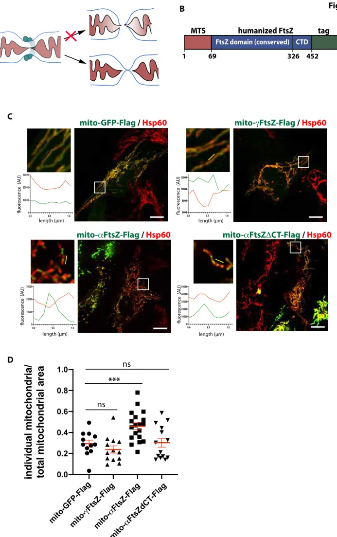
induce mitochondrial fission by quantifying mitochondrial morphology with the semi-151
automatic ImageJ plugin MiNA. This analysis showed that full length mt-aFtsZ induced 152
mitochondrial fission (Fig 1C, suppl Fig 1E). We validated our results by using a different 153
cell line (HeLa) and manually measuring mitochondrial length (suppl. Fig 1F), which 154 revealed a dose-dependent effect on mitochondrial fission. In addition, the slightly thicker 155 mitochondria of HeLa cells allowed us to discern mt-aFtsZ -labelled matrix constrictions 156 with non-constricted outer membrane, supporting previous reports showing that matrix 157 constriction can occur in the absence of outer membrane constriction25,26. 158
These data show that in contrast to mt-gFtsZ, mt-aFtsZ specifically localizes to 159
mitochondrial matrix constrictions and affects mitochondrial morphology, suggesting 160 that it labels mitochondrial matrix fission sites. 161 162 mt-aFtsZ localizes at mitochondrial fission sites prior to Drp1 recruitment 163 To assess whether mt-aFtsZ labeled constrictions indeed proceed to complete abscission, 164
we followed GFP tagged mt-aFtsZ in mitochondria of live cells. Full length mt-aFtsZ 165 labeled the vast majority of all fissions we observed (86.3%, n=73, N=6), accumulating at 166 prospective fission sites and often distributing to the tips of both daughter mitochondria 167 upon abscission (Fig 2A). In contrast, the C-terminal deletion mutant mt-aFtsZDCT did 168 not consistently label fission sites (Fig 2B) and was found at up to 1.3µm from the fission 169
site in 8 out of 11 fissions (N=4). In agreement with these findings, mt-aFtsZDCT 170
displayed an almost two-fold decrease in inner mitochondrial membrane localization 171
compared to full-length mt-aFtsZ as assessed by immune-electron microscopy (Fig 172
2C/D). In addition, we noticed that mt-aFtsZ induced a zipper-like phenotype with 173
regular, closely apposed cristae, which we also observed by high pressure freezing 174
electron microscopy. 175
Next, we investigated the spatiotemporal relationship between mt-aFtsZ and 176
Drp1. A fraction of endogenous Drp1 colocalized with flag-tagged mt-aFtsZ or 177 accumulated in its close proximity in fixed cells (Fig 2E, yellow and white arrowheads 178 respectively). Live cell imaging revealed that mt-aFtsZ precedes Drp1 recruitment during 179 mitochondrial fission (Fig 2F), suggesting that matrix constriction occurs prior to outer 180
membrane constriction by Drp1. In agreement with this hypothesis, we found matrix 181
constrictions that were labelled with mt-aFtsZ in the absence of outer membrane 182
constriction (suppl Fig 2). 183
Toghether, these data indicate that matrix-localized mt-aFtsZ is a bona fide marker 184 for mitochondrial fission sites and supports a fission model in which matrix constriction 185 precedes Drp1 recruitment and mitochondrial abscission. 186 187 Replication of the mitochondrial nucleoid is not necessary for mt-aFtsZ localization 188
The punctate pattern of mt-aFtsZ localization is reminiscent of nucleoids, which have 189
been shown to accumulate at the tips of mitochondria36,37. We therefore assessed the
190
spatial organization of mt-aFtsZ relative to nucleoids and found that mt-aFtsZ was 191
excluded from the area occupied by nucleoids (Fig 3A), in particular at mitochondrial 192 constrictions (suppl Fig 3). However, we also detected instances where mt-aFtsZ partially 193 colocalized with nucleoids (Fig 3A, arrowheads). We hypothesized that this subset could 194 represent replicating nucleoids, which have been estimated to amount to 9% of the total 195
nucleoid population38 and to mark fission sites in yeast and human39,40. We thus examined
196
whether mt-aFtsZ would colocalize with a red version of the mitochondrial DNA 197
polymerase processivity subunit 2 (POLG2-mScarlet), which labels actively replicating 198
mtDNA39. Surprisingly, mt-aFtsZ and POLG2 did not appear to substantially colocalize
199
(Fig 3B). 200
This prompted us to ask whether nucleoid replication was necessary for mt-aFtsZ 201
localization and/or fission. We inhibited mtDNA replication with dideoxycytosine 202
(ddC41). ddC treatment caused the nucleoid packing protein TFAM-GFP to label the entire
203
matrix compartment, reflecting mtDNA depletion (Fig 3C). In contrast, mt-aFtsZ 204
localization was not affected, marking mitochondrial fission sites in ddC treated and 205 control cells (Fig3C/D). Together, these data strongly suggest that mt-aFtsZ localization 206 is not dependent on nucleoid replication. 207 208 Identification of mitochondrial proteins that interact with mt-aFtsZ 209 The dynamic localization of mt-aFtsZ during mitochondrial fission suggests that it 210 interacts with a matrix-localized mitochondrial fission machinery. We therefore sought 211 to identify interaction partners of mt-aFtsZ. To this end, we immunoprecipitated Flag-212 tagged mt-aFtsZ or mt-GFP from transiently transfected HeLa cells in presence of non-213
ionic detergent (0.5% NP40), and identified co-precipitating proteins by quantitative 214
mass spectrometry. We identified 941 proteins, which only coprecipitated with mt-aFtsZ, 215
but not with mt-GFP. In addition, 119 proteins were detected in both samples, but were 216 significantly enriched in mt-aFtsZ immunoprecipitates (Fig 4A). 75% of the proteins we 217 identified were not mitochondrial, reflecting mt-aFtsZ interactions taking place prior to 218 its mitochondrial import and cells where mt-aFtsZ mislocalized to the cytoplasm due to 219 high overexpression. As mislocalized mt-aFtsZ colocalizes with microtubules (suppl Fig 220 1A/B), we were not surprised to detect numerous proteins associated with microtubules 221
among our hits. Interestingly, previous experiments have shown that E.coli FtsZ 222
expressed in the cytosol of mammalian cells does not spontaneously colocalize with 223
tubulin42, suggesting important structural differences between alpha- and
224
gammaproteobacterial FtsZ. Among the overall 1060 interactants of mt-aFtsZ, 31.7% 225 were organellar proteins and 25% (i.e. 269) mitochondrial according to the mitochondrial 226 protein database IMPI (Integrated Mitochondrial Protein Index, v2018_Q2), despite the 227 fact that we did not purify mitochondria prior to immunoprecipitation, reflecting an ~3 228 fold enrichment in mitochondrial proteins if compared with ~8% mitochondrial proteins 229 in the human genome43,44. Consistently, Gene Ontology term analysis showed significant 230 enrichment of organellar proteins, and in particular mitochondrial proteins (Fig 4B and 231
suppl fig 4). Our dataset contained several inner and outer mitochondrial membrane 232
proteins that have been linked to mitochondrial morphology or division (e.g. MICOS 233
complex proteins Mic60, Mic27 and Mic1945,46, Prohibitin 2 and SPY complex members
234
Yme1L and Stomatin-like protein 247, mitochondrial fission protein 1 (Fis148),
235
mitochondrial fission process protein 1 (MTFP1/MTP1849), ATAD3B50, SLC25A46A1 and
236
AFG3L251, reinforcing our hypothesis that mt-aFtsZ can interact with an endogenous
237
mitochondrial matrix fission machinery. Gene ontology analysis with the DAVID software 238
highlighted an enrichment in mitochondrial inner membrane proteins (suppl fig 4A). 239
Indeed, 74 of the 269 mitochondrial proteins (27.5%) we identified were predicted to 240
contain at least one transmembrane domain. Interestingly, previous in vitro 241
reconstitution experiments have shown that FtsZ cannot mediate unilamellar liposome 242
constriction without its membrane-anchoring partner FtsA52. We therefore chose to focus
243
on five highly enriched transmembrane proteins, the mitochondrial serine-threonine 244
phosphatase PGAM5, MTCH1 (mitochondrial carrier homolog 1), FAM210A, the ATP 245
synthase membrane subunit DAPIT (diabetes-associated protein in insulin-sensitive 246
tissue) and SFXN3 (Sideroflexin 3). We selected these proteins based on their enrichment 247
score and their detection in independent immunoprecipitation experiments, where beads 248
were used as a control (not shown). Among the selected candidates, MTCH1, DAPIT and 249 FAM210A have not been studied in the context of mitochondrial dynamics, while recent 250 data has implicated PGAM5 in mitochondrial dynamics53–55 and in the course of this study 251 members of the Sideroflexin family have been shown to act as serine transporters and 252 impact mitochondrial morphology56. 253
We employed Flag-tagged versions to confirm mitochondrial localization of the 254
five selected candidates and assess their impact on mitochondrial morphology. 255
Morphometric analysis (MiNa) revealed that PGAM5, MTCH1, FAM210A and SFXN3 256 induced significant mitochondrial fission (Fig 4C, suppl Fig 4B). This phenotype was not 257 a by-product of non-specific inner membrane perturbation due to overexpression, as even 258 strong overexpression of DAPIT had no detectable effect on mitochondrial morphology. 259 At very low expression levels, flag-tagged PGAM5, MTCH1, FAM210A and SFXN3 were 260 also found at mitochondrial matrix constrictions (Fig 4D), supporting a possible role in 261 matrix fission. Attempts to follow the sub-mitochondrial localization of these candidates 262 in live cells using GFP fusions failed because their overexpression induced mitochondrial 263
fission or substantially mislocalized to the cytoplasm (not shown). Silencing of the 264
individual proteins did not robustly induce mitochondrial hyperfusion, and did not 265
prevent localization of mt-aFtsZ to mitochondrial constrictions (not shown), suggesting 266
possible functional redundancy. Co-silencing of two or more proteins resulted in high 267 toxicity (not shown). 268 In conclusion, we propose that PGAM5, MTCH1, FAM210A and SFXN3 represent 269 novel candidate effectors of inner mitochondrial membrane fission. 270 Discussion 271 Fission of the mitochondrial matrix in the absence of outer membrane fission has been 272
shown in metazoans, both in vivo24 and in cellulo23,25,26. How this is achieved is unclear.
273
Although in several protozoa orthologs of the key bacterial fission protein FtsZ have been 274
shown to localize to the mitochondrial matrix and participate to mitochondrial 275
division12,19,20, FtsZ orthologs have not been detected in metazoans and fungi10,27. Here,
276
we show that when directed into human mitochondria, alphaproteobacterial FtsZ (mt-277
aFtsZ) localizes at mitochondrial fission sites and stimulates mitochondrial division. 278
Interestingly, expression of mitochondrial FtsZ from the brown alga Mallomonas 279
splendens in yeast was found to affect mitochondrial morphology19. Together, this data
suggest that human and yeast mitochondria contain a matrix-localized fission machinery 281
that is structurally similar to the bacterial division machinery, as it can accommodate 282 heterologously expressed FtsZ. indicating that mt-aFtsZ has retained features that allow 283 it to interact productively with the inner fission machinery of today’s mitochondria. 284 Surprisingly, when we compared FtsZ from an alphaproteobacterium (mt-aFtsZ) 285
with that from a gammaproteobacterium (mt-gFtsZ), only mt-aFtsZ formed stable 286 assemblies that localize at mitochondrial fission sites. Polymerization is not unexpected 287 per se, as purified FtsZ has been shown to spontaneously assemble into polymers upon 288 GTP addition57. One possibility is that mt-gFtsZ polymers are unstable, as they are unable 289 to interact with the mitochondrial matrix fission machinery, which manifests by lack of 290 localization to mitochondrial constrictions. Another possibility is that, coming from E.coli, 291 mt-gFtsZ has adapted to the diameter of the bacterium, (~0.5µm58) and therefore cannot 292 form rings in mitochondria due to spatial constrains imposed by the narrower diameter 293 of mitochondria (~0.2µm59). In contrast mt-aFtsZ may have an inherent ability to adapt 294 to smaller diameters, as Rickettsiae have diameters as small as 0,1µm60. 295 296 How are the sites of mt-aFtsZ assembly determined? 297 The question of how the division site is defined is central also in bacterial division. In E. 298 coli, the best-studied model, two negative regulatory mechanisms have been described, 299
based on nucleoid occlusion or on the Min system61. No orthologs for either of these
300
systems have been detected in mitochondria10, where the nucleoid has been suggested to
301
act as a spatial organizer of the mitochondrial fission machinery. Consistent with this 302
view, mitochondrial fission and mtDNA dynamics are tightly linked in the red alga 303
Cyanidioschizon merolae62. In mammalian cells, up to 70% of all fission events have been
304
found to occur in the vicinity of a nucleoid41 and components of the outer (cytosolic)
305
fission machinery have been suggested to sense the localization and replication status of 306
nucleoids39. In our hands the localization of mt-aFtsZ and mitochondrial morphology
307
were not affected by blocking mtDNA replication (Fig 3C), suggesting that this process is 308
not essential for IFM assembly and localization. Interestingly, in nucleoid-free E. coli 309 maxicells FtsZ localized to the midcell in a Min system - dependent manner63. As the Min 310 system is not conserved in human mitochondria10, how mt-aFtsZ localizes to fission sites 311 in the absence of nucleoids remains an open question. One possibility is that it is the inner 312 mitochondrial fission machinery and its associated proteins, such as the constituents of 313
contact sites or possibly lipid microdomains, that define mtDNA localization; this 314 situation is similar to what has been observed for the mtDNA helicase Twinkle, which can 315 associate with the inner mitochondrial membrane in the absence of mtDNA38. 316 317 How does mt-aFtsZ induce mitochondrial fission at the mechanistic level? 318 mt-aFtsZ clearly labels mitochondrial fission sites and stimulates fission, but we currently 319 do not know how if functions and whether it displaces components of the endogenous 320
IFM or not. In bacteria, FtsZ has been proposed to mediate constriction either 321
directly57,64,65, or indirectly, i.e. by recruiting the peptidoglycan synthesis machinery66–68.
322
Mitochondria have lost the peptidoglycan, but mt-aFtsZ might act by recruiting the lipid 323
biosynthesis machinery. In agreement with this hypothesis, we found several proteins 324
involved in lipid synthesis among the interactors of mt-aFtsZ. However, we cannot 325 exclude that mt-aFtsZ acts in a more direct manner, i.e. by physically pulling on the inner 326 membrane to promote fission of the matrix compartment. The interactions that allow the 327 recruitment of FtsZ to its membrane anchors FtsA, ZipA or SepF are mediated by the C-328 terminus of the protein, which is essential for promoting fission7. In agreement with this, 329
the C-terminus of mt-aFtsZ was essential to localize the protein to the mitochondrial 330
inner membrane and its deletion abolished mitochondrial fission induction. In vitro 331
experiments have shown that while FtsZ can self-assemble into contractile rings in the 332
absence of other proteins65, its recruitment to the membrane requires additional
333
proteins52. We employed an immunoprecipitation approach to identify mitochondrial
334 inner membrane proteins that could mediate the recruitment of mt-aFtsZ to the inner 335 mitochondrial membrane. With this approach, we could identify proteins that have been 336 previously implicated in mitochondrial fission and novel potential actors. 337
Here, we focused on five inner membrane proteins with an unclear role in 338
mitochondrial matrix fission: PGAM5, MTCH1, SFXN3, FAM210 and DAPIT. 339
Overexpression of DAPIT did not affect mitochondrial morphology, indicating that 340
overexpression does not per se alter mitochondrial morphology, even though the 341
mitochondrial inner membrane is one of the most protein-rich membranes69. In contrast,
342
mild overexpression of PGAM5, MTCH1, SFXN3 and FAM210 induced mitochondrial 343
fission. MTCH1 and FAM210 were not previously known to affect mitochondrial 344
dynamics. In our hands, even mild SFXN3 overexpression induced mitochondrial fission, 345
but previous data indicates that double deletion of SFXN3 and SFXN1 in Jurkat cells 346
caused a decrease mitochondrial length56. While further studies are needed to untangle
347
the precise role of SFXN3 in mitochondrial dynamics, our data confirms a role of PGAM5 348
in mitochondrial fission and adds FAM210 and MTCH1 to the growing list of inner 349 membrane proteins playing a role in mitochondrial fission. 350 It is intriguing to note that among the interactors of mt-aFtsZ we also found several 351 proteins that have been proposed to link the inner and outer membranes. One is ATAD3B, 352 a paralog of ATAD3A, an AAA+ ATPase shown to control mitochondrial dynamics and to 353 interact with both the inner and outer mitochondrial membranes70. We also found the 354 MICOS complex component Mic60, which has been shown to bind to the nucleoid71 and 355 link it with two outer membrane components (the SAM complex and Metaxins 1 and 2) 356
and with the cytosolic fission machinery45,46. Strikingly, we detected SAMM50 and
357
Metaxin 2 in mt-aFtsZ immunoprecipitates, suggesting at least partial integrity of the 358 complex and providing a mechanism for the observed spatiotemporal coordination of mt-359 aFtsZ and Drp1 recruitment. 360 361 Is mt-aFtsZ regulated? 362 Our live cell imaging experiments showed mt-aFtsZ localization at fission sites prior to 363
Drp1. While we detected mt-aFtsZ at virtually all fission events, not all mt-aFtsZ 364
assemblies underwent fission in a given time frame. This suggests that either only a subset 365
of mt-aFtsZ oligomers are functional, or that the IFM is preassembled and poised to act 366
upon specific triggering signals, similar to the cytosolic fission machinery based on 367
Drp172. A triggering signal for the IFM may be local calcium influx at ER-mitochondrial
368
contact sites. Indeed, calcium influx has been shown to induce mitochondrial matrix 369
fission, followed by outer membrane abscission26. Consistent with a role of calcium in the
370
regulation of mitochondrial matrix fission, we found several regulators of mitochondrial 371
calcium influx among the proteins that co-immunoprecipitated with mt-aFtsZ. 372
Incidentally, divalent cations (including calcium) also stimulate FtsZ assembly and 373 bundling in bacteria, pointing to an evolutionarily conserved regulatory mechanism of 374 division7,73. 375 376 In conclusion, our work suggests the presence of a mitochondrial fission machinery in the 377 mitochondrial matrix that retained sufficient structural conservation to accommodate a 378 heterologously expressed bacterial FtsZ. To our knowledge, this represents the first bona 379
fide marker of matrix fission sites described to date and paves the way for the molecular
380
characterization of the mitochondrial matrix fission machinery. Future experiments will 381
address whether FtsZ from other bacterial species have retained the ability to 382
consistently label mitochondrial fission sites and interact with the inner mitochondrial 383
fission machinery. Our system may thus also provide a novel “evolutionary cell biology” 384
approach to understand which bacteria represent the closest extant relatives of 385
mitochondria and shed light on the debated origin of the bacterial ancestor of 386 mitochondria. 387 388 Material and Methods 389 Cloning 390 To create humanized versions of gammaproteobacterial FtsZ, the E.coli FtsZ sequence was 391 completely re-coded according to human codon usage to optimize expression in human 392 cells. To comply with local regulations, for alphaproteobacterial FtsZ, we used the R.typhii 393
sequence as to re-code the N-terminal part (aa 2-326) of FtsZ, and the R.prowazekii 394
sequence to re-code the C-terminal domain. For both constructs, we deleted the initial 395
methionine to prevent internal initiation and the terminal stop codon was replaced with 396
a glycine-serine linker to allow in-frame expression of a Flag or a GFP tag. Re-coded 397 sequences were synthesized by Genecust or as gBlocks (Integrated DNA Technologies). 398 OM-mRuby was generated by in-frame fusion of the first 215bp of human TOM20 (gBlock, 399 Integrated DNA Technologies) with mRuby. All constructs are listed in table M1. 400 401 Reagents 402 Chemicals: Orange and Deep Red Mitotracker and secondary antibodies were purchased 403 from Thermo Fisher. All chemicals were obtained from Sigma-Aldrich/Merck. Complete 404 mini EDTA-free protease inhibitor and PhoStop phosphatase inhibitor tablets were from 405 Roche, anti-Flag M2 dynabeads were from Sigma-Aldrich/Merck. 406 Antibodies: Antibody sources are detailed in table M2. All antibodies were used according 407 to the manufacturer’s instructions unless otherwise stated. 408 409 Cell culture and transfection 410 HeLa and U2OS cells were obtained from ATCC and cultured under standard conditions; 411 media and additives were from Thermo Fisher. Cells were seeded on coverslips (#1.5, 412 Marienfeld) for immunofluorescence or MatTek dishes (MatTek Corporation) for live cell 413
imaging and transfected with FugeneHD (Roche) according to the manufacturer’s 414
instructions, at a DNA:transfectant rate of 1:3. The DNA quantities employed for each 415
construct are indicated in table M1. Cells were imaged or fixed and processed for 416 immunofluorescence 24h-36h posttransfection. 417 418 Immunofluorescence and imaging 419 Cells grown on coverslips were stained with mitotracker when necessary, fixed for 10 420
minutes in 4% paraformaldehyde (Electron Microscopy Sciences)/PBS, washed in PBS 421
and permeabilized for 5 minutes in 0.1% Triton X-100 in PBS and blocked for at least 30 422
minutes in 1% BSA and 10% goat serum. Primary antibodies (see table M2) were 423
incubated for 60 minutes in blocking buffer, followed by three washes in PBS (5 minutes) 424
and incubation with Hoechst 33258 and Alexa-labelled secondary antibodies (Thermo 425 Fisher) in blocking buffer for 30 minutes. Coverslips were then washed extensively in PBS 426 and mounted in Vectashield (Vector Laboratories). For live cell imaging, cells grown on 427 Mattek dishes were stained with mitotracker when necessary, then imaged in Fluorobrite 428
medium (Thermo Fisher) on a Roper spinning disk confocal system (Zeiss 429
AxioObserver.Z1 inverted fluorescence microscope equipped with an Evolve EM-CCD 430
camera (Photometrics) and a Yokogawa CSU-X1 spinning disc). Images were acquired at 431
37°C with a 100x NA 1.4 oil objective using MetaMorph. Cells were imaged every 20 432 seconds for 10 or 15 minutes. 433 434 Image analysis 435
All images were analyzed in ImageJ/Fiji (National Institutes of Health), including 436
adjustment of brightness and contrast. Overlays were assembled in Photoshop (Adobe) 437
and figure panels in Illustrator (Adobe). The ImageJ macro Mitochondrial Network 438
Analysis (MiNA) toolset74 was used to examine mitochondrial morphology. Single cells
439
were selected as regions of interest and pre-processing parameters were adjusted in 440
order to obtain optimal skeletonized images of the mitochondrial network, followed by 441
extraction of mitochondrial network features such as number of individuals, branch 442 length and mitochondrial area, referred to as “mitochondrial area”. To obtain a degree of 443 mitochondrial fragmentation that would be independent of the size of the mitochondrial 444 network we used the ratio of individual mitochondria and total mitochondrial area. 445 446 Statistical analysis 447
Results are expressed as means of at least three independent experiments, error bars 448 represent the standard error of the mean. For multiple comparisons, data were analyzed 449 with the Prism software (Graphpad) by one-way ANOVA, followed by Dunnett’s multiple 450 comparisons test to obtain the adjusted P value. Significance is indicated as p<0.05 (*), 451
p<0.01 (**) and p<0.005 (***), ns for p>0.05. N refers to the number of independent 452 experiments, n refers to the number of counted events (cells, individual mitochondria, 453 fissions). 454 455 Electron microscopy 456 Correlative light electron microscopy (CLEM) 457 To obtain landmarks for CLEM the pattern of an HF-15 finder grid (AGAR) was evaporated 458 with carbon on sapphire (3 mm diameter, 0.16 mm thickness, Wohlwendt instruments) 459 as described75. The discs with stabilized carbon pattern were sterilized by UV and coated 460 with poly-L-Lysine (Sigma-Aldrich) for cell culture. After transfection, cells were imaged 461 in medium containing 1mM Hepes after placing the disc in a glass bottom dish (MatTek) 462
using a Leica SP5 confocal microscope (Leica) and low expressing cells were selected. 463
After imaging the cells were frozen with an HPM 010 (Abra fluid). Samples were freeze 464
substituted in a Leica AFS2 (Leica microsystems) in 1% osmiumtetroxide, 0.1% 465
uranylacetate, 5% water, 2% MeOH in dry acetone with the following schedule: 1h at -466
90°C, 2.5°C/h for 16h, 30 min at -50°C, 15°C/h for 2h, 30 min at -20°C, 10°C/h for 2h, and 467
1h at 0°C. After substitution the dishes were infiltrated with epoxy resin (Agar) and 468 polymerization was done in flat bottom beam capsules at 60°C for 48h. After detachment 469 of the discs the sample was sectioned with a Leica UCT microtome (Leica microsystems) 470 with a nominal feed of 70 nm. Sections were picked up with slot grids and contrasted with 471
4% aqueous uranylacetate (Merck) and Leynolds lead citrate (Delta microscopies). 472
Images were taken with a Tecnai G2 microscope operated at 120 kV (Thermofisher), 473 equipped with an ultrascan 4000 CCD (Gatan Inc.). 474 Immuno electron microscopy 475 For immune-labeling on thawed cryo sections cells were fixed with 2% PFA (EMS) + 0.1% 476 glutaraldehyde (Sigma) in PHEM buffer, pH 7 (60 mM Pipes, 25 mM Hepes, 10 mM EGTA, 477 2 mM MgCl2) for 1 h at RT. Afterwards free aldehyde groups were quenched with 50 mM 478 NH4Cl in PBS and cells were removed from the culture plastic with a rubber policeman 479 and pelleted in a 1.5 ml eppendorf tube. The cell pellet was embedded in 12% gelatin 480
(TAAB) and after solidification on ice, small cubes of 1 mm3 were cut and infiltrated
481 overnight at 4°C with 2.3 M sucrose in PBS. The next day the cubes were mounted on 482 metal pins and frozen by immersion into liquid nitrogen. Thin sections of 60 nm nominal 483 feed were cut with a Leica UC6/FC6 cryo-microtome at -120°C. The sections were picked 484
up with a 1:1 mixture of 2% methylcellulose in water and 2.3M sucrose in PBS. After 485
thawing the sections were deposited on grids and labelled with rabbit anti GFP 486
(Rockland) followed by Protein A gold (CMC Utrecht). At the end of the labelling the 487
sections were contrasted with 0.4% uranylacetate in 1.8% methylcellulose and airdried 488
before observation with Tecnai G2 microscope. For the quantification of matrix/inner 489 membrane localization of mt-aFtsZ-GFP or its DCT version, 36 and 25 random sections 490 were quantified respectively. 491 492 Immunoprecipitation 493
Immunoprecipitation was performed as described76 with modifications. Briefly, HeLa
494 cells were seeded on 10 cm dishes and transfected with 7µg DNA. After 36h, cells were 495 washed three times in PBS and lysed for 30 min with 1 ml lysis buffer/10cm dish (20 mM 496 Tris, pH 7.4, 100 mM NaCl, 10% glycerol, 1,5mM MgOAc) supplemented with 0.5% NP-40 497 (Igepal), 1x protease and phosphatase inhibitors (Roche). Lysis and all subsequent steps 498 were performed at 4 °C. After lysate clarification at 13000xg for 10 minutes, the protein 499
concentration of the supernatant was determined by Bradford assay (Pierce). 1 mg of 500
lysate was incubated overnight with 20µl anti-Flag M2 magnetic Dynabeads (Sigma-501 Aldrich/Merck) under shaking. Magnetic beads were recovered, washed three times with 502 lysis buffer and four times with washing buffer (50 mM Tris, pH 7.4, 150 mM NaCl) and 503 eluted with 2x20µl 3xFlag peptide (100 mg/mL in washing buffer). The experiment was 504 performed in triplicate. For western blot, 5µl (10%) eluate was supplemented with 2x 505
Laemmli buffer, boiled for 10min resolved on a gradient SDS-PAGE (Biorad), and 506
subjected to western blotting via wet transfer to 0.45µm nitrocellulose membrane 507 (Millipore). Ten µg total lysate were loaded (corresponding to 1%) for the input. 508 509 Proteomic analysis 510
Protein digestion: Proteins were solubilized in urea 8 M, NH4HCO3 50 mM pH 7.5, then
511
disulfide bonds were reduced with 5 mM tris (2-carboxyethyl) phosphine (TCEP) for 30 512
min at 23°C and alkylated with 20 mM iodoacetamide for 30 min at room temperature in 513
the dark. Samples were diluted to 1 M urea with 50 mM NH4HCO3 pH 7.5, and Sequencing
514
Grade Modified Trypsin (Promega, Madison, WI, USA) was added to the sample at a ratio 515
of 50:1(w/w) of protein to enzyme for 8 h at 37°C. Proteolysis was stopped by adding 1% 516
formic acid. Resulting peptides were desalted using Sep-Pak SPE cartridge (Waters) 517
according to manufactures instructions. Peptides elution was done using a 50% 518
acetonitrile (ACN), 0.1% FA buffer. Eluted peptides were lyophilized and then store until 519
use. 520
LC-MS/MS analysis: a nanochromatographic system (Proxeon EASY‐nLC 1000, Thermo 521
Fisher Scientific) was coupled online to a Q Exactive™ Plus Mass Spectrometer (Thermo 522
Fisher Scientific). For each sample, 1µg of peptides was injected onto a 50 ‐ cm 523 homemade C18 column (1.9µm particles, 100 Å pore size, ReproSil‐Pur Basic C18, Dr. 524 Maisch GmbH) and separated with a multi‐step gradient from 2% to 45% ACN at a flow 525 rate of 250 nl/min over 180 min. The column temperature was set to 60°C. The data were 526 acquired as previously described77. 527 Data processing: Raw data were analyzed using MaxQuant software version 1.5.3.878 with 528 database search parameters as described in77. The MS/MS spectra were searched against 529 Uniprot proteome database of Human (January 13, 2015, 20,432 entries) and mt-aFtsZ-530
Flag protein, and usual MS contaminants. Data were quantified with the MaxLFQ 531
algorithm by requiring a minimum peptide ratio count of 2. The parameter “match 532
between run” was checked. Raw data have been deposited to the ProteomeXchange 533
Consortium via the PRIDE79 repository with the dataset identifier PXD016722.
534
Statistical and functional analysis: For the statistical analysis of one condition versus 535 another, proteins exhibiting fewer than 2 intensities in at least one condition were first 536 discarded. After log2 transformation, intensities were normalized by median centering 537 within conditions (normalizeD function of the R package DAPAR80). Proteins without any 538 intensity in one condition (quantitatively present in a condition, absent in another) were 539
considered as differentially abundant. Next, missing values were imputed using the 540
imp.norm function of the R package norm. Proteins with a log2(fold-change) inferior to 1
541
have been considered as proteins with no significant difference in abundance. Statistical 542
testing of the remaining proteins was conducted using a limma t-test81. An adaptive
543
Benjamini-Hochberg procedure was applied on the resulting p-values to select a set of 544
significantly differentially abundant proteins with a false discovery rate of 1%82. The
545
proteins of interest are therefore the proteins that emerge from this statistical test 546
supplemented by those being quantitatively absent from one condition and present in 547
another. Gene ontology analysis of mass spectrometry results was performed with the 548
online softwares Panther (http://pantherdb.org/) and DAVID 549
(https://david.ncifcrf.gov/), using standard settings. Transmembrane proteins were 550
predicted using the online software TMHMM
551 (http://www.cbs.dtu.dk/services/TMHMM/). 552 553 Data availability 554
The data that support the findings of this study are available from the corresponding 555 author on reasonable request. 556 557 Table M1: Plasmids 558 559
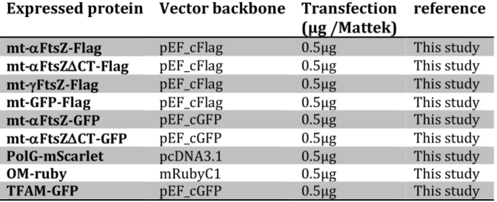
Expressed protein Vector backbone Transfection (µg /Mattek)
reference
mt-aFtsZ-Flag pEF_cFlag 0.5µg This study
mt-aFtsZDCT-Flag pEF_cFlag 0.5µg This study
mt-gFtsZ-Flag pEF_cFlag 0.5µg This study
mt-GFP-Flag pEF_cFlag 0.5µg This study
mt-aFtsZ-GFP pEF_cGFP 0.5µg This study
mt-aFtsZDCT-GFP pEF_cGFP 0.5µg This study
PolG-mScarlet pcDNA3.1 0.5µg This study
OM-ruby mRubyC1 0.5µg This study
PGAM5-myc-Flag pCMV6_Entry 0.25 µg Origene
MTCH1-myc-Flag pCMV6_Entry 0.25 µg Origene
FAM210A-myc-Flag pCMV6_Entry 0.25 µg Origene
SFXN3-myc-Flag pCMV6_Entry 0.25 µg Origene
DAPIT-myc-Flag pCMV6_Entry 0.25 µg Origene
pDsRed2-mito 0.25 µg Clontech Drp1-mCherry 0.5 µg 83 560 Table M2: Antibodies 561 562
Antibody Catalog number Provider
Tom20 Clone 29 BD Biosciences
Drp1 611112 BD Biosciences
tubulin Clone B5.1.2 Sigma-Aldrich/Merck
mtDNA Ac-30-10 Progen
Hsp60 D6F1 Cell Signaling
anti-FLAG Clone M2 Sigma-Aldrich/Merck
563
Author contributions 564
AS: Data curation, Formal analysis, Investigation, Methodology; MS: Investigation, 565
Methodology, Writing – review & editing; TNT: Investigation, Methodology; MM: 566
Methodology; FS and PC: Funding acquisition, Writing – review & editing; FS: 567
Conceptualization, Data curation, Formal analysis, Investigation, Methodology, 568 Validation, Visualization, Writing – original draft, Supervision. 569 570 Acknowledgements 571 We would like to thank Véronique Hourdel and Quentin Giai Gianetto for initial analysis 572 of mass spectrometry data and Ludmila Bonnand for help with live cell imaging analysis. 573 Alessandro Pagliuso, Jan Riemer and Tim Wai are thanked for discussion and Simonetta 574 Gribaldo, Bastian Huelsmann, Nika Pende, and Hans Spelbrink for critical reading of the 575 manuscript. This study was supported by the European Research Council (H2020-ERC-576
2014-ADG 670823-BacCellEpi to P.C.) and Institut Pasteur. A.S. was supported by a 577
BioSPC doctoral fellowship from the Université Paris Diderot. P.C. is a Senior International 578
Research Scholar of the Howard Hughes Medical Institute. F.S. is a CNRS permanent 579 researcher. 580 581 Conflict of Interest 582 The authors declare no conflict of interest. 583
584 Figure legends 585 586 Fig1: Expression of alphaproteobacterial FtsZ (mt-aFtsZ) in human mitochondria 587 induces mitochondrial fission. 588 A 589
Schematic representation of mitochondrial constriction (cross-section), where Drp1 is 590 depicted in turquoise, the outer membrane in blue, the inner membrane in black and the 591 matrix in burgundy. The top arrow points at a theoretical scenario, where fission entails 592 inner and outer membrane fusion. The bottom arrow points at the current fission model, 593
where inner membrane undergoes homotypic fusion, leading to matrix fission. This is 594
then followed by homotypic fusion of the outer membrane to achieve complete abscission 595
of the two daughter mitochondria. In vivo, matrix and outer membrane fission appear 596 closely linked in time and space. 597 B 598 General layout of the synthetic FtsZ constructs used in this study: a well-characterized 599 mitochondrial targeting sequence (MTS) from subunit 9 of the Fo-ATPase of N. crassa was 600 fused at the N-terminus of bacterial FtsZ, which was codon-optimized for expression in 601 human cells. A C-terminal Flag or GFP tag was added for detection. Numbers refer to the 602 alphaproteobacterial construct mt-aFtsZ. A control construct was created by replacing 603 the FtsZ sequence with GFP, resulting in mito-GFP-Flag. 604 C 605
Immunofluorescence of U2OS cells transfected with mito-GFP-Flag or mitochondrially 606
targeted FtsZ from a gamma- (mt-gFtsZ-Flag) or an alphaproteobacterium (mt-aFtsZ-607 Flag, C-terminal deletion mutant mt-aFtsZDCT-Flag, revealed in green) and mitochondria 608 (Hsp60, red). Scalebar: 10µm, insets are enlarged 4-fold. Linescan positions are indicated 609 by a yellow line. 610 D 611 Semiautomated quantification (MiNA plugin) of mitochondrial morphology expressed 612 by the amount of individual mitochondria/mitochondrial area in cells transfected as in 613 B. Three experiments were pooled. Mean and SEM are displayed in red, P=0.0002 by 614 one-way Anova, adjusted P-value = 0.0049. 615 616
617 Fig 2: mt-aFtsZ labels mitochondrial fission sites and precedes Drp1 recruitment 618 A 619 Live cell imaging of U2OS cells transfected with mt-aFtsZ-GFP or mt-aFtsZDCT-GFP. 620 Insets show an example of mitochondrial fission and are enlarged 2x. Linescans were 621 taken for each timepoint along the fission axis. Scalebar: 10µm. 622 B 623 Post-embedding immuno-EM of HeLa cells transfected with the above constructs, 624 stained with nanogold-anti-GFP. Scalebar: 100 nm. 625 C 626 Quantification of nanogold signal with respect to the inner mitochondrial membrane or 627 the matrix (i.e. >15 nm distance from the inner membrane). n indicates the number of 628 analyzed nanogold grains from 36 (mt-aFtsZ-GFP) or 25 random sections (mt-629 aFtsZDCT-GFP). 630 D 631 Colocalization of mt-aFtsZ-GFP (green) with Drp1 (red) in Hela cells. Mitochondria were 632 stained with mitotracker deep red (blue). Yellow arrowheads point at colocalization 633 between mt-aFtsZ-GFP and Drp1, white arrowheads point at apposition. Inset enlarged 634 2x. 635 E 636 Live cell imaging of U2OS cells co-transfected with mt-aFtsZ-GFP and Drp1-mCherry, 637 stained with mitotracker deep red. Insets show an example of mitochondrial fission and 638 are enlarged 2x. mt-aFtsZ-GFP (white arrowhead) is present prior to Drp1 (yellow 639 arrowhead). 640 641 Fig 3: mt-aFtsZ localizes in proximity of the nucleoid, but is independent of mtDNA 642 replication 643 A 644 U2OS cells transfected with mt-aFtsZ-GFP (green) and labeled for mtDNA (red) and 645 mitotracker deep red (blue). Inset enlarged 2x. Linescan showing juxtaposition of green 646 and red signal; arrowheads point at colocalization beteeen mt-aFtsZ and mtDNA. 647 Scalebar: 10µm. 648
649 B 650 Fluorescence images of U2OS cells transfected with mt-aFtsZ-GFP (green) and POLG-651 mScarlet (red). Mitochondria are shown in blue (mitotracker deep red). Inset enlarged 652 2x. Linescan showing alternating green and red signal. Scalebar: 10µm. 653 C 654 U2OS cells treated with 10µM ddC or vehicle for 48h, then transfected with mtDsRed 655 and mt-aFtsZ-GFP or TFAM-GFP and imaged 36h later. Still images show diffuse staining 656 of TFAM-GFP upon ddC treatment, indicating relocalization. mt-aFtsZ-GFP forms puncta 657 irrespective of ddC treatment. Inset enlarged 2x. Scalebar: 10µm. 658 D 659 Time lapse imaging of the same cells depicted in C, showing 4x enlarged insets with mt-660 aFtsZ-GFP localization at fission sites in both control and ddC treated cells. 661 662 Fig 4: Identification of mt-aFtsZ interaction partners that play a role in 663 mitochondrial fission 664 A 665 Number of proteins obtained by mass-spectrometry analysis of mt-aFtsZ-Flag versus 666 mtGFP-Flag immunoprecipitates. Grey indicates proteins identified in mt-aFtsZ-Flag and 667 absent in mtGFP-Flag immunoprecipitates, black indicates proteins that are significantly 668 enriched in mt-aFtsZ-Flag versus mtGFP-Flag immunoprecipitates. 669 B 670 GO-term analysis of proteins identified through mt-aFtsZ-Flag immunoprecipitatation 671 (Panther software). 672 C 673 Semiautomated quantification (MiNA plugin) of mitochondrial morphology expressed 674 by the amount of individual mitochondria/mitochondrial area in U2OS cells transfected 675 with mtGFP or inner membrane proteins PGAM5, FAM210A, MTCH1, SFXN3 and DAPIT. 676 Three experiments were pooled. Mean and SEM are displayed in red. P<0.0001 by one-677 way Anova, adjusted P-values < 0.0001. 678 D 679 U2OS cells expressing very low amounts of fission-inducing constructs PGAM5, 680 FAM210A, MTCH1, SFXN3, shown in green. Mitochondria are shown in red (Hsp60) . 681
Scalebar: 10µm, insets enlarged 4x. Linescans (yellow) show varying levels of 682 accumulation at constrictions. 683 684 Supplementary Figure legends 685 686 Suppl Fig1: Characterization of mt-aFtsZ localization and their impact on 687 mitochondrial morphology 688 A 689 Cytosolic mt-aFtsZ-Flag (green) colocalizes with tubulin (red). Scalebar: 10µm, insets 690 enlarged 3x. 691 B 692 Perinuclear aggregation of mitochondria in cells where mt-aFtsZ-GFP mislocalizes to the 693 cytosol. Immunofluorescence of U2OS cells transiently expressing mt-aFtsZ-GFP (green) 694 and labeled for Hsp60 (mitochondria, red). The percentage of cells displaying cytosolic 695 filaments increased to 20.2±10.5% (n=1361, N=4) when GFP was employed instead of 696 the flag tag. Scalebar: 10µm, insets enlarged 3x. 697 C 698 Representative example of a cell displaying diffuse mt-aFtsZDCT-Flag (green) staining in 699 mitochondria. Mitochondria are shown in red (Hsp60). Scalebar: 10µm, insets enlarged 700 3x. 701 D 702 Mitochondrial constrictions in Hela cells expressing intermediate levels of mt-aFtsZ-GFP 703 or mt-GFP analyzed by HPF-CLEM. Inner diameters (ID) and outer diameters (OD) are 704 indicated in yellow. Scalebar: 100nm. 705 E 706 MiNA analysis of mitochondrial branch length in U2OS cells transfected with mito-GFP-707 Flag or mitochondrially targeted FtsZ from a gamma- (mt-gFtsZ-Flag) or 708 alphaproteobacterium (mt-aFtsZ-Flag, C-terminal deletion mutant mt-aFtsZDCT-Flag). 709 The same dataset was used as in Fig 1C. Mean and SEM are displayed in red, p<0.0001 710 by one-way Anova, adjusted P-value <0.0001. 711 712 713 714
F 715 Manual measurement of the length of resolvable mitochondria in HeLa cells expressing 716 mt-aFtsZ-Flag and counterstained for mitochondria (Hsp60). n>60, N=5. Mean and SEM 717 are shown in red, P<0.0001 by one-way Anova, adjusted P-values <0.0001. 718 719 Suppl Fig 2: Differential effect of mt-aFtsZ on the outer membrane and the 720 mitochondrial matrix 721 HeLa cells labeled for mt-aFtsZ-Flag (green) display matrix constriction (Hsp60, red) in 722 the absence of outer membrane (Tom20, blue) constriction 723 724 Suppl Fig 3: mt-aFtsZ-labeled constrictions are flanked by mtDNA 725 U2OS cells transfected with mt-aFtsZ-GFP (green) and labeled for mtDNA (red) and 726 mitotracker deep red (blue). Insets enlarged 2x. Linescans showing mt-aFtsZ-GFP 727 localization at mitochondrial constrictions, flanked by mtDNA. 728 729 Suppl Fig 4: Functional annotation of mt-aFtsZ interaction partners and 730 overexpression of selected proteins 731 A 732 Functional annotation clusters (“Biological Process”, BP) of proteins co-733 immunoprecipitating with mt-aFtsZ-Flag assessed in the background of the human 734 proteome (DAVID software). 735 The 4 most enriched clusters are shown, with associated keywords and GO terms. Black 736 bars show % protein count and grey bars show fold enrichment. Benjamini scores are 737 shown on the far right. All p-values were<0.01. 738 B 739 Representative images of U2OS cells transfected with flag-tagged versions of the inner 740 membrane proteins FAM210A, MTCH1, PGAM5, SFXN3 and DAPIT (shown in green) and 741 quantified in Fig 4D. Mitochondria were labeled with Hsp60 (red) and appear 742 fragmented by FAM210A, MTCH1, PGAM5 or SFXN3 expression. 743 744 745 746 747
References 748 1. Sagan, L. On the origin of mitosing cells. J. Theor. Biol. 14, 255–274 (1967). 749 2. Muñoz-Gómez, S. A., Wideman, J. G., Roger, A. J. & Slamovits, C. H. The Origin of 750 Mitochondrial Cristae from Alphaproteobacteria. Mol. Biol. Evol. 34, 943–956 (2017). 751 3. Bi, E. F. & Lutkenhaus, J. FtsZ ring structure associated with division in Escherichia 752 coli. Nature 354, 161–164 (1991). 753 4. Löwe, J. & Amos, L. A. Crystal structure of the bacterial cell-division protein FtsZ. 754 Nature 391, 203–206 (1998). 755 5. Mukherjee, A. & Lutkenhaus, J. Purification, assembly, and localization of FtsZ. Meth. 756 Enzymol. 298, 296–305 (1998). 757 6. Egan, A. J. F. & Vollmer, W. The physiology of bacterial cell division. Ann. N. Y. Acad. 758 Sci. 1277, 8–28 (2013). 759 7. Du, S. & Lutkenhaus, J. At the Heart of Bacterial Cytokinesis: The Z Ring. Trends 760 Microbiol. 27, 781–791 (2019). 761 8. Pagliuso, A., Cossart, P. & Stavru, F. The ever-growing complexity of the 762 mitochondrial fission machinery. Cell Mol Life Sci 75, 355–374 (2018). 763 9. Wai, T. & Langer, T. Mitochondrial Dynamics and Metabolic Regulation. Trends in 764 endocrinology and metabolism: TEM 27, 105–117 (2016). 765 10. Leger, M. M. et al. An ancestral bacterial division system is widespread in eukaryotic 766 mitochondria. Proc. Natl. Acad. Sci. U.S.A. 112, 10239–10246 (2015). 767 11. Pernas, L. & Scorrano, L. Mito-Morphosis: Mitochondrial Fusion, Fission, and Cristae 768 Remodeling as Key Mediators of Cellular Function. Annu. Rev. Physiol. 78, 505–531 769 (2016). 770 12. Nishida, K. et al. Triple immunofluorescent labeling of FtsZ, dynamin, and EF-Tu 771 reveals a loose association between the inner and outer membrane mitochondrial 772 division machinery in the red alga Cyanidioschyzon merolae. J. Histochem. Cytochem. 773 52, 843–849 (2004). 774 13. Nishida, K. et al. Dynamic recruitment of dynamin for final mitochondrial severance 775 in a primitive red alga. Proc. Natl. Acad. Sci. U.S.A. 100, 2146–2151 (2003). 776 14. Lee, J. E., Westrate, L. M., Wu, H., Page, C. & Voeltz, G. K. Multiple dynamin family 777 members collaborate to drive mitochondrial division. Nature 540, 139–143 (2016). 778 15. Kamerkar, S. C., Kraus, F., Sharpe, A. J., Pucadyil, T. J. & Ryan, M. T. Dynamin-related 779 protein 1 has membrane constricting and severing abilities sufficient for 780 mitochondrial and peroxisomal fission. Nat Commun 9, 5239 (2018). 781 16. Fonseca, T. B., Sánchez-Guerrero, Á., Milosevic, I. & Raimundo, N. Mitochondrial 782 fission requires DRP1 but not dynamins. Nature 570, E34–E42 (2019). 783 17. Mahecic, D. et al. Membrane bending energy and tension govern mitochondrial 784 division. bioRxiv 255356 (2019) doi:10.1101/255356. 785 18. Takahara, M. et al. A putative mitochondrial ftsZ gene is present in the unicellular 786 primitive red alga Cyanidioschyzon merolae. Mol. Gen. Genet. 264, 452–460 (2000). 787 19. Beech, P. L. et al. Mitochondrial FtsZ in a chromophyte alga. Science 287, 1276–1279 788 (2000). 789 20. Gilson, P. R. et al. Two Dictyostelium orthologs of the prokaryotic cell division 790 protein FtsZ localize to mitochondria and are required for the maintenance of 791 normal mitochondrial morphology. Eukaryotic Cell 2, 1315–1326 (2003). 792 21. Kiefel, B. R., Gilson, P. R. & Beech, P. L. Diverse eukaryotes have retained 793 mitochondrial homologues of the bacterial division protein FtsZ. Protist 155, 105– 794 115 (2004). 795
22. Chan, D. C. Fusion and fission: interlinked processes critical for mitochondrial health. 796 Annu. Rev. Genet. 46, 265–287 (2012). 797 23. Legesse-Miller, A., Massol, R. H. & Kirchhausen, T. Constriction and Dnm1p 798 recruitment are distinct processes in mitochondrial fission. Mol. Biol. Cell 14, 1953– 799 1963 (2003). 800 24. Labrousse, A. M., Zappaterra, M. D., Rube, D. A. & van der Bliek, A. M. C. elegans 801 dynamin-related protein DRP-1 controls severing of the mitochondrial outer 802 membrane. Mol. Cell 4, 815–826 (1999). 803 25. Malka, F. et al. Separate fusion of outer and inner mitochondrial membranes. EMBO 804 Rep. 6, 853–859 (2005). 805 26. Cho, B. et al. Constriction of the mitochondrial inner compartment is a priming event 806 for mitochondrial division. Nat Commun 8, 15754 (2017). 807 27. Purkanti, R. & Thattai, M. Ancient dynamin segments capture early stages of host-808 mitochondrial integration. Proc. Natl. Acad. Sci. U.S.A. 112, 2800–2805 (2015). 809 28. Andersson, S. G. et al. The genome sequence of Rickettsia prowazekii and the origin 810 of mitochondria. Nature 396, 133–140 (1998). 811 29. Sassera, D. et al. Phylogenomic evidence for the presence of a flagellum and cbb(3) 812 oxidase in the free-living mitochondrial ancestor. Mol. Biol. Evol. 28, 3285–3296 813 (2011). 814 30. Wang, Z. & Wu, M. An integrated phylogenomic approach toward pinpointing the 815 origin of mitochondria. Sci Rep 5, 1–12 (2015). 816 31. Williams, K. P., Sobral, B. W. & Dickerman, A. W. A robust species tree for the 817 alphaproteobacteria. J. Bacteriol. 189, 4578–4586 (2007). 818 32. Fitzpatrick, D. A., Creevey, C. J. & McInerney, J. O. Genome phylogenies indicate a 819 meaningful alpha-proteobacterial phylogeny and support a grouping of the 820 mitochondria with the Rickettsiales. Mol. Biol. Evol. 23, 74–85 (2006). 821 33. Ferla, M. P., Thrash, J. C., Giovannoni, S. J. & Patrick, W. M. New rRNA gene-based 822 phylogenies of the Alphaproteobacteria provide perspective on major groups, 823 mitochondrial ancestry and phylogenetic instability. PLoS ONE 8, e83383 (2013). 824 34. Martijn, J., Vosseberg, J., Guy, L., Offre, P. & Ettema, T. J. G. Deep mitochondrial origin 825 outside the sampled alphaproteobacteria. Nature 557, 101–105 (2018). 826 35. Ma, X. & Margolin, W. Genetic and Functional Analyses of the Conserved C-Terminal 827 Core Domain of Escherichia coli FtsZ. Journal of Bacteriology 181, 7531 (1999). 828 36. Spelbrink, J. N. et al. Human mitochondrial DNA deletions associated with mutations 829 in the gene encoding Twinkle, a phage T7 gene 4-like protein localized in 830 mitochondria. Nat. Genet. 28, 223–231 (2001). 831 37. Margineantu, D. H. et al. Cell cycle dependent morphology changes and associated 832 mitochondrial DNA redistribution in mitochondria of human cell lines. 833 Mitochondrion 1, 425–435 (2002). 834 38. Rajala, N., Gerhold, J. M., Martinsson, P., Klymov, A. & Spelbrink, J. N. Replication 835 factors transiently associate with mtDNA at the mitochondrial inner membrane to 836 facilitate replication. Nucleic Acids Res 42, 952–967 (2014). 837 39. Lewis, S. C., Uchiyama, L. F. & Nunnari, J. ER-mitochondria contacts couple mtDNA 838 synthesis with mitochondrial division in human cells. Science 353, aaf5549 (2016). 839 40. Murley, A. et al. ER-associated mitochondrial division links the distribution of 840 mitochondria and mitochondrial DNA in yeast. Elife 2, e00422 (2013). 841 41. Ban-Ishihara, R., Ishihara, T., Sasaki, N., Mihara, K. & Ishihara, N. Dynamics of 842 nucleoid structure regulated by mitochondrial fission contributes to cristae 843
reformation and release of cytochrome c. Proc. Natl. Acad. Sci. U.S.A. 110, 11863– 844 11868 (2013). 845 42. Yu, X. C., Margolin, W., Gonzalez-Garay, M. L. & Cabral, F. Vinblastine induces an 846 interaction between FtsZ and tubulin in mammalian cells. J. Cell. Sci. 112 ( Pt 14), 847 2301–2311 (1999). 848 43. Calvo, S. E., Clauser, K. R. & Mootha, V. K. MitoCarta2.0: an updated inventory of 849 mammalian mitochondrial proteins. Nucleic Acids Res. 44, D1251-1257 (2016). 850 44. Smith, A. C. & Robinson, A. J. MitoMiner v3.1, an update on the mitochondrial 851 proteomics database. Nucleic Acids Res. 44, D1258-1261 (2016). 852 45. Itoh, K., Tamura, Y., Iijima, M. & Sesaki, H. Effects of Fcj1-Mos1 and mitochondrial 853 division on aggregation of mitochondrial DNA nucleoids and organelle morphology. 854 Mol. Biol. Cell 24, 1842–1851 (2013). 855 46. Li, H. et al. Mic60/Mitofilin determines MICOS assembly essential for mitochondrial 856 dynamics and mtDNA nucleoid organization. Cell Death Differ. 23, 380–392 (2016). 857 47. Wai, T. et al. The membrane scaffold SLP2 anchors a proteolytic hub in mitochondria 858 containing PARL and the i-AAA protease YME1L. EMBO Rep. 17, 1844–1856 (2016). 859 48. James, D. I., Parone, P. A., Mattenberger, Y. & Martinou, J.-C. hFis1, a novel component 860 of the mammalian mitochondrial fission machinery. J. Biol. Chem. 278, 36373–36379 861 (2003). 862 49. Tondera, D. et al. The mitochondrial protein MTP18 contributes to mitochondrial 863 fission in mammalian cells. J. Cell. Sci. 118, 3049–3059 (2005). 864 50. Harel, T. et al. Recurrent De Novo and Biallelic Variation of ATAD3A, Encoding a 865 Mitochondrial Membrane Protein, Results in Distinct Neurological Syndromes. Am. J. 866 Hum. Genet. 99, 831–845 (2016). 867 51. Banfi, S. et al. Identification and characterization of AFG3L2, a novel paraplegin-868 related gene. Genomics 59, 51–58 (1999). 869 52. Osawa, M. & Erickson, H. P. Liposome division by a simple bacterial division 870 machinery. Proc. Natl. Acad. Sci. U.S.A. 110, 11000–11004 (2013). 871 53. Sugo, M. et al. Syntaxin 17 regulates the localization and function of PGAM5 in 872 mitochondrial division and mitophagy. EMBO J. 37, (2018). 873 54. Ruiz, K. et al. Functional role of PGAM5 multimeric assemblies and their 874 polymerization into filaments. Nat Commun 10, 531 (2019). 875 55. Wilkins, J. M., McConnell, C., Tipton, P. A. & Hannink, M. A conserved motif mediates 876 both multimer formation and allosteric activation of phosphoglycerate mutase 5. J. 877 Biol. Chem. 289, 25137–25148 (2014). 878 56. Kory, N. et al. SFXN1 is a mitochondrial serine transporter required for one-carbon 879 metabolism. Science 362, (2018). 880 57. Erickson, H. P., Anderson, D. E. & Osawa, M. FtsZ in bacterial cytokinesis: 881 cytoskeleton and force generator all in one. Microbiol. Mol. Biol. Rev. 74, 504–528 882 (2010). 883 58. Trueba, F. J. & Woldringh, C. L. Changes in cell diameter during the division cycle of 884 Escherichia coli. J Bacteriol 142, 869–878 (1980). 885 59. Jans, D. C. et al. STED super-resolution microscopy reveals an array of MINOS 886 clusters along human mitochondria. Proc. Natl. Acad. Sci. U.S.A. 110, 8936–8941 887 (2013). 888 60. Manual of Clinical Microbiology, Eleventh Edition. (American Society of Microbiology, 889 2015). doi:10.1128/9781555817381. 890 61. Eswara, P. J. & Ramamurthi, K. S. Bacterial Cell Division: Nonmodels Poised to Take 891 the Spotlight. Annu. Rev. Microbiol. 71, 393–411 (2017). 892