740
O
besity has reached worldwide epidemic proportions. Therisk of developing chronic heart failure (HF) is higher in patients who are obese and, more specifically so, subjects with abdominal obesity (AO)1,2 independently of other
cardiovascu-lar risk factors such as hypertension and diabetes mellitus. In an effort to help healthcare providers with the early identifica-tion of patients who are at risk for developing HF, the American College of Cardiology/American Heart Association 2005 clas-sification3 of chronic HF has insisted on the early
asymptom-atic stages. However, the mechanisms underlying the transition from risk factors (stage A: patients at risk for HF but without structural heart disease or symptoms of HF) to early asymp-tomatic cardiac and vascular structural (stage B: patients with structural heart disease but without signs or symptoms of HF) and functional changes are still poorly understood.4 At least in
hypertension, early cardiac and vascular remodeling is the result
of pressure overload and interstitial fibrosis.5 Our group has also
reported that early changes in extracellular matrix biomarkers could be detected not only in patients with diabetes mellitus and hypertension6 but also in obese and otherwise healthy subjects.7
Whether subjects with AO develop adverse cardiac and vascular remodeling has not been investigated to date.
The aim of the present study is to assess whether changes in cardiac and arterial remodeling can be detected at an early stage in subjects with AO and otherwise healthy and normoten-sive and to investigate the contribution of blood pressure (BP) and myocardial fibrosis turnover to such potential changes.
Methods
Subjects’ Selection
White subjects with AO (waist circumference >94 cm for men and >80 cm for women)2 aged 40 to 65 years and age- and sex-matched healthy Abstract—Incidence and prevalence of abdominal obesity (AO) are growing exponentially. Subjects with AO are at higher
risk of developing heart failure. The purpose of the study was to investigate early changes in cardiac and arterial structure and function and extracellular matrix biomarkers in normotensive healthy subjects with AO. Subjects with AO and age- and sex-matched controls underwent echocardiography, MRI (cardiac remodeling index), carotid intima–media thickness, pulse wave velocity, and blood fibrosis biomarkers measurements. We enrolled 87 subjects with AO and 53 controls. Although normotensive, subjects with AO had higher systolic blood pressure (BP; 122±11 versus 116±11 mm Hg; P=0.003), left ventricular mass (94±24 versus 84±21 g; P=0.034), and cardiac remodeling index (0.67±0.16 versus 0.60±0.10 g/mL; P=0.026) but unchanged carotid intima–media thickness and pulse wave velocity. Diastolic dysfunction (E′ <10 cm/s) could be detected in 38% of subjects with AO (4% in controls). Left ventricular remodeling, as assessed by cardiac remodeling index, was positively and independently associated with higher BP (systolic BP and mean arterial pressure but not diastolic BP) and AO. Higher BP, AO, and procollagen-III-N-terminal peptide (≥2.4 ng/mL) concentrations (odds ratio, 4.15 [1.42–12.2]; P=0.01) were positively associated with diastolic dysfunction. Early cardiac structural remodeling, fibrosis, and diastolic dysfunction were detectable in healthy subjects with AO. Higher BP, procollagen-III-N-terminal peptide, and AO were independently associated with early cardiac structural and functional changes. It is to be investigated whether in subjects with AO, an early BP reduction, even if normotensive, combined with weight loss may avoid adverse cardiac remodeling and protect against progression to heart failure.
(Hypertension. 2014;63:740-746.)
•
Online Data SupplementKey Words: blood pressure ◼ heart failure, diastolic ◼ obesity, abdominal ◼ procollagen ◼ ventricular remodeling
Received September 17, 2013; first decision October 4, 2013; revision accepted December 26, 2013.
From the INSERM, Center d’Investigation Clinique CIC-P 9501, Nancy, France (R.E., P.R., A.K.S., C.A., K.K., R.F., F.Z.); CHU-Nancy, Department of Cardiology, Nancy, France (F.Z.); Université de Lorraine, Nancy, France (P.R., D.M., P.Y.M., F.Z.); INSERM, U1116, Nancy, France (P.R., P.Y.M., F.Z.); Clermont Université, Université d’Auvergne, UMR6284, Ferrand, France (R.E.); CHU Ferrand, Department of Cardiology, Clermont-Ferrand, France (R.E.); CHU-Nancy, Department of Radiology, Nancy, France (D.M.); CHU-Nancy, Department of Nuclear Medicine, Nancy, France (P.Y.M.); and INSERM, U947, Nancy, France (D.M.).
The online-only Data Supplement is available with this article at http://hyper.ahajournals.org/lookup/suppl/doi:10.1161/HYPERTENSIONAHA. 113.02419/-/DC1.
Correspondence to Faïez Zannad, CIC Plurithématique, Institut Lorrain du cœur et des vaisseaux, 4 rue du Morvan, 54500 Vandœuvre lès Nancy, France. E-mail f.zannad@chu-nancy.fr
Features of Cardiac Remodeling, Associated With Blood
Pressure and Fibrosis Biomarkers, Are Frequent in Subjects
With Abdominal Obesity
Romain Eschalier, Patrick Rossignol, Anna Kearney-Schwartz, Chris Adamopoulos,
Kyparissi Karatzidou, Renaud Fay, Damien Mandry, Pierre-Y. Marie, Faïez Zannad
© 2014 American Heart Association, Inc.
Hypertension is available at http://hyper.ahajournals.org DOI: 10.1161/HYPERTENSIONAHA.113.02419
volunteers without AO and body mass index <25 kg/m2 were consecu-tively recruited. Subjects with diabetes mellitus (taking antidiabetic agents or screening visit fasting glucose >7 mmol/L), hypertension (on antihypertensive therapy or BP >140/90 mm Hg at the screening visit or the enrollment visit), body mass index >40 kg/m2, history of cardiovascular, or endocrine, inflammatory, or malignant diseases were excluded. The study complied with the Declaration of Helsinki.8 Written informed consent was obtained from all subjects. Local Ethics Committee approved the study. No clinical trials.gov number was assigned to this study because it started before July 1, 2005.
Metabolic Phenotyping
Blood was sampled between 8 and 10 am after maintaining a supine position for 30 minutes and the following were assessed: fasting glucose, oral glucose tolerance test (to exclude patients with dia-betes mellitus), glycohemoglobin, serum creatinine (estimated glomerular filtration rate by the MDRD [Modification of the Diet in Renal Disease] formula),9 ultrasensitive C-reactive protein, ala-nine aminotransferase, lipid profile, leptin, and adiponectin (R&D Systems, Minneapolis, MN). Body composition was estimated from the attenuation of radiographs pulsed synchronously between 40 and 100 keV using a LUNARs DPX-IQ system (LUNARs Corporation, Madison, WI).10
Cardiac Phenotyping
Left ventricular (LV) diastolic function was assessed with transtho-racic Doppler echocardiography (HDI 5000), with measurements of peak E wave, peak A wave, E/A ratio, deceleration time of E wave, together with Doppler tissue imaging of the lateral part of mitral annulus: peak E′ wave, peak A′ wave, and E/E′ ratio. The European Society of Echocardiography guidelines were used to grade diastolic dysfunction,11 diagnosed if E′ was <10 cm/s.
Cardiac MRI was performed on a 1.5-T magnet (Signa Excite; GE Medical Systems, Milwaukee, WI) equipped with an 8-element phased-array surface coil. A steady-state free precession pulse se-quence was used to assess LV function in contiguous short-axis planes, as previously described in detail elsewhere.12 LV end-diastolic volume, LV end-systolic volume, LV stroke volume, LV ejection frac-tion (EF), and LV mass were determined on the contiguous short-axis slices using dedicated software (MASS, Medis, The Netherlands). LV mass was determined at end diastole, and papillary muscles and trabeculations were excluded for LV mass and LV volume measure-ments.12 Different scales were used to normalize LV mass: height1.7,13 height2.7,14 and fat-free mass.15 Cardiac remodeling index (CRI), in-dicating concentric LV remodeling, is represented by the ratio of LV mass/LV end-diastolic volume.16 LV hypertrophy, assessed by MRI, was defined according to Alfakih et al17 as follows: women ≥60 g/m2 and men ≥77 g/m2.
Arterial Phenotyping
During the screening visit as well as at the beginning of the echotrack-ing/MRI visit (≈1 month apart), specialist hypertension nurses mea-sured BP consecutively 3× (Dinamap oscillometry; cuff sizes, 27–35 and 33–47 cm; systolic BP [SBP], diastolic BP [DBP], and mean arte-rial pressure [MAP]). The mean of the last 2 readings was recorded for each series of measurements. Therefore, the 2 last measurements of the echotracking/MRI visit were taken into account in this analy-sis. All were performed after an extended rest period of ≥30 min-utes. Carotid intima–media thickness (IMT) and pulse wave velocity (PWV) were measured noninvasively as previously described.18 Extracellular Matrix Phenotyping
Radioimmunoassay kits (Orion Diagnostica, Espoo, Finland) were used for determination of serum collagen peptide concentrations (biomarkers of collagen synthesis: PINP [aminoterminal propeptide of type I procollagen] [reference range, 22–87 and 19–83 ng/mL in men and women, respectively], PICP [C-terminal propeptide of pro-collagen type I] [reference range, 69–163 ng/mL] was assayed using ELISA [Quidel Corporation, Santa Clara], aminoterminal propeptide
of type III procollagen [PIIINP; reference range, 2.3–6.4 ng/mL]; biomarker of collagen degradation: ICTP [type 1 collagen telopep-tide] [reference range, 3.2–3.5 ng/mL]) as previously reported19,20 with interassay variations <9.8%.
Statistical Analysis
All analyses were performed using SAS software 9.2 (SAS Institute, Cary, NC). The 2-tailed significance level was set at 5%. The sample size allowed to detect a difference ≥0.45 SD between groups with 80% power. The study being exploratory by nature, the overall error rate was not adjusted for multiple testing, and results were appreci-ated according to their consistency.
Between-group comparisons were performed using the non-parametric Mann–Whitney test or the χ2 test when appropriate. Multivariate linear regressions were performed on LV mass (g, g/m2, g/kg, g/height1.7, and g/height2.7), CRI, PWV, carotid IMT, E′, E′<10 cm/s, and E/E′. Only significant covariables from Table 1 (besides AO, which may be forced) were selected using an interactive back-ward stepwise method. Intercorrelated variables (eg, SBP, DBP, and MAP) were tested separately in the models. Each biomarker was test-ed individually in separate models. The conditions of validity of the models (linearity, normality of residuals, homoscedasticity, absence of interaction and colinearity, and impact of outliers) were thoroughly checked for each model. The factors associated with diastolic dys-function were identified using logistic regression. When the assump-tion of linearity of the associaassump-tion between diastolic dysfuncassump-tion and continuous covariable could not be met, the factor was dichotomized according to the Youden and closest to (0,1) criteria.21
The results are presented as mean±SD, regression coefficient, or odds ratio (95% confidence interval). A confirmatory sensitivity analysis was conducted on a subgroup of 50 patients with AO and 50 controls patients matched on age, sex, and mean BP according to their propensity score, followed by a second analysis restricted to subjects with BP <130/85 mm Hg (optimal or normal BP status according to the 2013 European guidelines of Arterial Hypertension management).
Results
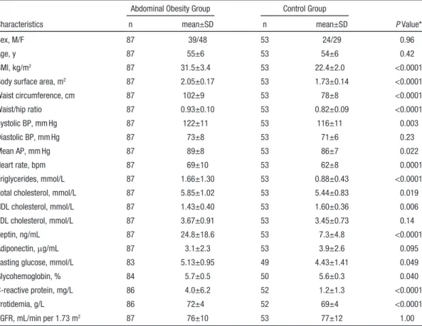
One hundred ninety-two subjects were recruited. Fifty-one (40 AO) were excluded: 41 (29 AO) because of BP >140/90 mm Hg, 4 took antihypertensive therapy, and 6 were on thyroid hormone medication (6 AO). One patient could not attend the second visit. Therefore, our final study population included 140 subjects: 87 subjects in the AO group and 53 controls. Anthropometric and Metabolic Characteristics Systolic BP (122±11 versus 116±11 mm Hg; P=0.003) and MAP (89±8 versus 86±7 mm Hg; P=0.022) were sig-nificantly higher in subjects with AO (not DBP, 73±8 versus 71±6 mm Hg; P=0.23)—although being still normotensive— along with a higher heart rate (P<0.0001). Leptin concentra-tions were significantly higher in AO group than in controls (24.8±18.6 versus 7.3±4.8 ng/mL; P<0.0001), and a trend for adiponectin concentrations in AO (3.1±2.3 versus 3.9±2.6;
P=0.095; Table 1).
Cardiac and Arterial Characteristics
Subjects with AO displayed a LV remodeling as assessed by a significant increase in LV mass (94±24 versus 84±21 g; P=0.034) without reaching LV hypertrophy17 and in CRI
(0.67±0.16 versus 0.60±0.10 g/mL; P=0.026) mainly because of the increase in LV mass. A significant increase in aminoter-minal propeptide of type I procollagen (P<0.0001) accompa-nied by a decrease in type 1 collagen telopeptide (P<0.0001) concentrations were observed in AO. C-terminal propeptide
of procollagen type I concentrations were higher in controls than in AO group (103±49 versus 87±52 ng/mL; P=0.001). In both groups LVEF was normal (P=0.85). In the AO group, E′ was significantly lower and E/E′ higher than that in controls (P<0.0001 and P=0.004, respectively; Table 2). Thirty-two patients with AO (38%) had diastolic dysfunction (grade I and II)11 compared with only 4% of controls. AO subjects with
dia-stolic dysfunction compared with AO without diadia-stolic dys-function displayed a higher BP (DBP and MAP, P=0.002 and
P=0.005, respectively, and a trend for SBP, P=0.077), waist circumference (P=0.028), and CRI (P=0.031; Table S1 in the online-only Data Supplement).
The 2 groups (AO versus controls) did not differ signifi-cantly in terms of arterial parameters (ie, PWV [P=0.26] and carotid IMT [P=0.33]). Only 4% of the total population pre-sented significant intima–media thickening (defined as carotid IMT >0.90 mm), and no subject had arterial stiffness as defined by a PWV >12 m/s.22 There was no association between PWV
or IMT on one hand and LV mass (LVM; scaled or not), CRI, and E′ on the other hand.
Structural and Functional Determinants of Cardiac and Arterial Remodeling
In multivariate analysis, SBP (regression coefficient±SEM, 0.33±0.13; P=0.013) and MAP (0.43±0.20; P=0.030) were positively and independently associated with LVM (or scaled LVM as expressed in g/kg of fat-free mass or g/m1.7, data not
shown) and MAP (0.34±0.16; P=0.035) with CRI. In multi-variate analysis, leptin concentrations were significantly asso-ciated with LV mass (−0.37±0.12; P=0.003) but not with CRI or diastolic dysfunction. Adiponectin concentrations were not found associated with LV mass, CRI, or diastolic dysfunc-tion. AO was independently associated with CRI (6.26±2.29;
P=0.007) and scaled LVM (g/m1.7 and g/m2.7, data not shown)
but not with LVM, which was found mainly associated with female sex and body surface area (Table 3).
In multivariate analysis, diastolic dysfunction was found positively and independently associated with PIIINP concen-trations above median (≥2.4 ng/mL; odds ratio [OR], 4.15 [1.42–12.2]; P=0.010), AO (OR, 13.3 [2.83–62.4]; P=0.001), and MAP ≥88 mm Hg (OR, 4.29 [1.57–11.7]; P=0.005; Table 4). In bivariate correlation analyses, waist circumfer-ence and waist/hip ratio were significantly associated with LV mass (r=0.52 and r=0.59; P<0.0001), CRI (r=0.37 and
r=0.49; P<0.0001), and E′ wave (r=−0.46 and −0.31, respec-tively; P=0.0002).
Finally, considering the higher (although in the normal range) BPs in the AO group, a first sensitivity analysis was performed in a subgroup of 50 patients with AO and 50 con-trol propensity score–matched patients (age, sex, and mean BP). Similar patterns were observed in this subgroup analy-sis, that is, higher LVM, CRI, and proportion of diastolic dysfunction, with however marginally significant differences (Tables S2 and S3). Multivariate analyses confirmed that (1)
Table 1. Anthropometric and Metabolic Characteristics of the Study Population
Characteristics
Abdominal Obesity Group Control Group
P Value*
n mean±SD n mean±SD
Sex, M/F 87 39/48 53 24/29 0.96
Age, y 87 55±6 53 54±6 0.42
BMI, kg/m2 87 31.5±3.4 53 22.4±2.0 <0.0001
Body surface area, m2 87 2.05±0.17 53 1.73±0.14 <0.0001
Waist circumference, cm 87 102±9 53 78±8 <0.0001 Waist/hip ratio 87 0.93±0.10 53 0.82±0.09 <0.0001 Systolic BP, mm Hg 87 122±11 53 116±11 0.003 Diastolic BP, mm Hg 87 73±8 53 71±6 0.23 Mean AP, mm Hg 87 89±8 53 86±7 0.022 Heart rate, bpm 87 69±10 53 62±8 0.0001 Triglycerides, mmol/L 87 1.66±1.30 53 0.88±0.43 <0.0001
Total cholesterol, mmol/L 87 5.85±1.02 53 5.44±0.83 0.019
HDL cholesterol, mmol/L 87 1.43±0.40 53 1.60±0.36 0.006
LDL cholesterol, mmol/L 87 3.67±0.91 53 3.45±0.73 0.14
Leptin, ng/mL 87 24.8±18.6 53 7.3±4.8 <0.0001
Adiponectin, μg/mL 87 3.1±2.3 53 3.9±2.6 0.095
Fasting glucose, mmol/L 83 5.13±0.95 49 4.43±1.41 0.049
Glycohemoglobin, % 84 5.7±0.5 50 5.6±0.3 0.040
C-reactive protein, mg/L 86 4.0±6.2 52 1.2±1.3 <0.0001
Protidemia, g/L 86 72±4 52 69±4 <0.0001
eGFR, mL/min per 1.73 m2 87 76±10 53 77±12 1.00
AP indicates arterial pressure; BMI, body mass index; BP, blood pressure; eGFR, estimated glomerular filtration rate (Modification in Diet Renal Disease 4-variable formula); F, female; HDL, high density lipoprotein; LDL, low-density lipoprotein; and M, male.
*P values from the Mann–Whitney or χ2 test as appropriate.
AO was associated with LVM, CRI, and diastolic dysfunction (the latter assessed by E′), and (2) PIIINP was associated with diastolic dysfunction (data not shown). A further sensitivity analysis in subjects with optimal/normal BP status (47 con-trols and 62 patients with AO with SBP/DBP <130/85 mm Hg) showed that AO (5.18±2.30; P=0.027) remained associated with CRI whereas BP did not and confirmed the relationship between diastolic dysfunction on one hand and AO (OR, 26.6 [3.04–233]; P=0.003), PIIINP ≥2.4 ng/mL (OR, 5.96 [1.55– 22.9]; P=0.01), and MAP ≥88 mm Hg (OR, 5.68 [1.62–19.9];
P=0.007) on the other hand.
Discussion
Cardiac and Arterial Remodeling: Changes in Structure and Function
The main and novel finding of our study is that in asymp-tomatic and normotensive healthy subjects with AO, cardiac remodeling (consisting of an increased LV mass) as well as features of cardiac concentric remodeling, which were
associated with the AO, and diastolic dysfunction are detect-able (the latter associated with increased collagen type III turnover). Importantly, we also examined the possible deter-minants of such early changes and found that, although in the normal range, BP was strongly associated with indices of cardiac remodeling and diastolic dysfunction in subjects with AO, suggesting a synergistic effect of AO amplifying the del-eterious effects of BP.
To analyze the specific effect of AO, we have carefully selected healthy asymptomatic young adult subjects with no hypertension and no known cardiovascular disease. We have also excluded patients with morbid obesity.
Although in elderly patients and patients with hyperten-sion LV hypertrophy,5 BP,23 and arterial stiffness24 are major
factors leading to diastolic dysfunction and subsequently to HF with preserved EF, in our AO asymptomatic subjects, we could show that increased LV mass and diastolic dysfunction could be detected early, before LV hypertrophy, hypertension, and arterial stiffening can be diagnosed.
Table 2. Cardiac and Arterial Characteristics of the Study Population
Characteristics
Abdominal Obesity Group Control Group
P Value* n mean±SD n mean±SD Cardiac characteristics PINP, ng/mL 87 34±15 53 22±14 <0.0001 PICP, ng/mL 87 87±52 53 103±49 0.001 PIIINP, ng/mL 81 2.5±1.4 53 3.4±6.7 0.16 ICTP, ng/mL 86 3.8±1.0 53 4.6±0.9 <0.0001 LVM, g† 72 94±24 47 84±21 0.034 LVMi, g/m2 72 45±9 47 48±10 0.068 LVM/FFM (Palmieri), g/kg 72 1.75±0.26 47 1.78±0.28 0.38 LVM (Chirinos), g/m1.7 72 38.9±7.6 47 34.7±7.3 0.003 LVM (De Simone), g/m2.7 72 23.3±4.0 47 20.6±4.1 0.0002 LVEF, %† 72 60±7 47 59±7 0.85 LVEDV, mL† 72 141±29 47 141±25 0.78 LVESV, mL† 72 58±21 47 57±15 0.63 LVEV, mL† 72 83±14 47 84±17 0.90 CO (LVEV×h), L/min 72 5.84±1.20 47 5.50±1.03 0.17 CRI (LVM/LVEDV), g/mL 72 0.67±0.16 47 0.60±0.10 0.026 E wave, cm/s‡ 87 68±17 52 74±15 0.043 A wave, cm/s‡ 87 59±14 52 53±10 0.007 E/A ratio 87 1.17±0.32 52 1.41±0.31 <0.0001 E′ wave, cm/s‡ 84 10.9±2.4 52 13.7±2.5 <0.0001 A′ wave, cm/s‡ 84 10.9±2.7 52 9.4±2.6 0.0010 E/E′ ratio 84 6.4±1.6 52 5.5±1.5 0.004 Arterial characteristics PWV, m/s 70 7.7±1.3 48 8.0±1.4 0.26 IMT, mm 73 0.64±0.15 48 0.61±0.12 0.33
CO indicates cardiac output; CRI, cardiac remodeling index; FFM, fat-free mass (assessed using DEXA); ICTP, type 1 collagen telopeptide; IMT, intima–media thickness; LVEDV, left ventricular end-diastolic volume; LVEF, LV ejection fraction; LVESV, LV end-systolic volume; LVEV, LV ejection volume; LVM, LV mass; PICP, carboxyterminal propeptide of type I procollagen; PINP, aminoterminal propeptide of type I procollagen; PIIINP, aminoterminal propeptide of type III procollagen; and PWV, pulse wave velocity.
*P values from the Mann–Whitney test. †Assessed by cardiac MRI.
‡Assessed by transthoracic echocardiography.
Furthermore, increased collagen type III turnover was observed in the study participants with diastolic dysfunction. We also investigated the relationship between LV geometric remodeling, LV function, and markers of myocardial colla-gen fibrosis indicating cardiac extracellular matrix remodel-ing. Irrespective of the study group, we were able to identify that collagen type III turnover was positively associated with diastolic dysfunction.
Our finding is novel but consistent with our previous report of increased PIIINP in asymptomatic obese subjects.7 In such
subjects, we had previously reported that PIIINP was inde-pendently associated with insulin resistance,7 which is a
com-mon state in AO, and could contribute specifically to increase myocardial fibrosis. Our results further suggest that enhanced collagen type III turnover is associated with early diastolic dysfunction independently from the adipokine pathways (the latter associated with LVM but not with diastolic dys-function). In our previous report, in obese healthy subjects,7
PIIINP and E/A ratio were significantly positively correlated. The transformation of the extracellular matrix into a more substantial collagen component potentiated by the increase in LV mass may alter ventricular filling, possibly contributing to the development of LV diastolic dysfunction in subjects with AO. Consistently in patients with hypertension, Martos et al25
showed that type 1 collagen telopeptide, C-terminal propep-tide of procollagen type I, and PIIINP concentrations were higher in patients with symptomatic HF with preserved EF. These findings in various categories of subjects suggest that PIIINP, which predict outcome in HF with preserved EF26,27
though not after adjusting for other predictors,27 could be also
a early biomarker of LV diastolic dysfunction. BP and AO as Therapeutic Targets to Prevent Adverse Cardiac Remodeling?
Subjects with AO had higher SBP and MAP than controls whereas remaining within the normal range and as such not currently eligible for an antihypertensive treatment. Systolic BP and MAP were positively associated with changes in cardiac structure (LV mass, scaled LV mass, and CRI). Furthermore, patients with MAP ≥88 mm Hg had a 4-fold increase in the rate
of diastolic dysfunction. Interestingly, BP may be a predomi-nant determipredomi-nant of structural (LVM and CRI) and functional (diastolic dysfunction) cardiac changes only in high-normal and hypertensive (SBP/DBP ≥ 130/85 mm Hg) subjects with AO but not strictly in normontensive ones.
Although it has already been repeatedly demonstrated that hypertension, via LV hypertrophy and arterial stiffness, leads to diastolic dysfunction, this is the first instance, to our knowl-edge, demonstrating that BP within the normal range is shown as a determinant of diastolic dysfunction. Some studies high-lighted that diastolic dysfunction could be associated with BP in normal range in general population28 but some were treated
for hypertension.29 Law et al30 emphasized the key role of
BP reduction in everyone to prevent cardiovascular diseases in the setting of the largest meta-analysis of randomized tri-als on hypertension management. Lowering SBP (by 10 mm Hg) or DBP (by 5 mm Hg) using any of the main classes of BP-lowering drugs reduced cardiovascular events (25% for HF) regardless of BP level before treatment. Furthermore, Julius et al31 (TROPHY [Trial of Preventing Hypertension]
Study) described that subjects at high risk to develop hyper-tension may benefit from an early intervention to reduce BP by RAAS (renin–angiotensin–aldosterone system) blockers to decrease the risk of incident hypertension and its conse-quences. In TROPHY Study, subjects were similar to our pres-ent study: they were young (48.6±7.9 years old), overweight (body mass index, 29.9±5.1 kg/m2), and most displayed a BP
in the high-normal category (SBP, 133.9±4.3 mm Hg; and
Table 3. Factors Associated With Left Ventricular Mass and Cardiac Remodeling Index in Multivariate Analysis
Factors Regression Coefficient±SEM P Value Variance Explained, %* LVM, g AO (yes vs no) −4.77±4.44 0.28 0.4 FS (yes vs no) −12.15±4.62 0.010 2.3 BSA, m2 56.4±12.2 <0.0001 7.2 SBP, mm Hg 0.33±0.13 0.013 2.1 Leptin, ng/mL −0.37±0.12 0.003 3.2 CRI, 10–2 g/mL AO (yes vs no) 6.26±2.29 0.007 4.5 FG (yes vs no) −11.26±2.45 <0.0001 12.6 MAP, mm Hg 0.34±0.16 0.035 2.7
P values are from linear regression. AO indicates abdominal obesity; BSA, body surface area; CRI, cardiac remodeling index; FS, female sex; LVM, left ventricular mass; MAP, mean arterial pressure; and SBP, systolic blood pressure.
*Independently of other factors.
Table 4. Factors Associated With Diastolic Dysfunction in Multivariate Analysis
Factor OR (95% CI) P Value*
AO (yes vs no) 13.3 (2.83–62.4) 0.001
PIIINP ≥2.4 ng/mL 4.15 (1.42–12.2) 0.010
MAP ≥88 mm Hg 4.29 (1.57–11.7) 0.005
P values are from logistic regression. AO indicates abdominal obesity; CI, confidence interval; DD, diastolic dysfunction (E′ <10 cm/s); MAP, mean arterial pressure; OR, odds ratio; and PIIINP, aminoterminal propeptide of type III procollagen.
DBP, 84.8±3.8 mm Hg). Interestingly, angiotensin-converting enzyme inhibitors and angiotensin receptor blocker antago-nists as well as the direct renin inhibitor, aliskiren,32 and the
MRA (mineralocorticoid receptor antagonist) eplerenone33
may regress LV hypertrophy in patients with hypertension.23
Furthermore, both angiotensin-converting enzyme inhibitors and angiotensin receptor blocker may decrease the incidence of HF in patients with high cardiovascular risk.34–37 We could
hypothesize that an aggressive and early management to reduce BP could possibly prevent cardiac structural and func-tional remodeling in subjects with AO.
Furthermore, that AO was independently associated with the cardiac structural (scaled LVM and CRI) and functional (diastolic dysfunction) remodeling (both in multivariate anal-yses led within the whole study population and after further adjustment based on propensity-score matching on age, sex, and mean BP in a sensitivity analysis) suggests that lifestyle changes leading to weight loss with reduction in waist circum-ference, independently from the use of BP-lowering drugs, could be a combination of paramount importance to achieve HF prevention in subjects with AO. Accordingly, in a general population, a waist circumference reduction was found associ-ated with a lower risk to develop hypertension.38
Study Limitations
Our study presents certain limitations. This study is a cross-sectional study, which prohibits from inferring a caus-ative link. Our results may not apply to patients with mor-bid obesity, which were not included herein. There is no consensus for normalization of LV mass by different types of scaling (height, fat-free mass, body surface area, etc), and parameters used are different according to echocardiography or MRI methods. However, our findings on LV remodeling were consistent throughout the different definitions used for LV mass scaling. BP status of our patients was only analyzed by office BPs although several standardized measurements were performed at each given visit. Furthermore, we previ-ously described thanks to an MRI substudy that there was no difference in aortic PWV between controls and patients with AO.39 We may hypothesize that arterial changes may not have
occurred yet in the subjects we have investigated, who were young and normotensives. Circulating collagen peptides are not specific to cardiac tissue, but previous histological studies observed a significant correlation between collagen peptides serum concentrations and cardiac fibrosis. Circulating colla-gen peptides are therefore an acceptable surrogate to evaluate cardiac fibrosis turnover. Of note, in the present study, no study participant exhibited abnormal alanine aminotransferase con-centrations (data not shown), therefore ruling out the potential confounding effect of nonalcoholic fatty liver disease, which was probably absent in our study population, on PIIINP con-centrations. To which extent sleep apnea disorders may have contributed to the observed cardiovascular features was not evaluated herein. Finally, no gadolinium tracer was injected, and thereby myocardial fibrosis could not be assessed in situ. Perspectives
Normotensive subjects with AO, but otherwise asymptomatic and healthy, exhibit early detectable features of cardiac concentric
remodeling as well as of enhanced collagen type III turnover associated with diastolic dysfunction without arterial changes. These alterations may help identifying subjects with AO at higher risk for developing HF with preserved EF and who could poten-tially benefit from early preventive interventions such as BP low-ering, even in normotensive subjects, and weight loss.
Acknowledgments
We thank Pierre Pothier for the editing of this article.Sources of Funding
This work was supported by French Program Hospitalier de Recherche Clinique, Inserm; the Région Lorraine; the Program National de Recherche Cardiovasculaire, the Société Française d’Hypertension artérielle; the Fédération Française de Cardiologie; the 7th EU-FP MEDIA project (261409); and HOMAGE (305507) projects.
Disclosures
None.References
1. Hu G, Jousilahti P, Antikainen R, Katzmarzyk PT, Tuomilehto J. Joint effects of physical activity, body mass index, waist circumfer-ence, and waist-to-hip ratio on the risk of heart failure. Circulation. 2010;121:237–244.
2. Holt RI. International Diabetes Federation re-defines the metabolic syn-drome. Diabetes Obes Metab. 2005;7:618–620.
3. Hunt SA; American College of Cardiology; American Heart Association Task Force on Practice Guidelines (Writing Committee to Update the 2001 Guidelines for the Evaluation and Management of Heart Failure). ACC/ AHA 2005 guideline update for the diagnosis and management of chronic heart failure in the adult: a report of the American College of Cardiology/ American Heart Association Task Force on Practice Guidelines (Writing Committee to Update the 2001 Guidelines for the Evaluation and Management of Heart Failure). J Am Coll Cardiol. 2005;46:e1–e82. 4. Kenchaiah S, Evans JC, Levy D, Wilson PW, Benjamin EJ, Larson MG,
Kannel WB, Vasan RS. Obesity and the risk of heart failure. N Engl J Med. 2002;347:305–313.
5. Janicki JS, Brower GL, Gardner JD, Chancey AL, Stewart JA Jr. The dynamic interaction between matrix metalloproteinase activity and adverse myocardial remodeling. Heart Fail Rev. 2004;9:33–42.
6. Alla F, Kearney-Schwartz A, Radauceanu A, Das Dores S, Dousset B, Zannad F. Early changes in serum markers of cardiac extra-cellular matrix turnover in patients with uncomplicated hypertension and type II diabetes. Eur J Heart Fail. 2006;8:147–153.
7. Quilliot D, Alla F, Böhme P, Bruntz JF, Hammadi M, Dousset B, Ziegler O, Zannad F. Myocardial collagen turnover in normotensive obese patients: relation to insulin resistance. Int J Obes (Lond). 2005;29:1321–1328. 8. Rickham PP. Human experimentation. Code of ethics of the world
medi-cal association. Declaration of Helsinki. Br Med J. 1964;2:177. 9. Levey AS, Coresh J, Balk E, Kausz AT, Levin A, Steffes MW, Hogg RJ,
Perrone RD, Lau J, Eknoyan G; National Kidney Foundation. National Kidney Foundation practice guidelines for chronic kidney disease: evalua-tion, classificaevalua-tion, and stratification. Ann Intern Med. 2003;139:137–147. 10. He M, Tan KC, Li ET, Kung AW. Body fat determination by dual energy
X-ray absorptiometry and its relation to body mass index and waist circumference in Hong Kong Chinese. Int J Obes Relat Metab Disord. 2001;25:748–752.
11. Nagueh SF, Appleton CP, Gillebert TC, Marino PN, Oh JK, Smiseth OA, Waggoner AD, Flachskampf FA, Pellikka PA, Evangelisa A. Recommendations for the evaluation of left ventricular diastolic function by echocardiography. Eur J Echocardiogr. 2009;10:165–193.
12. Mandry D, Lapicque F, Odille F, Djaballah W, Codreanu A, Escanyé JM, Felblinger J, Karcher G, Claudon M, Marie PY. Multicompartmental anal-ysis of late contrast enhancement in areas of myocardial infarction sup-plied by chronically occluded coronary arteries. J Magn Reson Imaging. 2009;29:78–85.
13. Chirinos JA, Segers P, De Buyzere ML, Kronmal RA, Raja MW, De Bacquer D, Claessens T, Gillebert TC, St John-Sutton M, Rietzschel ER. Left ventricular mass: allometric scaling, normative values, effect of obe-sity, and prognostic performance. Hypertension. 2010;56:91–98.
14. de Simone G, Izzo R, Trimarco B. Left ventricular hypertrophy: old marker, new problems and new possibilities. J Hypertens. 2011;29:1480–1482. 15. Bella JN, Devereux RB, Roman MJ, O’Grady MJ, Welty TK, Lee ET,
Fabsitz RR, Howard BV. Relations of left ventricular mass to fat-free and adipose body mass: the strong heart study. The Strong Heart Study Investigators. Circulation. 1998;98:2538–2544.
16. Bluemke DA, Kronmal RA, Lima JA, Liu K, Olson J, Burke GL, Folsom AR. The relationship of left ventricular mass and geometry to incident cardiovascular events: the MESA (Multi-Ethnic Study of Atherosclerosis) study. J Am Coll Cardiol. 2008;52:2148–2155.
17. Alfakih K, Plein S, Bloomer T, Jones T, Ridgway J, Sivananthan M. Comparison of right ventricular volume measurements between axial and short axis orientation using steady-state free precession magnetic reso-nance imaging. J Magn Reson Imaging. 2003;18:25–32.
18. Kearney-Schwartz A, Rossignol P, Bracard S, Felblinger J, Fay R, Boivin JM, Lecompte T, Lacolley P, Benetos A, Zannad F. Vascular structure and function is correlated to cognitive performance and white matter hyper-intensities in older hypertensive patients with subjective memory com-plaints. Stroke. 2009;40:1229–1236.
19. Zannad F, Rossignol P, Iraqi W. Extracellular matrix fibrotic markers in heart failure. Heart Fail Rev. 2010;15:319–329.
20. Iraqi W, Rossignol P, Angioi M, Fay R, Nuée J, Ketelslegers JM, Vincent J, Pitt B, Zannad F. Extracellular cardiac matrix biomarkers in patients with acute myocardial infarction complicated by left ventricular dysfunction and heart failure: insights from the Eplerenone Post-Acute Myocardial Infarction Heart Failure Efficacy and Survival Study (EPHESUS) study. Circulation. 2009;119:2471–2479.
21. Perkins NJ, Schisterman EF. The inconsistency of “optimal” cutpoints obtained using two criteria based on the receiver operating characteristic curve. Am J Epidemiol. 2006;163:670–675.
22. Mancia G, De Backer G, Dominiczak A, et al. 2007 Guidelines for the man-agement of arterial hypertension: the Task Force for the Manman-agement of Arterial Hypertension of the European Society of Hypertension (ESH) and of the European Society of Cardiology (ESC). Eur Heart J. 2007;28:1462–1536. 23. Wright JW, Mizutani S, Harding JW. Pathways involved in the transition
from hypertension to hypertrophy to heart failure. Treatment strategies. Heart Fail Rev. 2008;13:367–375.
24. Kang S, Fan HM, Li J, Fan LY, Miao AY, Bao Y, Wu LZ, Zhu Y, Zhang DF, Liu ZM. Relationship of arterial stiffness and early mild dia-stolic heart failure in general middle and aged population. Eur Heart J. 2010;31:2799–2807.
25. Martos R, Baugh J, Ledwidge M, O’Loughlin C, Conlon C, Patle A, Donnelly SC, McDonald K. Diastolic heart failure: evidence of increased myocardial collagen turnover linked to diastolic dysfunction. Circulation. 2007;115:888–895.
26. Barasch E, Gottdiener JS, Aurigemma G, Kitzman DW, Han J, Kop WJ, Tracy RP. The relationship between serum markers of collagen turnover and cardiovascular outcome in the elderly: the Cardiovascular Health Study. Circ Heart Fail. 2011;4:733–739.
27. Krum H, Elsik M, Schneider HG, Ptaszynska A, Black M, Carson PE, Komajda M, Massie BM, McKelvie RS, McMurray JJ, Zile MR, Anand IS. Relation of peripheral collagen markers to death and hospitalization in patients with heart failure and preserved ejection fraction: results of the I-PRESERVE collagen substudy. Circ Heart Fail. 2011;4:561–568.
28. Redfield MM, Jacobsen SJ, Burnett JC Jr, Mahoney DW, Bailey KR, Rodeheffer RJ. Burden of systolic and diastolic ventricular dysfunction in the community: appreciating the scope of the heart failure epidemic. JAMA. 2003;289:194–202.
29. Kuznetsova T, Herbots L, López B, Jin Y, Richart T, Thijs L, González A, Herregods MC, Fagard RH, Díez J, Staessen JA. Prevalence of left ventricular diastolic dysfunction in a general population. Circ Heart Fail. 2009;2:105–112.
30. Law MR, Morris JK, Wald NJ. Use of blood pressure lowering drugs in the prevention of cardiovascular disease: meta-analysis of 147 ran-domised trials in the context of expectations from prospective epidemio-logical studies. BMJ. 2009;338:b1665.
31. Julius S, Nesbitt SD, Egan BM, Weber MA, Michelson EL, Kaciroti N, Black HR, Grimm RH Jr, Messerli FH, Oparil S, Schork MA; Trial of Preventing Hypertension (TROPHY) Study Investigators. Feasibility of treating prehypertension with an angiotensin-receptor blocker. N Engl J Med. 2006;354:1685–1697.
32. Pitt B, Latini R, Maggioni AP, Solomon SD, Smith BA, Wright M, Prescott MF, McMurray JJ. Neurohumoral effects of aliskiren in patients with symptomatic heart failure receiving a mineralocorticoid receptor antagonist: the Aliskiren Observation of Heart Failure Treatment study. Eur J Heart Fail. 2011;13:755–764.
33. Pitt B, Reichek N, Willenbrock R, Zannad F, Phillips RA, Roniker B, Kleiman J, Krause S, Burns D, Williams GH. Effects of eplerenone, enala-pril, and eplerenone/enalapril in patients with essential hypertension and left ventricular hypertrophy: the 4E-left ventricular hypertrophy study. Circulation. 2003;108:1831–1838.
34. McMurray JJ, Adamopoulos S, Anker SD, et al; ESC Committee for Practice Guidelines. ESC Guidelines for the diagnosis and treatment of acute and chronic heart failure 2012: the Task Force for the Diagnosis and Treatment of Acute and Chronic Heart Failure 2012 of the European Society of Cardiology. Developed in collaboration with the Heart Failure Association (HFA) of the ESC. Eur Heart J. 2012;33:1787–1847. 35. Yusuf S, Sleight P, Pogue J, Bosch J, Davies R, Dagenais G. Effects of
an angiotensin-converting-enzyme inhibitor, ramipril, on cardiovascular events in high-risk patients. The Heart Outcomes Prevention Evaluation Study Investigators. N Engl J Med. 2000;342:145–153.
36. ONTARGET Investigators; Yusuf S, Teo KK, Pogue J, Dyal L, Copland I, Schumacher H, Dagenais G, Sleight P, Anderson C. Telmisartan, ramipril, or both in patients at high risk for vascular events. N Engl J Med. 2008;358:1547–1559.
37. Fox KM; EURopean trial On reduction of cardiac events with Perindopril in stable coronary Artery disease Investigators. Efficacy of perindopril in reduction of cardiovascular events among patients with stable coronary artery disease: randomised, double-blind, placebo-controlled, multicentre trial (the EUROPA study). Lancet. 2003;362:782–788.
38. Luo W, Guo Z, Hu X, Zhou Z, Mingwu, Zhang L, Liu J. A prospective study on association between 2 years change of waist circumference and incident hypertension in Han Chinese. Int J Cardiol. 2013;167:2781–2785. 39. Mandry D, Eschalier R, Kearney-Schwartz A, Rossignol P, Joly L,
Djaballah W, Böhme P, Escanyé JM, Vuissoz PA, Fay R, Zannad F, Marie PY. Comprehensive MRI analysis of early cardiac and vascular remodeling in middle-aged patients with abdominal obesity. J Hypertens. 2012;30:567–573.
What Is New?
•
It is the first report that in asymptomatic and normotensive healthy sub-jects with abdominal obesity, cardiac remodeling (structural, diastolic dysfunction, and myocardial fibrosis) is detectable.What Is Relevant?
•
Blood pressure in normal range, abdominal obesity, and aminoterminal propeptide of type III procollagen concentrations in nonhypertensive pa-tients with abdominal obesity were independently associated with dia-stolic dysfunction.Summary
Normal blood pressure and myocardial fibrosis in healthy, asymp-tomatic, nonhypertensive subjects with abdominal obesity were as-sociated with cardiac remodeling. Subjects with abdominal obesity could potentially benefit from early preventive interventions, such as BP lowering, even in normotensive subjects, and weight loss.