The biochemical basis for the cooperative action of microRNAs
By MAssACHUSE S ISTITUTE
By OFTECHNOLOGY >
Daniel Briskin
OCT
0 12019
B.S., Molecular Genetics (2012)
LIBRARIES
0
Ohio State University
SUBMITTED TO THE DEPARTMENT OF BIOLOGY IN PARTIAL FULFILLMENT OF
THE REQUIREMENTS FOR THE DEGREE OF DOCTOR OF PHILOSOPHY
AT THE
MASSACHUSETTS INSTITUTE OF TECHNOLOGY
-SEPTEMBER-2019- -fve e 7-1012
© 2019 Massachusetts Institute of Technology
All rights reserved
Signature redacted
Signature of Author: DanielBriskin
Department of Biology 17 September 2019
Signature redacted
Certified By:__ David P. Bartel Professor of Biology Thesis SupervisorAccepted By:
Signatureredacted
-V Stephen Bell
Uncas and Helen Whitaker Professor of Biology Investigator, Howard Hughes Medical Institute Co-Director, Biology Graduate Committee
The biochemical basis for the cooperative action of microRNAs by
Daniel Briskin
Submitted to the Department of Biology on 17 September 2019
In partial fulfillment of the requirements for the degree of doctor of philosophy Abstract
In metazoans, microRNAs (miRNAs) act to repress mRNAs through a combination of translational repression and target degradation. miRNAs predominantly pair within the 3' untranslated region (3' UTR) of the mRNA. In cells, closely spaced miRNA target sites within an mRNA can act cooperatively, leading to more repression of the target
mRNA than expected by independent action at each site.
This dissertation details the use of purified miRNA-AGO2 complexes, synthetic target RNAs, and a purified domain of TNRC6B that is able to simultaneously bind multiple AGO proteins. We examined the target site occupancy and affinities for miRNA-AGO2 binding in the absence and presence of TNRC6B, for target RNAs with a single miRNA site as well as multiple miRNA sites spaced at varying distances. As miRNA-AGO-binding to target correlates with target repression, our study assayed target miRNA-AGO-binding. Absent TNRC6B, miRNA-AGO2 complexes showed little if any cooperative binding. In the presence of the AGO-binding domain of TNRC6B, we observed strong cooperative binding to dual-site target RNAs. We went on to explore the miRNA site parameters suitable for cooperativity, investigating the spacing between sites as well as different miRNAs working alone or in combination with one another. To interrogate the
mechanism by which TNRC6B increases cooperativity, competitive slicing experiments were performed; results indicated that association rates between miRNA-AGO2
complexes and targets were not affected by TNRC6B, which implied that the improved affinities were due to reduced dissociation. Thus, the multivalent binding of TNRC6 enables cooperative binding of miRNA-AGO complexes to target RNAs, thereby explaining the basis of cooperative action.
Thesis advisor: David P. Bartel Title: Professor
Acknowledgments
I have been fortunate to work with a special group of people over the past five years of my graduate studies. First, I would like to thank Dave Bartel for his scientific guidance and rigor. Thanks to my thesis committee, Tom RajBhandary and Phil Sharp, for their guidance and support on my project. I would also like to thank the above group of
people for their instruction in the courses 7.77 and 7.60, where even before joining a lab I learned to think more deeply about the RNA-centric questions that interested me. Thank you to Gianpiero DiLeva, my first scientific mentor, who started to teach me how to think about and do science. Finally, I am sincerely grateful to Tom Tuschl, under whom I had the pleasure of working for two years as a technician. Tom is the person who got me excited about science; working in his lab, I began to understand the thrills of what research could illuminate and the impact that research can have outside of the lab. I would also like to thank other members of the Bartel lab. Laura and Asia are the
backbone of the lab who keep the day-to-day aspects running as smoothly as they do. Laura has, on more than one occasion, helped me to rush a rad order, allowing me to complete some critical, last experiment before lab meeting. Asia wrangles the
cornucopia of equipment we have in the lab, some component of which is always
breaking down. Nonetheless, she works to make sure that we always have whatever we need to do our work. (Furthermore, she takes on the daunting task of interfacing with the vendors.) I also thank Bartel lab graduate students, postdocs, and technicians. The attention that lab mates give to each others' projects is exceptional; I have received help from all of my peers and to them I am deeply grateful. Thanks, Papa Sean, my blood
brother. I would also like to specifically thank my friends in 623, Tim, Jamie
McKwasnieski, Michael, (Glenn and Emir, RIP) and my bay mate Matt. They make 623 the most fun room in Bartel lab. From discussing science, to double blocks, I always looked forward to coming in and talking about what is happening in our lives and the world. I'd like to thank and "thank" Matt for encouraging my running. It's the most fun and rewarding aspect of my life, but also responsible for an unfortunate number of trips to MIT Medical. I'd also like to thank Breakfast Club (Elena) for a friendly way to start the day. Thanks, Swim Club (paradoxically, the most exclusive and least elite swim team in the world), for giving me one less way to die. And thank you to Run Club for traveling near and far to run beautiful and challenging courses.
I'd like to thank my classmates Laurens and Nolan for their friendship over the years. Thank you to dead te me people Glassmates housemates friends at 6 Antrim St., Chris,
Grace, John, Josh, Santi, and Spencer. They have been an exceptional group of people to joke, commiserate, and live with. I have learned a multitude from these friends: from what lawyers are, to Caligula's travails, to being more woke, and to naht take my half
out of the middle. I can always count on them to slap some sense into me.
My family, Jeanne, Dan, Joe, Lily, Jackie, Ken, Olivia, Grandma, Grandpa, Benjamin, Emily, Mom and Dad, for your unwavering love and support, I love and thank you.
Table of contents A b s tra c t... 3 Acknowledgm ents... 5 Table of contents ... 7 Chapter 1. Introduction ... 9 m iRNA biogenesis... 9
Figure 1. m iRNA biogenesis... . 12
m iRNA-m ediated target repression m echanism ... . 15
Figure 2. m iRNA-m RNA target site types... . 16
Figure 3. m iRNA-m ediated target repression... . 18
T N R C 6 ... 1 9 P-bodies and phase separation... . 25
m iRNAs targeting, regulation, and dysregulation ... 29
R e fe re n c e s ... 3 5 Chapter 2. The biochemical basis for the cooperative action of microRNAs... 45
Chapter 3. Future directions ... . 83
Chapter 1. Introduction miRNA biogenesis
MicroRNAs (miRNAs) are short, non-coding RNAs approximately 22 nucleotides (nt) long that complex with Argonaute (AGO) proteins to posttranscriptionally regulate target gene expression through a combination of translational repression and mRNA destabilization. miRNAs target mRNAs through Watson-Crick pairing between the miRNA and mRNA target at sites typically within the 3' untranslated region (3' UTR) of the mRNA. AGO proteins mediate mRNA degradation through three distinct
mechanisms. One mechanism, unique to AGO2, one of four AGO paralogues in mammals, is using its catalytically active RNase H-like domain that, upon extensive complementarity between the miRNA guide and target mRNA, is used to cleave, or slice, the target, leading to the target's degradation (Elbashir et al., 2001; Schirle and MacRae, 2012; Song et al., 2004). The other human AGO paralogues, AGO1, 3, and 4, are loaded with miRNAs but do not cleave their targets (Liu et al., 2004; Meister et al., 2004). In addition, the vast majority of miRNAs do not pair extensively enough with their target as required to induce cleavage but rather bind with complementarity to the
miRNA seed region (miRNA nucleotides 2-7) (Bartel, 2009). These non-cleavage competent interactions repress mRNA through other modes: translational repression and mRNA destabilization.
Early characterization of miRNAs was done in C. elegans where the lin-4 gene was found to regulate lin-14 activity, which plays a role in C. elegans development (Lee et al., 1993; Wightman et al., 1993). The product of lin-4 is not a protein but rather a 21
nt RNA with complementarity to multiple sites in the 3' UTR of lin-14. Since this early work, miRNAs have been found throughout eukaryotes, including plants and animals (Bartel, 2004; Lagos-Quintana et al., 2001).
In metazoans, canonical miRNAs are transcribed by RNA polymerase 11 as part of a primary transcript that is capped and polyadenylated (Fig. 1) (Cai et al., 2004; Lee et al., 2004). miRNAs may be found in the transcribed sequence of protein-coding genes, typically in introns, or have their own promoters (Ha and Kim, 2014). The primary miRNA transcript (pri-miRNA) has regions of sequence with reverse
complementarity such that the RNA folds back on itself and makes a stem-loop. This stem-loop is then processed by a heterotrimeric complex called microprocessor, consisting of two DiGeorge syndrome critical region 8 (DGCR8) proteins and one Drosha. DGCR8 acts as a cofactor for Drosha, which has two RNase Ill domains, each of which interacts with one DGCR8 (Kwon et al., 2016). One RNase IlIl domain cleaves the 5' strand of the stem-loop; the other RNase Ill domain cleaves the 3' strand of the stem-loop (Han et al., 2004). Features that improve processing of pri-miRNAs include both structural and primary sequence elements (Fang and Bartel, 2015). Structurally, pri-miRNA processing is favored by a 35 base pair (bp) stem length with an apical loop of a10 nt and basal single stranded RNA (Fang and Bartel, 2015). Drosha functions as
a ruler to measure 11 bp from the basal junction for one cut site; cutting both strands of RNA duplex produces a hairpin with a 5' phosphate and 2 nt 3' hydroxy overhang; this species is known as the pre-miRNA (Kwon et al., 2016). The pre-miRNA is then
exported from the nucleus by exportin 5 in complex with RAN-GTP (Lund et al., 2004; Yi et al., 2003).
Once in the cytoplasm, the pre-miRNA is processed by Dicer. Dicer recognizes the 5' and 3' ends of the pre-miRNA and cleaves the pre-miRNA at two places to
generate an RNA duplex typified by two -22 nt RNA molecules, each with 5' phosphate and 3' hydroxy termini and a 2 nt 3' overhang on each end (Bernstein et al., 2001;
Hutvagner et al., 2001; Park et al., 2011; Tian et al., 2014; Zhang et al., 2002). Finally, one of the two strands of RNA is loaded into AGO. The more unstable 5' end of the duplex is favored for AGO loading (Khvorova et al., 2003; Schwarz et al., 2003). Additionally, AGO favors loading strands with 5' adenosine or 5' uridine (Suzuki et al., 2015). However, some duplexes exhibit essentially equal loading between the originally 5' and 3' strands, which become the 5p and 3p strands, respectively. The strand that is loaded into AGO is termed the guide strand, while the strand that is not loaded is
termed the passenger, or star, strand. The unloaded strand is degraded by endogenous RNases.
Pol 11 transcription Microprocessor VGCR %rosha DGCR 3' HO Exportin5-RAN-GTP Nucleus Cytoplasm 5'-~1 5'-p " OH-3' 31-HO p-5'
AGO loading, star strand degradation,
mRNA targeting AGO
Figure 1. miRNA biogenesis
miRNAs are transcribed by RNA polymerase II, resulting in a capped and
polyadenylated transcript. Drosha (salmon) works in combination with two DGCR8 (green) to cleave the pri-miRNA at indicated sites (salmon arrows) to generate the pre-miRNA. Exportin5-RAN-GTP exports the pre-miRNA from the nucleus to the cytoplasm where Dicer (purple) further cleaves (purple arrows) to generate the mature miRNA. Finally, the guide miRNA strand (red) is loaded into AGO protein (brown) to target an mRNA (black). The passenger strand (blue) is degraded.
Some miRNAs are processed in a Dicer-independent way, such as miR-451. miR-451 is transcribed as a relatively short hairpin such that it is too short for proper Dicer processing (Siolas et al., 2005). Thus, the hairpin is loaded directly into an AGO,
although AGO2 is uniquely capable of processing the miR-451 due to the catalytic domain of AGO2. The hairpin is cleaved by AGO2, the short 3'cleavage fragment is ejected, and the 3' end of the loaded miRNA is trimmed back by endogenous nucleases (Cheloufi et al., 2010; Cifuentes et al., 2010; Yoda et al., 2013). Similarly, miR-486, although Dicer dependent, requires AGO2 catalytic activity to slice and eject the guide strand to generate mature miR-486-AGO2 (Jee et al., 2018).
Argonaute proteins are divided into four globular domains, termed N, MID, PWI,
and PAZ, and two linker domains, Li and L2 (Song et al., 2004; Swarts et al., 2014b). The 5' terminus of the miRNA is tethered to the MID domain of AGO (Elkayam et al., 2012; Ma et al., 2005; Parker et al., 2005; Schirle and MacRae, 2012). The 5'
nucleotide inserts into a binding pocket on AGO such that it is not available for target pairing (Elkayam et al., 2012; Ma et al., 2005; Schirle et al., 2015; Wang et al., 2009). Target mRNAs often have an adenosine at their first position, and this base specifically goes into a pocket between the MID and L2 domains (Lewis et al., 2005; Schirle et al., 2015; Schirle et al., 2014). Nucleotides 2-6 of the guide RNA are prearranged in A-form
helix and exposed to the solvent, which helps speed miRNA target searching (Bartel, 2018; Chandradoss et al., 2015; Elkayam et al., 2012; Salomon et al., 2015; Schirle and MacRae, 2012). Not only does AGO confer shape to and nuclease protection upon the loaded miRNA, but the loaded miRNA also confers stability upon AGO, increasing AGO resistance to protease (Elkayam et al., 2012). Only limited interactions are observed between AGO and the Watson-Crick face of nucleotides, but interactions are observed
with the backbone of the miRNA, including Van der Waals interactions with the 2' hydroxy of the ribose (Schirle and MacRae, 2012).
The PIWI domain of AGO contains the active site DEDH tetrad reminiscent of RNase H enzymes, which uses Mg2
+ as a cofactor (Liu et al., 2004; Nakanishi et al.,
2012). Although AGO2 is considered the only biological catalytically active of the four human paralogues, AGO3 has the same four amino acid residues in its catalytic tetrad as AGO2 and, under the right guide-target sequence conditions, AGO3 can catalyze cleavage of target RNA, albeit less effectively than AGO2 (Park et al., 2017). The greatly reduced cleavage activity of AGO3 is due to an immature, or less rigidly
structured, nucleic acid binding channel between the N and PWI domains (Hauptmann et al., 2013; Park et al., 2017). AGO1 does not conserve the catalytic tetrad of AGO2 (inactive DEDR in AGO1 rather than the active DEDH of AGO2) and therefore cannot slice target (Faehnle et al., 2013; Nakanishi et al., 2013). AGO4 is also incapable of slicing target, having a catalytically inactive DEGR at the would-be active site (Park et al., 2019).
The central region of miRNAs can be largely unpaired from target, even given complementarity to target (Sheu-Gruttadauria et al., 2019). Indeed, AGO can
accommodate a bridge of up to 15 upaired target nucleotides across from the central region of a miRNA while still effectively repressing target (Sheu-Gruttadauria et al., 2019). The 3' half of the miRNA interacts with the PAZ domain of AGO. Guide nucleotides 14-18 are fed through a channel between the PAZ and N domains. miRNA-AGO interactions perturb base stacking; the Watson-Crick edges of guide
nucleotides 15, 17, and 18 face toward AGO, away from the solvent, and are therefore unavailable for target pairing in the solved structure (Schirle et al., 2014). Supplemental pairing, pairing of miRNA nucleotides 13-16 to target mRNA, requires a conformational change in AGO whereby guide nucleotides 13-16 are exposed for pairing and a
supplementary chamber of AGO is opened to accommodate base pairing to target (Sheu-Gruttadauria et al., 2019).
miRNA-mediated target repression mechanism
The mammalian miRNA-AGO complex targets mRNAs, typically in the 3' UTR, through complementary base pairing. Compared to naked RNA, AGO increases the
association rate, kon, of miRNA pairing with target (Salomon et al., 2015). This increase in kon is executed by AGO structurally prearranging seed of the loaded miRNA into an A-form helix (Bartel, 2018; Elkayam et al., 2012; Schirle and MacRae, 2012). Although AGO increases kon, of miRNA to target, it also increases dissociation rate, koff of fully
paired target, relative to naked RNA pairing (Wee et al., 2012). Overall, miRNA-AGO has lower affinity for target RNA in comparison to the pairing of two naked RNA molecules (Salomon et al., 2015; Wee et al., 2012).
More than 60% of human mRNAs have conserved target sites of miRNAs
(Friedman et al., 2009). Like many genes, miRNA expression is temporally and spatially regulated (Landgraf et al., 2007). The more productive canonical mammalian miRNA target sites have 6-7 nt of contiguous base pairing, including nucleotides 2-7 or 2-8 of the miRNA (termed 6mer and 7mer-m8 sites, respectively) (Fig. 2) (Bartel, 2009, 2018).
If the target mRNA has an adenosine residue across from position 1 of the miRNA and nucleotides 2-7 or 2-8 are paired, the sites, respectively, are called a 7mer-A1 or 8mer. Other miRNA-target pairing regimes include supplementary pairing, which often
includes pairing across from nucleotides 13-16 of the miRNA, and compensatory pairing, in which imperfect pairing to the miRNA seed can be somewhat compensated for by additional pairing to the 3' region of the miRNA often including nucleotides 13-16
(Brennecke et al., 2005; Doench and Sharp, 2004; Grimson et al., 2007). Seed
87654321
3'-NNNNNNNNNNNNNNNNNNNNN-5' miRNA
1111111
8mer 5'...NNNNNNNNNNNNNNNNNNNNNNNANN ... 3 ' mRNA
7mer-m8 5'...NNNNNNNNNNNNNNNNNNNNNNNBNN ... 3' 7mer-A15'. . .NNNNNNNNNNNNNNNNNNNNNNNANN ... 3' 6mer 5'...NNNNNNNNNNNNNNNNNNNNNNNBNN...3' Seed 87654321 3'-NNNNNNNNNNNNNNNNNNNNN-5' miRNA liii 1111111 3'Supplementary 5'...NNNNNNNNNNNNNNNNNNNNNNNNNN ... 3 'mRNA Seed 87654321 3'-NNNNNNNNNNNNNNNNNNNN-5' miRNA 3' Compensatory 5' . . . NNNNNNNNNNNNNNNNNNNNNNNNNN... 3 'mRNA
Figure 2. miRNA-mRNA target site types
The miRNA (top) is shown with its seed (red) base paired to target mRNA. In cells, the miRNA is loaded into an Argonaute protein (not shown). For various mRNA pairing sites, the nucleotides involved in base pairing are diagramed in blue. 8mer, 7mer-m8, 7mer-A1, and 6mer sites are canonical site types diagramed in descending order of pairing stability. 3' supplementary and compensatory sites are atypical and are diagramed in descending order of pairing stability.
In rare instances in mammals, miRNA-AGO2 cleaves target mRNA, creating unprotected 5' and 3' termini that are subject to endogenous RNase decay, thereby degrading the RNA (Shin et al., 2010; Yekta et al., 2004). More typically, miRNA-AGO does not function alone but rather recruits additional protein factors. After miRNA-AGO
binds to target RNA, TNRC6 binds to AGO (Fig. 3). TNRC6, GW182 in flies, is a-182
kDa protein rich in glycine (G) and tryptophan (W); there are three TNRC6 paralogues in humans, TNRC6A-C. The mammalian gene name TNRC6 stands for trinucleotide-repeat containing protein 6. TNRC6 was show to interact with AGO through reciprocal co-IP experiments (Landthaler et al., 2008; Meister et al., 2005). The N-terminal half of TNRC6 is the AGO-binding domain (ABD). It has three annotated AGO-binding
hotspots, is largely unstructured, and is rich in tryptophan residues (Pfaff et al., 2013). Furthermore, TNRC6 has been shown to simultaneously interact with up to three AGO proteins (Elkayam et al., 2017; Takimoto et al., 2009). AGO-TNRC6 interactions are mediated via three W-binding pockets on the surface of AGO (Sheu-Gruttadauria and MacRae, 2018).
The C-terminal half of TNRC6, termed the silencing domain, has motifs that recruit factors responsible for the repression of mRNA targets (Braun et al., 2011;
Chekulaeva et al., 2011; Chen et al., 2014; Jonas and Izaurralde, 2015; Lazzaretti et al., 2009). Through other tryptophan-mediated interactions, the silencing domain directly
interacts with PAN2/PAN3, which are non-processive deadenylases that shorten the poly(A) tail of an mRNA (Christie et al., 2013). Via tryptophan-mediated binding interactions in the silencing domain of TNRC6, NOT proteins are recruited to the targeted mRNA. The CCR4/NOT complex has two deadenylases that shorten the poly(A) tail (Basquin et al., 2012; Jonas and Izaurralde, 2015). NOT proteins also bind DDX6, which represses translation (Chen et al., 2014; Kuzuoglu-Ozturk et al., 2016; Mathys et al., 2014). Further protein-protein interactions recruit decapping enzymes,
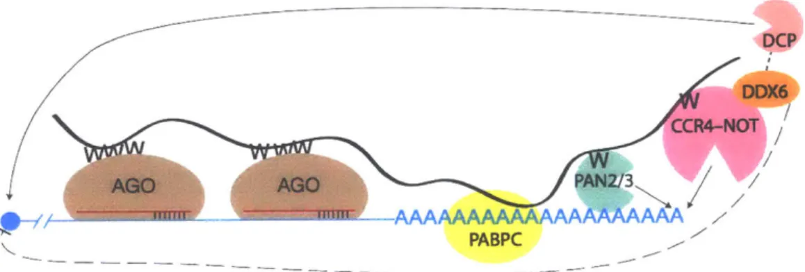
including DCP2, which removes the 5'm7G cap from the mRNA, leaving the mRNA for XRN1-mediated 5'-to-3' degradation (Jonas and Izaurralde, 2015). Thus, miRNA-AGO works to degrade an mRNA from both ends, first through deadenylation and then through decapping (Chen et al., 2009).
DCP
CCR4-NOT
AGOAG PAN2/3A W
AMI n AAAAAAAAAAAALAA.
PABPC
Figure 3. miRNA-mediated target repression
miRNA-AGO (red and brown, respectively) bind mRNA (blue). TNRC6 (black) binds miRNA-AGO through tryptophan-binding pockets on AGO and tryptophan residues (W) on TNRC6. TNRC6 also binds PABPC (yellow) which is presumably already bound to the mRNA poly(A) tail. TNRC6 recruits PAN2/PAN3 deadenylases (green) and CCR4-NOT deadenylation complex (pink) which both deadenylate the mRNA. DDX6 (orange) binds CCR4-NOT and translationally represses the mRNA. DCP (salmon) also interacts with repressive machinery and decaps the mRNA. Decapped mRNA is subject to
XRN1-mediated 5'-to-3' decay.
Inasmuch as miRNA-AGO-TNRC6 recruits factors for both translational repression as well as deadenylation and decapping, studies have been carried out to tease apart the contributions of each on posttranscriptional gene regulation. Ribosome footprint profiling has been performed in combination with mRNA sequencing to assess
concomitant changes in transcript levels and translation. Initial studies in human and mouse showed that miRNAs predominately downregulated gene expression via
decay accounted for at least 84% of repression (Guo et al., 2010). In certain contexts, such as early zebrafish development 4 h post-fertilization (hpf), translational repression was found to be the dominant effect of miRNAs (Bazzini et al., 2012). Further
investigation into early developmental stages of zebrafish and frog revealed that
translational efficiency is coupled to poly(A)-tail length, but only for zebrafish up to 4 hpf, and only for Xenopus laevis embryos up to stage 9 (Subtelny et al., 2014). The yeast, mouse, and human samples examined failed to reveal any correlation between poly(A)-tail length and translational efficiency (Subtelny et al., 2014). This correlation between poly(A)-tail length and translational efficiency can be attributed to miRNAs shortening poly(A)-tails and some feedback between tail length and translation. In zebrafish, after the window of coupling between poly(A)-tail length and translational efficiency, i.e. at 6 hpf, miRNAs again predominantly act to decrease mRNA levels (Subtelny et al., 2014). Examination of additional mammalian cell types and contexts found that
miRNA-mediated target degradation explained most of miRNA-miRNA-mediated repression, and that if translational repression occurs, by the time target is substantively repressed,
degradation is the main cause of repression (Eichhorn et al., 2014).
TNRC6
As discussed above, TNRC6 is a scaffold that recruits translational repression and degradation machinery to target mRNAs. The C-terminal half of TNRC6 has
additional domains that do not interact with deadenylases, including an RNA-recognition motif (RRM), poly(A)-binding protein (PABP)-interacting motif (PAM2 domain on
TNRC6), a glutamine (Q)-rich region, and a ubiquitin-associated (UBA) domain (Jonas and Izaurralde, 2015). Mutating the PAM2 domain of TNRC6 impedes the ability of miRNA-AGO-TNRC6 to repress targets (Huntzinger et al., 2013). The functions of the
UBA and RRM have yet to be elucidated; mutating either domain has not been shown
to alter target repression (Jonas and Izaurralde, 2015).
Both the N- and C-terminal halves of TNRC6 are necessary to elicit proper miRNA-mediated target silencing. The N-terminus binds AGO but the C-terminal
silencing domain of TNRC6 cannot. However, if recombinant N-fusion TNRC6 protein is tethered to the 3' UTR of a reporter mRNA with BoxB sites, that mRNA will be
repressed, as observed as a decrease in fluorescence reporter activity (Lazzaretti et al.,
2009). Residues 599-683 of TNRC6B (AGO-APP) have been shown to strongly bind to
human AGO2 (Hauptmann et al., 2015; Pfaff et al., 2013). If that peptide is expressed at high levels from a plasmid in HeLa cells, a global de-repression of target mRNAs is observed (Hauptmann et al., 2015). This de-repression occurs when AGO-APP outcompetes endogenous TNRC6. As AGO-APP does not have the silencing domain necessary for recruitment of repressive machinery, target mRNAs are no longer subject to miRNA-mediated repression.
TNRC6A has a nuclear export signal (NES) and a nuclear localization signal
(NLS); TNRC6B and TNRC6C do not have an NLS (Nishi et al., 2013). Thus, while in
mammals the action of miRNAs is largely in the cytoplasm, TNRC6A can be shuttled between the nucleus and cytoplasm. TNRC6 and AGO colocalize to cytoplasmic
for P-body localization in TNRC6B, but not TNRC6A (Lazzaretti et al., 2009). Deleting the Q-rich domain from TNRC6C has an intermediate effect, impairing by ~50% the
localization of TNRC6C to P-bodies.
The interaction between TNRC6-ABD and AGO has been explored and found to be more nuanced than tryptophan-tryptophan-binding pocket. Binding of TNRC6 to AGO causes a structural change in AGO to close a gate around the tryptophan of TNRC6 to help secure the AGO-TNRC6 interaction (Elkayam et al., 2017). Although glycine residues often flank tryptophan residues in the ABD, neither the glycine nor other non-tryptophan residues on TNRC6 have been found to interact with AGO
(Elkayam et al., 2017). Rather, in order to avoid steric clashes, the absence of bulky or aromatic side chains around the tryptophan enhances AGO-TNRC6 interactions (Pfaff et al., 2013). Each of the three tryptophan-binding pockets on the surface of AGO is -25
A
from its two neighboring tryptophan-binding pockets (Sheu-Gruttadauria and MacRae, 2018). This three-dimensional distance corresponds to a flexible amino acid linker distance of 10-15 amino acid residues; indeed, tryptophan residues within the ABD of TNRC6 are spaced at a mean distance of 11.7 amino acids apart (Sheu-Gruttadauria and MacRae, 2018).Further dissecting the interaction between individual binding pockets and TNRC6 proteins reveals a complex relationship. A 2016 study from the Izaurralde group, when only two tryptophan-binding pockets on AGO had been described, showed mutating one pocket on AGO had either very little effect (i.e. AGO2, pocket 1 mutant binds TNRC6B at almost native levels) or a very strong effect (i.e. AGO2, pocket 2 mutant has
extremely weak interaction with TNRC6C), depending on what pocket was mutated and which TNRC6 paralogue was binding (Kuzuoglu-Ozturk et al., 2016).
A 2018 study from the MacRae group, describing the three tryptophan-binding pockets on AGO2, investigated AGO-TNRC6B from both a TNRC6-centric and AGO2-centric approach. Different mutations in tryptophan residues of TNRC6B-ABD have a
similarly complex relationship with AGO2 binding. Purified TNRC6B-ABD association with AGO2 was assessed with WT TNRC6B-ABD, and TNRC6B-ABD in which every tryptophan was mutated to an alanine (A). In the W to A mutant, no AGO2-TNRC6B-ABD interaction was observed (Sheu-Gruttadauria and MacRae, 2018). Restoring two
W residues was sufficient to restore AGO-TNRC6B-ABD interaction. This restored interaction could again be ablated by mutating either of two tryptophan-binding pockets on the surface of AGO; mutating the third tryptophan-binding pocket does not impede AGO-TNRC6B-ABD (Sheu-Gruttadauria and MacRae, 2018). Alternatively, restoring three different W residues from A in a different stretch of TRNC6B-ABD was able to restore AGO-TRNC6B-ABD interactions with wild type AGO2. This TNRC6B-ABD peptide with three restored W residues was then tested for binding to each of the three AGO2 W pocket mutants; two pockets were found to be essential for binding and one
pocket was found to be dispensable, but the dispensable pocket for these three W
residues was different than the dispensable pocket for two different W residues (Sheu-Gruttadauria and MacRae, 2018). Thus, each tryptophan pocket on AGO2 has different
preferences has not been explained by neighboring side chains or alternate AGO2-TNRC6 interactions.
Further nuances factor in to the affinities between AGO and TNRC6. The affinity of one AGO-binding hotspot, the "AGO hook" of TNRC6A (residues 821-841 of
TNRC6A) was measured for binding AGO1 and for AGO2, each AGO either loaded or unloaded. Loaded AGO bound TNRC6 with -5-8 times higher affinity than did unloaded AGO (Elkayam et al., 2017). A cellular benefit of a higher affinity for loaded AGO is that
a loaded AGO engaged with target would better interact with TNRC6 and associated repressive machinery, leading to productive targeted degradation of an mRNA. An unloaded AGO-binding TNRC6 would not be able to direct repressive machinery to any specific target.
Additionally, although the N-terminal ABD of TNRC6 is thought of as largely unstructured, regulation of this region is possible. The affinity of any of the three AGO-binding hotspots alone at its tightest is 47 nM to AGO2 (Elkayam et al., 2017). However,
the entire TNRC6A-ABD has an affinity of 7.7 nM for its first instance of binding to AGO2. The second AGO2 to bind the same molecule of TNRC6A-ABD does so at an affinity of 1 pM, despite the next tightest of the AGO hotspots having, in isolation, an affinity of 120 nM to AGO2 (Elkayam et al., 2017). Thus, binding of one AGO decreases the affinity of TNRC6 for subsequent AGO-binding. If an mRNA has two miRNA target sites spaced closely enough to bind the same TNRC6, binding the second AGO with decreased affinity may be biologically advantageous. If the first and second AGO-binding events had equal affinity, the cell may need to preload TNRC6 with the specific
miRNA-AGO complexes loaded with the specific miRNAs targeting the neighboring miRNA sites on a single target mRNA. Given the many different miRNA species
expressed in one cell type at any given time, such prearrangement on TNRC6 would be statistically unlikely. Allowing miRNA-AGO to bind to target with pM affinity, and then recruit TNRC6, however, eliminates the need to preassemble specific miRNA-AGO on one TNRC6.
Although the above describes the mechanism of action of miRNA-AGO in the context of mammals, Argonaute was first described in plants as important for general plant architecture and leaf shape (Bohmert et al., 1998). In plants and yeast, AGO can work not only posttranscriptionally, but also on the chromatin level (Baulcombe, 2004; Verdel et al., 2004; Volpe et al., 2002). Argonaute proteins have been found across domains of life, from bacteria to mammals (Swarts et al., 2014b). The evolutionary origin of AGO is thought to be a defense mechanism against invading nucleic acid. Prokaryotic AGO can target mobile genetic elements as well as foreign DNA (Makarova et al., 2009; Olovnikov et al., 2013; Sheng et al., 2014; Swarts et al., 2014a). While eukaryotic AGOs use RNA guides to target RNA, prokaryotic AGOs can be loaded with DNA and/or RNA and can target DNA and/or RNA (Hegge et al., 2019; Swarts et al., 2015; Wang et al., 2008; Wang et al., 2009). Thermus thermophilus AGO decreases
intracellular plasmid levels and plasmid transfection efficiency (Swarts et al., 2014a). The C. elegans genome encodes 27 Argonaute genes. C. elegans have a
commensurate diversity of small RNAs with diverse targets and functions including gene regulation similar to that in mammals-worms produce miRNAs that act through
AIN-1 and AIN-2, which are TNRC6 orthologues-as well as small interfering RNAs (siRNAs) and secondary siRNAs generated via RNA-dependent RNA polymerases, which are found in plants and fungi but not mammals (Kuzuoglu-Ozturk et al., 2012; Yigit et al., 2006; Youngman and Claycomb, 2014). Overall, while AGO proteins are found throughout evolution, they have adapted varying means of regulating gene expression.
P-bodies and phase separation
Eukaryotic P-bodies are cytoplasmic foci enriched for mRNA decay factors (Decker and Parker, 2012). These foci are biomolecular condensates, or liquid-liquid phase-separated entities (Banani et al., 2017). Phase-separated liquid droplets are prone to occur through multivalent interactions between RNA and protein; intrinsically
disordered regions (IRDs) also help promote phase separation (Banani et al., 2017; Lin et al., 2015). Phase separation occurs as a function of the solubility of macromolecules. If interactions between macromolecules and water are stronger than between
macromolecules, molecules stay dissolved in water. However, if macromolecule-macromolecule interactions are stronger than those of macromolecule-macromolecule-water, phase separation will occur (Banani et al., 2017). Thus, many weak
macromolecule-macromolecule interactions that may be mediated through the aforementioned multivalent interactions can contribute to phase separation.
Liquid-liquid phase-separated bodies have liquid like properties, including the ability to form droplets that can fuse with one another or bud off (Banani et al., 2017).
Phase-separated bodies are able, to varying extents, to exchange materials with their surroundings (Lin et al., 2015). Exchange with surroundings can be measured by fluorescence recovery after photobleaching (FRAP). A fluorescently labeled protein or
RNA is mixed under the proper conditions to cause phase separation. Then, a laser is applied to photobleach the droplet and recovery of signal over time is measured. Exchange with surroundings may depend on the age of a droplet, as some droplets glassify over time and lose ability to exchange with surroundings (Lin et al., 2015).
Indeed, TNRC6 has properties typical of proteins that are prone to phase separation, including an IDR (the N-terminal ABD), RNA-binding (an RRM as well as AGO-mediated RNA binding), and multivalent interactions (binding multiple AGO
proteins simultaneously). The properties of TNRC6B phase separation has been studied in vitro and in vivo (Sheu-Gruttadauria and MacRae, 2018). In vitro, purified
human AGO2 was titrated into purified human TNRC6B-ABD and was shown to induce phase-separated bodies that included both TNRC6B-ABD and AGO2; these bodies were able to merge with one another over time. FRAP showed that these bodies exchanged both TNRC6B-ABD and AGO2 with surroundings, although AGO2
recovered more quickly and to a greater extent than TNRC6B-ABD (Sheu-Gruttadauria and MacRae, 2018).
Functional experiments by the MacRae group attempted to explore the idea that target mRNA deadenylation is enhanced in GW bodies. GW bodies were formed in vitro with 0.5 pM miRNA-AGO2, 1 pM TNRC6B, and soluble cellular lysate from HEK293 cells. Target RNA specifically bearing target sites to the miRNA with which AGO2 was
loaded was also sequestered by these droplets (Sheu-Gruttadauria and MacRae, 2018). AGO2 was active in these phase-separated bodies, as measured by its ability to cleave synthetic target RNA bearing a target site. Furthermore, components of the CCR4-NOT deadenylation complex, including CNOT7 and CNOT9, were present in these droplets whereas proteins present in cellular lysate but not part of
miRNA-mediated target repression, such as Actin, were not in droplets (Sheu-Gruttadauria and MacRae, 2018).
To test if phase separation affects target deadenylation rates, miRNA-AGO2, TNRC6B, and cellular lysate were mixed at 200 nM miRNA-AGO2, 20 nM TNRC6B. However, at those relatively lower protein concentrations, droplets do not form. Thus, PEG8000 was added at 5% w/v to induce liquid-liquid phase separation
(Sheu-Gruttadauria and MacRae, 2018). While synthetic target deadenylation was enhanced in these phase-separated droplets, PEG is a molecular crowding reagent which
enhances reaction rates. Thus, under these conditions, it cannot be known whether or not increased target deadenylation was due to the generic crowding effects of PEG increasing reaction rate or to the enrichment of deadenylation machinery and activity in the phase-separated droplets.
TNRC6-phase-separation was also studied in vivo. GFP-tagged TNRC6B has been shown to form foci in human cells (Baillat and Shiekhattar, 2009;
Sheu-Gruttadauria and MacRae, 2018). While these droplets can fuse with one another, they do not always merge after coming in contact, potentially indicating glassification (Sheu-Gruttadauria and MacRae, 2018). FRAP experiments of these bodies do show,
however, that these GW bodies are able to exchange contents with their surroundings (Sheu-Gruttadauria and MacRae, 2018). This study did not further examine the in vivo properties of miRNA-mediated target deadenylation in the context of TNRC6-mediated phase separation.
Although P-bodies were hypothesized to be sites of mRNA decay, knocking out various components required to microscopically visualize P-bodies does not necessarily impede mRNA decay (Luo et al., 2018). In an effort to use high-throughput techniques to characterize P-bodies, the Weil group developed a method called
fluorescence-activated particle sorting (FAPS) to enrich P-bodies and subject them to mass
spectroscopy and RNA-seq (Hubstenberger et al., 2017). A canonical P-body marker, LSM14A, was made as a transgene fused with GFP, and expressed in HEK293 cells. Cells were lysed, depleted of nuclei, and P-bodies were enriched by adapting a cell sorter to select GFP-positive particles on the size scale of P-bodies; a GFP-LSM14A
mutant that does not localize to P-bodies was used as a negative control. Mass spec revealed an enrichment of proteins in P-bodies that are annotated to play a role in mRNA repression and decay, including DDX6, eIF4E-T, and AGO1, AGO2, and various
DCP decapping enzymes. P-bodies were found to be translationally repressed and are depleted for ribosomes (Hubstenberger et al., 2017).
To investigate the decay status of mRNAs enriched in P-bodies, RNA-seq was performed on FAPS enriched particles and total mRNA. mRNAs depleted from P-bodies showed modestly decreased read density toward the 5' end of the mRNA, relative to P-body enriched mRNAs. mRNAs enriched in P-bodies showed modestly decreased read
density toward the 3' end. The authors suggest that the weakness of the signal indicates 3' decay or 5' protection are not enriched in P-bodies, but rather those
characteristics might help induce a transcript to be shuttled to a P-body (Hubstenberger et al., 2017). However, during the P-body purification protocol, nucleases may remain active and degrade mRNAs with 5'-to-3' decay pathways and that the degraded mRNA will then not appear in sequencing libraries.
A different investigation by the Chao group used single molecule imaging to assess mRNA decay in P-bodies. Using a reporter RNA with a 5' end and 3' end each separately visible in live-cell or fixed-cell imaging, the authors were able to visualize mRNA decay in P-bodies and cytoplasm. The 3' end is protected from 5'-to-3' decay by
a pseudoknot resistant to Xrn1-mediated decay. They observed that, for this one reporter mRNA, mRNAs localized to P-bodies are not degraded, as evidenced by the fact that the 5' and 3' ends of the mRNAs colocalize in P-bodies; if P-bodies were sites of decay, the 3' end would be expected to accumulate (Horvathova et al., 2017).
miRNAs targeting, regulation, and dysregulation
Since the initial characterization of a miRNA as a developmental regulator in C.
elegans, the scope of miRNA and AGO biology has greatly expanded. Following the
discovery of lin-4 as a regulator of heterochronic genes, broad developmental roles of miRNAs have been described. In zebrafish, miR-430 is expressed at the start of zygotic transcription and functions in the maternal-to-zygotic transition (MZT) by aiding in the clearance of hundreds of maternally deposited mRNAs (Giraldez et al., 2006). In
contrast to the widespread effects of miR-430 in zebrafish MZT, in C. elegans, the miRNA Isy-6 has been shown to function in the specification of one specific neuron (Alberti and Cochella, 2017; Cochella and Hobert, 2012).
Hundreds of miRNAs have been identified in humans, flies, worms, and other species (Bartel, 2018). Integral to understanding miRNA biology is knowing what
miRNAs are expressed, where, and when, and what specific mRNAs are targeted. Early efforts to identify miRNA targeting showed that evolutionary conservation of miRNA sites on mRNAs was sufficient to predict miRNA targets above false-positive rates
(Lewis et al., 2003). High-throughput sequencing of miRNAs has helped identify expression and regulation patterns in time and space (de Rie et al., 2017; Landgraf et
al., 2007; Shenoy and Blelloch, 2014). In addition to identifying when miRNAs are expressed, knowing which mRNAs miRNAs are actively targeting has been an area of
interest; various methods have been pioneered to attempt to identify specific
endogenous miRNA-AGO-target interactions. Such techniques often pair crosslinking with high-throughput sequencing. For example, PAR-CLIP takes advantage of the
photoactivatable ribonucleoside 4-thiouridine (4SU), which can be incorporated into RNAs by cellular polymerases and, upon exposure to 365 nm wavelength UV light, crosslinks protein to RNA. After pulling down tagged AGO protein and sequencing, the specific site of crosslinking can be identified (Hafner et al., 2010). Thus, one can pull on AGO and see where exactly in the transcriptome it has bound. Interestingly, while miRNAs predominantly act to target the 3' UTR, 50% of crosslinked reads mapped to CDS and 46% to 3' UTR (Bartel, 2009; Hafner et al., 2010). However, those CDS sites
targeted by miRNAs were found to be only marginally effective. Similar techniques and analyses have attempted to identify miRNA-AGO-target interactions to assess target sites and pairing motifs (Broughton et al., 2016; Grosswendt et al., 2014; Helwak et al., 2013). Other large-scale efforts to predict miRNA targets have used data from a
combination of miRNA transfections and miRNA knockouts to look, respectively, at downregulated and upregulated mRNAs (Agarwal et al., 2015; Grimson et al., 2007;
Gumienny and Zavolan, 2015).
miRNA target prediction remains an open area of pursuit. The ever-expanding knowledge base on miRNAs simultaneously increases understanding while adding complexity. While in humans different miRNAs have not been shown to preferentially sort into different AGO paralogues, the miRNAs that are in different AGO proteins are of different lengths, with 3' end differences likely due to tailing and trimming of the miRNA (Dueck et al., 2012). Furthermore, work from the Thompson lab showed that miRNA abundances do not always correlate with repression (La Rocca et al., 2015). In a survey of healthy adult mouse tissues, miRNA-Ago has been shown to be found in high
molecular weight (HMW) complexes of >2 MDa, and in low molecular weight (LMW) complexes of -100 kDa, as separated by size exclusion chromatography and visualized by Western blots against Ago2 (La Rocca et al., 2015). Some tissues, such as heart, skeletal muscle, and erythrocytes, only showed Ago2 in the LMW fraction. Brain, kidney, lymph nodes, and thymocytes showed Ago2 in both HMW and LMW fractions. Four of four cell lines showed Ago2 only in HMW fractions. RNase treatment showed that RNA-mediated interactions were necessary for migration at HWM; adding RNase
shifted Ago2 to an intermediate fraction size of -500 kDa. TNRC6A-C also contributed to migration at HMW as siRNAs against TNRC6A-C shifted some of the HMW fraction to LMW.
T cell stimulation was then used to interrogate how miRNAs shift between LMW and HMW populations under different physiological conditions. Indeed, T cell stimulation caused miRNAs to redistribute between LMW and HMW. For tested miRNAs that
redistributed to the HMW fraction but did not change expression levels, reporter assays showed that shifting to the HMW population increased ability of that miRNA to repress a
reporter mRNA (La Rocca et al., 2015). These data point to the idea that some AGO is associated with TNRC6 and repressive machinery whereas other AGO is not, and that there may be cellular mechanisms for redistributing which expressed miRNAs are active under different cellular conditions.
An additional level of targeting regulation is the phosphorylation state of AGO.
AGO has multiple annotated sites of phosphorylation; some sites affect AGO
localization during stress conditions while other sites affect AGO targeting (Golden et al., 2017; Quevillon Huberdeau et al., 2017; Zeng et al., 2008). Five specific sites of phosphorylation on AGO were implicated in target binding; these residues are in the PIWI domain of AGO but are structurally unresolved. Serines and threonines are phosphorylated by the kinase CSNK1A1 while phosphatases ANKRD52 and PPP6C dephosphorylate the residues targeted by CSNK1Al. A phosphorylation cycle is established whereby the binding of miRNA-AGO to target triggers phosphorylation. Phosphorylation then decreases target binding; the residues that are phosphorylated
are near the miRNA-target interface (Golden et al., 2017). Furthermore, when AGO is phosphorylated, it binds to targets more selectively. Per eCLIP experiments,
dephosphorylated AGO binds twice as many target sites as phosphorylated AGO. Dephosphorylated AGO binds the same repertoire of genes as phosphorylated AGO while also binding additional target genes. The authors posit that this forms a cycle of phosphorylation wherein dephosphorylated AGO binds to target. CSNK1A1 then
phosphorylates AGO, which causes it to disengage from target, then ANKRD52/PPP6C
dephosphorylates AGO such that it can reengage another target. Disruption of this cycle globally impairs miRNA-mediated target repression (Golden et al., 2017).
Given the impact miRNAs can have on normal cellular functions, it is
unsurprising that miRNAs can be dysregulated in disease, in both functional ways and as biomarkers for disease state (Cullen, 2013; Lai et al., 2015; Max et al., 2018; Renwick et al., 2013; Wang et al., 2016). For example, the Herpesvirus saimiri(HVS) transcript HSUR1 has target sites for miR-27 that cause the miRNA to be degraded and the targets of miR-27 to increase in abundance (Cazalla et al., 2010). The Herpesvirus saimiritranscript HSUR2 employs a unique mechanism of regulating host mRNA levels.
HSUR2 has a target site for each of miR-16 and miR-142-3p. Also, as identified through psoralen-mediated crosslinking and sequencing experiments, HSUR2 base-pairs to a
subset of cellular mRNAs (Gorbea et al., 2017). The effect of this base pairing is to tether the miRNA-AGO to HSUR2-bound mRNA, leading to the downregulation of HSUR2-bound mRNAs. mRNAs bound by HSUR2 were enriched for genes involved in p53 signaling. Overall, HSUR2 works to decrease cellular apoptosis of infected cells as
compared to cells infected with HVS that lacks HSUR2 (Gorbea et al., 2017). Whereas AGO proteins and RNAi are thought to have evolved as a host defense against invading nucleic acids, the evolutionary arms race has progressed such that virus is now
hijacking AGO to abet cellular infection.
This thesis focuses on an additional aspect of miRNA targeting and efficacy: the cooperative action of closely spaced miRNA binding events (Grimson et al., 2007; Saetrom et al., 2007). When two miRNAs target the same transcript and the binding sites for those sites are < 40 and > 7 nt apart (counting between the 3' end of the
upstream site and the 5' end of the downstream site), those two sites act synergistically to repress the mRNA more than expected if the two sites were to act independently
(Grimson et al., 2007). Previous studies have phenomenologically investigated parameters for cooperatively in vivo using mRNA reporter assays (Broderick et al., 2011; Doench et al., 2003). This dissertation uses purified components and in vitro assays to biochemically dissect the molecular factors responsible for cooperative repression of mRNAs with closely spaced miRNA target sites.
References
Agarwal, V., Bell, G.W., Nam, J.W., and Bartel, D.P. (2015). Predicting effective microRNA target sites in mammalian mRNAs. eLife 4.
Alberti, C., and Cochella, L. (2017). A framework for understanding the roles of miRNAs in animal development. Development 144, 2548-2559.
Baillat, D., and Shiekhattar, R. (2009). Functional dissection of the human TNRC6 (GW182-related) family of proteins. Mol Cell Biol 29, 4144-4155.
Banani, S.F., Lee, H.O., Hyman, A.A., and Rosen, M.K. (2017). Biomolecular
condensates: organizers of cellular biochemistry. Nat Rev Mol Cell Biol 18, 285-298.
Bartel, D.P. (2004). MicroRNAs. Cell 116,281-297.
Bartel, D.P. (2009). MicroRNAs: target recognition and regulatory functions. Cell 136, 215-233.
Bartel, D.P. (2018). Metazoan MicroRNAs. Cell 173, 20-51.
Basquin, J., Roudko, V.V., Rode, M., Basquin, C., Seraphin, B., and Conti, E. (2012). Architecture of the nuclease module of the yeast Ccr4-not complex: the Not1-Caf1-Ccr4 interaction. Molecular cell 48, 207-218.
Baulcombe, D. (2004). RNA silencing in plants. Nature 431, 356-363.
Bazzini, A.A., Lee, M.T., and Giraldez, A.J. (2012). Ribosome profiling shows that miR-430 reduces translation before causing mRNA decay in zebrafish. Science 336, 233-237.
Bernstein, E., Caudy, A.A., Hammond, S.M., and Hannon, G.J. (2001). Role for a
bidentate ribonuclease in the initiation step of RNA interference. Nature 409, 363-366.
Bohmert, K., Camus, I., Bellini, C., Bouchez, D., Caboche, M., and Benning, C. (1998). AGO1 defines a novel locus of Arabidopsis controlling leaf development. The EMBO journal 17, 170-180.
Braun, J.E., Huntzinger, E., Fauser, M., and Izaurralde, E. (2011). GW182 proteins directly recruit cytoplasmic deadenylase complexes to miRNA targets. Molecular cell 44, 120-133.
Brennecke, J., Stark, A., Russell, R.B., and Cohen, S.M. (2005). Principles of microRNA-target recognition. PLoS Biol 3, e85.
Broderick, J.A., Salomon, W.E., Ryder, S.P., Aronin, N., and Zamore, P.D. (2011). Argonaute protein identity and pairing geometry determine cooperativity in mammalian RNA silencing. Rna 17,1858-1869.
Broughton, J.P., Lovci, M.T., Huang, J.L., Yeo, G.W., and Pasquinelli, A.E. (2016). Pairing beyond the Seed Supports MicroRNA Targeting Specificity. Molecular cell 64, 320-333.
Cai, X., Hagedorn, C.H., and Cullen, B.R. (2004). Human microRNAs are processed from capped, polyadenylated transcripts that can also function as mRNAs. Rna
10, 1957-1966.
Cazalla, D., Yario, T., and Steitz, J.A. (2010). Down-regulation of a host microRNA by a
Chandradoss, S.D., Schirle, N.T., Szczepaniak, M., MacRae, I.J., and Joo, C. (2015). A Dynamic Search Process Underlies MicroRNA Targeting. Cell 162, 96-107. Chekulaeva, M., Mathys, H., Zipprich, J.T., Attig, J., Colic, M., Parker, R., and
Filipowicz, W. (2011). miRNA repression involves GW182-mediated recruitment of CCR4-NOT through conserved W-containing motifs. Nature structural &
molecular biology 18, 1218-1226.
Cheloufi, S., Dos Santos, C.O., Chong, M.M., and Hannon, G.J. (2010). A
dicer-independent miRNA biogenesis pathway that requires Ago catalysis. Nature 465, 584-589.
Chen, C.Y., Zheng, D., Xia, Z., and Shyu, A.B. (2009). Ago-TNRC6 triggers microRNA-mediated decay by promoting two deadenylation steps. Nature structural &
molecular biology 16, 1160-1166.
Chen, Y., Boland, A., Kuzuoglu-Ozturk, D., Bawankar, P., Loh, B., Chang, C.T.,
Weichenrieder, 0., and Izaurralde, E. (2014). A DDX6-CNOT1 complex and W-binding pockets in CNOT9 reveal direct links between miRNA target recognition and silencing. Molecular cell 54, 737-750.
Christie, M., Boland, A., Huntzinger, E., Weichenrieder, 0., and Izaurralde, E. (2013). Structure of the PAN3 pseudokinase reveals the basis for interactions with the PAN2 deadenylase and the GW182 proteins. Molecular cell 51, 360-373. Cifuentes, D., Xue, H., Taylor, D.W., Patnode, H., Mishima, Y., Cheloufi, S., Ma, E.,
Mane, S., Hannon, G.J., Lawson, N.D., et al. (2010). A novel miRNA processing pathway independent of Dicer requires Argonaute2 catalytic activity. Science 328,1694-1698.
Cochella, L., and Hobert, 0. (2012). Embryonic priming of a miRNA locus
predetermines postmitotic neuronal left/right asymmetry in C. elegans. Cell 151, 1229-1242.
Cullen, B.R. (2013). MicroRNAs as mediators of viral evasion of the immune system. Nature immunology 14, 205-210.
de Rie, D., Abugessaisa, I., Alam, T., Arner, E., Arner, P., Ashoor, H., Astrom, G., Babina, M., Bertin, N., Burroughs, A.M., et al. (2017). An integrated expression atlas of miRNAs and their promoters in human and mouse. Nature biotechnology 35,872-878.
Decker, C.J., and Parker, R. (2012). P-bodies and stress granules: possible roles in the control of translation and mRNA degradation. Cold Spring Harb Perspect Biol 4, aO12286.
Doench, J.G., Petersen, C.P., and Sharp, P.A. (2003). siRNAs can function as miRNAs. Genes & development 17, 438-442.
Doench, J.G., and Sharp, P.A. (2004). Specificity of microRNA target selection in translational repression. Genes & development 18, 504-511.
Dueck, A., Ziegler, C., Eichner, A., Berezikov, E., and Meister, G. (2012). microRNAs associated with the different human Argonaute proteins. Nucleic acids research
40, 9850-9862.
Eichhorn, S.W., Guo, H., McGeary, S.E., Rodriguez-Mias, R.A., Shin, C., Baek, D., Hsu, S.H., Ghoshal, K., Villen, J., and Bartel, D.P. (2014). mRNA destabilization is the
dominant effect of mammalian microRNAs by the time substantial repression ensues. Molecular cell 56,104-115.
Elbashir, S.M., Lendeckel, W., and Tuschl, T. (2001). RNA interference is mediated by 21- and 22-nucleotide RNAs. Genes & development 15, 188-200.
Elkayam, E., Faehnle, C.R., Morales, M., Sun, J., Li, H., and Joshua-Tor, L. (2017). Multivalent Recruitment of Human Argonaute by GW182. Molecular cell 67, 646-658 e643.
Elkayam, E., Kuhn, C.D., Tocilj, A., Haase, A.D., Greene, E.M., Hannon, G.J., and Joshua-Tor, L. (2012). The structure of human argonaute-2 in complex with miR-20a. Cell 150, 100-110.
Faehnle, C.R., Elkayam, E., Haase, A.D., Hannon, G.J., and Joshua-Tor, L. (2013). The making of a slicer: activation of human Argonaute-1. Cell reports 3, 1901-1909. Fang, W., and Bartel, D.P. (2015). The Menu of Features that Define Primary
MicroRNAs and Enable De Novo Design of MicroRNA Genes. Molecular cell 60, 131-145.
Friedman, R.C., Farh, K.K., Burge, C.B., and Bartel, D.P. (2009). Most mammalian mRNAs are conserved targets of microRNAs. Genome research 19, 92-105. Giraldez, A.J., Mishima, Y., Rihel, J., Grocock, R.J., Van Dongen, S., Inoue, K., Enright,
A.J., and Schier, A.F. (2006). Zebrafish MiR-430 promotes deadenylation and clearance of maternal mRNAs. Science 312, 75-79.
Golden, R.J., Chen, B., Li, T., Braun, J., Manjunath, H., Chen, X., Wu, J., Schmid, V., Chang, T.C., Kopp, F., et al. (2017). An Argonaute phosphorylation cycle promotes microRNA-mediated silencing. Nature 542, 197-202.
Gorbea, C., Mosbruger, T., and Cazalla, D. (2017). A viral Sm-class RNA base-pairs with mRNAs and recruits microRNAs to inhibit apoptosis. Nature 550, 275-279. Grimson, A., Farh, K.K., Johnston, W.K., Garrett-Engele, P., Lim, L.P., and Bartel, D.P.
(2007). MicroRNA targeting specificity in mammals: determinants beyond seed pairing. Molecular cell 27, 91-105.
Grosswendt, S., Filipchyk, A., Manzano, M., Klironomos, F., Schilling, M., Herzog, M., Gottwein, E., and Rajewsky, N. (2014). Unambiguous identification of
miRNA:target site interactions by different types of ligation reactions. Molecular cell 54, 1042-1054.
Gumienny, R., and Zavolan, M. (2015). Accurate transcriptome-wide prediction of microRNA targets and small interfering RNA off-targets with MIRZA-G. Nucleic acids research 43, 1380-1391.
Guo, H., Ingolia, N.T., Weissman, J.S., and Bartel, D.P. (2010). Mammalian microRNAs predominantly act to decrease target mRNA levels. Nature 466, 835-840.
Ha, M., and Kim, V.N. (2014). Regulation of microRNA biogenesis. Nat Rev Mol Cell Biol 15, 509-524.
Hafner, M., Landthaler, M., Burger, L., Khorshid, M., Hausser, J., Berninger, P.,
Rothballer, A., Ascano, M., Jr., Jungkamp, A.C., Munschauer, M., et al. (2010). Transcriptome-wide identification of RNA-binding protein and microRNA target sites by PAR-CLIP. Cell 141,129-141.
Han, J., Lee, Y., Yeom, K.H., Kim, Y.K., Jin, H., and Kim, V.N. (2004). The Drosha-DGCR8 complex in primary microRNA processing. Genes & development 18, 3016-3027.
Hauptmann, J., Dueck, A., Harlander, S., Pfaff, J., Merkl, R., and Meister, G. (2013). Turning catalytically inactive human Argonaute proteins into active slicer enzymes. Nature structural & molecular biology 20, 814-817.
Hauptmann, J., Schraivogel, D., Bruckmann, A., Manickavel, S., Jakob, L., Eichner, N., Pfaff, J., Urban, M., Sprunck, S., Hafner, M., etal. (2015). Biochemical isolation of Argonaute protein complexes by Ago-APP. Proceedings of the National Academy of Sciences of the United States of America 112, 11841-11845. Hegge, J.W., Swarts, D.C., Chandradoss, S.D., Cui, T.J., Kneppers, J., Jinek, M., Joo,
C., and van der Oost, J. (2019). DNA-guided DNA cleavage at moderate temperatures by Clostridium butyricum Argonaute. Nucleic acids research 47, 5809-5821.
Helwak, A., Kudla, G., Dudnakova, T., and Tollervey, D. (2013). Mapping the human miRNA interactome by CLASH reveals frequent noncanonical binding. Cell 153, 654-665.
Horvathova, I., Voigt, F., Kotrys, A.V., Zhan, Y., Artus-Revel, C.G., Eglinger, J., Stadler, M.B., Giorgetti, L., and Chao, J.A. (2017). The Dynamics of mRNA Turnover Revealed by Single-Molecule Imaging in Single Cells. Molecular cell 68,615-625 e619.
Hubstenberger, A., Courel, M., Benard, M., Souquere, S., Ernoult-Lange, M., Chouaib, R., Yi, Z., Morlot, J.B., Munier, A., Fradet, M., et al. (2017). P-Body Purification Reveals the Condensation of Repressed mRNA Regulons. Molecular cell 68, 144-157 e145.
Huntzinger, E., Kuzuoglu-Ozturk, D., Braun, J.E., Eulalio, A., Wohlbold, L., and Izaurralde, E. (2013). The interactions of GW182 proteins with PABP and deadenylases are required for both translational repression and degradation of
miRNA targets. Nucleic acids research 41, 978-994.
Hutvagner, G., McLachlan, J., Pasquinelli, A.E., Balint, E., Tuschl, T., and Zamore, P.D. (2001). A cellular function for the RNA-interference enzyme Dicer in the
maturation of the let-7 small temporal RNA. Science 293, 834-838.
Jee, D., Yang, J.S., Park, S.M., Farmer, D.T., Wen, J., Chou, T., Chow, A., McManus,
M.T., Kharas, M.G., and Lai, E.C. (2018). Dual Strategies for
Argonaute2-Mediated Biogenesis of Erythroid miRNAs Underlie Conserved Requirements for Slicing in Mammals. Molecular cell 69,265-278 e266.
Jonas, S., and Izaurralde, E. (2015). Towards a molecular understanding of microRNA-mediated gene silencing. Nature reviews Genetics 16, 421-433.
Khvorova, A., Reynolds, A., and Jayasena, S.D. (2003). Functional siRNAs and miRNAs Exhibit Strand Bias. Cell 115, 209-216.
Kuzuoglu-Ozturk, D., Bhandari, D., Huntzinger, E., Fauser, M., Helms, S., and Izaurralde, E. (2016). miRISC and the CCR4-NOT complex silence mRNA targets independently of 43S ribosomal scanning. The EMBO journal.