HAL Id: tel-02499706
https://tel.archives-ouvertes.fr/tel-02499706
Submitted on 5 Mar 2020HAL is a multi-disciplinary open access archive for the deposit and dissemination of sci-entific research documents, whether they are pub-lished or not. The documents may come from teaching and research institutions in France or abroad, or from public or private research centers.
L’archive ouverte pluridisciplinaire HAL, est destinée au dépôt et à la diffusion de documents scientifiques de niveau recherche, publiés ou non, émanant des établissements d’enseignement et de recherche français ou étrangers, des laboratoires publics ou privés.
Fragmentation dynamics and geometrical arrangement
of diatomic molecular clusters
Vishant Kumar
To cite this version:
Vishant Kumar. Fragmentation dynamics and geometrical arrangement of diatomic molecular clusters. Physics [physics]. Normandie Université, 2019. English. �NNT : 2019NORMC245�. �tel-02499706�
THÈSE
Pour obtenir le diplôme de doctorat
Spécialité PHYSIQUE
Préparée au sein de l'Université de Caen Normandie
Fragmentatiοn dynamics and geοmetrical arrangement οf
diatοmic mοlecular clusters
Présentée et soutenue par
Vishant KUMAR
Thèse soutenue publiquement le 10/12/2019 devant le jury composé de
M. ALAIN DUBOIS Professeur des universités, Université Paris 6 Pierre et Marie Curie Rapporteur du jury Mme WANIA WOLFF Professeur associé, Université fédérale de Rio de Janeiro Rapporteur du jury M. JIMMY RANGAMA Chargé de recherche au CNRS, 14 GANIL de CAEN Membre du jury
Thèse dirigée par AMINE CASSIMI, Centre de recherche sur les ions, les matériaux et la photonique (Caen)
THESE
Pour obtenir le diplôme de doctorat
Spécialité Physique
Préparée au sein de
l’Université de Caen Normandie
Fragmentation dynamics and geometrical arrangement of diatomic
molecular clusters
Présentée et soutenue par
Vishant KUMAR
Thèse dirigée par Amine CASSIMI, Centre de recherche sur les ions, les matériaux et la
photonique (Caen)
Thèse soutenue publiquement le XX Décembre 2019 devant le jury composé de
M. Alain DUBOIS Professeur des Universités, UPMC, Paris Rapporteur
Mme. Wania WOLFF Professeur des Universités, UFRJ, Rio de Janeiro,
Brasil Rapporteur
M. Amine CASSIMI Ingénieur de recherche, CEA, CIMAP, Caen Directeur de thèse
Contents
1 Introduction 1
2 Context 5
2.1 Structure of molecular clusters . . . 6
2.1.1 Covalent bonds . . . 7
2.1.2 van der Waals bonds . . . 8
2.1.3 Geometry of molecular clusters . . . 9
2.1.3.1 𝑁2 dimers . . . 10
2.1.3.2 𝐶𝑂 dimers . . . 11
2.2 Collision with Low Energy Highly Charged Ion (LE-HCI) . . . 13
2.2.1 Electron capture in dimers . . . 15
2.2.2 Fragmentation pathways . . . 19
2.3 Coulomb explosion imaging technique . . . 20
2.3.1 van der Waals bond cleavage . . . 21
2.3.2 Fragmentation of molecules and dimers . . . 22
3 Experimental Technique and Calibration 25 3.1 General description . . . 26
3.2 COLd Target Recoil Ion Momentum Spectroscopy . . . 27
3.3 Target Preparation . . . 29
3.3.1 Cold Target . . . 29
3.3.1.1 Supersonic gas jet . . . 29
3.3.2 Jet Parameters . . . 31
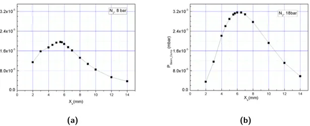
3.3.3 Optimization of cluster production . . . 33
3.4 Projectile . . . 35
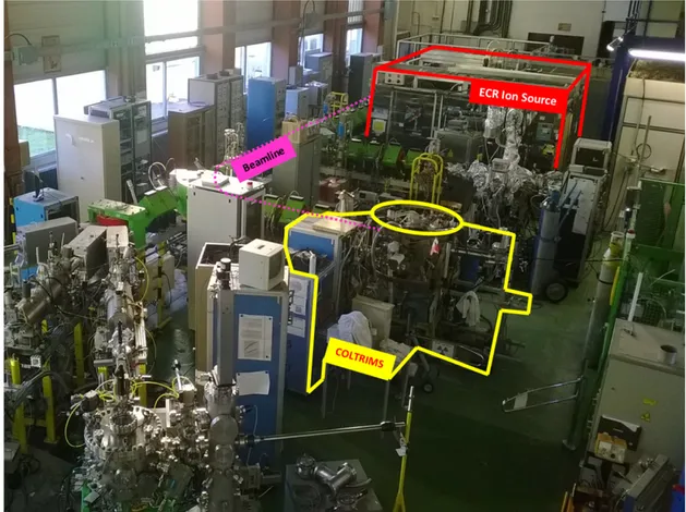
3.4.1 GANIL: Low energy facility ARIBE . . . 35
3.4.2 ECR Ion Source and Beam Line at ARIBE . . . 35
Contents
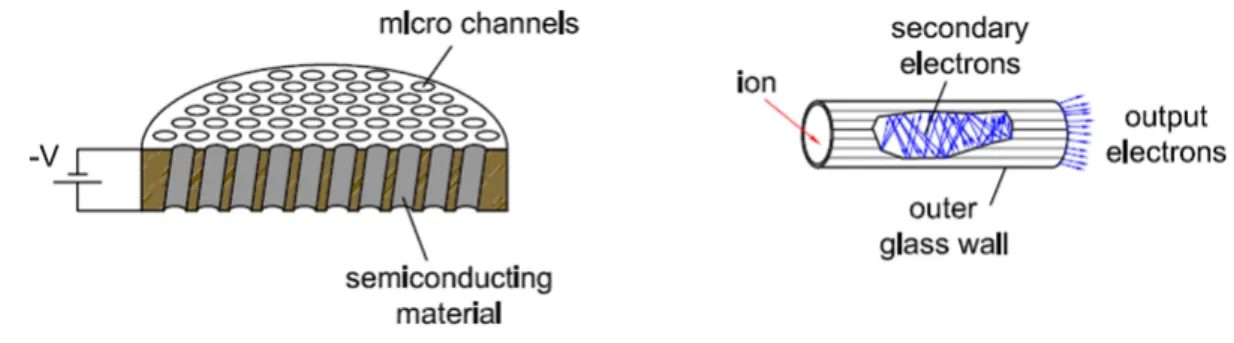
3.5 Detection . . . 39
3.5.1 Position Sensitive Detector (PSD) . . . 39
3.5.2 Comparison between standard and tapered MCP . . . 42
3.6 Acquisition system . . . 44
3.6.1 FASTER (Analog to Digital) . . . 44
3.6.2 Internal clock, Trigger window . . . 44
3.7 Energy calibration . . . 46
3.7.1 TOF calibration . . . 46
3.7.2 Position calibration . . . 48
3.7.3 Comparative calibration through the role of electric field, extraction length and 𝐶𝐷𝐿𝐷 . . . 51
4 Fragmentation Dynamics of 𝑁2 and CO dimers 55 4.1 2-Body Coulomb explosion of (𝑁2)2+2 and (𝐶𝑂) 2+ 2 dication . . . 56
4.2 3-body Fragmentation of (𝑁2)3+2 and (𝐶𝑂) 3+ 2 trication . . . 58
4.2.1 3-body dissociation of (𝑁2)3+2 . . . 58
4.2.1.1 Dissociation mechanism . . . 58
4.2.1.2 Effect of a charged environment on the dissociation of 𝑁2+ 2 dication . . . 59
4.2.2 3-body dissociation of (𝐶𝑂)3+ 2 . . . 60
4.2.2.1 Dissociation mechanism . . . 61
4.2.2.2 Direct and sequential: Newton diagram comparison . . . 62
4.2.3 Identification of short and long lived states of 𝐶𝑂2+ . . . . 63
4.2.3.1 Role of a neighbouring ion on molecular fragmentation . . . 64
4.2.3.2 Direct and sequential fragmentation . . . 65
4.3 Conclusion . . . 67
5 Geometrical analysis of 𝑁2 and CO dimers 69 5.1 Geometry of the 𝑁2 dimer . . . 70
5.1.1 2-body dissociation channel: Inter molecular distance . . . 72
5.1.2 3-body dissociation channel: Molecular orientation in the dimer . . . . 72
5.1.2.1 Numerical simulation of 3-body dissociation . . . 73
5.1.3 Results: Experimental vs Simulation . . . 79
5.2 Geometry of the 𝐶𝑂 dimer . . . . 80
5.2.1 2-body dissociation channel: Inter molecular distance . . . 81
5.2.2 3-body dissociation channel: Molecular orientation in the dimer . . . . 82
5.3 Conclusion . . . 84
Contents
6 Geometrical Analysis of 𝑁2 and CO trimers 87
6.1 3-body fragmentation of 𝑁2 trimers . . . 89
6.1.1 KER Spectra of (𝑁2)3+3 . . . 89
6.1.2 Newton Diagram . . . 90
6.1.3 Dalitz plot . . . 92
6.2 3-body fragmentation of 𝐶𝑂 trimers . . . . 94
6.2.1 KER Spectrum of (𝐶𝑂)3+ 3 . . . 94
6.2.2 Newton diagram and Dalitz plot . . . 96
6.3 Conclusion . . . 98
7 Conclusion 101 Bibliographie 105 A Annex 1 119 A.1 Optics basics and TOF equations . . . 119
B Annex 2 121 C Annex 3 123 C.1 Acquisition system . . . 123
C.1.1 FASTER . . . 123
Introduction
1
Atomics and molecular physics generates a fundamental knowledge base for our understanding in disciplines, such as atmospheric, environmental, chemical, biomedical and even astrophysical sciences. However, it also improves our quality of life by providing for example the primary inputs necessary for developing new instrumentation and techniques for medical applications, such as computer tomography, magnetic resonance imaging, positron emission tomography, laser induced surgeries and radiation therapy for cancer treatments etc. Today, many groups around the world study gas phase biomolecular fragmentation induced by ion collision with the aim to provide an exhaustive data base for radiobiology and hadrontherapy. However, these molecules being in the gas phase is a slight concern for biologists as the role of the environment of target molecules on its fragmentation is known to be of major importance.
Over the years, the "Atoms, Molecules and Clustesr" group in CIMAP has been studying various collisional systems ranging from atoms to large molecules and clusters. Thanks to the variable range of projectiles in GANIL, our group have not only studied the primary collision processes but also the relaxation mechanisms of the target after the collision. In the recent studies the rare gas dimers in collision with three different projectiles it has been found that due to the large interatomic bond length a dimer can be considered in a first approximation as a two independent atoms during the collision. Due to very low electron mobility inside such van der Waals dimers, two specific relaxation mechanisms (Radiative charge transfer and interatomic coulombic decay) have been identified which differs drastically from the case of covalent molecules where fast electron rearrangement usually takes place before fragmentation. In a next step, increasing the complexity of the target intermediately from atomic dimers to diatomic molecular dimers or trimers will open up the possibilities to answer various questions regarding the fragmentation dynamics and geometry of such systems. As the diatomic molec-ular cluster consist of both van der Waals and covalent bonds, it is interesting to compare the molecular fragmentation inside the dimer with the one of an isolated molecule. Such systems offer an unique tool to investigate the transition from gas phase molecules to the condensed phase.
nitro-1 Introduction
gen (𝑁2) and carbon monoxide (𝐶𝑂) molecules. In the last decade, there had been a lot of ab-initio calculations along with several infrared spectroscopic measurement done to speculate the three dimensional geometry of such diatomic molecular clusters. Even with the constant advancement in the computational and experimental methods no definitive conclusion had been provided so far leaving it as an open question for the investigation.
The experiments presented in this thesis concern collision between low energy multiply charged projectiles and diatomic molecular clusters (dimers and trimers). Clusters of two dif-ferent targets 𝑁2 and 𝐶𝑂 were used during the experiment with an 135 q.keV 𝐴𝑟9+ projectile
ions. Such highly charged ions are used to produce multiple ionized targets which then un-dergoes dissociation due to charge repulsion. The recording of the products from the collision system (recoil ions from the target and the scattered projectile) is ensured by a Cold Target Recoil Ion Momentum Spectroscopy (COLTRIMS) setup. The coincident detection of all frag-ments generated by Coulomb explosion of a multiply charged target allows the reconstruction of the three dimensional momentum vectors of each fragment. Using these momentum vec-tors we can indirectly deduce the initial structure of the neutral cluster. The experimental results are then compared to a Monte-Carlo based numerical simulation which computes the trajectories of three repulsing ionic fragments. Unlike rare gas dimers, the molecular dimers comprises of two different types of bond which upon cleavage can be used to investigate dif-ferent fragmentation pathways. In our analysis we have thoroughly analyzed 2 and 3-body fragmentation channels from (𝑁2)𝑛+2 and (𝐶𝑂)
𝑛+
2 , where 𝑛 Ȃ 2. The 2-body fragmentation
channel is due to cleavage of van der Waals bond only and is used to deduce the intermolecular separation in dimers. Additionally, the 3-body fragmentation channels are used for investigating the dynamics of fragmentation of a molecule in a controlled environment and different frag-mentation pathways upon the cleavage of both van der Waals and covalent bond inside a dimer. This thesis is organized as follows. The second chapter is a bibliographic summary in-cluding a general description of molecules and clusters and focusing on the geometry of 𝑁2 and 𝐶𝑂 dimers. Then an introduction about the collision of the low energy multiply charged projectile with the dimer is presented within the framework of the Classical Over-the-Barrier Model and showing the importance of multiple electron capture in such collisional systems. A description of the Coulomb Explosion Imaging Technique is also given at the end of this section. The third chapter consists of a presentation of the experimental setup used to perform this work. It encapsulated the features of the momentum spectrometer and description of the time of flight and position measurements of the fragments. Another part of this chapter comprises a description of the supersonic gas jet target and projectile ion beam used in this experiment. At the end, a detailed discussion about the energy calibration methods used for kinetic energy release measurements for both (𝑁2)2+2 and (𝐶𝑂)2+2 is given.
Chapter four, five and six expose all the results obtained during this work, concerning respectively the fragmentation dynamics of the 𝑁2 and 𝐶𝑂 dimers, the three dimensional structure of these dimers and the geometry of 𝑁2 and 𝐶𝑂 trimers.
Finally, the conclusion summarizes the work done in this thesis and presents a discussion on its relevance and future directions.
Context
2
Contents
2.1 Structure of molecular clusters . . . 6
2.1.1 Covalent bonds . . . 7
2.1.2 van der Waals bonds. . . 8
2.1.3 Geometry of molecular clusters . . . 9
2.1.3.1 𝑁2dimers . . . 10
2.1.3.2 𝐶𝑂 dimers . . . 11
2.2 Collision with Low Energy Highly Charged Ion (LE-HCI) . . . 13
2.2.1 Electron capture in dimers . . . 15
2.2.2 Fragmentation pathways . . . 19
2.3 Coulomb explosion imaging technique . . . 20
2.3.1 van der Waals bond cleavage . . . 21
2 Context
Collisions involving ions, molecules or clusters play a key role in various gaseous and plasma environments. The study of such collisions is required for fundamental understanding and have applications in fields of astrophysics, planetary atmosphere [1], biological matter and medical treatments [2], thermonuclear fusions [3] and semiconductor industry. Ion collisions have also been studied to compare the fundamental processes from the gaseous phase to the condensed phase of atomic or molecular targets. Since the early 30’s, the properties of various rare gas clusters and their relative comparison with the diatomic molecules in fundamental aspects has been investigated [4]. Since then the study of the van der Waals complexes has became an in-teresting tool to enhance knowledge of intermolecular potentials and therefore provides further insight in the specific properties of clusters, geometry of these complexes and their fragmenta-tion dynamics [5–16].
The collision between an ion and a molecular target is more complex in comparison to an atomic target, due to the presence of the more degrees of freedom (vibration and rotation of the molecule) in the former system and also due to the dependence of the collision process with the orientation of the molecule with respect to the ion beam. Very recently, the multiple electron capture probability as a function of the impact parameter in the case of two rare gas dimers have been studied with a Monte-Carlo based classical over the barrier model [17]. A brief introduction of this model and its relevance with our target system will be discussed in later in this chapter.
Finally, the investigations of atomic and molecular clusters have grown enormously and become possible especially with the spectacular developments and advancement in the spectroscopic techniques as well as computational methods. Among the state-of-the-art experimental meth-ods, Coulomb explosion imaging technique allows to investigate the initial structure of the molecules and clusters as well as the relaxation mechanisms leading to the fragmentation thanks to the coincident detection of all emitted particles.
A brief review of the existing data about the geometry of 𝑁2 and 𝐶𝑂 dimers using various
theoretical and experimental methods is presented in this chapter. In addition, a compilation of the work done on the molecular fragmentation using Coulomb explosion imaging technique is also discussed at the end of this chapter.
2.1 Structure of molecular clusters
The history of atomic and molecular clusters dates back to very early times in 1930’s [4]. These clusters serve as a bridge between the independent study of atoms or molecules in gas phase to the condensed phase. This thesis is devoted to the study of molecular dimers and trimers. Such systems are composed of two or three covalently bound molecules (𝑁2or 𝐶𝑂 in our case) which are weakly interacting thanks to the van der Waals forces.
Chemical systems such as molecules or clusters are held together by different types of forces and only the valence electrons (electron from the outermost shell) take part in the binding
2.1 Structure of molecular clusters
process. Several types of bonding exist depending on the nature and the electronic structure of the constitutive atoms. They are classified in four categories: the covalent bond, the ionic bond, the van der Waals bond and the hydrogen bond. In the following, we will describe only the covalent and van der Waals bonds as well as the structure of the 𝑁2 or 𝐶𝑂 dimers.
2.1.1 Covalent bonds
The covalent bond is based on a sharing of the valence electrons of two atoms. Several bonds can be formed between the two centers depending on the number of valence electrons of each atom. Such molecules are best explained by the Molecular Orbital (MO) theory which can estimate the energies of the electrons in a molecule along with the probable location of these electrons. Unlike in valence bond theory, MO theory uses the combination of atomic orbitals to yield molecular orbitals which are delocalized over the entire molecule rather than being localized on the corresponding atoms.
For example, the 𝑁2 molecule has three bonds (triple bond) because the nitrogen atom lacks three valence electrons to complete its outer shell. The potential energy curve (PEC) of the ground state of the neutral 𝑁2 is shown in figure 2.1.1 (a). The 𝑁2 molecule has a triple bond leaving one lone pair of electrons. The equilibrium bond length (R) of 𝑁2 molecule is 1.1
Å and its bond dissociation energy (BDE) is about 9.8 eV. Being homonuclear this molecule has no permanent dipole moment.
Figure 2.1.1 : Potential energy curves of the ground state of 𝑁2 and 𝐶𝑂 molecules [18]. (1eV = 8100 cm−1)
Usually each atom provides one electron for a single covalent bond, however it is not true necessarily. If one of the atom is more electronegative in the covalent bond then the electron density is partially shifted towards the more electronegative atom. This generates a polarity making the covalent bond polar as it is the case for 𝐶𝑂. Due to this polarity the molecule attains a permanent dipole. 𝐶𝑂 also has the same number of valence electrons as 𝑁2. It also
makes a triple bond with one lone pair of electrons. However, the one pair of shared electrons are provided from the oxygen atom. Oxygen being more electronegative the electron density is
2 Context
shifted towards it causing a permanent dipole moment of 0.11 D. The PEC curve of the ground state of 𝐶𝑂 is shown in figure 2.1.1 (b) showing the bond length of about 1.13 Å with a BDE of 14.5 eV making it the strongest bond in neutral molecules.
2.1.2 van der Waals bonds
In rare gas dimers the MO theory predicts that there is no possibility of covalent bonds between the atoms because they have a full outer shell of valence electrons. The binding is then only possible via van der Waals bond. Due to its polarizability, an atom or molecule may have a permanent or induced electrostatic dipole moment (𝜇). In presence of another atom or molecule, a dipole-dipole attraction may create a weak bond between the two atoms or molecules. This bond is known as van der Waals bond and is not chemical in nature as there is no participation of electrons although the polarization from the electron cloud.
A dipole-dipole interaction could result from a permanent or induced dipole interaction inside the atoms or molecules. A dipole may interact with its electrostatic environment, including other dipoles, and the strength of these interactions depends directly the nature of the neighbor. As we can see in the figure 2.1.2 the arrangement of dipole due to presence of another dipole.
Figure 2.1.2 : Alignment of dipole due to the presence of the other dipole in vicinity. On the left is an anti parallel configuration and on the right is a co-linear configuration.
In the case of rare gas dimers, the bonding results from an induced dipole interaction as the atoms do not have a permanent dipole moment. Figure 2.1.3b shows the comparison between the absolute bond strength and bond length of all the rare gas dimers [19]. The van der Waals bonds have a much smaller binding energy and longer bond length than covalent bonds. Typical binding energies are about 10 meV and bond length are 3 or 4 Å. Due to its low binding energy, the van der Waals bond can exist wherever there is no possibility of other bond formation. An example of comparison between bond strength of covalently bonded 𝑁2 and van der Waals
bonded (𝑁2)2 is shown in the figure 2.1.3a [18,20]. The covalent bond is upto 650 times more stable as the van der Waals bond and the repulsive bond length differs by a factor of about 4.
2.1 Structure of molecular clusters
(a)
(b)
Figure 2.1.3 : a)A comparison of potential energy curves of covalent bond between 𝑁 − 𝑁 inside 𝑁2 molecule and the van der Waals bond between 𝑁2− 𝑁2 inside the (𝑁2)2 dimer. In this case the
covalent bond is upto 650 times stable than its van der Waals bond. The diagram has been modified using the work of Powell in [18] and Gomez in [20] to give an qualitative comparison between the strength and separation of the two bonds. b) Potential energy curves of all the rare gas dimers [19]. The general shape of the PEC for the van der Waals bonding can be described by the Lennard-Jones potential which is shown in equation 2.1.1 below:
𝑉 = 𝜀{(𝜎 𝑅 )12 − 2 (𝜎 𝑅 )6} (2.1.1) where 𝜀 is the depth of the potential well located at the distance 𝜎 and 𝑅 is the inter-atomic/intermolecular distance. However, the precise calculation of the potential energy in the molecular dimers is quite complex as it depends on various other factors including the three dimensional geometrical arrangement of the molecules inside the dimer.
2.1.3 Geometry of molecular clusters
One of the main topic of this thesis is the geometry of the 𝑁2 and 𝐶𝑂 dimers and trimers.
There have been many theoretical works done to predict the geometry of these molecular dimers.
2 Context
2.1.3.1 𝑁2 dimers
The first studies of the geometry of 𝑁2 dimer are dated back in the early 1970’s, where Long et al. used infrared (IR) spectroscopy to comment over the intermolecular distance and its geometry [5]. Earlier, the same team studied the van der Waals complexes of 𝐴𝑟 − 𝑁2, 𝐴𝑟 − 𝑂2
and found that equilibrium geometry is a "T" configuration where the 𝑁2 (or 𝑂2) molecule is oriented perpendicular to the dimer axis (see figure 2.1.5 (d)). The similar conclusion had been made for the 𝑁2 dimer. The infrared spectrum was also used to predict the intermolecular distance and was found to be 3.7 Å. In conclusion from this work they felt the need of new theoretical models for such complexes in order to conclude on the geometry. In another study conducted by Carnavole et al. using the photoelectron spectroscopy of (𝑁2) dimer [21], the author suggested an "X" shaped (see figure 2.1.5 (b)) configuration with a dimer bond length of 3.8 Å.
Figure 2.1.4 :General representation for a dimer of two diatomic molecules [22]. 𝑅 is the distance between the center of mass of the two monomers 𝑀1 and 𝑀2. 𝜃1 and 𝜃2 are the angles between the
molecular axis of 𝑀1 or 𝑀2 and the dimer axis. 𝜙 is the dihedral angle between the two monomers 𝑀1 and 𝑀2.
Since then there have been several works on the geometry of (𝑁2)dimer [20,22–27], which predict five possible three dimensional geometries. These three dimentional structures can be described using the "internal" parameters ((𝑅, 𝜃1, 𝜃2, 𝜙), see figure 2.1.4) introduced in [22].
Several ab-inito calculations were performed in order to determine the global minimum of the potential energy surface of the neutral 𝑁2 dimer [23,24]. A summary of the different predicted
structures is shown in figure 2.1.5. For "H" and "X" geometries the intermolecular distance is in the range of 3.6-3.8 Å, for "Z" and "T" shapes this distance varies between 4-4.1 Å and the linear "L" configuration is always predicted above 4.3 Å. However, the "Z" and "T" configurations are found to be the most stable in most of the calculations [20, 22, 25–27].
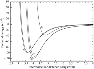
Figure 2.1.6 shows the PEC’s for all the five possible geometries studied in the work of Gomez et al. [20]. On the basis of intermolecular distance there are three main configurations where, "H" and "X" are centered around 3.6 Å, "T" and "Z" are around 4 Å and "L" is at 4.6 Å. As the predicted difference between the intermolecular distances of the "H" and "X" shapes is very low, therefore it will be necessary to access the torsional angle 𝜙 in order to distinguish between the two configurations ("H" corresponds to 𝜙=0Ԃ and "X" corresponds to 𝜙=90Ԃ).
2.1 Structure of molecular clusters
Figure 2.1.5 : Summary of the five structures of (𝑁2) dimers predicted in various theoretical studies [20, 22–27]. a)Planar "H" shaped, b) non planar "X" shaped, c) canted parallel "Z" shaped, d) "T" shaped and e) linear "L" shaped. The corresponding "internal" coordinates are indicated in brackets.
Contrarily, the "T" and "Z" configurations differs from their (𝜃1,𝜃2) angles and are expected
to be more easily distinguished in our experiments. These predicted geometries will be used as reference and compared to our experimental results in the Chapter 5.
Figure 2.1.6 : Potential energy curves of (𝑁2)2 dimer calculated in [20] for all the five predicted
geometrical configurations.
2.1.3.2 𝐶𝑂 dimers
The carbon monoxide being an heteronuclear molecule, 𝐶𝑂 dimer makes itself a good target to be studied with IR spectroscopy. The three dimensional geometry of 𝐶𝑂 dimers is also
2 Context
described using the "internal" parameter as introduced for 𝑁2 dimers. However, a geometry where the carbon atoms of the each molecule are oriented toward the center of the dimer is now called "C-bonded" (For eg. 𝜃1=135Ԃ ,𝜃2=45Ԃ). Similarly, a "O-bonded" geometry correspond
to the case of two "inner" oriented oxygen atoms (For eg. 𝜃1=45Ԃ ,𝜃
2=135Ԃ). In last two
decades, there have been several IR spectroscopic measurements [28–31] done to determine the geometry of the 𝐶𝑂 dimer. In the work by Brookes et al. the 𝐶𝑂 dimer IR spectrum in the 2139-2152 cm−1 region has been studied, showing the presence of two possible different
overlapping isomers of 𝐶𝑂 dimer. The author suggested two different configurations both corresponding to the "T" shape, C-bonded and O-bonded with intermolecular distance of 4.4 Å and 4 Å(see figure 2.1.7 (c) and (d)). However, not so later in 2003 Surin et al. [32] observed the millimeter wave spectrum and still could not differentiate between the two isomers due to the low energy difference between them. Another group from South Africa used a Fourier transform interferometer to record the IR spectrum of 𝐶𝑂 dimers trapped in nitrogen and ar-gon [29,30]. In parallel they have also performed theoretical calculation for five distinct possible structures, shown in figure 2.1.7. From the agreement between experimental and calculated dimer-monomer wave number shifts they highly suggest an "T" shaped O-bonded configuration with intermolecular separation of around 4 Å. Although, due to very small energy difference in the minima of various configuration in potential energy surface (PES), they suggest not to completely excluded the C-bonded isomer with separation around 4.4 Å. In another work done very recently by Rezaei et al., the existence of the slipped anti-parallel configurations have been reported [31].
Figure 2.1.7 : Representation of all the possible geometries predicted using the IR or millimeter wave spectroscopy and PES calculations in [14,28–35]. Five most favorable geometries are a) slipped anti-parallel C-bonded b) slipped anti-parallel O-bonded c) "T" shaped C-bonded d) "T" shaped O-bonded and finally e) O-bonded linear shaped.
Over the time the need of much accurate PES for 𝐶𝑂 dimer became of high importance. In parallel to all the IR and millimeter wave spectroscopy, the advancement in the PES and rovibrational calculations were reported by various groups [14, 32, 33]. Up to the begining of
2.2 Collision with Low Energy Highly Charged Ion (LE-HCI)
2000, the calculation conclude to the existance of both C and O-bonded, slipped anti-parallel and "T" shaped isomers. But the confirmation of a slipped anti-parallel configuration became more evident with the most recent PES calculations in [34, 35]. In contrast, the two most favorable predictions for geometry of 𝐶𝑂 dimer are: a C-bonded antiparallel geometry with
𝜃1 Ȃ 135Ԃ, 𝜃
2 Ȃ 45Ԃ and bond length 𝑅= 4.4 Å, and an O-bonded antiparallel geometry with
𝜃1 Ȃ 45Ԃ, 𝜃
2 Ȃ 135Ԃ and a bond length of 𝑅= 4 Å.
For both the 𝑁2 and 𝐶𝑂 dimers a detailed comparison of proposed geometries and the experimental data using a numerical simulation is explained in chapter 5.
2.2 Collision with Low Energy Highly Charged Ion
(LE-HCI)
In the past, atomic and molecular complexes have been studied experimentally using various ionisation or excitation processes: photoionization using synchrotron radiation [36–38], elec-tron [39–43], laser [44–46] and multiply charged ion beams [47–58]. In this thesis we will only focus on the ion-molecule/cluster collision leading to charge and energy exchange between the collision partners via three elementary processes: electron capture, excitation and ionization. The importance of these processes depends on the comparison of the projectile velocity (𝑣𝑝) with the classical orbital velocity of valence electrons (𝑣𝑒) of the target [48] and also on the col-lision asymmetry which is the ratio between projectile and target atomic numbers (𝑍𝑝Ȃ𝑍𝑡) [59]. Combined together they give the collision strength parameter 𝜅 = (𝑣𝑒Ȃ𝑣𝑝) × (𝑍𝑡Ȃ𝑍𝑝). If the
𝜅 Ȃ 1 the electron capture will be the most probable process and for 𝜅 Ȃ 1 ionization and the excitation dominates. In certain cases where 𝑍𝑝 Ȃ 𝑍𝑡, these dominant processes can be categorized just by the energy of projectile. In our case for 𝐴𝑟9+ at 15 q.keV colliding with 𝑁
2
or 𝐶𝑂 targets, 𝜅 Ȃ 2.7 Ȃ 1 implying the electron capture as the most probable process during collision.
The interaction time is another point of interest in the ion induced dissociation of molecular and dimer targets. As we use the Coulomb explosion imaging technique to measure the initial structure of the neutral target, the interaction time has to be much smaller than the nuclear or molecular motion in order to obtain a reliable measurement. As an example, it has been shown that using too long laser pulses to measure the geometry of triatomic molecules 𝐷2𝑂 results in a deviation from the expected values [60]. Here the time scale of ionization was comparable to the time scale of the molecular dynamics which allows the ions to move from their initial position after ionization and causing the deviation. Although, in the case of the low energy ion collisions (few tens of keV range) the ionization time is less than 1 fs. For example in our case, we use 𝐴𝑟9+ with a velocity of 8 Å/fs (0.37 a.u.), the target dimer bond length is of
2 Context
gives an interaction time of about 7 + 4
8 = 1.4 fs in the worst case possible. This value is much shorter than the time scale of molecular vibration (10 fs) and rotation (1 ps) [47]. Due to this time difference, the nuclear motion is considered as frozen during the collision leading to excitation through vertical transition in the Frank Condon region (see figure 2.2.1) and validate our approach using Coulomb explosion imaging technique.
Figure 2.2.1 : Schematic representation of the transition from the initial ground state to a disso-ciative state of the ionized and/or excited dimer and then its dissociation into two ionic fragments. The vertical transition due to the collision takes place in the Franck Condon region.
For many years, theoretical and experimental investigations of collision mechanisms involv-ing ionic projectiles and neutral gas phase targets (atom, molecule or clusters) have became of prime importance. In the case of molecule and cluster as targets, there is a huge depen-dency of bond type of the target on the primary and relaxation mechanisms. In case of a diatomic covalent molecule in collision with the ionic projectile, the total charges of the tran-sient molecular ions are generally shared among the dissociating atomic fragments [61]. This is due to the delocalization of the valence electrons through out the molecule. On the other hand for a van der Waals dimer for example 𝐴𝑟2, this rearrangement of charge is weak due to the long interatomic distance. In addition a highly sensitive orientation dependence has been found in this 𝐴𝑟2 dimer for asymmetric channels [53, 54]. In a previous thesis in our group,
it was found that for asymmetric fragmentation channels, due to low charge mobility between the two atomic sites of dimer, the projectile is scattered in the direction of the most charged fragment [55]. This indicates that the projectile mainly capture electron to the atom closest to its trajectory. Furthermore, new relaxation mechanisms were also observed in comparison to covalent molecules due to the asymmetric distribution of charges in a dimer: Radiative Charge Transfer (RCT) and Interatomic Coulombic Decay (ICD) [53, 56]. These mechanisms involve either the electron transfer or the energy transfer to the neighbour neutral atom from the ion-ized atom. For further investigation of the electron capture mechanism a theoretical approach
2.2 Collision with Low Energy Highly Charged Ion (LE-HCI)
has been designed which is detailed in the section below [17].
2.2.1 Electron capture in dimers
For a case of low energy ion (400 eV/amuȂ 0.13 a.u.) atom collision, the first Classical Over-the-Barrier Model (COBM) was developed by several groups based on the idea that the electrons can transfer from target to projectile at a given internuclear distance [62–65]. At this distance the potential barrier between the two nuclei is lower than the Stark shifted binding energy of the transit electrons. After the successful agreement in case of low energy ion atom collisions, a similar approach is used for molecular targets. A three center COBM has been developed to understand the asymmetric charge distribution in Coulomb explosion of 𝑁2 with upto 100 eV/amu 𝐾𝑟8+ ions [66]. A similar approach was later used to update this model
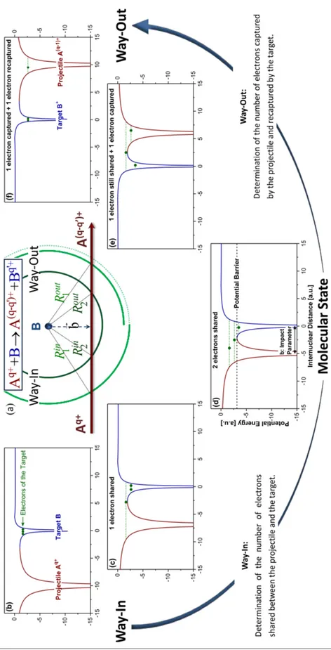
for rare gas dimers which is one of the basis of the COBM approach used in [67]. In this approach to perform a complete treatment of collision, the model was divided into two steps, the way-in and the way-out. This was one of the adaption from the model of Niehaus for atomic targets [65]. Additionally, a Monte Carlo approach is used to account for the different orientations of the dimer and impact parameters and aid the comparison between simulation and experimental data.
In this model, the projectile approaches slowly the target and the potential barrier of the target is lowered by the proximity of the incoming highly charged projectile ion. This leads to the creation of quasi-molecular state, where projectile and target are sharing electrons. In this quasi-molecular state, the electrons can move from the target to the projectile. In the way-out (when the projectile is getting away from the target) the electron can be captured by the projectile or go back to the target. The probability of capturing depends on the impact parameter, the projectile charge state and ionization energies of the target.
In figure 2.2.2, a schematic of an ion-atom collision is shown as described in this model. In 2.2.2 (a) a projectile 𝐴𝑞+ with a charge state of 𝑞+ is approaching the neutral target atom 𝐵, at different instances the internuclear distance between the two is presented as 𝑅𝑖𝑛
𝑖 (for way-in step) and 𝑅𝑜𝑢𝑡
𝑖 (for way-out step). The final state of the projectile is represented as 𝐴
(𝑞−𝑞′)+
, where 𝑞′ is the number of electron stripped from the atom at the way-out. Much detailed
understanding of this model and its agreement with the experimental results can be found in the corresponding publication [67].
2 Context
Figure 2.2.2 : Schematic representation of the ion (𝐴𝑞+) and target atom (𝐵) collision along with
the evolution of potential barriers in the way-in and the way-out of the collision [67]. See details in the text.
2.2 Collision with Low Energy Highly Charged Ion (LE-HCI)
Further, this MC-COBM model has been extended to the case of a van der Waals rare gas dimer where the dimer is approximated by two quasi independent atoms. Now the similar approach as in the case of the atomic target can be used with both the atoms separately con-sidering two independent collisions but during which the effective projectile charge state may change because of electron capture on each site of the dimer. Indeed, for highly charged ions the capture radii are larger than the van der Waals bond and electrons can be captured from the two sites of the dimer depending on the impact parameter.
Figure 2.2.3 represents the two steps of collision where step 1) shows the collision between a projectile 𝐴𝑞+ and a (𝑀)
2 dimer. Following electron capture, the system can be represented
as: 𝐴𝑞+ + (𝑀)
2 Ă 𝐴(𝑞−(𝑥+𝑦))+ + 𝑀𝑥+ + 𝑀𝑦+. Once the target is ionized it may relax via
fragmentation due to the repulsive Coulomb force acting between the 𝑀𝑥+ and 𝑀𝑦+.
Figure 2.2.3 : (a): Representation of the collision between a low energy projectile ion (𝐴𝑞+) and
a dimer target ((𝑀)2) in a 3-Dimensional Cartesian system,(b): Relaxation of ionized target via
fragmentation due to Coulomb explosion.
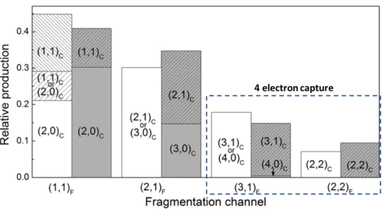
Figure 2.2.4 shows the relative intensities of the different fragmentation channel observed in the 𝐴𝑟9++𝐴𝑟
2 collision system. The MC-COBM (in gray) reproduces the measured yield and
validates the hypothesis that the dimer can be in first approximation considered as two inde-pendent atoms. In particular, the dominance of asymmetric fragmentation following quadruple electron capture is explained by an higher probability of asymmetric electron capture and by the slow electronic rearrangement between two atomic sites.
The projectile impact parameter has been shown to play a crucial role on the electron capture probabilities from both sites of the dimer. These capture probabilities depend on the ionisation energies and capture radii of the target. Table 2.2.1 shows a comparison between the successive capture radii for 𝐴𝑟 atom and for 𝑁2and 𝐶𝑂 molecules. Since, the first, second and
third ionization potential of both 𝑁2 and 𝐶𝑂 molecules are very close to that of argon atom, therefore the capture radii for 𝐴𝑟9+ projectiles are also found similar for all targets. Note that
2 Context
Figure 2.2.4 : Relative yields for the different electron capture and fragmentation channels (from 𝐴𝑟9++𝐴𝑟
2collision system) extracted from the experimental data (white) and results from MC-COBM
calculations (gray) [55].
due to unavailability of third ionization potential for both the 𝑁2 and 𝐶𝑂 molecules, we have used the third ionization potential of nitrogen atom to have a fair approximation of capture radii.
Table 2.2.1 : Estimation of the capture radii for one, two or three electrons capture from 𝐴𝑟, 𝑁2 and 𝐶𝑂 targets using the 𝐴𝑟9+as the projectile. 𝐼𝑃
𝑖represents the ionization potential for 𝑖𝑡Ăelectron
and 𝑅𝑐
𝑖 is the capture radius for that electron using COBM model. Target 𝐼𝑃1 (eV) 𝑅𝑐 1 (Å) 𝐼𝑃2 (eV) 𝑅 𝑐 2 (Å) 𝐼𝑃3 (eV) 𝑅 𝑐 3 (Å) 𝐴𝑟 15.8 6.39 27.6 5.46 40.7 4.73 𝑁2 15.6 6.46 27.2 [68] 5.55 47.4 4.06 𝐶𝑂 14.5 6.95 27 [69] 5.59 -
-Moreover, the equilibrium intermolecular distance for both 𝑁2 and 𝐶𝑂 dimer are of the same order of interatomic distance of 𝐴𝑟2 dimer, which is around 4 Å. As the size of these
dimers is smaller than the capture radii for 𝐴𝑟9+, this implies that the multiple electron capture
may occur on each site of the molecular dimers. Depending on the number of electrons captured on each molecular site of the dimer, molecular fragmentation may also occur in addition to the cleavage of the weak van der Waals bond. This leads to the existence of various fragmentation channels of the multi-ionized molecular dimers.
2.2 Collision with Low Energy Highly Charged Ion (LE-HCI)
2.2.2 Fragmentation pathways
This thesis focuses on ion collision with diatomic molecular clusters (dimer and trimer). Such polyatomic targets comprise both covalent and van der Waals bonds. Until now we have es-tablished that multiple electron capture may lead to various fragmentation channels because of the Coulomb repulsion between the monomers. For a collision system:
𝐴𝑞++ (𝑀)
2 Ă𝐴(𝑞−(𝑥+𝑦))++ 𝑀𝑥++ 𝑀𝑦+
where 𝐴𝑞+ is the projectile ion, 𝑀 is the diatomic molecule and 𝑥, 𝑦 are the number of electrons captured from each site of the (𝑀)2 dimer. From here several fragmentation channels can be expected depending upon the value of 𝑥 and 𝑦.
Case 1: If both 𝑥 and 𝑦 are equal to 1, this corresponds to the case where one electron has been captured on each monomer. Consequently, the dimer fragments via a 2-body channel where two singly charged 𝑀+ ions are emitted. In this case, the 𝑀++ 𝑀+ dissociation follows
a pure Coulomb law and the intermolecular bond length can be deduced from the kinetic energy released (see 2.3.1 for more details).
Case 2: If 𝑥 Ȃ2 and 𝑦=1. For a simplest case lets consider 𝑥=2 and 𝑦=1, where two electrons are captured from one site/molecule of the dimer. In such a case, a molecular dication is produced inside the dimer. Although this triply ionized dimer may fragment via a 3-body fragmentation channel where both the van der Waals and covalent bond breaks. However, this fragmentation of the molecular dication (covalent bond cleavage) inside the dimer depends on the presence of metastable states in the molecule. For example in the case of 𝑁2+
2 and 𝐶𝑂 2+
the lifetimes of the electronic states are totally different. The lower excited states of the 𝑁2+ 2
dication are short lived as the density of metastable states is very low. On the other hand, the 𝐶𝑂2+ dication have abundance of such metastable vibrational states. Figure 2.2.5 (a)
shows the potential energy curves of four lowest lying electronic metastable states of 𝐶𝑂2+,
they are 𝐴3̀+, 𝑏1̀, 𝑎1̀+ and 𝑋3̀. The lifetimes of all the metastable states in these four
states are depicted in 2.2.5 (b). There are at least more than 70 metastable vibrational states associated to these electronic states having lifetimes longer than 1 ps [70]. Due to the presence of these long lived states the 𝐶𝑂2+ dication, the dimer trication (𝐶𝑂)3+
2 , might induce two
distinct dissociation mechanisms direct and sequential. Direct fragmentation corresponds to a synchronous cleavage of the van der Waals and covalent bonds and is associated to lifetimes of the dication shorter than 1 ps. Sequential fragmentation refers to the case where the van der Waals bond explodes followed by the cleavage of the covalent bond in the molecular dication because of the presence of the metastable states with lifetimes larger than 1 ps. Finally, if the lifetime are larger than few 𝜇𝑠, the experiment will record a 2-body 𝑀++ 𝑀2+ channel.
If 𝑥 Ȃ3 and 𝑦=1, this would also result in a 3-body fragmentation channel. However, there will be mostly the participation of pure dissociative states in the fragmentation of the molecular trication. Therefore, sequential fragmentation is not expected to occur in this case.
2 Context
Figure 2.2.5 : (a): Representation of the potential enery curves of four lowest electronic states of 𝐶𝑂2+, (b): life times of all the metastable vibrational states inside the potential well of the four
lowest lying electronic states of 𝐶𝑂2+ [70].
Such 3-body fragmentation channels are used to investigate the effect of a charged environ-ment on molecular fragenviron-mentation (see chapter 4). These channels are also used to reconstruct the initial structure of the molecular dimer using the Coulomb explosion imaging technique (see 2.3.1, 2.3.2 and chapter 5).
Case 3: If both 𝑥 and 𝑦 Ȃ 2. In this case, we expect a 4-body fragmentation channel with breaking of the van der Waals bond along with both covalent bonds on both sites of the molecular dimer. The production cross section and absolute detection efficiency of this channel make these 4-body channels hard to investigate experimentally. Even if these 4-body channels have an direct application in determination of the three dimensional geometry of the diatomic molecular dimers, they will not be discussed in this thesis due to the very low statistics.
2.3 Coulomb explosion imaging technique
Until now various instrumental advancements have been carried out to collect the required information needed to describe the collisions and fragmentation with details. In general this requires to measure the charge, mass and the velocity of the emitted particles. A review of such detectors used in the spectroscopic measurements is given in the article by Medhe [71].
Coulomb explosion technique were first introduced in 1980’s for determining the stereo-chemical structure of molecular ions accelerated to energies of about 1 MeV. These ions were
2.3 Coulomb explosion imaging technique
send through a foil and the exiting multiply ionized molecule then undergoes Coulomb ex-plosion. Its initial structure is deduced from the energy and angular shifts measurement of the emitted fragments [72, 73]. At the same time the new developments in the spectroscopic techniques had also been carried to collect all the ions and electrons produced during colli-sion with the best resolution possible [74, 75]. These are known as recoil ion spectroscopy, recoil ion momentum spectroscopy (RIMS) or cold target recoil ion momentum spectroscopy (COLTRIMS). The first measurement performed on RIMS was to observe projectile deflection in terms of scattering angle from an atomic target [76]. Several teams together have con-tributed in developments to achieve the most efficient spectroscopic instrument for measuring the kinematics of projectile and target collisions [74, 75, 77, 78]. The kinematically complete measurements in COLTRIMS involves, the detection of the projectile ions, along with the recoil ions (fragments) and electrons in multiple coincidence with a 4𝜋 solid angle collection efficiency. For highly charged low energy projectiles in few hundred keV regime the electron capture is the dominant process and may lead to multiple electron capture. For molecular or dimer targets, electron can be captured from both sites resulting in a Coulomb explosion of the tar-get. Collection of these recoil ions and measurement of their momentum vectors to access the initial geometry is known as Coulomb imaging technique. As explained in the previous sec-tion, the rapid ionization of the target is a key requirement in the Coulomb explosion imaging (CEI) technique to ensure that the nuclear motion before explosion is negligible. Recently, it has proven to be a powerful tool in investigation of the fragmentation dynamics of covalent molecules [57, 60, 79–81].
Ideally, the complete determination of the three dimensional structure of diatomic molecular dimers would require the full atomisation of the target i.e. the detection of the four constitutive atomic fragment ions. However,we will show in chapter 4 that a partial information on this structure can be deduced from the 2-body and 3-body fragmentation channels.
2.3.1 van der Waals bond cleavage
Molecular van der Waals complexes are dedicated polyatomic systems for enhancing our knowl-edge of intermolecular potentials, which helps in understanding fragmentation dynamics, envi-ronmental perturbation inside clusters and the geometry of these clusters. As already estab-lished, the collision between slow (low energy) highly charged projectile ion and a dimer can be explained in 2 steps where the first step is the capture process and the second step is the relaxation process.
For direct transitions from the fundamental state to a dissociative state, in which the pro-jectile removes one electron from the two centres of the dimer, the doubly charged dimer will fragment via the (𝑀2)2+ Ă𝑀++ 𝑀+ channel. This process is called Coulomb explosion.
Be-2 Context
cause of the large intermolecular distance (Ȃ 2.1 Å), the 𝑀++ 𝑀+ dissociating curve can be
fairly approximated by a pure Coulomb curve [82]. The sum of the kinetic energy of each frag-ment called Kinetic Energy Release (KER) can then be calculated using the Coulomb potential:
𝐾𝐸𝑅 = 14.4 × 𝑞1𝑞2 𝑅 ,
where the 𝑞1=1 and 𝑞2=1 are the charges on each fragment and 𝑅 is the intermolecular
separation. Experimentally, the position of the KER peak for such a 2-body channel will be used to derive the intermolecular distance.
2.3.2 Fragmentation of molecules and dimers
Previously in our group in CIMAP, the fragmentation of diatomic and triatomic molecules have been studied with both low energy and high energy ionic projectiles [83, 84]. The work done in the thesis of M. Terisien studies the dependence of the projectile velocity (11.4 MeV/u 𝑂7+
and 4 keV/u 𝑂7+) on the population of excited states of 𝐶𝑂2+ [51].
Multiple ionization followed by Coulomb dissociation has been extensively studied for molecules and dimers under intense laser fields [45, 57, 79, 81, 82, 85–87], pulsed electron beam [88] and ionic projectiles [57, 80]. Here is a brief review of various experimental re-sults obtained using the Coulomb explosion imaging technique. In a study concerning the vibrational states of 𝑂3 using two laser pulse duration, short 9 fs and long 40 fs, with an
inten-sity of 2×10¹⁵ 𝑊 Ȃ𝑐𝑚2, it has been found that the nuclear dynamics are induced in the longer
pulse duration [79]. The triply ionized 𝑂3 molecule has shown stretching along all the three vibrational coordinates including anti symmetric stretching among all coordinates.
Using a triatomic molecule such as 𝐶𝑂2, the many body structure increases the internal degrees of freedom as well as the number of fragmentation pathways. In 2010, Neumann et al. used 3.2 keV/u 𝐴𝑟8+ projectiles to collide with a 𝐶𝑂
2 target jet [80]. The 3-body
fragmenta-tion channel from the 𝐶𝑂3+
2 trication had been investigated and three different fragmentation
pathways were observed: synchronous direct fragmentation, sequential fragmentation and asyn-chronous fragmentation. The asynasyn-chronous fragmentation arises from the molecular bending and asymmetric stretching. Similar distinction between sequential and non sequential fragmen-tation have been observed by the Wu et al. using femtosecond laser [82] and Wong et al. using pulsed electron beam [88]. For another triply ionized polyatomic molecule 𝑂𝐶𝑆, the separation of direct and sequential fragmentation has been provided using the intermediate 𝐶𝑂2+ or 𝐶𝑆2+
rotation before its unimolecular fragmentation [81]. Note that all these experiments use the KER measurement as a tool to distinguish between the different fragmentation pathways.
2.3 Coulomb explosion imaging technique
There had also been several studies on molecular and mixed clusters using the CEI tech-nique, all of them have used phase controlled femtosecond laser with intensity varying between 1014-1015 𝑊 Ȃ𝑐𝑚2. In 2014 Xie et al. studied the 𝐶𝑂
2 dimer, trimer and tetramer by using
the KER distribution for all the 2, 3 and 4-body channel respectively. They found that the (𝐶𝑂2)3 trimer has essentially an equilateral triangular geometry and the (𝐶𝑂2)4 has a regular
tetrahedral geometry. Moreover, it has been found out that the intermolecular distance is the same in all the three clusters [86]. Such properties have already been reported for rare gas clusters 𝐴𝑟𝑛Ȃ4 and 𝑁𝑒𝑛Ȃ4 [85,87]. These similarities between rare gas and non-polar molecular
𝐶𝑂2 clusters seem to indicate that the internal structure of the constituents plays a minor role on the geometry of the clusters. The structure of mixed cluster 𝑁2𝐴𝑟 have also been determined using 2 and 3-body channels of the van der Waals complex [89]. The 𝑁2 molecule
is found to make an angle of 90Ԃ with the dimer axis at an intermolecular distance of around
3.7 Å. Similary, a T-shaped geometry was observed for 𝐴𝑟𝐶𝑂 dimer [45].
In this thesis, we used a similar CEI technique to access the information about fragmentation dynamics and geometry of diatomic molecular clusters induced by collision with slow highly charged ions. Our experimental setup and the results will be detailed in the next chapters.
Experimental Technique and
Calibration
3
Contents
3.1 General description . . . 26 3.2 COLd Target Recoil Ion Momentum Spectroscopy . . . 27 3.3 Target Preparation . . . 29
3.3.1 Cold Target . . . 29
3.3.1.1 Supersonic gas jet . . . 29
3.3.2 Jet Parameters . . . 31
3.3.3 Optimization of cluster production . . . 33
3.4 Projectile . . . 35
3.4.1 GANIL: Low energy facility ARIBE . . . 35
3.4.2 ECR Ion Source and Beam Line at ARIBE . . . 35
3.4.3 Collimator and parallel plate analyzer . . . 38
3.5 Detection . . . 39
3.5.1 Position Sensitive Detector (PSD). . . 39
3.5.2 Comparison between standard and tapered MCP . . . 42
3.6 Acquisition system. . . 44
3.6.1 FASTER (Analog to Digital) . . . 44
3.6.2 Internal clock, Trigger window . . . 44
3.7 Energy calibration . . . 46
3.7.1 TOF calibration . . . 46
3.7.2 Position calibration . . . 48
3.7.3 Comparative calibration through the role of electric field, extraction length
3 Experimental Technique and Calibration
We use a COLTRIMS (COLd Target Recoil Momentum Spectroscopy) [74] setup to measure in coincidence the charged fragments emitted following the collision of a molecular cluster target and slow highly charged ions (HCI). In this chapter, I will present the whole experimental technique and briefly describe the different parts of our experiment. It includes the low energy HCI projectile beam provided by the GANIL’s (Grand Accelerateur National d’Ions Lourds) ARIBE (Accelerateur de Recherche avec les Ions de Basse Energie) facility. In the next section, preparation of the target jet beam is discussed followed by characterization of this jet with the help of a quadrapole mass selector (QMS). After this, I will briefly introduce our spectrometer along with the details about the detectors used in our setup and give an introduction to our new digital acquisition system. At the end, time of flight (TOF) and position calibrations will be discussed to support the confidence in our measured data.
3.1 General description
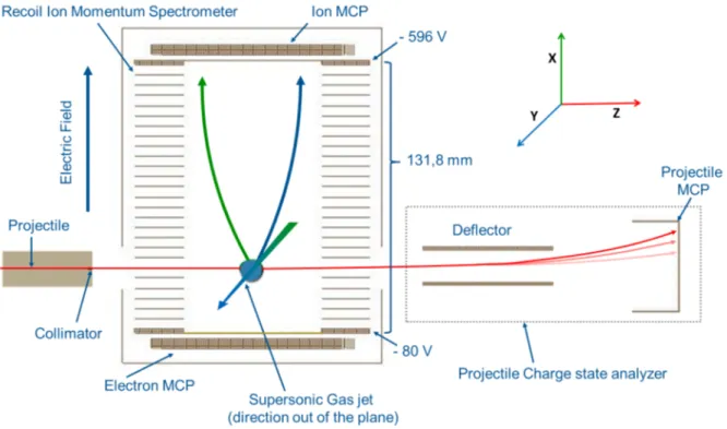
Figure 3.1.1 : Recoil Ion Momentum Spectrometer in its chamber with highlighted sections, the target jet is in the plane perpendicular to figure
3.2 COLd Target Recoil Ion Momentum Spectroscopy
The figure 3.1.1 is a picture of our spectrometer placed inside the chamber showing the spectrometer as well as the detectors for the recoil ions and electrons. On the left side there is a cylindrical tube with a collimator of 600 𝜇𝑚, through which the projectile ions enter the chamber. The projectile used are 𝐴𝑟9+ and 𝑋𝑒20+ of 15 qkeV energy, produced by an 14.5 GHz
ECR ion source of the very low energy facility of GANIL. Once the projectile enters the collision region of the spectrometer, it collides with the target beam produced by a supersonic gas jet. We can see the collision region in figure 3.1.2 where the target molecule/cluster get ionized by the projectile, mainly through electron capture, followed by fragmentation of the target. These fragments (as seen in the figure 3.1.2) are then extracted using a homogeneous electric field of 40 V/cm towards a position sensitive detector (PSD) for recoil ions. In addition, after collision the projectile may have different charge states (𝑞𝑜-1, 𝑞𝑜-2, 𝑞𝑜-3 etc) which are analyzed using a charge state analyzer consisting of a parallel plate analyzer (PPA) and a PSD detector located about 1.25 m away from the center of the collision region. As soon as the projectile reaches its PSD, it gives the START signal for the TOF measurements. Moreover the final charge state of the projectile is deduced by its impact position (X,Y) on this detector. TOF is measured in coincidence and calculated as the difference between the detection of the projectile and the detection of a recoil ion; each ion detection acts as a STOP signal. The electron detector was not used during the experiment performed during my thesis so it will not be discussed in the manuscript.
Although the relevant TOF is the time required for ions to fly from collision region to the ion micro channel plate (MCP). Note that the flight time of the projectile from the collision region to its detector is about 1-2 𝜇𝑠 (depending on its mass and the kinetic energy of the projectile). This time is long in comparison to TOF therefore it can not be neglected. Further discussion about this time delay is done in TOF calibration later in this chapter.
Using the TOF and position coordinates we compute the 3D momentum vectors of each fragment using the equation of motion in the homogeneous electric field. Moreover, a data selection is applied using the conservation of momentum law, see Annex A. Once we have the desired set of 3D momentum vector for each and every ion, we can calculate the associated kinetic energy.
3.2 COLd Target Recoil Ion Momentum Spectroscopy
COLTRIMS; is a momentum spectroscopy technique which enables to measure 3D momentum vectors of recoil ions with high resolution and 4𝜋 solid angle, making it good for the coincidence measurements [37]. Mainly in this section I will discuss about the spectrometer’s design, as well as a description of the recoil ion detector used for TOF and position measurement using Micro Channel Plate-Delay Line Detector. Although MCP detector will be discussed later in
3 Experimental Technique and Calibration
Figure 3.1.2 : Schematic of experimental setup highlighting the reference axis, X=Extraction axis, Y=Supersonic gas jet axis, Z=Projectile axis. Typical voltages applied to both grids before detector are also shown. A resistor chain ensures a constant voltage difference between each electrode of the detector resulting in an homogeneous electric field inside the spectrometer volume.
this chapter. Figure 3.1.2 also shows the mechanical aspect of the spectrometer used in our experiment. The region where the projectile and target are crossing is 10 mm gap between two electrodes. Starting from the center of collision region and moving up towards recoil ion PSD, we have a set of 19 equidistant electrodes of 1 mm thickness with a 4 mm separation between them using ceramic spacers. To have a uniform electric field across the extraction region, there is 2.75𝑀̀ resistance between each electrode. In this region the uniform electric field gives an acceleration to recoil ions to reach the ion MCP detector. The two upper and lower electrodes (in front of the ion and electron detectors) are 2 mm thick and had a 90% transmission grid to ensure the homogeneous field. This provides the total length of our extraction region (for Ions) as 𝐿𝑑 Ȃ 100𝑚𝑚 from the center of the collision region to detector.
Following the collision of the projectile and the molecular or cluster target, positively charged fragment ions are accelerated towards the ion detector. During the experiment the electric field is set to a minimal value which is calculated by taking the maximum kinetic energy of fragment to be detected [84]. For a spectrometer of length 𝐿𝑑 using a homogeneous electric field of strength 𝐸 and a detector of diameter 𝜙𝑑, one can demonstrate that 100% collection is achieved for recoil ions of charge 𝑞 and maximum kinetic energy 𝐾𝑚𝑎𝑥 using relation below:
3.3 Target Preparation 𝐾𝑚𝑎𝑥 = 𝑞.𝐸 16 × 𝜙2 𝑑 𝐿𝑑. (3.2.1)
In our case, using 𝐿𝑑= 100 mm, 𝐸= 40 V/cm and 𝜙𝑑=80 mm, we find a limit of 16 q.eV using equation 3.2.1. All these ions have characteristic TOF under the influence of uniform electric field which depends on the mass and charge, 𝑇 𝑂𝐹 Ȃ√𝑚𝑞. The TOF also depends on the velocity component of the ion fragment in the X direction (𝑣𝑜𝑥) of the spectrometer (see figure 3.1.2). After the extraction region between the front side of the MCP and the top grid is the post acceleration region, the voltage on the MCP front in nearly equal to -3000 V to ensure the maximum detection efficiency of ions with 𝑚
𝑞<28 [90].
3.3 Target Preparation
3.3.1 Cold Target
The "COLT" in COLTRIMS refers to the cold target, that is the other section of our exper-iment setup used to prepare the neutral target beam. This target beam could be monomer (atom/molecule), dimer, trimer or higher order cluster. For preparing this neutral beam we use a supersonic gas jet which has a nozzle of few tens of microns diameter. Since the 70’s many studies have been done to determine the relationship between jet properties and the experi-mental parameters. [91–94]. Mostly in past, supersonic jets were used to cover the studies of translational, rovibrational and even electronic relaxation, atomic and molecular spectroscopy, scattering processes in gas (target) and projectile (photon, laser, ions) and physics in van der Waals complexes or clusters [95]. There were many other application in field of surface inter-action, nuclear fusion, aerospace studies etc. In most of these important applications, it is of interest to obtain, narrow velocity spreads (or very low temperatures) in the jet, high density, and a variable kinetic energy from the thermal to the electronvolt range [96]. In our case, the supersonic jet is used to achieve the velocity spread much lower than the fragmentation velocity so that the maximum possible momentum resolution can be achieved. Secondly, this technique also allows the formation of small molecular clusters in the supersonic expansion of the molecular gas.
3.3.1.1 Supersonic gas jet
The supersonic jet is obtained by adiabatic expansion of the target gas which is initially under high pressure 𝑃0 and move in a low pressure region [91]. Different gases can have different jet characteristics depending upon experimental parameters, such as temperature of the nozzle (𝑇0), inlet pressure (𝑃0), backing pressure (𝑃1) and few other physical adjustment which will be mentioned in the next section.
3 Experimental Technique and Calibration
Figure 3.3.1 : Comparison of velocity components according to the axis (TOF axis) of extraction between a gas cell (with Gaussian fit in blue) and a supersonic jet of Argon [83]. The TOF of 𝐴𝑟+
is directly proportional to the 𝑣𝑜𝑥 component of the recoil ions. The width of these peaks is due to
the initial thermal motion of the 𝐴𝑟 atoms, therefore slow initial velocity spread of supersonic gas jet provides good momentum resolution.
The gas is allowed to expand through an orifice nozzle of diameter 𝐷, in such a way that equation 3.3.1 is followed:
𝜆0
𝐷 Ȃ 1 (3.3.1)
where 𝜆0 is the average mean free path of a gas atom or molecule under pressure 𝑃0. Due
to multiple collisions the flow along the nozzle converts the random thermal agitated movement into a directed translational flow along the jet axis.
When gas expands in the low pressure region the thermal agitation becomes so weak that molecule start to lose the velocity component perpendicular to jet axis. Due to transfer of this thermal energy in the final velocity of the jet we have a high velocity jet (more than sonic, Mach number 𝑀 > 1) which is almost monokinetic. This eventually brings the translational tempuratures down to 10−2K (see figure 3.3.1 [83]). For the case of polyatomic molecules we
have more degrees of freedom in form of the rotational and vibrational energies. It has been shown that the vibrational and rotational degrees of freedom are also cooled down as transla-tional motion. Therefore, the vibratransla-tional temperature near few K could be achieved [97, 98].
Figure 3.3.2 show the schematic of different sections of the supersonic gas jet setup. After the expansion the cold beam passes from a first stage to a second stage and in this second differential chamber we use a collimator of 500𝜇𝑚 to fix the size of the target beam before it enters the collision chamber. 𝑃1, 𝑃2 and 𝑃3 represents the pressures in three different chambers
from which the jet passes. In the first stage/chamber where the expansion took place, we use series of root pumps to achieve the pressure 𝑃1low enough down to 10−2− 10−3 mbar (primary
3.3 Target Preparation
Figure 3.3.2 : Campargue type supersonic gas jet schematic, Along the nozzle contour, a boundary layer forms, it is a thin flow layer attached to the nozzle walls that connects the high velocity flow in the volume of the flow domain with flow boundary that is immediately attached to the confining walls and therefore has zero velocity. These boundary walls are often termed as shock structure, as they shock the particle back to the flow domain. The gas jet flowed in the first stage is extracted by a sharp skimmer placed in the "zone-of-silence" without breaking its characteristics [92].
vacuum). These pumps have pumping capacity of 40 𝑚3ȂĂ, 253 𝑚3ȂĂ and 2050 𝑚3ȂĂ. Then
in stage 2, which is a differential pumping chamber with vacuum of the order of 10−5 mbar, a
primary pump along with a turbo pump (capacity 400 𝑙Ȃ𝑠) is used. Just before the collision chamber there is valve to prevent the entry of jet before the desired vacuum is reached. Finally beam enters the collision chamber where the vacuum is of the order of 10−7 mbar achieved
with the turbo pump and an additional liquid 𝑁2 screen is added to achieve better vacuum
conditions down to 10−9 mbar. Although, the primary objective of this screen is to reduce the
water vapours from the background inside the collision chamber. Figure 3.3.3 shows a mechani-cal schematic of the supersonic gas jet setup and the pumping system for the collision chamber. To avoid the diffusion of the jet in the collision chamber we use a beam dump with a turbo pump along jet axis. A quadrupole mass selector is also placed in the this beam dump and serves as a diagnostic to characterize the jet properties (see later).
3.3.2 Jet Parameters
Before we move to the characterization and optimization of the target jet, here we discuss very briefly about the size and the velocity of the beam. Due to the design of the setup, the diameter of the jet 𝐷𝐶𝑃 at the point of collision depends on geometrical parameters. In figure 3.3.4 we can see a schematic on how the diameter of jet and its divergence depends on the nozzle, skimmer and collimator assembly.

![Figure 3.3.3 : Mechanical details of the gas jet setup and the pumping system [78].](https://thumb-eu.123doks.com/thumbv2/123doknet/12843606.367389/41.892.84.763.153.556/figure-mechanical-details-gas-jet-setup-pumping.webp)
![Figure 3.5.4 : SEM image of a standard and tapered MCP showing difference in OAR to improve detection efficiency [104]](https://thumb-eu.123doks.com/thumbv2/123doknet/12843606.367389/51.892.71.750.468.633/figure-standard-tapered-showing-difference-improve-detection-efficiency.webp)
