Endothelial Cells and in a Reconstituted Purified Protein System by
Eric A. Osborn
B.S.E. Biomedical Engineering Duke University, 1998
Submitted to the Department of Mechanical Engineering in Partial Fulfillment of the
Requirements for the Degree of
Masters of Science in Mechanical Engineering at the
Massachusetts Institute of Technology February 2001
2001 Massachusetts Institute of Technology All rights reserved
Signature of Author
Department of Mechanical Engineering January 19, 2001 Certified by
C. Forbes Dewey, Jr. Professor of Mechanical Engineering Thesis Supervisor Certified by
John H. Hartwig Associate Professor of Cell Biology, Harvard Medical School Thesis Supervisor Accepted by
Ain Sonin Chairman, Department Committee on Graduate Students
Endothelial Cells and in a Reconstituted Purified Protein System by
Eric A. Osborn
Submitted to the Department of Mechanical Engineering on January 19, 2001 in Partial Fulfillment of the Requirements for the
Degree of Master of Science in Mechanical Engineering ABSTRACT
Cell motility and shape change are complex processes that depend primarily on the cytoplasmic dynamics and distribution of actin monomer and polymer. Proteins that regulate actin cycling control cellular architecture and movement. One method to measure parameters that characterize actin dynamics is photoactivation of fluorescence (PAF), which can simultaneously estimate the fraction of total actin polymerized (PF) and the lifetime of actin filaments (τ). By deciphering the relationships between actin dynamics and regulatory proteins, the complicated motions of cells and biological consequences of these movements can be better understood.
In purified actin solutions at steady-state, actin filament dynamics can be analyzed with PAF at long times following photoactivation. By increasing the width of the photoactivated band, actin filament turnover (τ ~ 8 hours) can be distinguished from actin filament diffusion. Proteins believed to stabilize actin filaments against depolymerization markedly slow actin filament turnover in wide photoactivated bands (τ ~ 65 hours). Decreasing the band width causes photoactivated fluorescence to decay more rapidly (τ ~ 3 hours) due to a combination of actin filament diffusion and turnover. Addition of actin-binding protein forms crosslinked actin gels that hinder filament diffusion and slow filament turnover (τ ~ 12 hours) in narrow photoactivated bands.
Endothelial cells decrease τ and PF in order to accelerate their migration speed, consistent with mechanisms attributed to ADF/cofilin in vitro. Removal of gelsolin in fibroblasts produces a similar correlation between motility, τ, and PF. Consistent with increased actin filament severing, fast-moving endothelial cells have an increased number of short actin filaments and more uncapped barbed ends, but paradoxically bind less cofilin. A mechanism of increasing endothelial cell motility is proposed that relies on actin filament severing to create uncapped pointed ends for ADF/cofilin-mediated depolymerization.
Thesis Supervisor: C. Forbes Dewey, Jr. Title: Professor of Mechanical Engineering
Thesis Supervisor: John H. Hartwig
I would like to thank my advisors, John H. Hartwig and C. Forbes Dewey, Jr. for their wonderful ideas, advice, and support throughout the process of shaping this research. I am also grateful for the continued mentorship and collaboration of James L. McGrath, who has immeasurably helped me focus and guide my thoughts about actin dynamics and cell movement. As well, I am indebted to the tireless effort of Sarah K. Chalos for her contributions to the studies with the purified protein system. Finally, I would like to thank the students and faculty of the Harvard–MIT Division of Health Sciences and Technology, a group with which I am grateful to be associated.
Eric Alan Osborn was born on October 2, 1975 in Battle Creek, MI to Larry and Margie Osborn. In 1994, Eric graduated from Port Huron Northern High School in Port Huron, MI and proceeded to attend college at Duke University in Durham, NC where he graduated in 1998 with a B.S.E. in Biomedical Engineering. In 1999, Eric began graduate study in the Department of Mechanical Engineering at MIT and the Medical Engineering/Medical Physics program at the Harvard–MIT Division of Health Sciences and Technology. Currently, he is continuing his doctoral research in this program at the Hematology Division of Brigham and Women’s Hospital located in Boston, MA.
Abstract ...3
Acknowledgements...5
Biography...7
Table of Contents...9
List of Figures ...11
Chapter I: Background and Literature Review Actin monomer ...13
Actin filaments...15
Actin filament turnover...16
Regulation of actin dynamics...17
The actin cytoskeleton ...22
Cellular actin dynamics...23
Development of fluorescence methods to measure actin dynamics ...25
Actin dynamics and movement in endothelial cells ...27
Actin dynamics in subconfluent gelsolin knockout fibroblasts ...29
The importance of actin dynamics and endothelial cells in physiology ...30
Goals of this thesis ...31
References...33
Chapter II: Modeling Actin Filament Dynamics in Purified Solutions Introduction...39
Methods Photoactivation of Fluorescence ...40
The Tardy Model ...40
General solution ...44
Simplified solution...45
Dynamics at short times...46
Actin monomer diffusion...46
Dynamics at long times...47
Actin filament diffusion...47
Actin filament turnover...48
Computation and simulations ...50
Results Diffusion coefficients for actin monomer and filaments ...51
Diffusion times for actin monomer and filaments ...51
Turnover times for actin filaments...52
Isolating actin filament turnover from actin filament diffusion...52
Narrow photoactivated bands ...53
References...58
Chapter III: Actin Filament Dynamics in Purified Solutions Introduction...61
Methods Preparation of caged resorufin actin ...62
Optimization of BSA glass coating to limit actin binding...64
Photoactivation of fluorescence (PAF)...65
PAF experiments with purified proteins ...66
Analysis of PAF experiments ...67
Calculation of the actin filament pointed end subunit dissociation rate ...69
Deleterious effects of photoactivation on actin ...69
ATP levels required for actin filament turnover ...70
Measurement of actin filament lengths...71
Results Synthesis of caged resorufin actin ...73
BSA coating optimization...75
Actin monomer diffusion...77
Actin filament diffusion and turnover...79
Actin filament turnover...83
Stabilizing actin filaments in wide photoactivated bands...87
Crosslinking actin filaments in narrow photoactivated bands ...89
Discussion ...90
References...91
Chapter IV: Actin Dynamics and Regulation in Endothelial Cells Introduction...97
Methods Cell culture...98
Actin assembly measurements...98
Actin quantitation...100
Cofilin quantitation ...101
Results Actin polymer fraction...103
Actin filament end counts ...105
Actin filament lengths...106
Cofilin content ...106
Discussion ...107
Description Page
Fig. 1-1: Atomic structure of the actin monomer ...14
Fig. 1-2: De novo actin polymerization ...15
Fig. 1-3: Actin filament treadmilling ...17
Fig. 1-4: Regulation of actin dynamics...19
Fig. 1-5: The actin cytoskeleton...23
Fig. 1-6: Endothelial cell speed correlates with morphological changes...27
Table 1-1: Endothelial cell actin dynamics correlate with cell speed...29
Fig. 1-7: Actin dynamics in subconfluent fibroblasts upon removal of gelsolin...30
Fig. 2-1: Simplified cell for analysis of PAF experiments ...41
Fig. 2-2: β dependence of fluorescence decay...45
Fig. 2-3: Diffusion coefficient of actin monomer and filaments ...51
Fig. 2-4: Theoretical purified actin diffusion time scales ...51
Fig. 2-5: Separating actin filament diffusion and turnover...53
Fig. 2-6: Narrow photoactivated band Tardy Model simulations...55
Fig. 2-7: Wide photoactivated band Tardy Model simulations ...57
Fig. 3-1: Caged resorufin actin fluorescence and polymerization properties ...73
Fig. 3-2: Actin binding to BSA coated glass surfaces ...75
Fig. 3-3: Actin monomer diffusion from a wide photoactivated band ...77
Fig. 3-4: Negatively stained purified actin filaments...79
Fig. 3-5: Narrow photoactivated band actin dynamics ...81
Fig. 3-6: Wide photoactivated band actin dynamics...85
Fig. 3-7: Phallacidin-actin dynamics in wide photoactivated bands...87
Fig. 3-8: ABP-120-actin dynamics in narrow photoactivated bands...89
Fig. 4-1: Actin monomer and filament concentration in BAECs ...103
Fig. 4-2: BAEC actin filament ends...105
Background and Literature Review
Despite the complexity of signals and proteins that are required to coordinate cellular movement, the final substrate is the protein actin, which coexists inside cells in both monomeric (G-actin) and filamentous (F-actin) states. Inside the cell, individual actin filaments are linked together to form a dense three-dimensional network called the actin cytoskeleton, which gives the cell shape and mechanical structure. Actin filaments are also highly dynamic, continuously adding and losing monomeric subunits from their ends in a process known as actin filament turnover. In order for cells to move, they must selectively break down and reassemble the actin cytoskeleton in specific cellular regions to produce coordinated extension and retraction of the leading edge, cell body, and tail. At the periphery of a crawling cell, membrane protrusion is driven by actin polymerization. At the cell interior in the bulk cytoskeleton, actin filaments are depolymerized to recycle actin monomers. At steady state, cells crawl by balanced actin polymerization and depolymerization in a cycle tightly regulated by a variety of actin-associated proteins. These include proteins that control the kinetics and access to actin filament ends, nucleate or sever actin filaments, sequester actin monomers, or alter actin monomer nucleotide exchange. Actin dynamics, structure, and regulation are important to a wide variety of normal and pathological situations because cells require an intact actin cytoskeleton and appropriate regulation of this structure for proper biological function. Since actin dynamics and cell movement are linked, understanding the relationships between actin dynamics and actin regulatory proteins provides insight into cell motility, morphology, and function.
Actin monomer
Actin is a globular 42 kDa protein [1] that has been highly conserved throughout evolution [2]. As one of the most abundant proteins in eukaryotic cells [3], actin is of
considerable biological significance due to its involvement in a large number of important cellular functions. There are six different isoforms of actin monomer known to exist in cells: α-cardiac, α-skeletal, α-vascular smooth muscle, β-non-muscle, γ -non-muscle, and γ-smooth muscle [4]. The actin isoforms are all ~ 375 amino acids in length and differ little from each other in sequence except near their amino terminus, which may confer isoform-specific actin-regulatory protein binding and filament assembly properties. The α- isoforms are found predominately in muscle cells, whereas the β- and γ- isoforms are predominant in non-muscle cells. However, each isoform may be expressed to varying degrees in spatially and temporally regulated patterns in many different cells [5].
The structure of the actin monomer has been solved by x-ray crystallography [6]. An actin monomer is approximately of dimensions 5.5 nm × 5.5 nm × 3.5 nm and forms two domains, one small and one large, which are separated by a binding pocket that accepts a divalent cation and nucleotide (Fig. 1-1). Actin monomer has one high-affinity divalent cation (Ca2+
or Mg2+
) binding site and four low-affinity binding sites [7]. In vivo, the high-affinity site is thought to be bound to Mg2+
, since the polymerization kinetics are enhanced for Mg2+
-actin over Ca2+
-actin [8]. The conformation and properties of the actin monomer are different depending on whether Mg2+ or Ca2+ occupies this site [8, 9]. In addition, actin monomers bind one of
Fig. 1-1. Atomic structure of the actin monomer. The actin monomer is divided into two domains, separated by a nucleotide and divalent cation binding cleft (from Ref. [6]).
three nucleotide species: ATP, ADP•Pi, or ADP. The dynamics, protein binding affinities, structure, and regulation of actin are highly dependent on the nucleotide species composition of the actin monomer population.
Actin filaments
In the presence of physiological salts and pH, actin monomers spontaneously self-assemble into polymeric filaments. The self-assembly of actin filaments is a result of energetic considerations. Above a particular concentration, actin monomers disturb the disordered nature of water molecules enough to favor the entropic formation of actin filaments and restore water molecules to their native state. The concentration at which actin monomers will self-assemble under physiological conditions is known as the critical concentration, which, for ATP-actin, is Cc-ATP≈ 0.1 µM [10, 11]. Therefore, actin filament formation does not require the input of cellular energy (ATP) as long as the appropriate actin concentration and buffer conditions are met.
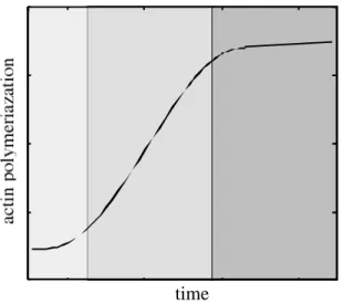
In purified actin solutions, polymerization begins with the formation of actin trimers, which serve as nucleation sites for further assembly of monomers [12]. Trimer formation is the rate-limiting step in polymerization, since the addition of actin filament seeds (pre-formed nucleation sites) eliminates the lag phase. Once actin trimers are created, rapid elongation occurs at the ends of the filament. The kinetics of filament elongation is strongly influenced by the nucleotide species bound to actin monomers [10, 13]. Eventually actin
Fig. 1-2. De novo actin polymerization. The three shaded sections, from left to right, represent the lag, elongation, and steady state phases of actin filament formation.
filaments reach a steady state in which no net elongation of the existing filament population is observed (Fig. 1-2).
Actin filaments are helical polymers that are stabilized by multiple, noncovalent contacts between adjacent monomeric subunits. The actin filament structure can be described by either a one-start left-handed genetic helix of 5.9 nm pitch or a two-start right-handed helix of 72 nm pitch with a filament diameter of ~7 nm [14, 15]. Actin filaments are polarized: the two ends of the polymer can be differentiated both geometrically and biochemically. Filament ends are distinguished by their appearance in the electron microscope after labeling with myosin subfragment-1 (myosin-S1), a proteolytic fragment of heavy meromyosin. Actin filaments decorated in this manner exhibit a bipolar appearance in which the myosin-S1 groups align along the filament in a head-to-tail fashion, giving it an arrowhead appearance. In light of this observation, the ends of the actin filament are differentiated as ‘barbed’ and ‘pointed’. Biochemically, filament ends are distinct, and the rate constants for actin monomer assembly and disassembly bound to each nucleotide species must be considered independently.
Actin filament turnover
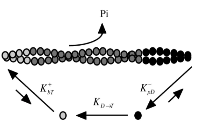
At long times in vitro, polymerized actin reaches a steady state in which there is no net change in actin filament lengths (Fig. 1-2). In the presence of excess ATP, due to different affinities for actin monomer at the barbed and pointed ends of the filament, actin filaments slowly elongate from the barbed end and shrink from the pointed end. The result is a net F-actin subunit flux from the barbed to pointed end [16], a mechanism known as turnover or treadmilling. Actin filament turnover is a cyclic process that relies on ATP hydrolysis to provide sufficient chemical energy to maintain the different filament end kinetics (Fig. 1-3) [10]. In the treadmilling model, actin monomers charged with ATP add to the barbed end where ATP-actin is hydrolyzed to an ADP•Pi-actin intermediate. Since actin is an ATPase, hydrolysis is accomplished intrinsically by actin filament subunits. ADP•Pi-actin is a stable intermediate state in actin filaments, but this
state slowly decays as Pi dissociates from the actin subunit while it cycles through the filament [17]. Therefore, as actin filament subunits approach the pointed end, they are predominately bound to ADP, an unstable state that favors F-actin subunit disassembly. Once depolymerized, free actin monomers exchange ADP for ATP to recharge their energy supply, thus completing the actin cycle.
The time required for an actin subunit to traverse a filament from barbed to pointed end defines the characteristic lifetime of the actin filament, also known as the actin filament turnover time (τ). This quantity is determined by the association and dissociation kinetics of actin monomers at the filament ends. Cellular actin filament turnover has been estimated from seconds to minutes, depending on the cell type and conditions [18-23]. However, in vitro, the actin filament lifetime is long (on the order of hours) because of the slow dissociation rate for monomers at the pointed end [24], and the absence of proteins that enhance filament dynamics.
Regulation of actin dynamics
Many different cytoplasmic proteins bind directly, or associate with factors that bind, to actin in order to influence actin dynamics. Actin-regulatory proteins have been studied in depth in vitro and are classified by their influence on the actin cycle. In general, these proteins control the availability of the ends of filaments to diffusing actin monomer, or directly influence the kinetics of monomer association, dissociation, and nucleotide
KpD− Pi
KbT+
KD→T
Fig. 1-3. Actin filament treadmilling. ATP-actin monomers ( ) assemble at the barbed end, are hydrolyzed to ADP•Pi-actin ( ) as they flux through the actin filament, and dissasemble as ADP-actin ( ) at the pointed end.
exchange. For simplicity, the actions of actin-associated proteins can be generalized into families of mechanisms: barbed and pointed end capping, acceleration of filament
depolymerization filament severing and nucleation, nucleotide exchange enhancement, and monomer sequestration. The proteins and mechanisms currently thought to be of major importance in regulating and enhancing the actin cycle are shown in Fig. 1-4.
Many proteins, such as gelsolin and CapZ, bind to the barbed end of actin filaments, thus ‘capping’ this end and blocking actin monomer association. This greatly slows actin filament elongation and turnover, since the barbed end is the primary assembly site for actin monomers. Gelsolin binds tightly to the barbed end of actin filaments but is released by membrane polyphosphoinositides, a mechanism that promotes rapid actin filament polymerization [25, 26].
Pointed end capping in non-muscle cells occurs via actions of the Arp2/3 complex, which binds to this site with nM affinity [27], and tropomodulin [28]. Recently, however, the degree of pointed end capping achieved by the Arp2/3 complex has been questioned [29]. Due to the large excess of barbed end capping proteins, actin filaments that are additionally capped at pointed ends are likely to be extremely stable in cells, as this is the primary site of F-actin subunit disassembly.
Not only does the Arp2/3 complex bind to actin filament pointed ends, but it binds to the sides of pre-existing actin filaments and nucleates new filaments at 70° angles by associating with free actin monomers to form a barbed end polymerization site [27]. Nucleation by the Arp2/3 complex is activated by association with members of the
Fig. 1-4. Regulation of actin dynamics. Many different proteins affect the dynamics of actin as monomers and polymer. Protein mechanisms of action are highlighted in the text.
WASp family of proteins [30]. Due to its ability to form a barbed end nucleation site, the Arp2/3 complex has recently been implicated in numerous models of cell membrane protrusion involving dendritic actin networks [27, 31].
When barbed ends are capped, actin filament disassembly is slow, but it can be accelerated by actions of the ADF/cofilin protein family. ADF/cofilin binds to the sides of filaments, preferentially to ADP-actin subunits and, therefore, most likely near the pointed end, enhancing the kinetics of ADP-actin depolymerization ~25-fold in vitro [32]. Accelerated depolymerization by ADF/cofilin, in cooperation with barbed end capping, may be responsible for the high rates of actin filament turnover observed in vivo [33].
Actin filament severing is accomplished mainly by the gelsolin protein family, which, besides gelsolin, includes such proteins as adseverin, CapG, flightless, and villin [34]. In the presence of µM Ca2+ levels, gelsolin binds to the sides of actin filaments, severs them, and remains tightly associated with the newly formed barbed ends [35-37]. Therefore, gelsolin is a major response protein that affects both actin cytoskeletal architecture and dynamics in signaling cascades involving calcium by altering filament length and end exposure.
The intrinsic rate of nucleotide exchange on actin monomer can be accelerated in solution by proteins such as profilin. Profilin binds to free actin monomer in solution, exchanges ADP for ATP ~140x faster than monomer alone, and shuttles ATP-actin monomer to uncapped barbed ends [38, 39]. The intrinsic rate of nucleotide exchange by actin during rapid filament turnover may be limiting, and profilin provides a mechanism to overcome this barrier.
Monomer sequestering proteins, such as thymosin-β4, bind unpolymerized actin monomer at higher affinity than pointed actin filament ends, but not barbed ends [40]. Therefore, through sequestration, actin monomer concentrations can be held much higher
than the critical concentration in vivo, allowing discrete control of the availability of monomer for incorporation into filaments.
Recently the necessary components for reconstituting actin-based motility in the bacterial pathogens Lysteria monocytogenes and Shigella were discovered [41]. The required actin-binding proteins include the Arp2/3 complex, capping protein, and ADF/cofilin. However, the results of these experiments have been questioned by other investigators as possibly containing contaminating protein factors. Despite this progress, the reconstitution of actin dynamics with purified actin and regulatory proteins is currently unable to achieve the levels measured in cells. The reasons for this dichotomy between in vitro and in vivo characteristics of actin dynamics have yet to be determined.
The actin cytoskeleton
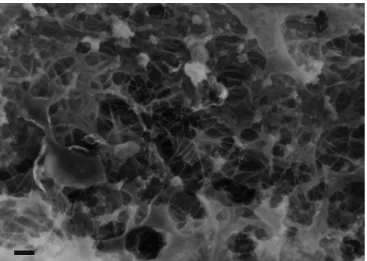
In cells, actin filaments are linked together to form a three-dimensional support structure that fills the cytoplasmic space. This dense meshwork of actin polymer and bound regulatory proteins is known as the actin cytoskeleton (Fig. 1-5). The actin cytoskeleton gives the cell shape, stiffness, and a mechanical scaffold that helps to fix the position of organelles and provides pathways for intracellular transport. The interfilament spacing of the cortical actin cytoskeleton has been modeled as an orthogonal lattice with a width of 20 to 100 nm [42, 43], through which the cell transports cytoplasmic solute and proteins. The filamin protein family is responsible for creating the structure of the actin cytoskeleton in cells by binding two actin filaments and crosslinking them into a gel [44]. Filamin-A, expressed primarily in non-muscle cells, is also present in many diverse structures within the cell, such as broad lamellae in which filamin-A promotes the formation of orthogonal filament arrays [44, 45] and at the base of filopodia [46]. The importance of filamin-A in cytoskeletal organization and structure is evident in a natural line of human melanoma cells that lack this protein. Filamin-A-null cells crawl poorly via
tubular projections and blebbing, while rescuing these cells with filamin-A cDNA results in a normal motile phenotype and the reappearance of lamellar protrusions and membrane ruffles [21, 47].
Dissolution of the actin cytoskeleton within a cell by decreased filament crosslinking and massive filament severing causes the cell cytoplasm to rapidly flow, known as a ‘gel-sol’ transition. An example of the rapid nature in which cells can change their physical state is the morphology of crawling neutrophils. In response to chemotactic signals, these cells move quickly across a substrate, constantly dissolving their internal network at the leading edge of the crawling cell and reforming this cell structure in the cell body [36, 48]. As in neutrophils and most other cells, the degree of crosslinking by filamin proteins is high [44]. There is some evidence that small actin filaments are able to diffuse through cytoskeletal pores [22] and are mobile near the plasma membrane [49]. In general, however, actin filaments are considered well anchored to each other in the cell cytoskeleton.
Cellular actin dynamics
In cells, the actin cytoskeleton is not simply a static structure, but one that must rapidly remodel when a cell changes shape or moves. Therefore, the bulk of cellular actin is dynamic via physical processes such as diffusion, turnover, severing, and nucleation. Tight spatial and temporal control of these dynamics is required to coordinate cell movement. When cells change shape, they recruit unpolymerized actin for assembly into
Fig. 1-5. The actin cytoskeleton. As typical for non-muscle cells, the endothelial cell actin cytoskeleton is a dense mesh of highly crosslinked actin filaments. Bar = 200 nm.
new or elongating filaments, and, at steady state, actin polymerization must be balanced by depolymerization elsewhere. Actin polymerized at the cell periphery in crawling cells is transported centripetally toward the cell body, implying that it is depolymerized in the bulk of the cytoskeleton [18-20, 50]. Therefore, cell movement and cytoskeletal actin remodeling are linked. A model of cell crawling involves actin polymerization and membrane protrusion at the leading edge, retrograde flow of F-actin to the cell interior, contraction of the cell body and tail, and depolymerization of actin filaments in the bulk, interior cytoplasm.
The lifetimes of actin filaments in cells are quite short as compared to purified actin solutions. Actin filament turnover is accelerated in cells due to the large number of regulatory proteins and signaling pathways that enhance actin dynamics. Fluorescence measurements in the lamellae of slowly moving fibroblasts [18] and highly motile keratocytes [19] provide estimates of actin filament lifetimes on the order of minutes and seconds, respectively, correlating actin filament turnover and the rate of cell movement. Because cellular actin is tightly regulated by numerous proteins and mechanisms, understanding the functions of these proteins provides the key to understanding how actin is remodeled in moving cells.
Development of fluorescence methods to measure actin dynamics
Actin dynamics can be measured in cells with variable precision. Two analogous fluorescence techniques offer the best estimates of parameters that describe the dynamic nature of cellular actin: photoactivation of fluorescence (PAF) and fluorescence recovery after photobleaching (FRAP). In PAF, a non-fluorescent, caged precursor actin molecule is uncaged by exposure to ultraviolet (UV) light in a small spot or rectangular region within the cell. FRAP is the inverse technique, in which the fluorescence of a labeled actin molecule is quenched with high-intensity light at the excitation wavelength of the fluorophore. Fluorescence decay or recovery is then measured in the subcellular photoactivated or photobleached area, respectively, and interpreted with mathematical
models that describe cellular actin transport. One theory relating labeled actin dynamics and photoactivated fluorescence decay is the Tardy Model, which computes of the mobility of actin monomer (Dm), the fraction of total actin polymerized (PF), and the actin filament turnover time (τ) [51]. The Tardy Model has been used to quantitate actin dynamics in various cell types such as endothelium, fibroblasts, and melanoma cells [21, 22], and the resulting measurements are consistent with those previously published by other investigators [18-20, 23].
Actin dynamics and movement in endothelial cells
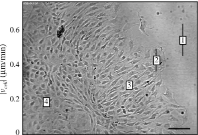
Following mechanical wounding of confluent monolayers of endothelial cells and overnight recovery, distinct morphological and motile subpopulations of endothelial cells are observed in different regions with respect to the wound (Fig. 1-6) [21]. Wounding a confluent endothelial monolayer removes the inhibitory effects of intercellular contacts on cells near the wound, increasing cellular spacing, and activating the cell to crawl. In the undisturbed, confluent endothelial monolayer far away from the wound (zone 4), the cells retain the characteristic cobblestone morphology and low rate of movement expected for quiescent endothelial cells. Endothelium intermediate to the wound and the confluent region (zone 3) exhibits an elongated morphology perpendicular to the wound
1 2 3 4 0.6 0.4 0.2 0
Fig. 1-6. Endothelial cell speed correlates with morphological
changes. After overnight recovery from wounding, endothelial
cells organize into distinct morphological zones: (1) subconfluent, (2) wound edge, (3) elongated, and (4) confluent. Average cell speed (|vcell|) decreases with increased distance from
the wound (from Ref. [21]). Bar = 100 µm.
|vcell
| (
µ
edge and a faster rate of movement. At the wound edge (zone 2), endothelial cells remain in contact with the monolayer, but rapidly extend cellular processes into the wound, increase their speed, and assume a more spread morphology. The fastest moving endothelial cells are subconfluent or free from cellular contact in the wound (zone 1), and assume the phenotype of a typical motile cell with broad lamellipodia and filopodia extending from the leading edge.
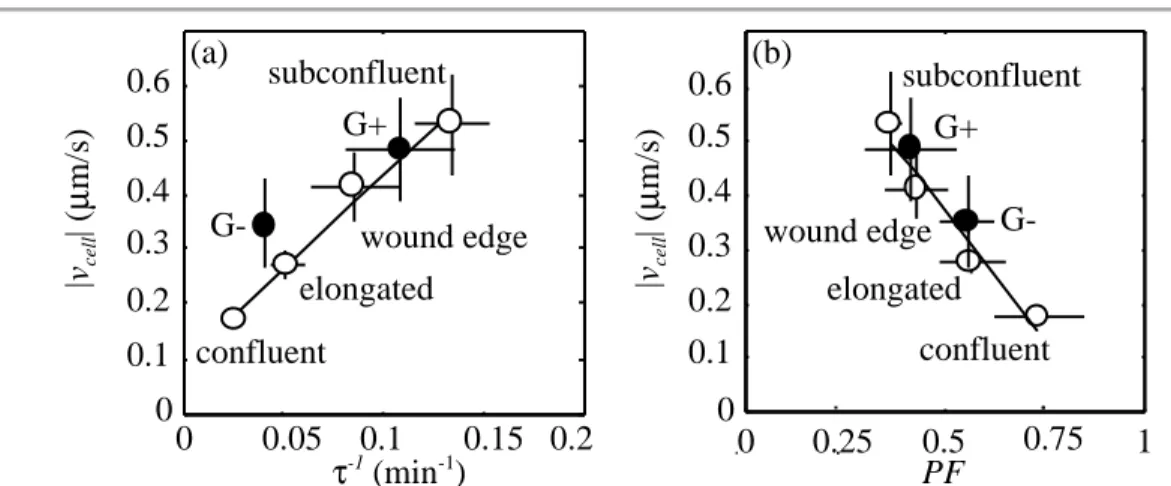
In addition to morphological differences, root-mean-square (RMS) cell speed decreases with increasing distance from the wound (Fig. 1-6). PAF measurements of endothelial cell actin dynamics exhibit a positive correlation between cell speed, polymer fraction, and actin filament turnover (Table 1-1). The slowest moving (confluent) and fastest moving (subconfluent) endothelial cells differ in PF by ~50% and in τ by more than a factor of five. Thus, individual endothelial cells accelerate actin filament turnover and decrease the amount of polymerized actin in order to increase steady state motility [21]. Actin dynamics in subconfluent gelsolin knockout fibroblasts
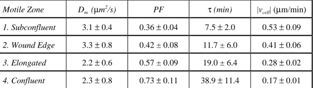
Fibroblasts isolated from gelsolin knockout (G-) transgenic mice crawl poorly in comparison to wild-type cells (G+) [52]. This implies the importance of gelsolin, a potent actin filament severing and barbed end capping protein, in the coordination of normal Motile Zone Dm (µm
2
/s) PF τ (min) |vcell| (µm/min) 1. Subconfluent 3.1 ± 0.4 0.36 ± 0.04 7.5 ± 2.0 0.53 ± 0.09 2. Wound Edge 3.3 ± 0.8 0.42 ± 0.08 11.7 ± 6.0 0.41 ± 0.06 3. Elongated 2.2 ± 0.6 0.57 ± 0.09 19.0 ± 6.4 0.28 ± 0.02 4. Confluent 2.3 ± 0.8 0.73 ± 0.11 38.9 ± 11.4 0.17 ± 0.01
Table 1-1. Endothelial actin dynamics correlate with cell speed. Monomer diffusion coefficient (Dm), polymer fraction (PF), filament turnover time (τ), and cell speed (|vcell|) measured in the different motile
cellular movement. The observed changes in motility between subconfluent G- and G+ fibroblasts are similar to the transition between motile phenotypes observed in wounded BAEC monolayers. PAF measurements reveal that the slower moving G- fibroblasts exhibit slower actin filament turnover (τ = 25.0 ± 6.1 min) and a greater fraction of actin polymerized (PF ~ 55 ± 7%) than the faster moving G+ phenotype (τ = 9.3 ± 4.7 min; PF ~ 41 ± 11%) [21]. The changes in actin dynamics that occur in subconfluent fibroblasts upon the removal of gelsolin show positive correlations with cell speed similar to measurements in endothelial cells (Fig. 1-7). This result indicates that the capping and severing activities of gelsolin are consistent with the mechanisms by which actin dynamics are regulated in endothelial cells [21].
The importance of actin dynamics and endothelial cells in physiology
Actin dynamics are integral to many normal and pathological processes in the body and are therefore essential to understanding physiology [53]. All animal cells crawl or change their shape throughout their lives, and these motile and structural changes are directly related to dynamic changes in the cellular actin cytoskeleton. Not only does the actin
0 0.2 0 0.1 0.2 0.3 0.4 0.5 0.6 0.1 0.05 0.15 (a) subconfluent wound edge elongated confluent G-G+ 0 0.1 0.2 0.3 0.4 0.5 0.6 (b) 0.75 0 0.25 0.5 1 G+ G-subconfluent wound edge elongated confluent |vcell | ( µ m/s) |vcell | ( µ m/s) τ-1 (min-1 ) PF
Fig. 1-7. Actin dynamics in subconfluent fibroblasts ( ) upon removal of gelsolin. Gelsolin knockout fibroblasts (G-) crawl slower than wild-type (G+) and exhibit changes in actin dynamics consistent with measurements in the endothelial cell ( )
motile zones from Fig. 1-6. (a) Actin filament turnover rates (τ-1) markedly decrease in
G- fibroblasts, coinciding with (b) an increase in the fraction of total actin polymerized (PF). (from Ref. [21])
architecture change in moving cells, but the ratio of actin monomer to polymer, the rate of actin filament turnover, the length of actin filaments, and the exposure of actin filament ends are all tightly regulated during these processes. In fetal development, there are numerous examples of cells altering their shape and motile state (e.g. gastrulation) as the embryo forms by patterned cellular differentiation and proliferation. Activation of blood platelets in the coagulation cascade following injury to the vascular wall is another example of the importance of actin dynamics in physiology: resting, discoid platelets circulating in the blood must rapidly alter their shape in order to reestablish the structure of a compromised blood vessel and facilitate vessel repair [54]. Blocking actin dynamics eliminates this important platelet function.
Endothelial cells, which line the inner surface of the vasculature, also depend on proper actin cytoskeletal structure and dynamics. Situated at the barrier between flowing blood and soft tissue, the endothelium senses fluid forces and extracellular chemical signals in the blood to regulate macromolecule permeability, maintain vascular tone, and provide a surface resistant to blood clot formation [55]. Damage to the endothelial lining promotes thrombotic episodes, potentially resulting in myocardial infarct and stroke. In response to vascular wounding and signals promoting new blood vessel formation such as angiogenesis, endothelial cells are stimulated to move, redefining and remodeling their actin cytoskeletons. The fraction of total cellular actin incorporated into filaments, the lifetime of these filaments, the structure of the actin cytoskeleton, and the linkages between actin and cell-substrate adhesion sites determine the cell’s shape, stiffness, potential to crawl, and integrity of attachment to the artery wall, all of which affect endothelial function in vivo.
Goals of this thesis
The overall thrust of this research is to further understand the dynamics and regulation of the actin cytoskeleton by actin-associated proteins in the context of cellular shape change and motility. This is an ambitious undertaking, since the regulation of actin dynamics is
an extremely complex problem involving synergy between many different proteins. In order to narrow the scope of this research, from the many model systems for studying actin dynamics such as cellular extracts, bacterial pathogens Lysteria monocytogenes and Shigella, and every known cell type, this thesis will focus on examination and comparison of actin dynamics in a reconstituted purified protein system and in endothelial cells. Similarly, the focus on actin regulation is narrowed to make the problem tractable by investigating only a few specific actin-associated proteins believed to be important in actin cycling. Powerful fluorescence techniques are used to measure actin polymer fraction, diffusivity, and filament turnover. These methods are supplemented with classical biochemical techniques to analyze actin polymerization and protein levels in cells and purified solutions.
Chapters II and III of this thesis develop the theory and experimental results for a system to measure actin filament dynamics in purified actin solutions with PAF. This reconstituted purified protein system is novel in that it allows the direct observation of the dynamic processes at work via the fluorescence decay of labeled actin monomers. It also is the ideal system to noninvasively analyze actin filament dynamics at a true steady state, outside of the complex cellular environment. The ultimate aspirations of this work are to reconstitute a purified model of cellular actin architecture and dynamics in vitro, while learning more about the function of the actin regulatory proteins involved in cellular processes.
Actin dynamics and regulation in endothelial cells moving at different speeds are investigated in Chapter IV. When endothelial cells migrate from their normal resting state, they must change their speed, shape, and, therefore, actin dynamics. The mechanism that regulates this transition is unknown but is most likely related to the functions of known actin-associated proteins. By understanding how the state of actin changes and examining the distributions of certain actin-regulatory proteins, mechanisms of altering endothelial cell speed by these proteins can be determined.
REFERENCES
1. Elzinga M, Collins JH, Kuehl WM, Adelstein RS: Complete amino-acid sequence of
actin of rabbit skeletal muscle. Proc Natl Acad Sci U S A 1973; 70(9): 2687-91.
2. Hightower RC, Meagher RB: The molecular evolution of actin. Genetics 1986; 114(1):
315-32.
3. Pollard TD: Actin. Curr Opin Cell Biol 1990; 2(1): 33-40.
4. Vandekerckhove J, Weber K: At least six different actins are expressed in a higher
mammal: an analysis based on the amino acid sequence of the amino-terminal tryptic peptide. J Mol Biol 1978; 126(4): 783-802.
5. Herman IM: Actin isoforms. Curr Opin Cell Biol 1993; 5(1): 48-55.
6. Kabsch W, Mannherz HG, Suck D, Pai EF, Holmes KC: Atomic structure of the
actin:DNase I complex. Nature 1990; 347(6288): 37-44.
7. Carlier MF, Pantaloni D, Korn ED: Fluorescence measurements of the binding of
cations to high-affinity and low-affinity sites on ATP-G-actin. J Biol Chem 1986; 261(23): 10778-84.
8. Carlier MF, Pantaloni D, Korn ED: The effects of Mg2+ at the high-affinity and
low-affinity sites on the polymerization of actin and associated ATP hydrolysis. J Biol Chem 1986; 261(23): 10785-92.
9. Frieden C, Lieberman D, Gilbert HR: A fluorescent probe for conformational changes
in skeletal muscle G- actin. J Biol Chem 1980; 255(19): 8991-3.
10. Korn ED, Carlier MF, Pantaloni D: Actin polymerization and ATP hydrolysis.
11. Bonder EM, Fishkind DJ, Mooseker MS: Direct measurement of critical
concentrations and assembly rate constants at the two ends of an actin filament. Cell 1983; 34(2): 491-501.
12. Wegner A, Engel J: Kinetics of the cooperative association of actin to actin filaments.
Biophys Chem 1975; 3(3): 215-25.
13. Pollard TD: Rate constants for the reactions of ATP- and ADP-actin with the ends of
actin filaments. J Cell Biol 1986; 103(6 Pt 2): 2747-54.
14. Milligan RA, Whittaker M, Safer D: Molecular structure of F-actin and location of
surface binding sites. Nature 1990; 348(6298): 217-21.
15. Amos LA: Structure of muscle filaments studied by electron microscopy. Annu Rev
Biophys Biophys Chem 1985; 14: 291-313.
16. Wegner A: Head to tail polymerization of actin. J Mol Biol 1976; 108(1): 139-50. 17. Carlier MF, Pantaloni D: Direct evidence for ADP-Pi-F-actin as the major
intermediate in ATP- actin polymerization. Rate of dissociation of Pi from actin filaments. Biochemistry 1986; 25(24): 7789-92.
18. Wang YL: Exchange of actin subunits at the leading edge of living fibroblasts:
possible role of treadmilling. J Cell Biol 1985; 101(2): 597-602.
19. Theriot JA, Mitchison TJ: Actin microfilament dynamics in locomoting cells. Nature
1991; 352(6331): 126-31.
20. Theriot JA, Mitchison TJ: Comparison of actin and cell surface dynamics in motile
fibroblasts. J Cell Biol 1992; 119(2): 367-77.
21. McGrath JL, Osborn EA, Tardy YS, Dewey CF, Jr., Hartwig JH: Regulation of the
actin cycle in vivo by actin filament severing. Proc Natl Acad Sci U S A 2000; 97(12): 6532-6537.
22. McGrath JL, Tardy Y, Dewey CF, Jr., Meister JJ, Hartwig JH: Simultaneous
measurements of actin filament turnover, filament fraction, and monomer diffusion in endothelial cells. Biophys J 1998; 75(4): 2070-8.
23. Kreis TE, Geiger B, Schlessinger J: Mobility of microinjected rhodamine actin within
living chicken gizzard cells determined by fluorescence photobleaching recovery. Cell 1982; 29(3): 835-45.
24. Selve N, Wegner A: Rate of treadmilling of actin filaments in vitro. J Mol Biol 1986;
187(4): 627-31.
25. Janmey PA, Iida K, Yin HL, Stossel TP: Polyphosphoinositide micelles and
polyphosphoinositide-containing vesicles dissociate endogenous gelsolin-actin complexes and promote actin assembly from the fast-growing end of actin filaments blocked by gelsolin. J Biol Chem 1987; 262(25): 12228-36.
26. Janmey PA, Stossel TP: Modulation of gelsolin function by phosphatidylinositol
4,5-bisphosphate. Nature 1987; 325(6102): 362-4.
27. Mullins RD, Heuser JA, Pollard TD: The interaction of Arp2/3 complex with actin:
nucleation, high affinity pointed end capping, and formation of branching networks of filaments. Proc Natl Acad Sci U S A 1998; 95(11): 6181-6.
28. Fowler VM: Tropomodulin: a cytoskeletal protein that binds to the end of erythrocyte
tropomyosin and inhibits tropomyosin binding to actin. J Cell Biol 1990; 111(2): 471-81.
29. Pantaloni D, Boujemaa R, Didry D, Gounon P, Carlier MF: The Arp2/3 complex
branches filament barbed ends: functional antagonism with capping proteins. Nat Cell Biol 2000; 2(7): 385-91.
30. Machesky LM, Mullins RD, Higgs HN, et al.: Scar, a WASp-related protein, activates
nucleation of actin filaments by the Arp2/3 complex. Proc Natl Acad Sci U S A 1999; 96(7): 3739-44.
31. Svitkina TM, Borisy GG: Arp2/3 complex and actin depolymerizing factor/cofilin in
dendritic organization and treadmilling of actin filament array in lamellipodia. J Cell Biol 1999; 145(5): 1009-26.
32. Carlier MF, Laurent V, Santolini J, et al.: Actin depolymerizing factor (ADF/cofilin)
enhances the rate of filament turnover: implication in actin-based motility. J Cell Biol 1997; 136(6): 1307-22.
33. Carlier MF, Pantaloni D: Control of actin dynamics in cell motility. J Mol Biol 1997;
269(4): 459-67.
34. Kwiatkowski DJ: Functions of gelsolin: motility, signaling, apoptosis, cancer. Curr
Opin Cell Biol 1999; 11(1): 103-8.
35. Janmey PA, Chaponnier C, Lind SE, Zaner KS, Stossel TP, Yin HL: Interactions of
gelsolin and gelsolin-actin complexes with actin. Effects of calcium on actin nucleation, filament severing, and end blocking. Biochemistry 1985; 24(14): 3714-23.
36. Yin HL, Stossel TP: Control of cytoplasmic actin gel-sol transformation by gelsolin, a
calcium-dependent regulatory protein. Nature 1979; 281(5732): 583-6.
37. Yin HL, Zaner KS, Stossel TP: Ca2+ control of actin gelation. Interaction of gelsolin
with actin filaments and regulation of actin gelation. J Biol Chem 1980; 255(19): 9494-500.
38. Selden LA, Kinosian HJ, Estes JE, Gershman LC: Impact of profilin on actin-bound
nucleotide exchange and actin polymerization dynamics. Biochemistry 1999; 38(9): 2769-78.
39. Goldschmidt-Clermont PJ, Machesky LM, Doberstein SK, Pollard TD: Mechanism of
the interaction of human platelet profilin with actin. J Cell Biol 1991; 113(5): 1081-9.
40. Safer D, Nachmias VT: Beta thymosins as actin binding peptides [published erratum
41. Loisel TP, Boujemaa R, Pantaloni D, Carlier MF: Reconstitution of actin-based
motility of Listeria and Shigella using pure proteins. Nature 1999; 401(6753): 613-6.
42. Luby-Phelps K, Taylor DL, Lanni F: Probing the structure of cytoplasm. J Cell Biol
1986; 102(6): 2015-22.
43. Satcher RL, Jr., Dewey CF, Jr.: Theoretical estimates of mechanical properties of the
endothelial cell cytoskeleton [see comments]. Biophys J 1996; 71(1): 109-18.
44. Hartwig JH, Shevlin P: The architecture of actin filaments and the ultrastructural
location of actin-binding protein in the periphery of lung macrophages. J Cell Biol 1986; 103(3): 1007-20.
45. Hartwig JH, Tyler J, Stossel TP: Actin-binding protein promotes the bipolar and
perpendicular branching of actin filaments. J Cell Biol 1980; 87(3 Pt 1): 841-8.
46. Ohta Y, Suzuki N, Nakamura S, Hartwig JH, Stossel TP: The small GTPase RalA
targets filamin to induce filopodia. Proc Natl Acad Sci U S A 1999; 96(5): 2122-8.
47. Cunningham CC, Gorlin JB, Kwiatkowski DJ, et al.: Actin-binding protein
requirement for cortical stability and efficient locomotion. Science 1992; 255(5042): 325-7.
48. Nunnally MH, Powell LD, Craig SW: Reconstitution and regulation of actin gel-sol
transformation with purified filamin and villin. J Biol Chem 1981; 256(5): 2083-6.
49. Sund SE, Axelrod D: Actin dynamics at the living cell submembrane imaged by total
internal reflection fluorescence photobleaching. Biophys J 2000; 79(3): 1655-69.
50. Fisher GW, Conrad PA, DeBiasio RL, Taylor DL: Centripetal transport of cytoplasm,
actin, and the cell surface in lamellipodia of fibroblasts. Cell Motil Cytoskeleton 1988; 11(4): 235-47.
51. Tardy Y, McGrath JL, Hartwig JH, Dewey CF: Interpreting photoactivated
fluorescence microscopy measurements of steady-state actin dynamics. Biophys J 1995; 69(5): 1674-82.
52. Witke W, Sharpe AH, Hartwig JH, Azuma T, Stossel TP, Kwiatkowski DJ:
Hemostatic, inflammatory, and fibroblast responses are blunted in mice lacking gelsolin. Cell 1995; 81(1): 41-51.
53. Janmey PA, Chaponnier C: Medical aspects of the actin cytoskeleton. Curr Opin Cell
Biol 1995; 7(1): 111-7.
54. Hartwig JH: Mechanisms of actin rearrangements mediating platelet activation. J Cell
Biol 1992; 118(6): 1421-42.
55. Davies PF: Flow-mediated endothelial mechanotransduction. Physiol Rev 1995;
Modeling Actin Filament Dynamics in Purified Solutions
Actin filaments fill the cell cytoplasm in vivo and form a dynamic network that is actively remodeled when a cell moves. Various actin-associated proteins (reviewed in Chapter I) drive actin dynamics inside cells. In cells, actin filaments are predominately crosslinked by proteins that bind adjacent filaments [1], thereby fixing them in space and hindering their mobility. Therefore, filament dynamics in cells are primarily a result of turnover of actin polymer. In purified solutions of actin, however, no such crosslink-mediated restrictions on filament motion occur, and thus filaments may diffuse through the entangled F-actin network in addition to cycling their subunits.
In purified solutions of actin at physiological salt and ATP levels, actin spontaneously polymerizes into filaments of various lengths. At long times after initiation of polymerization, a steady state is reached in which there is no net actin polymerization and the actin filament length distribution is stable. In the absence of convection, purified actin gels exhibit three dynamic processes. First, unpolymerized monomer may diffuse throughout the buffer. Second, filaments may cycle in an ATP-dependent process, predominately adding actin monomer onto the barbed end and losing monomer from the pointed end. In this way, actin subunits flux through filaments and exhibit treadmilling behavior. Finally, without crosslinking proteins, actin filaments may diffuse. If these processes can be distinguished from one another in reconstituted, purified actin networks, then analysis of the mechanisms controlling actin dynamics is possible.
This chapter develops theory to isolate the three dynamic processes of purified actin in photoactivation of fluorescence (PAF) experiments. This is accomplished by examining the theoretical time scale of each of these events and their dependence on physical parameters, such as the properties of actin and the width of the photoactivated band. Using this knowledge, experiments are developed in Chapter III to extract information
about actin dynamics in purified solutions and compare how these processes are altered by the addition of actin regulatory proteins.
METHODS
Photoactivation of fluorescence
Photoactivation of fluorescence (PAF) involves liberating the fluorescence of a non-fluorescent precursor molecule in a small region and measuring the fluorescence decay in this region over time. For measurement of purified actin dynamics, actin monomers are fluorescently labeled and ‘caged’ by coupling an ultraviolet (UV) light sensitive moiety to the photosensitive group on the fluorophore, causing the normally fluorescent species to become non-fluorescent [2, 3]. PAF experiments are initiated by exposing caged actin to UV light in a specified geometry (typically circular or rectangular). Once the actin is uncaged, the fluorescence at the center of the photoactivated region is monitored over time. Using an interpretive model that describes the physics of the actin transport processes underlying the fluorescence decay, appropriate physical parameters can be extracted and analyzed [4].
The Tardy Model
A model describing PAF-based actin dynamics in cells, the Tardy Model [4], allows calculation of the fraction of total actin incorporated into actin filaments (PF), the diffusion coefficient of actin monomer (Dm), and the average time for actin filament turnover (τ). These three parameters are computed from the fluorescence decay in a rectangular, photoactivated band. Although this model was developed to analyze cellular actin dynamics, the simplified geometry and assumptions inherent in the theory are even better suited for analyzing purified actin dynamics.
The Tardy Model employs certain simplifying assumptions to create a tractable mathematical formulation of the problem.
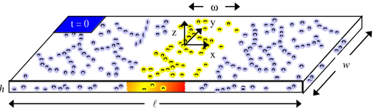
1. The cell is modeled as a rectangle of constant thickness (Fig. 2-1). Photoactivation across the entire width and thickness of the model cell results in one-dimensional actin dynamics in the x-direction. The nucleus and cellular organelles are not included in the model. Since purified solutions of actin in glass tubes are this shape and lack organelles to disrupt the actin structure, this simplification is extremely accurate.
2. Actin is distributed homogeneously across the model cell, disregarding actin concentrates or bundles that exist in cells, particularly in the cell cortex. Again, this assumption is valid in purified actin solutions because actin is uniformly distributed and does not contain actin bundling proteins.
3. Two interacting pools of actin, monomeric and filamentous, are dynamically coupled to each other. Actin filaments cycle subunits on and off their ends in direct exchange with the monomer pool. Actin monomer is free to diffuse, but filaments are assumed to be non-diffusible. In cells, the idealization of immobile filaments is valid since cortical actin networks are highly crosslinked by filamin
Fig. 2-1. Simplified cell for analysis of PAF experiments. The geometry of the cell is as shown, where h << l, w. The shaded region in the center of the model cell represents an uncaged, fluorescent actin band of width, ω, immediately after photoactivation. (adapted from Ref. [4])
t = 0 l w ω h x y z
[1] and filaments diffuse much slower than the rates of cellular actin filament turnover. In purified solutions, the restriction on actin filament diffusion must be relaxed since filament turnover and diffusion may occur on similar time scales. For example, if nearly all of the actin is polymerized and the rate of filament turnover is much slower than filament diffusion, then the actin monomer diffusion coefficient calculated with the Tardy Model corresponds to the actin filament diffusion coefficient.
4. Actin dynamics are at steady state. This simplification requires that there is no net growth or diminishment of the actin monomer and filamentous pools, change in the length distributions of filaments, and generation or degradation of actin. This condition must hold on the global level within cells, since cellular movements occur on time scales much shorter than protein degradation and synthesis. However, local heterogeneities and actin transients within cells challenge this idealization. In purified, reconstituted actin networks at long times, actin dynamics are inherently at steady state as described by the kinetics of actin polymerization (Fig. 1-2).
5. The rate of actin exchange between monomer and filaments is assumed uniform throughout the model cell. This is a simplifying assumption for actual cells, but in a purified solution of proteins, a uniform rate of actin turnover should be extremely valid.
The general mathematical problem defined in the Tardy Model involves a set of unsteady, coupled partial differential equations that describe actin monomer and filament transport in the model cell. These equations are derived from first principles given the previously mentioned simplifying assumptions.
∂cum ∂t =Dm ∂2 cum ∂x2 +g ˙ cuf cf −cum cm (2.1)
∂cuf ∂t = −g ˙ cuf cf −cum cm (2.2)
In these equations, cum and cuf describe the uncaged actin monomer and filament concentrations, respectively, whereas cm and cf describe the total actin monomer and filament concentrations, respectively. Dm is the actin monomer diffusion coefficient. The uniform rate of monomer exchange between the monomeric and filamentous pools is denoted ˙ g . These equations are subject to boundary conditions stipulating that the spatial rate of change of uncaged monomer is zero at the model cell boundaries,
∂cum
∂x x=0,l
=0 (2.3)
and initial conditions where monomer residing outside of the photoactivated band is nonfluorescent, while that contained within the band is fluorescent.
cum=cuf =0 cum= αcm cuf = αcf
∈ x−x0 >1
2ω (2.4)
In these equations, α represents the fraction of total actin in the photoactivated band that is uncaged. In purified protein experiments α ~ 0.6, given that ~ 60% of the actin in the band is labeled, and assuming 100% of that actin is uncaged.
Solution of the general Tardy Model. The general solution to the Tardy Model is found via a Fourier-LaPlace transform, and is represented as an infinite series for both actin monomer and filament concentrations [4]. The fluorescence intensities, F(t), measured in PAF experiments are proportional to a weighted sum of the actin monomer and polymer concentrations, the excitation light intensity (Io), and the quantum efficiency (Q) of the fluorophore.
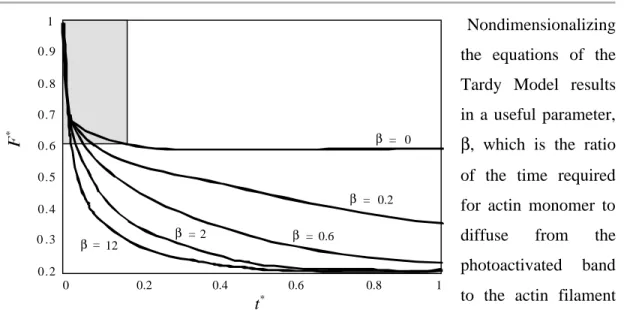
Nondimensionalizing the equations of the Tardy Model results in a useful parameter, β, which is the ratio of the time required for actin monomer to diffuse from the photoactivated band to the actin filament turnover time.
The general solution at long times depends strongly on β (Fig. 2-2). The fluorescence decay is biphasic, in which the rapid decay is primarily due to actin monomer diffusion, and the slow decay primarily due to actin filament turnover. The rates of decay, and the distribution of fluorescence between the two phases determines PF, τ, and Dm.
Simplification of the general Tardy Model solution. If actin monomer diffusion is fast enough compared to actin filament turnover, expressed as the criterion β < 4, then the solution to the general Tardy Model may be simplified to analyze fluorescence decay once actin monomer diffusion is complete (t > 4τDm) [4]. This equation is simplified further if ω<< l. F∗= F Fo = cf cm +cf −t 4τ
e
(2.7)When actin dynamics are not diffusion limited, the fluorescence evolution in a photoactivated band at long times decays as a simple exponential with time constant, τ,
0.2 0 .3 0.4 0.5 0.6 0.7 0.8 0.9 1 0 0.2 0.4 0.6 0.8 1 β = 0 β = 0.2 β = 0.6 β = β= 12 2 * t*
Fig. 2-2. β dependence of fluorescence decay. Simulations with the
Tardy Model (γ = 1;
ωl = 0.2) predict a biphasic fluorescence decay; the
shaded area corresponds to the actin monomer diffsuion regime, and the decay at long times is dependent on the rate of actin filament turnover. (from Ref. [4])
and y-intercept PF. For a typical PAF experiment, l ~ 60 µm, Dm ~ 3 µm
2
/s, and τ> 10 min; therefore β < 4 and the simplified model applies. The values for most in vitro and in vivo PAF experiments correspond to β < 4.
Dynamics at short times
At short times, monomer diffusion from the photoactivated band dominates actin dynamics. This is due to the relatively small size and shape of an actin monomer with respect to that of polymeric actin filaments, a parameter quantitated by the diffusion coefficient of each species. The time scale at which monomer diffuses can be approximated using physical arguments.
Actin monomer diffusion. Monomer diffusion from photoactivated bands is modeled as a one-dimensional, unsteady process. At constant temperature in a stationary fluid with no chemical reactions or mass generation, the transport of monomer is described by a second order partial differential equation.
∂cum ∂t =Dm
∂2
cum
∂x2 (2.8)
In this equation, cum is the uncaged actin monomer concentration in the photoactivated band; Dm is the diffusion coefficient for actin monomer; and x is the distance along the length of the model cell measured from the centerline of the photoactivated band (Fig. 2-1).
In order to determine the time scale at which actin monomer diffuses from a photoactivated band, the following scaling is introduced. Time is scaled according to a characteristic diffusion time for actin monomer (τDm). The initial amount of uncaged monomer in the photoactivated band ( cum
0) scales cum. The appropriate length scale for
a physical constant, determined experimentally, describing monomer mobility in a particular fluid. Substituting into Eq. 2.8.
τDm ~
ω2
Dm
(2.9) Therefore, the characteristic time for actin monomer diffusion from a photoactivated band is dependant on the width of the photoactivated band and the diffusion coefficient of actin monomer.
Dynamics at long times
At long times, two dynamic processes compete in photoactivated band fluorescence decay: actin filament diffusion and actin filament turnover. The time scales describing these processes can be estimated based on knowledge of the kinetics and physics underlying actin filament dynamics.
Actin filament diffusion. Actin filaments, like many semi-flexible polymers, diffuse along their length by reptation, which is a snake-like motion of the actin filament through an entangled network driven by thermal energy [5]. Actin filament diffusion perpendicular to the long filament axis is negligible [6]. The governing transport equation for one-dimensional, unsteady actin filament diffusion in a fluid at rest under constant temperature with no actin generation or chemical reactions is analogous to that developed for monomer diffusion (Eq. 2.8).
∂cuf ∂t =Df
∂2
cuf
∂x2 (2.10)
The concentration of uncaged actin monomer incorporated into filaments in the photoactivated band is denoted cuf, and Df is the diffusion coefficient for actin filaments along their length.
The time scale for filament diffusion from a photoactivated band can be estimated from Eq. 2.10. Time is scaled by a characteristic filament diffusion time (τDf), and the uncaged polymerized actin monomer concentration (cuf) is scaled according to the initial amount present in the photoactivated band ( cuf
0). As described for scaling monomer
diffusion, the appropriate length scale for filament diffusion is the width of the photoactivated band (ω). The actin filament diffusion coefficient (Df) describes the mobility of filaments along their length, and depends strongly on the actin concentration (ca) and average actin filament length (Lavg) in semi-dilute solutions (2 µM < ca < 50 µM) [6]. Assuming the diffusion of semi-flexible actin filaments is similar to stiff rods allows determination of Df for any actin filament length but overestimates experimental filament mobility within a factor of ~20 [6].
Df = kBT ln Lavg b 2πηLavg (2.11) The viscosity of water at T = 20°C (293 K) is η = 1×10⁻⁹ kg/(µm·s), and Boltzman’s Constant is kB = 1.38×10
-23
J/K. The diameter of an actin filament is b ≈ 7 nm. Scaling Eq. 2.10 according to τD
f, Df, and ω allows computation of the characteristic time scale
for actin filament diffusion from a photoactivated band. τDf ~
ω2
Df (2.12)
Therefore, the actin filament diffusion time depends on Df, a function of the average actin filament length and the width of the photoactivated band.
Actin filament turnover. At steady state, actin filaments cycle their individual monomeric subunits at known rates. On average, assuming a treadmilling model of actin filament turnover, actin monomers charged with ATP predominately assemble at the barbed end because of the ~10-fold greater affinity of ATP-actin at this end of the filament [7]. After
assembly, ATP-actin is hydrolyzed to ADP•Pi-actin, followed by slow dissociation of Pi from actin filament subunits as they flux towards the pointed end. At the actin filament pointed end, ADP F-actin subunits dissociate, and are subsequently recharged with ATP in the actin monomer pool. While the true nature of actin dynamics is more complex, the treadmilling approximation for actin filament turnover is considered valid for low concentrations of actin, near the critical concentration of ATP-actin for the pointed end ( Ccp = 0.6 µM) [7-9]. Under these conditions, filament elongation is only observed at the barbed end of actin filaments [10].
The time rate of change of actin filament length at the barbed and pointed ends can be described by first order differential equations accounting for the kinetics of actin monomer association and dissociation.
dLb dt =KbT + cm dLp dt = −KpD − (2.13)
Lb and Lp are the actin filament lengths at the barbed and pointed ends, respectively; cm is the actin monomer concentration; KbT+ is the ATP-actin monomer association rate constant at the barbed end; and K−pD is the rate constant for ADP-actin subunit dissociation at the pointed end. At steady state, because there is no net actin polymerization, the rate of actin filament elongation at the barbed end equals filament disassembly at the pointed end.
dLb dt =
dLp
dt (2.14)
For simplicity, the characteristic time scale for actin filament turnover (τt) at steady state will be formulated according to the rate of subunit loss at the pointed end of the actin filament, rather than the kinetics at the barbed end, since these are equivalent. The average length of actin filaments (Lavg) scales Lp, and the kinetic rate constant KpD
−
determined experimentally. Given the above arguments for a treadmilling model at steady state, the characteristic actin filament turnover time is proportional to the rate of ADP-actin subunit dissociation at the pointed end and the average length of ADP-actin filaments.
τt~ Lavg
KpD− (2.15)
Values for the ADP-actin pointed end dissociation rate constant are taken from the published values as K−pD = 0.03 - 0.4 s-1
, depending on the reference source [7, 8, 11, 12].
Computation and simulations
The computational software package Matlab 5.2 (v. 1.62, The Mathworks, Natick, MA) was used to model the time scales of actin dynamics and perform Tardy Model simulations. The Tardy Model was coded in C++, compiled, and run remotely via a Matlab script. Computer algorithms were executed on a Macintosh G3 computer (Apple, Cupertino, CA).
RESULTS
At long times in PAF experiments, actin filament diffusion and filament turnover determine the fluorescence decay properties of photoactivated bands. If these two processes can be isolated, the functions of purified actin-regulatory proteins on filament dynamics can be examined to either confirm previously proposed mechanisms of action or infer new, undiscovered functions. Based on the modeling, isolation of actin filament turnover and diffusion is theoretically possible simply by altering the width of the photoactivated band.
Diffusion coefficients for actin monomer and filaments
In a given solute, actin monomer has a constant diffusivity, while the mobility of actin filaments is dependent on filament length. These physical constants are determined experimentally. PAF experiments on unpolymerized, purified actin (see Chapter III) estimate the actin monomer diffusion coefficient at Dm ≈ 56 µm2/s. This value is consistent with the consensus value for monomer mobility in aqueous solution of Dm = 71.5 µm
2
/s [13]. Actin filament diffusion coefficients, as predicted by Eq. 2.11, are orders of magnitude less than for monomer at any filament length (Fig. 2-3).
Diffusion times for actin monomer and filaments
The time scale for a one-dimensional, purely diffusive process in a photoactivated band depends explicitly on the width of the photoactivated band and the diffusion coefficient of the mobile species. Eqs. 2.9 and 2.12 describe the characteristic time
0 100 200 300 0.1 1 10 100 0.01 0.001 0.0001 actin monomer Lavg actin filaments τD (hrs)
Fig. 2-4. Theoretical purified actin diffusion time scales. Actin monomer diffusion is on the order of seconds and is orders of magnitude faster than actin filament diffusion.
ω (µm)
2
Lavg (µm)
Fig. 2-3. Diffusion coefficient of actin monomer and
filaments. Actin monomer is approximately three
orders of magnitude more mobile than filaments. (data from Refs. [6, 13])
*
0 1 2 3 4 actin monomer actin filaments 100 10 1 0.1 0.01scales of these processes. According to these approximations, actin monomers diffuse from a photoactivated band on the order of seconds, while filaments require hours (Fig. 2-4). Therefore, monomer diffusion and filament diffusion can be isolated from each other (τDm << τDf); within minutes after photoactivation, monomer will have completely diffused from the photoactivated band and subsequent diffusion can be attributed solely to filaments.
Turnover times for actin filaments
In photoactivated bands, actin filaments turnover and lose fluorescent subunits from their ends. According to Eq. 2.15, the filament turnover time at steady state depends on the length of actin filaments and the rate of loss at the pointed end, but is independent of the width of the photoactivated band as long as the criterion τt >> τDm is satisfied. Therefore, the characteristic time for actin filaments to turnover at a given length is on the order to hours (Fig. 2-5).
Isolating actin filament turnover from actin filament diffusion
The time scales for actin filament diffusion and turnover at steady state (Eqs. 2.12 and 2.15) are both related to the length of actin filaments, but only the filament diffusion time is dependent on the width of the photoactivated band. Therefore, altering the width of the photoactivated band can preferentially select one dynamic process to occur more rapidly than the other. This implies that, in an experiment with the same average filament length, by using different sized photoactivated bands actin filament diffusion and turnover can be isolated from one another. Comparison of the time scales of actin filament diffusion and turnover allows selection of appropriate photoactivated band widths (Fig. 2-5). Choice of a wide, 230 µm photoactivated band slows actin filament diffusion tremendously, resulting in fluorescence decay due solely to actin filament turnover. Alternatively, selecting a narrow, 30 µm photoactivated band theoretically produces actin filament