CASE REPORT
Pseudoprogression after proton beam irradiation for a choroid
plexus carcinoma in pediatric patient: MRI and PET imaging
patterns
Amine M. Korchi&Valentina Garibotto&
Marc Ansari&Laura Merlini
Received: 22 October 2012 / Accepted: 1 November 2012 / Published online: 15 November 2012 # Springer-Verlag Berlin Heidelberg 2012
Abstract
Purpose Pseudoprogression is a rare complication of radia-tion therapy, and discriminaradia-tion between true progression and pseudoprogression is of paramount importance for fur-ther medical care. We present a case of intra-axial pseudo-progression following complementary proton radiation therapy for a choroid plexus carcinoma in a child. We aim to highlight radiological patterns of pseudoprogression after proton beam therapy.
Case report A 6-year-old girl presented with choroid plexus carcinoma, manifesting as change in behavior, tremor, and balance disorder. Partial resection and chemotherapy were performed. Complementary localized proton beam therapy (54 Gy) was administered on the residual tumor. Eight month follow-up MRI showed an abnormal, irregular, rim-like enhancement in the pons and both temporal lobes within the field of irradiation. These lesions had a low cerebral blood volume (CBV) on perfusion MR imaging
and no restricted diffusion. However, the lesions were hypermetabolic on O-(2-[18F]fluoroethyl)-L-tyrosine (FET)-PET MRI. Follow-up MRI showed disappearance of these lesions confirming the perfusion MR diagnosis of pseudoprogression.
Conclusion Based on this case, radiological patterns of pseudoprogression after proton beam therapy may be a low CBV and no restricted diffusion. Lesions can be hyper-metabolic on FET-PET imaging.
Keywords Radiotherapy . Proton . Tumor . Radiation injury . MRI . PET
Introduction
Proton beam therapy (PBT) is an emerging and promising technique in neuro-oncology. Its fundamental principles originate from particle physics research during World War II, which led to the development of the early charged parti-cle radiation therapy devices. The dosimetric characteristics of protons allow the delivery of a high-dose radiation in a limited area of the body [1].
Today, proton beam therapy has shown benefits in the treatment of skull base tumors, pituitary adenomas, acoustic neuromas, uveal melanoma, optic pathway gliomas, and nasopharynx, paranasal sinuses, and spinal cord tumors [1]. Pseudoprogression is a rare complication of radiation therapy consisting of new abnormal enhancing lesions appearing often within 2 months after the completion of radiochemotherapy and resolving spontaneously without any treatment [2]. The incidence of pseudoprogression after conventional photon therapy ranges from 5 to 24 % [3,4]. Many previous studies have described pseudoprogression after photon radiation therapy associated with temozolomide chemotherapy for glioblastoma in adults. Subacute radiation
A. M. Korchi
Department of Diagnostic and Interventional Radiology, University Hospitals of Geneva, Geneva, Switzerland V. Garibotto
Department of Radiology, Nuclear Medicine Unit, University Hospitals of Geneva, Geneva, Switzerland M. Ansari
Department of Pediatrics, Onco-hematology unit, Geneva University Hospitals, Geneva, Switzerland L. Merlini
Department of Radiology, Pediatric Radiology Unit, University Hospitals of Geneva, Geneva, Switzerland A. M. Korchi (*)
Department of Diagnostic and Interventional Radiology, Geneva University Hospitals, Rue Gabrielle-Perret-Gentil 4, 1211 Geneva 14, Switzerland
e-mail: [email protected] Childs Nerv Syst (2013) 29:509–512 DOI 10.1007/s00381-012-1967-6
injury in childhood gliomas treated by conventional ra-diotherapy with or without chemotherapy has also been described [5, 6], but only one case of pseudoprogression after PBT for an intra-axial low-grade glioma in a teen-ager has been reported so far [7]. The discrimination between pseudoprogression and tumoral progression is a radiological challenge and is of great importance for further medical care.
We present a case of intra-axial pseudoprogression following adjuvant proton radiation therapy for a cho-roid plexus carcinoma. We aim to highlight radiological patterns of pseudoprogression after proton beam therapy in a pediatric case.
Case
A 6-year-old girl, with unremarkable medical and family history, presented with behavior changes, tremor, and balance disorder. Neurological and ophthalmological examinations showed ataxia with a widened base, bilat-eral papillary edema, decreased visual acuity (6/10 on Snellen chart), and concentric visual field narrowing. The initial brain MRI showed hydrocephalus and a large and heterogeneous mass centered on the left lateral ventricle (Fig. 1). This mass had a mixed tissular and cystic composition with strong and heterogeneous en-hancement after gadolinium administration. The lesion was surrounded by parenchymal edema. The patient underwent partial surgical resection of the lesion with ventriculoperitoneal shunt placement. Cerebrospinal fluid was free of any abnormality. The postoperative histo-logical diagnosis was OMS grade III choroid plexus carcinoma. A six-course chemotherapy according to the
protocol SIOP-CPT-2000, comprising carboplatin, etopo-side, and vincristine, was administered from October to July 2010. Proton beam radiation therapy (PBT) was administered from March to May 2010 at a total dose of 54 Gy on the residual tumor. She then received temozolomide from October to December 2010 (1 month and 11 days). The MRI follow-up (1.5 T) 8 months after the start of radiation therapy showed the appear-ance within the field of irradiation of abnormal hetero-geneous enhancements in the pons and in both temporal lobes with rim-like appearance. The lesions were hyper-intense on T2W and FLAIR and hypohyper-intense on T1W (Figs. 2 and 3). At this point, the differential diagnosis was tumoral relapse versus pseudoprogression. An FDG PET was performed and showed no hypermetabolism, in particular within the abnormal enhancements. Then, a O-(2-[18F]fluoroethyl)-L-tyrosine (FET)-PET MRI was performed
and showed a clear hypermetabolism in the lesions of the pons and the left temporo-polar lobe orienting the diagnosis toward tumor recurrence. PET acquisitions were performed on a hybrid PET/MRI tomograph (Philips Ingenuity TF PET/MR 3 T). However, at MRI, there was no restricted diffusion on diffusion-weighted imaging (DWI) and the lesions presented a low cerebral blood volume (CBV) on the perfusion sequence, indicating radiation-induced modifications.
The follow-up MRI at 11 and 29 months (Figs.2and3) showed disappearance of the lesions and confirmed the diagnosis of pseudoprogression. The patient has kept post-operation sequelae such as complete blindness as well as frequent complex partial seizures controlled by medication. The last follow-up MRI (26 months since the end of PBT) showed no sign of relapse.
Discussion
To our knowledge this is the first reported case of intra-axial pseudoprogression following proton beam therapy of an extra-axial ventricular tumor in a pediatric patient. In the presented case, diagnosis of pseudoprogression was confirmed by the disappearance of lesions on MRI follow-up.
Lesions were located bilaterally within the field of irra-diation and appeared 8 months after the beginning of PBT. The time of occurrence of pseudoprogression in our case is delayed in comparison to the 2 months described in the literature for adult gliomas treated by photon radiation ther-apy and chemotherther-apy [2,5, 8]. However it is concordant with the only report of pseudoprogression after exclusive PBT of a partially resected low-grade glioma in a teenager by Meyzer and colleagues [7] who described a lapse of 6 months. Since our patient has been treated by chemother-apy and PBT, and the case reported in the literature has been
Fig. 1 Brain MRI at presentation: axial T2-weighted image (a) and T1-weighted image after gadolinium administration (b) showing a large and heterogeneous mass centered on the left lateral ventricle, with tissular and cystic components, strong and heterogeneous en-hancement, mass effect, and perifocal parenchymal edema. Histologi-cal diagnosis was OMS grade III choroid plexus carcinoma
treated exclusively by PBT after surgery [7], we can assume that the late appearance of pseudoprogression is related to the use of PBT.
The lesions appeared hypointense on T1W and hyperin-tense on T2W and FLAIR which is not specific. The rele-vant MRI characteristics of these lesions are a low CBV on the dynamic susceptibility contrast perfusion MR imaging and absence of restricted diffusion on DWI. Our findings are concordant with the literature, as Kang and colleagues have shown that new lesions with restricted diffusion (low ADC values) after radiosurgery for brain intra-axial metastasis are more likely to be recurrent tumors than radiation-induced injury [9]. Regarding CBV, our findings are in further agreement with Essig and colleagues [10] who have shown that a low CBV value is suggestive of tumor response in a group of 18 patients with treated brain metastasis. Our findings are also concordant with the study of Hoefnagels and colleagues who showed that high relative CBV is sug-gestive of tumor recurrence in a group of 20 tumor recur-rences and 14 tumor necrosis following brain metastasis treated by stereotaxic radiosurgery [11].
These studies are related to pseudoprogression after pho-ton radiation therapy of intra-axial tumors and found similar imaging characteristics in our case of extra-axial tumor treated by proton radiation therapy in a child. So we can suppose that pseudoprogression is a process which seems
unrelated to the type or location of the primary tumor, and independent of the patient age, and its imaging patterns are a low CBV with no restricted diffusion.
In our case, FET-PET imaging showed hypermetabolism in two out of three lesions and was suggestive of tumor progression, so it did not help to diagnose pseudoprogres-sion. Many studies support that amino acid tracers, such as FET and 11C-methionine, might be a valuable modality to differentiate between tumor recurrence and pseudoprogres-sion [12, 13]. In the latter studies, however, the primary tumors were intra-axial gliomas treated by photon radiation therapy, in opposition to our case describing a ventricular extra-axial tumor treated by proton radiation therapy. In agreement with our results, there are a few reports showing PET positive findings in radiation necrosis lesions [14,15]. The discrepancy of results regarding PET imaging findings can possibly be explained by the different location of pri-mary tumor and/or the radiation modality. Presumably, the uptake intensity could support differential diagnosis, but large series are required in order to validate a specific cutoff. Analysis of CBV and diffusion-weighted imaging is helpful in the differential diagnosis of relapse versus radiation injury. Based on this case, it may be suggested that low CBV and absence of restricted diffusion are patterns of pseudoprogression, while FET-PET can be positive in these cases.
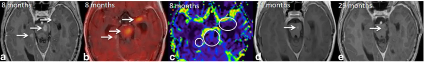
Fig. 2 Follow-up MRI 8 months (a, b), 11 months (c), and 29 months (d) after beginning of complementary proton beam irradiation. a Axial T2-weighted showing an hyperintense lesion within the pons. b Axial T1-weighted after gadolinium showing appearance of abnormal
rim-like enhancement (white arrow) with T2 hypersignal (a). The lesion
shows a low CBV (circle, a) and is hypermetabolic on FET-PET/MRI
(black arrow, b). Follow-up MRI at 11 and 29 months shows almost
disappearance of the lesion (white arrows, c, d) confirming the
diag-nosis of pseudoprogression
Fig. 3 Follow-up MRI 8 months (a–c), 11 months (d), and 29 months
(e) after beginning of complementary proton beam irradiation showing appearance of abnormal rim-like enhancements on T1W after
gadolin-ium within the pons and both temporal lobes (arrows, a). The lesions
show a low CBV (circles, c) and two of them are hypermetabolic on
FET-PET/MRI (arrows, b). Follow-up MRI at 11 and 29 months
shows disappearance of temporal lesions and almost disappearance of
the pontine lesion (white arrows, d, e) confirming the diagnosis of
pseudoprogression
Conflict of interest No competing interest.
References
1. Khuntia D, Tomé WA, Mehta MP (2009) Radiation techniques in
neuro-oncology. Neurotherapeutics 6:487–499
2. Brandsma D, Stalpers L, Taal W, Sminia P, van den Bent MJ (2008) Clinical features, mechanisms, and management of
pseu-doprogression in malignant gliomas. Lancet Oncol 9:453–461
3. Marks JE, Baglan RJ, Prassad SC, Blank WF (1981) Cerebral radionecrosis: incidence and risk in relation to dose, time,
fraction-ation and volume. Int J Radiat Oncol Biol Phys 7:243–252
4. Burger PC, Mahley MS Jr, Dudka L, Vogel FS (1979) The mor-phologic effects of radiation administered therapeutically for
intra-cranial gliomas: a postmortem study of 25 cases. Cancer 44:1256–
1272
5. Bakardjiev AI, Barnes PD, Goumnerova LC, Black PM, Scott RM, Pomeroy SL et al (1996) Magnetic resonance imaging changes after stereotactic radiation therapy for childhood low grade astro-cytoma. Cancer 78:864–873
6. Spreafico F, Gandola L, Marchiano A, Simonetti F, Poggi G, Adduci A et al (2008) Brain magnetic resonance imaging after high-dose chemotherapy and radiotherapy for childhood brain
tumors. Int J Radiat Oncol Biol Phys 70:1011–1019
7. Meyzer C, Dhermain F, Ducreux D, Habrand JL, Varlet P, Sainte-Rose C (2010) A case report of pseudoprogression followed by complete remission after proton-beam irradiation for a low-grade glioma in a teenager: the value of dynamic contrast-enhanced MRI. Radiat Oncol 5:9
8. W. Taal, D. Brandsma, HG de Bruin, J. E. Bromberg, A. T. Swaak-Kragten, W. M. Eijkenboom et al. (2007) The incidence of pseudo-progression in a cohort of malignant glioma patients treated with chemo-radiation with temozolomide. J Clin Oncol 25 (18S) 9. Kang TW, Kim ST, Byun HS, Jeon P, Kim K, Kim H, Lee JI
(2009) Morphological and functional MRI, MRS, perfusion and diffusion changes after radiosurgery of brain metastasis. Eur J Radiol 72:370–380
10. Essig M, Waschkies M, Wenz F, Debus J, Hentrich HR, Knopp MV (2003) Assessment of brain metastases with dynamic susceptibility-weighted contrast-enhanced MR imaging: initial
results. Radiology 228:193–199
11. Hoefnagels FW, Lagerwaard FJ, Sanchez E, Haasbeek CJ, Knol DL, Slotman BJ et al (2009) Radiological progression of cerebral metastases after radiosurgery: assessment of perfusion MRI for differentiating between necrosis and recurrence. J Neurol
256:878–887
12. Pöpperl G, Götz C, Rachinger W, Gildehaus FJ, Tonn JC, Tatsch K
(2004) Value of O-(2-[18F]fluoroethyl)-L-tyrosine PET for the
diagnosis of recurrent glioma. Eur J Nucl Med Mol Imaging
31:1464–1470
13. Terakawa Y, Tsuyuguchi N, Iwai Y, Yamanaka K, Higashiyama S, Takami T et al (2008) Diagnostic accuracy of 11C-methionine PET for differentiation of recurrent brain tumors from radiation necrosis after radiotherapy. J Nucl Med 49:694–699
14. Garibotto V, Haller S, Vargas MI et Al. (2012) Increased uptake of
18F-fluoroethyl-L-tyrosine in radiation-induced brain necrosis
(abstract). Eur J Nucl Med Mol Imaging 39:S382
15. Kim YH, Oh SW, Lim YJ, Park CK, Lee SH, Kang KW et al (2010) Differentiating radiation necrosis from tumor recurrence in high-grade gliomas: assessing the efficacy of 18F-FDG PET, 11C-methionine PET and perfusion MRI. Clin Neurol Neurosurg
112:758–765