HAL Id: hal-02990232
https://hal.archives-ouvertes.fr/hal-02990232
Submitted on 10 Nov 2020HAL is a multi-disciplinary open access archive for the deposit and dissemination of sci-entific research documents, whether they are pub-lished or not. The documents may come from teaching and research institutions in France or abroad, or from public or private research centers.
L’archive ouverte pluridisciplinaire HAL, est destinée au dépôt et à la diffusion de documents scientifiques de niveau recherche, publiés ou non, émanant des établissements d’enseignement et de recherche français ou étrangers, des laboratoires publics ou privés.
Biotinylated non-ionic amphipols for GPCR ligands
screening
Michaël Bosco, Marjorie Damian, Vinay Chauhan, Mélanie Roche, Pierre
Guillet, Jean-Alain Fehrentz, Françoise Bonnete, Ange Polidori, Jean-Louis
Banères, Grégory Durand
To cite this version:
Michaël Bosco, Marjorie Damian, Vinay Chauhan, Mélanie Roche, Pierre Guillet, et al.. Biotiny-lated non-ionic amphipols for GPCR ligands screening. Methods, Elsevier, 2020, 180, pp.69-78. �10.1016/j.ymeth.2020.06.001�. �hal-02990232�
Biotinylated non-ionic amphipols for GPCR Ligands
Screening
Michaël Bosco,a Marjorie Damian,b Vinay Chauhan,a Mélanie Roche,b Pierre Guillet,a Jean-Alain Fehrentz,b Françoise Bonneté,a,c Ange Polidori,a Jean-Louis Banères,b Grégory Duranda*
aInstitut des Biomolécules Max Mousseron (UMR 5247 UM-CNRS-ENSCM) & Avignon University, Equipe Chimie Bioorganique et Systèmes amphiphiles ; 301 rue Baruch de Spinoza – 84916 Avignon cedex 9 (France) ;
bInstitut des Biomolécules Max Mousseron (UMR 5247 UM-CNRS-ENSCM); Faculté de pharmacie, 15, avenue Charles Flahault, 34000 Montpellier, France ;
CCurrent address: Institut de Biologie Physico-Chimique (UMR 7099 CNRS Université de Paris), Laboratoire de Biologie Physico-Chimique des Protéines Membranaires, 13 rue Pierre et Marie Curie, F75005 Paris, France
Corresponding Author.
Abstract
We present herein the synthesis of biotin-functionalized polymers (BNAPols) that have
been developed for the fixation of membrane proteins (MPs) onto surfaces. BNAPols
were synthesized by free-radical polymerization of a
tris(hydroxymethyl)acrylamidomethane (THAM)-derived amphiphilic monomer in the
presence of a thiol-based transfer agent with an azido group. Then a
Huisgen-cycloaddition reaction was performed with Biotin-(PEG)8-alkyne that resulted in
formation of the biotinylated polymers. The designed structure of BNAPols was
confirmed by NMR spectroscopy, and a HABA/avidin assay was used for estimating the
percentage of biotin grafted on the polymer end chain. The colloidal characterization of
these biotin-functionalized polymers was done using both dynamic light scattering (DLS)
and small angle X-ray scattering (SAXS) techniques. These BNAPols were used to
stabilize a model G protein-coupled receptor (GPCR), the human Growth Hormone
Secretagogue Receptor (GHSR), out of its membrane environment. Subsequent
immobilization of the BNAPols:GHSR complex onto a streptavidin-coated surface
allowed screening of ligands based both on their ability to bind the immobilized receptor
and to trigger GHSR conformational changes using the fluorescence energy transfer
(FRET)-based assay. This opens the way to the use of biotinylated NAPols to immobilize
functional, unmodified, membrane proteins, providing original sensor devices for
1. Introduction
Membrane proteins (MPs) play fundamental roles in biology because they control
communication and material transfer within and between living cells and their environment.
Traditionally, MPs are solubilized and studied in detergent micelles, although micellar
assemblies are poor mimics of biological membranes. Detergents have major drawbacks for
manipulating isolated membrane proteins that include denaturing properties and a negative
impact on protein dynamics [1]. Alternative approaches have been developed to circumvent the
destabilizing/denaturing effects of detergents. This includes facial amphiphiles [2],
neopentyl-based detergents [3] as well as cyclic [4] and fluorinated detergents [5] to name but a few.
Heterogeneous systems consisting in amphiphilic polymers have also been developed in
parallel and have shown very promising properties. Among them one can cite amphipols
(APols) [6] or styrene maleic acid (SMA) [7].
APols are amphipathic polymers that were designed and validated as mild alternatives
to classical detergents. Accordingly, most integral membrane proteins are much more stable in
APols than they are in detergent solutions. The first designed polyacrylate-based A8-35 [6] has
been so far the most widely used amphipol [8]. However, acidic pH or the presence of
multivalent cations can result in A8-35 aggregation. Besides, the ionic character of A8-35 can
be a major drawback in the context of screening assays, as non-specific interactions between
the polymer and charged compounds cannot be ruled out. These constraints prompted the
development of chemically different APols (reviewed in reference [9], including zwitterionic
APols [10], sulfonated APols (SAPols) [11], and nonionic amphipols (NAPols) [12-13]. The
stabilizing efficiency of APols has been shown to inversely correlate with the charge density of
the polymers, the NAPols being the most stabilizing derivatives [14]. NAPols therefore
represent new, milder tools for the manipulation of membrane proteins.
half of all drug targets [16]. Therefore, the screening of ligands for drug discovery has been of
increasing interest over the recent years. In many cases, screening of specific ligands with
isolated proteins involves their immobilization onto surfaces. For membrane proteins,
functional immobilization can be carried out by covering bare and modified surfaces with native
and reconstituted membrane fragments or by using proteins with engineered tags. This have
been extensively reviewed in reference [17]. More recently, functionalized β-sheet peptides
with stabilizing properties were successfully used for the immobilization of MPs onto solid
supports [18]. Covalent immobilization of a MP on silica-based carriers was also reported [19].
In this context, amphipols offer an alternative and appear as highly versatile means for
membrane protein immobilization [20-22]. Indeed, by grafting affinity tags on the polymer,
MP/APol complexes have been successfully attached onto solid supports. The first example of
such a functionalized amphipol was the biotinylated derivative of A8-35 called BAPol (Figure
1), which was used to immobilize a model membrane protein for SPR and fluorescence
microscopy studies [20]. Functionalization of A8-35 was further extended to poly-histidine and
Figure 1. Structure of BAPol (biotinylated amphipol with w=0.7-1.4%, x=30-35%,
y=21-25% and z=36-41%), B-PCAPol (biotinylated phosphorylcholine amphipol with x=29%, y=29% and z=42%) and BNAPol (biotinylated non-ionic amphipol).
However, one of the drawbacks of tagged A8-35 is the random distribution of the tag
along the polymer chain, as the synthesis protocol does not allow a control of the position of
the tag on the polymer chain. To relieve this drawback, biotinylated phosphorylcholine
amphipols B-PCAPols (Figure 1) have been synthesized by RAFT polymerization in the
presence of a transfer agent onto which the biotin group was further grafted [24]. This allowed
attaching only one biotin group at the end of the polymer chain. B-PCAPols have been
successfully used to study the interaction between E. coli outer membrane protein FhuA and a
bacteriophage tail protein by SPR and Electrochemical Impedance Spectroscopy [24].
However, synthesis of these polymers is quite complex, and this limits large-scale preparation.
In addition, ionic- and phosphocholine-based amphipols have a high electrostatic charge
density that can induce non-specific binding or electrostatic repulsion [25]. This can be a
limitation when using amphipol-trapped proteins for analyzing protein:ligand binding events
where ionic interactions can have a prominent role. Development of non-ionic tagged
of an efficient, robust and versatile biosensor. We report herein the synthesis of the first series
of biotinylated NAPols and the study of their colloidal properties by Dynamic Light Scattering
(DLS) and Small Angle X-Ray Scattering (SAXS). These biotinylated NAPols were next
investigated for their ability to allow immobilization onto a surface of a model membrane
protein from the G protein-coupled receptor (GPCR) family. In particular, we investigated
whether the polymer preserved the pharmacological properties of the receptor even after
attachment to a functionalized surface and whether the immobilized receptor could then be used
for subsequent screening of ligands.
2. Materials & methods
2.1. Synthesis. All starting materials were commercially available and were used without
further purification. 1-ethoxycarbonyl-2-ethoxy-1,2-dihydroquinoline (EEDQ), triethylsilane
and sodium chloride were purchased from Acros Organics. Triphenylmethyl chloride (TrCl)
and trifluoroacetic acid were purchased from TCI. 3-mercaptopropionic acid,
tris(hydroxymethyl)aminomethane, 2-bromoethylisocyanate, diisopropylethylamine (DIEA),
triethylamine (Et3N), sodium azide (NaN3), sodium bicarbonate (NaHCO3), sodium sulfate
(Na2SO4) and copper iodide were purchased from Sigma-Aldrich. Biotin-(PEG)8-alkyne was
purchased from SiChem (Germany). Monomer DGC11 i.e. N-(1,1-(2 ,3 ,4 ,6 -Tetra-O-acetyl-β-D-glucopyranosyloxymethyl)-1-(undecylcarbamoyloxymethyl)-methyl)acrylamide was
synthesized according to earlier reported procedure [26]. All solvents were of reagent grade and
used as received unless otherwise indicated. Tetrahydrofuran (THF) was dried over sodium and
benzophenone, methanol (MeOH) over sodium, dichloromethane (CH2Cl2) over CaH2 under
argon atmosphere. Commercial anhydrous N,N-Dimethylformamide (DMF) was stored in the
presence of activated molecular sieves 3 Å. Azobisisobutyronitrile (AIBN) was recrystallized
twice from ethanol. The progress of the reactions was monitored by thin layer chromatography
exposure to ultraviolet light (254 nm) or by spraying with sulfuric acid (5% ethanol) and/or
ninhydrin (5% ethanol), followed by heating at ~150°C. Flash chromatography purifications
were carried out on silica gel (pore size 60 Å, 230-400 mesh particle size, 40-63 μm particle
size). Size exclusion chromatography purifications were carried out on Sephadex LH-20 resin.
1H and 13C spectra were recorded on a Bruker AC 250 and AC 400 spectrometer at 250 and 400
MHz for 1H and 62.86 and 100 MHz for 13C respectively. Chemical shifts (δ values) were
reported in ppm downfield from internal residual solvent as a heteronuclear reference. HR-MS
(ESI+) was determined on a QStar Elite mass spectrometer. UV-Visible spectra were recorded
on a Cary Win Varian Spectrophotometer with a double-compartment quartz cell of 10-mm
length (Suprasil).
2.1.1. Synthesis of 3-(tritylthio)propionic acid (2). To a solution of triphenylmethyl chloride
(17.6 g, 63.1 mmol) in dichloromethane (150 mL) under argon atmosphere,
3-mercaptopropionic acid (6.0 mL, 69.4 mmol, 1.1 eq.) was added dropwise. The precipitate
formed was collected by filtration and dried under vacuum to give compound 2 (20.0 g, 57.4
mmol, 90%) as a white powder. 1H NMR (250 MHz, DMSO) δ 7.56 – 6.88 (m, 15H), 2.29 (t,
J = 6.6 Hz, 2H), 2.17 (t, J = 6.6 Hz, 2H). 13C NMR (62.86 MHz, DMSO) δ 172.8, 144.4, 129.1,
128.1, 126.8, 66.2, 32.9, 26.7.
2.1.2. Synthesis of
N-(1,3-dihydroxy-2-(hydroxymethyl)propan-2-yl)-3-(tritylthio)propanamide (3). To a solution of 3-tritylsulfanyl-propionic acid 2 (17.2 g, 49.2
mmol) in ethanol (80 mL) were added tris(hydroxymethyl)aminomethane (6.6 g, 54.1 mmol,
1.1 eq.) and EDDQ (14.6 g, 59.1 mmol, 1.2 eq.). After being stirred at 55°C for 16 hours, the
reaction mixture was concentrated under vacuum. The crude mixture was recrystallized from
ethanol to give compound 3 (19.1 g, 42.3 mmol, 86%) as a white solid. 1H NMR (250 MHz,
1.94 (t, J = 7.0 Hz, 2H); 13C NMR (62.86 MHz, CDCl
3) δ 173.1, 144.6, 129.7, 128.1, 126.9,
67.1, 62.0, 61.9, 35.9, 27.9.
2.1.3. Synthesis of 2-((((2-azidoethyl)carbamoyl)oxy)methyl)-2-(3-(tritylthio)propanamido)propane-1,3-diyl dibenzoate (4). To a mixture of dimethylformamide (10 mL) and diisopropylethylamine (770 µL, 3.32 mmol, 1 eq.) compound
3 (2.0 g, 4.43 mmol, 1.33 eq.) was added portion wise, then 2-bromoethylisocyanate (300 µL,
3.31 mmol, 1 eq.) was added dropwise. After stirring for 4h, sodium azide (1.07 g, 16.5 mmol,
5 eq.) was added to the reaction mixture and the stirring was continued for 16 hours at 50°C.
The solvent was evaporated under vacuum and the resulting residue was solubilized in
dichloromethane (100 mL). The organic layer was washed with a saturated solution of sodium
bicarbonate (100 mL), brine (100 mL), then dried over sodium sulfate, filtered off and
evaporated under vacuum to afford compound 4a. Compound 4a was directly used in the next
step without purification. To a solution of compound 4a in dichloromethane (25 mL),
triethylamine (4.63 mL, 33.3 mmol, 10 eq.) and benzoyl chloride (2.6 mL, 22.2 mmol, 6.7 eq.)
was added dropwise. After being stirred for 16h, the reaction was diluted with dichloromethane
(75 mL). The organic layer was washed with water (100 mL) and brine (100 mL), dried over
sodium sulfate, filtered off and evaporated under vacuum. The crude mixture was purified by
flash chromatography on silica gel, eluting with ethylacetate/cyclohexane (1:9 to 3:7, v/v) to
afford compound 4 (1.36 g, 1.77 mmol, 53%) as a colorless oil. Rf 0.37
(ethylacetate/cyclohexane, 3:7, v/v); 1H NMR (250 MHz, CDCl3) δ 8.16 – 6.96 (m, 25H), 6.29
(s, 1H), 5.12 (t, J = 5.9 Hz, 1H), 4.77 (s, 4H), 4.57 (s, 2H), 3.32 (t, J = 7 Hz, 2H), 3.25 – 3.06
(m, 2H), 2.49 (t, J = 7.0 Hz, 2H), 2.00 (t, J = 7.0 Hz, 2H); 13C NMR (62.86 MHz, CDCl3) δ
171.6, 166.2, 156.2, 144.6, 133.5, 130.2, 129.8, 129.6, 129.4, 128.6, 128.1, 126.8, 67.0, 64.1,
2.1.4. Synthesis of 2-((((2-azidoethyl)carbamoyl)oxy)methyl)-2-(3-mercaptopropanamido)propane-1,3-diyl dibenzoate (5). To a solution of compound 4 (0.4
g, 518 µmol) in a 2:8 mixture of trifluoroacetic acid and dichloromethane (4 mL, v/v)
triethylsilane (92 µL, 570 µmol, 1.1 eq.) was added dropwise under argon atmosphere at 0°C.
After being stirred at room temperature for 1 hour, the solvents were evaporated under vacuum.
The crude mixture was purified by flash chromatography on silica gel, eluting with
ethylacetate/cyclohexane (2:8 to 3:7, v/v) to afford compound 5 (0.21 g, 396 µmol, 76%) as a
colorless oil. Rf 0.57 (ethylacetate/cyclohexane, 4/6, v/v); 1H NMR (250 MHz, CDCl3) δ 8.13
– 7.30 (m, 10H), 6.78 (s, 1H), 5.47 (t, J = 5.7 Hz, 1H), 4.83 (s, 4H), 4.63 (s, 2H), 3.44 – 3.24
(m, 4H), 2.83 – 2.65 (m, 2H), 2.51 (t, J = 6.6 Hz, 2H), 1.59 (t, J = 8.3 Hz, 1H); 13C NMR (62.86
MHz, CDCl3) δ 171.4, 166.2, 156.5, 133.5, 129.8, 129.4, 128.6, 64.3, 63.3, 59.2, 50.9, 40.9,
40.6, 20.3; ESI-MS: Calcd for C24H28N5O7S+ [M+H]+: 530.2. Found: 530.3; ESI-HRMS: Calcd
for C24H28N5O7S+ [M+H]+: 530.1709. Found: 530.1705.
2.1.5. Synthesis of BNAPol. The synthesis of biotinylated NAPoL involved three steps. (i) NAPol-N3. DGC11 (1.0 g, 0.97 mmol, 15.0 eq.), compound 5 (34.2 mg, 64.6 µmol, 1.0 eq.)
and AIBN (5.3 mg, 32.3 µmol, 0.5 eq.) were dissolved in dry THF (1.0 mL) in a Schlenk tube.
This solution was degassed by 5 freeze-pump-thaw cycles. Then the reaction mixture was
refluxed under argon for 4 hours. The reaction mixture was concentrated under vacuum and the
crude was purified by gravity size-exclusion chromatography on SephadexTM LH-20 resin
eluting with a 1:1 (v/v) MeOH/CH2Cl2 mixture. The solvent was evaporated under vacuum to
give the protected NAPol-N3 as a white powder. Assuming that the conversion is complete
(confirmed by 1H-NMR of the reaction crude), the polymerization yield was determined from
the ratio of the amount determined by gravimetric analysis after the purification process
(Sephadex) versus the theoretical expected amount of polymer. 0.85 g of NAPol-N3 (Batch
aromatic), 5.2-4.1 (m, glucose unit), 3.1 (m, -NH-CH2), 2.1-1.9 (bs, -OCOCH3), 1.7-1.2 (m,
CH2 of the alkyl chain), 0.8 (t, CH3 of the alkyl chain).
(ii) BNAPol. To a solution of NAPol-N3 (0.200 g, 11.2 µmol, 1 eq.) in dry THF (5 mL),
diisopropylethylamine (2.63 µL, 15.2 µmol, 1.4 eq.), copper iodide (1.4 mg, 7.6 µmol, 0.7 eq.)
and Biotin-(PEG)8-alkyne (9.6 mg, 15.2 µmol, 1.4 eq.) was added. Then the reaction mixture
was refluxed for 36 hours under argon. The reaction mixture was concentrated under vacuum
and the resulting crude polymer was purified by gravity size-exclusion chromatography on
SephadexTM LH-20 resin eluting with a 1:1 (v/v) MeOH/CH2Cl2 mixture. The solvent was
evaporated under vacuum to give the protected BNAPol as a white powder (0.170 g, 80%). 1H
NMR (400 MHz, CDCl3) δ 8.1-7.4 (m, aromatic), 5.2-4.1 (m, glucose unit), 3.6 (m, PEG), 3.1
(m, -NH-CH2), 2.1-1.9 (bs, -OCOCH3), 1.7-1.2 (m, CH2 of the alkyl chain), 0.8 (t, CH3 of the
alkyl chain).
(iii) Deprotection of BNAPol. To a solution of protected BNAPol (0.170 g) in dry methanol (20
mL) under argon atmosphere, a catalytic amount of sodium methoxide was added and the
mixture was stirred overnight at room temperature. The reaction solution was neutralized by
addition of a spatula of acidic resin (IRC 50) (pH8) and the solution was shaken for 15 minutes. The solution was filtered off, and then concentrated under vacuum. The crude polymer
was dissolved in methanol (10 mL) and precipitated into cold ether (200 mL). The precipitate
was filtered off and the solvent was evaporated under vacuum. Finally, the resulting BNAPol
was solubilized in milliQ water at 10 g/L, filtered through a 0.45 µm PVDF syringe filter and
then subjected for 24 hours and under continuous stirring to dialysis against milliQ water
(membrane MW Cut Off 6-8 kDa). The resulting dialyzed solution was freeze-dried to give the
BNAPol (0.092 g, 80%, batch NV12-02) as a white powder. 1H NMR (400 MHz, MeOD) δ
5.2-4.1 (m, glucose unit), 3.1 (m, -NH-CH2 ), 1.7-1.2 (m, CH2 of the alkyl chain), 0.8 (t, CH3
2.2. Polymer analysis.
2.2.1. 1H NMR. The number-average molecular weight of polymers was obtained by 1H NMR
calibration, by comparing the integral area of protons from the phenyl groups at 7.4-8.1 ppm with those of the terminal methyl protons at δ 0.8 ppm of each amphiphilic monomer
constituting the polymer backbone. NAPol-N3 and BNAPols were analyzed at 5 g/L in CDCl3.
For BNAPols the appearance of peak at 3.6 ppm corresponds to PEGylated protons of biotin
that was further estimated by HABA analysis.
2.2.2. HABA test [27]. In a 1 mL cuvette, 0.9 mL of the red/orange solution of HABA/avidin
reagent was analyzed. The absorbance was measured at λ = 500 nm i.e. A492. Subsequently, 0.1
mL of biotinylated polymer solution was added into of the HABA/Avidin solution and then
A500 was measured. The concentration of polymer was adjusted (from 1 to 5 g/L) so as to keep
the decrease in the absorbance between 0.1 and 0.4. 2 minutes later A500 was measured again
in order to make sure that the measurement was the same. Each measurement was repeated
three times. The data were treated with the online HABA calculator from ThermoFischer
Scientific and were expressed as a percentage of biotine per polymer chain.
2.3. Colloidal characterization.
2.3.1. Dynamic light scattering. The hydrodynamic particle size distribution and
polydispersity of NAPols, BNAPols and deprotected monomer DGC11 were determined using
a Zetasizer Nano-S model 1600 (Malvern Instruments Ltd., UK) equipped with a He-Ne laser
(λ = 633 nm, 4.0 mW). The time-dependent correlation function of the scattered light intensity
was measured at a scattering angle of 173° relative to the laser source. The stock solutions were
prepared at 5.0 g/L in Milli-Q water and were stored overnight at room temperature. On the day
of the experiment the solutions were centrifuged at room temperature (10000 rpm) for 30
concentration, and the size of the particles was measured 1 h after filtration. Other conditions
were as reported elsewhere [28].
2.3.2. Small-angle X-ray scattering. Self-assemblies were characterized on the bioSAXS
beamline BM29 at the European Synchrotron Radiation Facility (Grenoble, France). The
sample–detector distance was 2.85 m and the X-ray wavelength λ = 0.1008 nm leading to a
q-range of 0.032–4.55 nm-1. The beamline was equipped with a 2D-detector (Pilatus 1M) and an
automated sample changer. A stock solution was prepared at 10.0 g/L in Milli-Q water and a
concentration series prepared by dilution down to 2.5 g/L. The sample storage and measurement
temperatures were fixed at 20.0°C. To prevent radiation damage during the scattering
experiments, data were collected in 10 successive 2-s frames, and the solution was moved in
the capillary during exposure. All data reduction and analysis were performed using a standard
procedure via the ISPyB interface [29] and the program package PRIMUS [30].
2.4. Biochemical validation.
2.4.1 GHSR preparation and assembly in BNAPols. The ghrelin receptor was produced and
reconstituted in NAPols and in NAPols/BNAPols mixtures using the protocol previously
described [14]. Briefly, the ghrelin receptor was expressed in E. coli inclusion bodies as a fusion
protein with the 5 integrin fragment [31]. The receptor was then purified under denaturing conditions, i.e. in the presence of 0.8% SDS. NAPols or NAPols/BNAPols mixtures at different
functionalized-to-non functionalized polymer weight ratios (see text) were added at a ratio of
10g of total polymer per gram of SDS-unfolded protein. Asolectin (0.2:1 (w/w) lipid:amphipol
ratio) and cholesteryl hemisuccinate (0.02%, w/v) were then added. After incubation at room
temperature for 30 min, folding was initiated by precipitating dodecyl sulfate (SDS) with 200
mM KCl [32]. After 30 min, the precipitate was removed by two 10 min centrifugation runs at
16,100g and the supernatant was dialyzed against a 50 mM potassium phosphate, 150 mM KCl,
with a GHSR ligand immobilized on a chromatography column [33]. To this end, the protein
after assembly into amphipols was loaded on the column and the proteins bound to the matrix
were subsequently recovered by washing the column with a 25 mM HEPES, 150 mM KCl, pH
7.5 buffer containing 0.5 mM of the neutral antagonist JMV2959. The latter was removed
through extensive dialysis in a 25 mM HEPES, 150 mM KCl, 0.5 mM EDTA, pH 7.5 buffer.
Monomeric GHSR was finally separated from protein aggregates through a size-exclusion
chromatography step on a Superdex 200 increase 10/300 GL column (GE Healthcare) using a
25 mM Na-HEPES, 150 mM KCl, 0.5 mM EDTA, pH 7.4 buffer as the eluent. For ligand
binding assays, the ghrelin receptor was labeled at its N-terminus with Lumi4-NHS (CisBio),
as previously described [33].
2.4.3. Receptor immobilization. The ghrelin receptor was immobilized onto 96-well
streptavidin coated high-capacity plates (Pierce) following manufacturer instructions. Briefly,
each well was washed with 200 µL of a 25 mM Tris-HCl, 150 mM NaCl, pH 7.5, 0.1% BSA
buffer. The receptor was then added and incubated for two hours at room temperature and the
plates washed. To optimize the coating efficiency, different dilutions of biotin-tagged receptor
preparation were tested ranging from 1 to 0.001 g/L.
2.4.4. Ligand binding assays. Ligand binding was monitored through the homogenous
time-resolved fluorescence (HTRF) signal between GHSR labeled with Lumi4-NHS at its
N-terminus (see above) and a ghrelin peptide labeled with dy647 at its C-N-terminus. The
immobilized receptor was incubated with the labeled ghrelin peptide (50 nM concentration
range) and the compounds to be tested. Plates were then incubated at 4°C for 3 h before signal
detection. The signal was detected using a fluorescence microplate reader (PHERAstar plus,
BMG Labtech) equipped with a HTRF optic module allowing a donor excitation at 337 nm and
a signal collection at 620 nm.
3.1 Synthesis of BNAPols.
Functionalization of NAPols was achieved by post-functionalization of the
polymer, which consists in the grafting of a tag of interest at the end of the polymer chain
once the polymer is synthesized. To achieve such an end chain post-functionalization, a
thiol-based transfer agent also called telogen, bearing an azido group, was first
synthesized as described in Scheme 1. 3-Mercapto propionic acid was first protected by
trityl chloride to afford compound 2. Tris(hydroxymethyl)aminomethane was next
condensated to compound 2 in the presence of EEDQ as a coupling agent to afford
compound 3. Bromoethylisocyanate was next put in reaction with compound 3 in the
presence of diisopropylethylamine, then sodium azide was added to afford compound 4a.
Compound 4a was directly used for protection of the two hydroxyl groups by benzoyl
groups to lead after purification by flash chromatography to compound 4. Finally,
deprotection of the trityl group led to the transfer agent 5.
Scheme 1. a) TrCl, CH2Cl2, RT, 3.5 h; b) Tris(hydroxymethyl)aminomethane, EDDQ, EtOH,
55°C, 16 h; c) BrCH2CH2N=C=O, DIEA, DMF; d) NaN3, 50°C; e) BzCl, Et3N, CH2Cl2, RT,
14 h; f) Et3SiH, CF3COOH, CH2Cl2, 4 h, 0°C.
BNAPols were therefore synthesized by free-radical telomerization of the amphiphilic
monomer DGC11 in the presence of the azido transfer agent 5 in refluxing THF and in the
polymerization (DPn) were synthesized by varying the ratio of transfer agent (TA) and
amphiphilic monomers (Table 1). The reaction was monitored by thin layer chromatography
and carried on until full disappearance of the monomer spot. The number-average degree of
polymerization (DPn) was set by the initial ratio of DGC11 to transfer agent TA (R0),
considering, in a first approximation, the transfer constant of 5 to DGC11 (Ct) to be close to 1
[34]. The observed DPn was rather close to the initial ratio (Table 1) suggesting that in THF Ct
is close to 1, as usually observed during homotelomerization of THAM monomers [35-37].
Table 1. Represents the initial ratio of monomer (R0), number-average degree of
polymerization (DPn), percentage of biotin grafting, number-average molecular weight (Mn)
for azido-functionalized NAPols and their corresponding biotinylated version (BNAPol).
Polymers R0 DPn Biotin grafting (%) Number-average Molecular Weight. (kDa) Name Batch
NAPol 20 20 N/A 14.1 NA14 MB105
60 52 N/A 36.5 NA36 MB136
20 24 N/A 17.0 NA17 MAS-C68762-EF
NAPol-N3(OAc)a BNAPol BNAPol 20 20 N/A 17 40 21.1 14.9 14.9 N/A BNA15_17 BNA15_40 MB134 MB156 MB160 NAPol-N3(OAc)a BNAPol 60 50 N/A 31 52.1 35.9 N/A BN36_31 MB135 MB159 NAPol-N3(OAc)a BNAPol BNAPol 15 17 N/A 40 14 17.9 12.6 12.6 N/A BNA13_40 BNA13_14 NV08 NV12-02 NV12-03 NAPol-N3(OAc)a BNAPol 15 20 N/A 28 21.1 14.9 N/A BNA15_28 NV08-1 NV12-04 aProtected NAPol from which biotin was grafted before removal of the acetyl groups.
The resulting azido functionalized NAPol was purified by size-exclusion
chromatography on sephadex LH20 resin and the chemical structure was confirmed by 1H NMR
spectroscopy. In the second step NAPol-N3 was reacted with the Biotin-(PEG)8-alkyne in THF
through a Huisgen-cycloaddition reaction in the presence of copper and the resulting
biotinylated polymer was purified by size-exclusion chromatography. Then the final step
consisted in the removal of the benzoyl and acetyl ester groups using the Zemplén method in
BNAPol. BNAPol was further purified by dialysis through cellulose-based membrane for 24
hours to lead after freeze drying to a dry powder of pure BNAPol.
3.2. Polymer analysis.
Figure 2. Represents the NMR spectra of: (top) pure biotinylated polymer (5 g/L in ), (middle)
NAPol-N3 (5 g/L in CDCl3) and (bottom) Biotin-(PEG)8-alkyne (5 g/L in DMSO-d6). Yellow
circles designate the PEG chain protons in biotinylated polymer (OAc) & Biotin-(PEG)8
-alkyne. Green circles designate aromatic protons of the benzoyl groups.
As shown in Figure 2, the presence of the biotin group on the polymer chain was
confirmed by 1H NMR. The PEGylated chain of the Biotin-(PEG)8-alkyne was easily monitored
by 1H NMR and was also observed in BNAPols at δ 3.54 ppm, indicating the grafting of biotin
to the polymer. The presence of the benzoyl protons both in the protected N3-NAPol and its
polymer chain (Table 1). The grafting of the biotin group onto the polymer chain was further
estimated using spectrophotometric avidin - HABA test and was found to be between 14-40%
(Table 1). The binding of HABA to avidin and the ability of biotin to displace HABA in
stoichiometric proportions was monitored. A standard solution was titrated with BNAPol
solutions and the decrease in absorption spectra of HABA/Avidin solutions witness the
presence of biotin on the polymer (Figure 3). Throughout the present article, BNAPols are
denoted by a short name reflecting their chemical structure: the letters BNA stand for
"biotinylated non-ionic amphiphilic polymer", followed by the average molecular weight
expressed in kDa and the percentage of biotin group grafted onto the polymer chain. Batch
numbers are given in Table 1.
Figure 3. Absorption spectra of avidin-HABA solution (standard) with addition of two different
BNAPols. BNAPols solution were prepared at 5 mg/mL for BNA15_17 and at 1 mg/mL for BNA15_40.
Scheme 2. a) DGC11 (15 eq.), 5 (1 eq.), AIBN (0.5 eq.), THF, reflux, 16h, 78%; b) Biotin-(PEG)8-alkyne (2 eq.), NAPol-N3 (1eq.), CuI (1
3.3. Colloidal Characterization.
The colloidal properties of BNAPols were determined both by dynamic light scattering
(DLS) and Small-angle X ray scattering (SAXS) methods in aqueous solution. For the sake of
comparison, the deprotected monomer DGC11 was included in the study as well as two NAPols
with no biotin group. Data are reported in table 2.
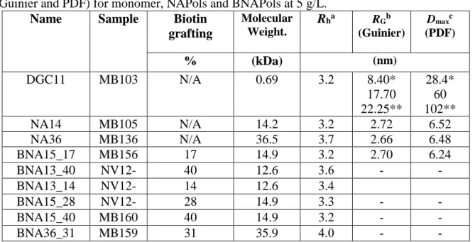
Table 2. Structural parameters obtained from DLS (hydrodynamic radius) and from SAXS
(Guinier and PDF) for monomer, NAPols and BNAPols at 5 g/L.
Name Sample Biotin grafting Molecular Weight. Rha RGb (Guinier) Dmaxc (PDF) % (kDa) (nm) DGC11 MB103 N/A 0.69 3.2 8.40* 17.70 22.25** 28.4* 60 102** NA14 MB105 N/A 14.2 3.2 2.72 6.52 NA36 MB136 N/A 36.5 3.7 2.66 6.48 BNA15_17 MB156 17 14.9 3.2 2.70 6.24 BNA13_40 NV12-02 40 12.6 3.6 - - BNA13_14 NV12-03 14 12.6 3.4 BNA15_28 NV12-04 28 14.9 3.3 - - BNA15_40 MB160 40 14.9 3.2 - - BNA36_31 MB159 31 35.9 4.0 - -
aHydrodynamic radius, values are the average of 5 experiments and are given ± 0.1 nm bRadius of gyration from SAXS, values are given ± 0.1 nm
cMaximum diameter from pair distribution fnctions (D
max), values are given ± 0.2 nm.
*2.5 g/L ; ** 10g/L
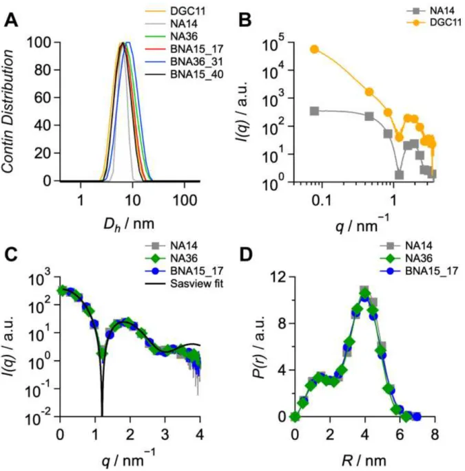
3.3.1. Dynamic light scattering. DLS measurements were carried out at 5 g/L. The
morphology of BNAPol aggregates in aqueous solution indicates the formation of small size
aggregates having hydrodynamic diameter ranging from 6.4 to 8.0 nm (Figure 4). A slight
increase in the hydrodynamic diameter is observed when the DPn increases while the percentage
of biotin grafted seems to have no particular effect on the self-aggregation. This is in full
agreement with the previous report on the classical NAPols where hydrodynamic diameter of
about 6 nm were observed.
3.3.2. Small-angle X-ray scattering. Self-assemblies were further characterized by SAXS. The
approximation LnI(q) = LnI(0) − (qRg)2/3, assuming that qRg < 1. The pair distribution
functions (PDF) and the maximum particle dimensions Dmax were determined by inverse
Fourier transformation of scattering intensity I(q) using the program GNOM [38], to evaluate
the geometry of the micelles. Finally, the SAXS data were fitted using SasView software
(http://www.sasview.org/).
Figure 4. (A) hydrodynamic volume distribution of DGC11, NAPols and BNAPols in aqueous
phase at 5 g/L. (B) SAXS patterns of NAPol (DPn 20) and DGC11 assemblies at 5 g/L (the
curves for each series are shifted for more clarity). (C) SAXS patterns of NAPol (DPn 20
■
and50 ) and BNAPol (DPn 20
●
) at 5 g/L superimposed with a SASVIEW fit of a core-shellsphere model (black line). (D) Pair distribution function of NAPols (DPn 20
■
and 50 ) andAs already suggested by DLS, SAXS experiments showed that whatever the DPn,
NAPols and BNAPols adopt similar spherical shapes with radii of gyration of 2.70 nm and
maximal dimensions of 6.2 to 6.5 nm. The shape of the PDF (Figure 4C) suggests a core-shell
sphere model, which fits well the experimental SAXS curves (Figure 4B). The core radius fitted
with SasView equal to 0.7nm with the hydrophilic thickness of 2.42 nm are consistent with the
value of Dmax and hydrodynamic radius obtained from DLS. The presence of a biotin group on
the polymer chain does not alter the shape and dimensions of the self-assemblies. While the
polymers do not show any concentration effect on the shape of the spherical assemblies, the
monomer micelles exhibit an elliptic core-shell shape that enlarges with concentration to an
elongated core-shell cylinder (Table 2).
3.4. Biochemical validation.
3.4.1. GHSR assembly into BNAPols. To assess whether BNAPols could be efficiently used
for stabilizing the native fold of membrane proteins (MP) and their subsequent immobilization
on functionalized surfaces, we used here the Growth Hormone Secretagogue Receptor GHSR
of ghrelin as a model. Besides being a typical MP, GHSR is a model for rhodopsin-like GPCRs,
a class of most challenging proteins because of their intrinsic instability out of the membrane.
We have previously demonstrated that GHSR could be efficiently refolded in NAPols [14]. The
same folding protocol, based on SDS precipitation in the presence of the polymer, was used
here with BNAPols (see experimental section).
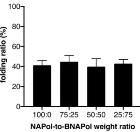
To first assess the impact of the biotin moiety on the folding efficiency, GHSR was
folded in non ionic amphipols at varying NAPol-to-BNAPol ratios while keeping the
protein-to-polymer ratio identical (1:10, w/w). As shown in Figure 5, the yield of functional receptor
after amphipol-mediated folding was very similar whatever the ratio of non
does not significantly affect the ability of NAPols to fold the ghrelin receptor to its native state.
To reach a sufficient immobilization rate, a constant 1:1 NAPol-to-BNAPol ratio was used
throughout this work. Such mixtures were successfully used with the functionalized A8-35
amphipol. [20] Such mixtures allowed an efficient immobilization of functional membrane
proteins on surfaces. Another consideration for using mixtures of functionalized and
non-functionalized amphipols is that multiplying the attachment points on the surface for a single
protein-amphipol complex may be detrimental to its dynamics and therefore to the functional
analysis (dynamics are essential in the binding process in the case of GPCRs).
Figure 5. Impact of NAPol-to-BNAPol ratio on the ability to fold GHSR. Polymers NA16 and
BNA15_40 were used. Folding of GHSR from its SDS-state was carried out as described in the experimental section at varying NAPol-to-BNAPol weight ratios. Data are presented as the mean ± SEM of three experiments.
3.4.2. GHSR immobilization. As GHSR could be refolded in BNAPols, we subsequently
tested whether the receptor:polymer complex could be immobilized onto streptavidin plates.
Immobilization was carried out under standard conditions (see experimental section). We
estimated the amount of receptor immobilized under such conditions. To this end, the receptor
was labeled at its N-terminus with Lumi4-Tb NHS and the emission intensity recovered in the
signal initially present was recovered after the immobilization process. This indicates that about
80% of the receptor could be immobilized on the streptavidin plates under these conditions.
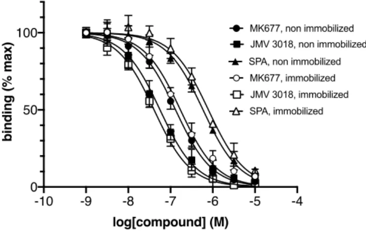
We then analyzed whether the immobilization process affected the receptor
ligand-binding properties. GHSR ligand-ligand-binding assays were performed using the fluorescence energy
transfer (FRET)-based assay we previously developed [33]. This assay is based on measuring
the FRET signal between the immobilized receptor labeled with lumi4-Tb at its N-terminus and
ghrelin labeled with dy647 at its C-terminus in the presence of varying concentrations in a
competing compound. Three pharmacologically-distinct compounds were tested here, i.e. the
full agonist MK 0677, the antagonist JMV 3018 and the inverse agonist SPA. As shown in
Figure 6, the competition profile obtained for all three compounds were very closely related for
the non-immobilized and immobilized receptor. This clearly suggests that immobilization onto
the streptavidin surface through the polymer belt does not significantly imparts the
pharmacological properties of the ghrelin receptor, as far as ligand binding is considered.
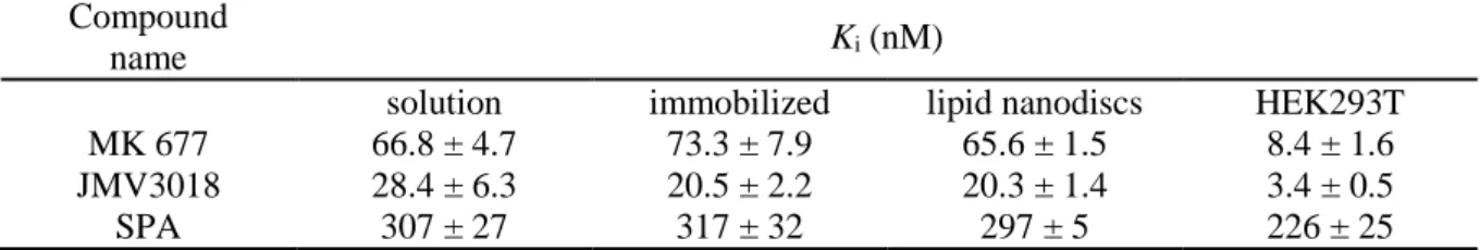
Figure 6. Impact of immobilization on the ligand-binding properties of GHSR. FRET-monitored competition curves obtained with GHSR assembled into NAPols:BNAPols (1:1 weight ratio, polymers NA16 and BNA15_40 were used) in solution (closed symbols) or immobilized on a streptavidin plate (open symbols). Data are presented as the mean ± SEM of three experiments.
The values for solution and immobilized receptor were inferred from the titration plot in Figure
6 and are summarized in Table 3. BNAPol-mediated immobilization of GHSR does not
the sake of comparison, we also included in Table 3 the values obtained for the recombinant
GHSR in nanodiscs [39] and those obtained in HEK293T cells [40]. To be noted, the Ki values
obtained for MK0677 and JMV3018 are systematically higher for the isolated receptor than for
GHSR expressed at the surface of HEK intact cells. This is consistent, as both compounds are
agonists, full agonist for MK0677, and partial agonist for JMV3018 [41], and the isolated
receptor is in a low affinity state, as it is uncoupled from its G protein partner. We previously
demonstrated that high-affinity agonist binding can be restored upon adding isolated G proteins
to the purified receptor [33].
Table 3. Ligand-binding properties of GHSR in NAPols, in solution and immobilized
compared to lipid nanodisc and HEK293T cells. In all cases, errors span the range of values observed in triplicate experiments.
Compound
name Ki (nM)
solution immobilized lipid nanodiscs HEK293T
MK 677 66.8 ± 4.7 73.3 ± 7.9 65.6 ± 1.5 8.4 ± 1.6
JMV3018 28.4 ± 6.3 20.5 ± 2.2 20.3 ± 1.4 3.4 ± 0.5
SPA 307 ± 27 317 ± 32 297 ± 5 226 ± 25
As the ligand-binding properties were not significantly affected by the immobilization
process, we then analyzed whether the ghrelin receptor assembled into BNAPols and
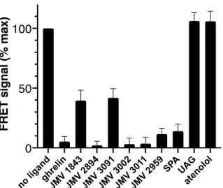
immobilized onto a streptavidin surface could be used as a ligand screening device. To this end,
we selected a set of known GHSR ligands. These include the natural full agonist ghrelin, a
affinity synthetic agonist (JMV 2894), a low-affinity synthetic agonist (JMV 3091), a
high-affinity synthetic antagonist (JMV 3002), a low-high-affinity synthetic antagonist (JMV 2959), the
low-affinity inverse agonist SPA and JMV 1843, an agonist pseudopeptide which got the
approval of the FDA for the diagnosis of GH defficiency in adults. All JMV compounds belong
to the series of 1,2,4-triazole derivatives [42-45], with the exception of the pseudopeptide JMV
1843 compound [45]. In addition, two negative controls were included in the assay, i.e.
unacylated ghrelin (UAG) that has been shown to be unable to bind GHSR because of the lack
in Figure 7, a specific decrease in the ghrelin:GHSR FRET signal was observed for all ligands
known to bind the receptor. Interestingly, the extent in the decrease in the FRET signal was
directly related to the affinity of the compound considered. Overall, this indicates that the FRET
signal obtained under such conditions indeed reports for the ability of the compound to compete
with labeled ghrelin for binding GHSR.
Figure 7. Ligand-binding screening assay with the immobilized ghrelin receptor.
FRET-monitored competition for binding GHSR assembled into NAPols:BNAPols (1:1 weight ratio, polymers NA16 and BNA15_40 were used) and immobilized on a streptavidin plate. Competing ligands were used at a 10 µM concentration while the fluorescent ghrelin tracer was used at 50 nM. All data are presented as the mean ± SEM of three experiments.
4. Conclusion
We have synthesized a series of biotin-functionalized non-ionic amphipols called
BNAPols by free-radical telomerization of an amphiphilic monomer in the presence of a
transfer agent that bears an azido group. By using a Huisgen-cycloaddition reaction the biotin
group was grated on the polymer end chain. The BNAPols were found freely soluble in water
and formed rather homogenous and globular micellar aggregates of about 6 nm in diameter.
The reconstitution of the ghrelin receptor GHSR in BNAPols was further demonstrated.
Moreover, under our conditions, the receptor was efficiently bound to the surface while keeping
represent a valuable approach to immobilize functional GPCRs, and probably other MPs, onto
surfaces for subsequent screening assays. As such, this immobilization strategy of isolated MP
could represent a valuable alternative to cellular systems in screening assays to identify a new
generation of drugs from compound libraries. In particular, this assay could be well-adapted to
fragment screening that is actually out of the scope of classical cell-based assays. Moreover, in
contrast to other strategies already described, the method presented here is based on the use of
a native, unmodified receptor, which can avoid any possible bias related to stabilizing
mutations. Moreover, as the immobilization process involves the surfactant rather than the
protein, such a process could be extended to untagged proteins as well.
Acknowledgements
This work was supported by the Agence Nationale de la Recherche (ANR) through grants no.
ANR-2010-BLAN-1535 and ANR-17-CE18-0022. We acknowledge the financial support of the European Regional Development Fund, the French Government, the “Région Provence
Alpes Côte d'Azur”, the “Département de Vaucluse” and the “Communauté d’agglomération
Grand Avignon” for access to the NMR platform (CPER 3A). We thank the European
Synchrotron Radiation Facility (ESRF) for access to synchrotron radiation facilities and Petra
Pernot for assistance in using the beamline BM29. This work benefited from the use of the
SasView application, originally developed under NSF award DMR-0520547. SasView contains code developed with funding from the European Union’s Horizon 2020 research and innovation
References
[1] J. -L. Popot, Amphipols, nanodiscs, and fluorinated surfactants: three nonconventional
approaches to studying membrane proteins in aqueous solutions, Ann. Rev. Biochem. 79 (2010) 737-775.
[2] Q. Zhang, X. Ma, A. Ward, W.-X. Hong, V.-P. Jaakola, R. C. Stevens, M. G. Finn, G.Chang, Designing Facial Amphiphiles for the Stabilization of Integral Membrane Proteins. Angew. Chem. Int. Ed., 46 (2007) 7023-7025.
[3] P. S. Chae, S. G. F. Rasmussen, R. R. Rana, K. Gotfryd, R. Chandra, M. A. Goren, A. C. Kruse, S. Nurva, C. J. Loland, Y. Pierre, D. Drew, J.-L. Popot, D. Picot, B. G. Fox, L. Guan, U. Gether, B. Byrne, B. Kobilka, S. H. Gellman, Maltose-neopentyl glycol (MNG) amphiphiles for solubilization, stabilization and crystallization of membrane proteins. Nat. Meth., 7 (2010) 1003-1008.
[4] J. Hovers, M. Potschies, A. Polidori, B. Pucci, S. Raynal, F. Bonneté, M. J. Serrano-Vega, C. G. Tate, D. Picot, Y. Pierre, J. L. Popot, R. Nehmé, M. Bidet, I. Mus-Veteau, H. Bußkamp, K.-H. Jung, A. Marx, P. A. Timmins, W. Welte, A class of mild surfactants that keep integral membrane proteins water-soluble for functional studies and crystallization. Mol. Membr. Biol., 28 (2011) 171-181.
[5] C. Breyton, F. Gabel, M. Abla, Y. Pierre, F. Lebaupain, G. Durand, J.-L. Popot, C. Ebel, B. Pucci, Micellar and biochemical properties of (hemi)fluorinated surfactants are controlled by the size of the polar head. Biophys. J., 97 (2009) 1-10.
[6] C. Tribet, R. Audebert, J. -L. Popot, Amphipols: polymers that keep membrane proteins soluble in aqueous solutions, P. Natl. Acad. Sci. U. S. A. 93 (1996) 15047-15050.
[7] M. Jamshad, Lin, Y. P.; Knowles, T. J.; Parslow, R. A.; Harris, C.; Wheatley, M.; Poyner, D. R.; Bill, R. M.; Thomas, O. R.; Overduin, M.; Dafforn, T. R., Surfactant-free purification of membrane proteins with intact native membrane environment. Biochem Soc Trans 2011, 39 (3), 813-8.
[8] J.-L. Popot, T. Althoff, D. Bagnard, J.-L. Banères, P. Bazzacco, E. Billon-Denis, L. J. Catoire, P. Champeil, D. Charvolin, M. J. Cocco, G. Crémel, T. Dahmane, L. M. de la Maza, C. Ebel, F. Gabel, F. Giusti, Y. Gohon, E. Goormaghtigh, E. Guittet, J. H. Kleinschmidt, W. Kühlbrandt, C. Le Bon, K. L. Martinez, M. Picard, B. Pucci, J. N. Sachs, C. Tribet, C. van Heijenoort, F. Wien, F. Zito, M. Zoonens, Amphipols From A to Z. Ann. Rev. Biophys. 40 (2011) 379-408.
[9] M. Zoonens, J. -L. Popot, Amphipols for each season, J. Membr. Biol. 247 (2014) 759-96. [10] B. M. Gorzelle, A. K. Hoffman, M. H. Keyes, D. N. Gray, D. G. Ray, C. R. Sanders,
Amphipols Can Support the Activity of a Membrane Enzyme. J. Am. Chem. Soc. 124 (2002) 11594-11595
[11] T. Dahmane, F. Giusti, L. J. Catoire, J.-L. Popot, Sulfonated Amphipols: Synthesis, Properties, and Applications. Biopolymers 95 (2011) 811-823
[12] K. S. Sharma, G. Durand, F. Gabel, P. Bazzacco, C. Le Bon, E. Billon-Denis, L. J. Catoire, J.-L. Popot, C. Ebel, B. Pucci, Non-Ionic Amphiphilic Homopolymers: Synthesis, Solution Properties, and
Biochemical Validation, Langmuir. 28 (2012) 4625-4639.
[13] K. S. Sharma, G. Durand, F. Giusti, B. Olivier, A.-S. Fabiano, P. Bazzacco, T. Dahmane, C. Ebel, J.-L. Popot, B. Pucci, Glucose-Based Amphiphilic Telomers Designed to Keep Membrane Proteins Soluble in Aqueous Solutions: Synthesis and Physicochemical Characterization, Langmuir. 24 (2008) 13581-13590.
[14] P. Bazzacco, E. Billon-Denis, K. S. Sharma, L. J. Catoire, S. Mary, C. Le Bon, E. Point, J. L. Baneres, G. Durand, F. Zito, B. Pucci, J. -L. Popot, Nonionic homopolymeric amphipols: application to
membrane protein folding, cell-free synthesis, and solution nuclear magnetic resonance, Biochemistry. 51 (2012) 1416-1430.
[15] L. Fagerberg, K. Jonasson, G. von Heijne, M. Uhlen, L. Berglund, Prediction of the human membrane proteome, Proteomics. 10 (2010) 1141-1149.
[16] J. P. Overington, B. Al-Lazikani, A. L. Hopkins, How many drug targets are there?, Nat. Rev. Drug Discov. 5 (2006) 993-996.
[17] V. Fruh, I. J. AP, G. Siegal, How to catch a membrane protein in action: a review of functional membrane protein immobilization strategies and their applications, Chem. Rev. 111 (2011) 640-656.
[18] H. Hoi, A. Jimenez Castellanos, M. Aminpour, Y. He, H. Zhou, S. Abraham, C. D. Montemagno, Immobilization of membrane proteins on solid supports using functionalized beta-sheet peptides and click chemistry. Chem. Comm. 54 (2018) 1889-1892.
[19] M. Oelschlägel, A. Riedel, A. Zniszczoł, K. Szymańska, A. B. Jarzębski, M. Schlömann, D. Tischler, Immobilization of an integral membrane protein for biotechnological phenylacetaldehyde
production. J. Biotech. 174 (2014) 7-13.
[20] D. Charvolin, J.-B. Perez, F. Rouvière, F. Giusti, P. Bazzacco, A. Abdine, F. Rappaport, K. L.
Martinez, J.-L. Popot, The use of amphipols as universal molecular adapters to immobilize membrane proteins onto solid supports Proc. Natl. Acad. Sci. U. S. A. 106 (2009) 405-410 .
[21] C. Le Bon, J. L. Popot, F. Giusti, Labeling and functionalizing amphipols for biological applications, J. Membr. Biol. 247 (2014) 797-814.
[22] F. Giusti, P. Kessler, R. W. Hansen, E. A. Della Pia, C. Le Bon, G. Mourier, J. L. Popot, K. L. Martinez, M. Zoonens, Synthesis of a Polyhistidine-bearing Amphipol and its Use for Immobilizing Membrane Proteins, Biomacromolecules 16 (2015) 3751-3761.
[23] C. Le Bon, E. A. Della Pia, F. Giusti, N. Lloret, M. Zoonens, K. L. Martinez, J. -L. Popot, 2014. Synthesis of an oligonucleotide-derivatized amphipol and its use to trap and immobilize membrane proteins. Nucleic acids research, (2014) 42, e83.
[24] H. Basit, K. S. Sharma, A. Van der Heyden, C. Gondran, C. Breyton, P. Dumy, F. M. Winnik, P. Labbe, Amphipol mediated surface immobilization of FhuA: a platform for label-free detection of the bacteriophage protein, Chem. Comm. 48 (2012) 48, 6037-6039.
[25] Y. Ferrandez, M. Dezi, M. Bosco, A. Urvoas, M. Valerio-Lepiniec, C. Le Bon, F. Giusti, I. Broutin, G. Durand, A. Polidori, J. -L. Popot, M. Picard, P. Minard, Amphipol-mediated screening of molecular orthoses specific for membrane protein targets, J. Membre. Biol. 247 (2014) 925-940.
[26] K. S. Sharma, G. Durand, B. Pucci, Synthesis and Determination of Polymerization Rate Constants of Glucose-Based Monomers, Des. Monomers Polym. 14 (2011) 499-513.
[27] E. Garanger, R. Weissleder, L. Josephson, A Multifunctional Single-Attachment-Point Reagent for Controlled Protein Biotinylation, Bioconjug Chem. 20 (2009) 170-173.
[28] M. Abla, G. Durand, B. Pucci, Glucose-based surfactants with hydrogenated, fluorinated, or hemifluorinated tails: synthesis and comparative physical-chemical characterization, J. Org. Chem. 73 (2008) 8142–8153.
[29] S. Delagenière, P. Brenchereau, L. Launer, A. W. Ashton, R. Leal, S. Veyrier, J. Gabadinho, E. J. Gordon, S. D. Jones, K. E. Levik, S. M. McSweeney, S. Monaco, M. Nanao, D. Spruce, O. Svensson, M. A. Walsh, G. A. Leonard, ISPyB: an Information Management System for Synchrotron Macromolecular Crystallography, Bioinformatics. 27 (2011) 3186-3192.
[30] P. V. Konarev, V. V. Volkov, A. V. Sokolova, M. H. J. Koch, D. I. Svergun, PRIMUS: a Windows PC-based system for small-angle scattering data analysis, J. Appl. Crystallogr. 36 (2003) 1277-1282. [
[31] M. Damian, J. Marie, J.-P. Leyris, J.-A. Fehrentz, P. Verdié, J. Martinez, J.-A. Banères, S. Mary, High Constitutive Activity Is an Intrinsic Feature of Ghrelin Receptor Protein, J. Biol. Chem 287 (2012) 3630-3641.
[32] T. Dahmane, M. Damian, S. Mary, J.-L. Popot, J.-L. Banères, Amphipol-Assisted in Vitro Folding of G Protein-Coupled Receptors, Biochemistry 48 (2009) 6516-6521.
[33] G. Ferré, M. Louet, O. Saurel, B. Delort, G. Czaplicki, C. M’Kadmi, M. Damian, P. Renault, S. Cantel, L. Gavara, P. Demange, J. Marie, J.-A. Fehrentz, N. Floquet, A. Milon, J.-L. Banères, Structure and dynamics of G protein-coupled receptor–bound ghrelin reveal the critical role of the octanoyl chain, Proc. Natl. Acad. Sci. U. S. A. 116 (2019) 17525-17530.
[34] C. M. Starks, Free radical telomerization, Academic Press, Inc., New York, 1974.
[35] A. A. Pavia, B. Pucci, J. G. Riess, L. Zarif, New perfluoroalkyl telomeric non-ionic surfactants: synthesis, physicochemical and biological properties, Makromol. Chem. 193 (1992) 2505-2517. [36] K. S. Sharma, G. Durand, F. Giusti, B. Olivier, A.-S. Fabiano, P. Bazzacco, T. Dahmane, C. Ebel, J.-L. Popot, B. Pucci, Glucose-based amphiphilic telomers designed to keep membrane proteins soluble in
aqueous solutions: synthesis and physicochemical characterization, Langmuir. 24 (2008) 13581-13590.
[37] P. Bazzacco, K. S. Sharma, G. Durand, F. Giusti, C. Ebel, J.-L. Popot, B. Pucci, Trapping and stabilization of integral membrane proteins by hydrophobically grafted glucose-based telomers, Biomacromolecules. 10 (2009) 3317-3326.
[38] D. I. Svergun, Determination of the Regularization Parameter in Indirect-Transform Methods Using Perceptual Criteria, J. Appl. Crystallogr. 25 (1992) 495-503.
[39] M. Damian, S. Mary, M. Maingot, C. M'Kadmi, D. Gagne, J.-P. Leyris, S. Denoyelle, G. Gaibelet, L.Gavara, M. Garcia de Souza Costa, D. Perahia, E. Trinquet, B. Mouillac, S. Galandrin, C. Galès, J.-A Fehrentz., N. Floquet, J. Martinez, J. Marie, J.-L.Banères, Ghrelin receptor conformational dynamics regulate the transition from a preassembled to an active receptor:Gq complex. Proc. Natl. Acad. Sci. USA, 112 (2015) 1601-1606.
[40] J.-P Leyris., T. Roux, E. Trinquet, P. Verdié, J.-A. Fehrentz, N. Oueslati, S. Douzon, E. Bourrier, L. Lamarque, D. Gagne, J.-C. Galleyrand, C. M'kadmi, J. Martinez, S. Mary, J.-L.Banères, J. Marie, Homogeneous time-resolved fluorescence-based assay to screen for ligands targeting the growth hormone secretagogue receptor type 1a. Anal. Biochem., 408 (2011) 253-262.
[41] C. M'Kadmi, J.-P. Leyris, L. Onfroy, C. Galés, A. Saulière, D. Gagne, M. Damian, S. Mary, M. Maingot, S. Denoyelle, P. Verdié, J.-A. Fehrentz, J. Martinez, J.-L. Banères, J. Marie, Agonism, Antagonism, and Inverse Agonism Bias at the Ghrelin Receptor Signaling. J. Biol. Chem., 290 (2015) 27021-39.
[42] L. Demange, D. Boeglin, A. Moulin, D. Mousseaux, J. Ryan, G. Bergé, D. Gagne, A. Heitz, D. Perrissoud, V. Locatelli, A. Torsello, J.-C. Galleyrand, J.-A. Fehrentz, J. Martinez, Synthesis and Pharmacological in Vitro and in Vivo Evaluations of Novel Triazole Derivatives as Ligands of the Ghrelin Receptor, J. Med. Chem. 50 (2007) 1939-1957.
[43] A. Moulin, L. Demange, J. Ryan, C. M’Kadmi, J.-C. Galleyrand, J. Martinez, J.-A. Fehrentz, Trisubstituted 1,2,4-triazoles as ligands for the ghrelin receptor: On the significance of the orientation and substitution at position 3, Bioorg. Med. Chem. Lett. 18 (2008) 164-168.
[44] A. Moulin, L. Demange, J. Ryan, D. Mousseaux, P. Sanchez, G. Bergé, D. Gagne, D. Perrissoud, V. Locatelli, A. Torsello, J.-C. Galleyrand, J.-A. Fehrentz, J. Martinez, New Trisubstituted 1,2,4-Triazole Derivatives as Potent Ghrelin Receptor Antagonists. 3. Synthesis and Pharmacological in Vitro and in Vivo Evaluations, J. Med. Chem. 51 (2008) 689-693.
[45] V. Guerlavais, D. Boeglin, D. Mousseaux, C. Oiry, A. Heitz, R. Deghenghi, V. Locatelli, A. Torsello, C. Ghé, F. Catapano, G. Muccioli, J.-C. Galleyrand, J.-A. Fehrentz, J. Martinez, New Active Series of Growth Hormone Secretagogues, J. Med. Chem. 46 (2003) 1191-1203.
[46] M. Van Craenenbroeck, F. Gregoire, P. De Neef, P. Robberecht, J. Perret, Ala-scan of ghrelin (1– 14): interaction with the recombinant human ghrelin receptor, Peptides 25 (2004) 959-965.