MUSCULOSKELETAL
Oedema and fatty degeneration of the soleus
and gastrocnemius muscles on MR images in patients
with achilles tendon abnormalities
Adrienne Hoffmann&Nadja Mamisch&
Florian M. Buck&Norman Espinosa&
Christian W. A. Pfirrmann&Marco Zanetti
Received: 4 January 2011 / Revised: 14 March 2011 / Accepted: 16 April 2011 / Published online: 6 May 2011 # European Society of Radiology 2011
Abstract
Objective The purpose of this study was to evaluate the frequency of oedema and fatty degeneration of the soleus and gastrocnemius muscles in patients with Achilles tendon abnormalities.
Methods Forty-five consecutive patients (mean 51 years; range 14–84 years) with achillodynia were examined with magnetic resonance (MR) images of the calf. The frequency of oedema and fatty degeneration in the soleus and gastrocnemius muscles was determined in patients with normal tendons, tendinopathy and in patients with a partial tear or a complete tear of the Achilles tendon.
Results Oedema was encountered in 35% (7/20) of the patients with tendinopathy (n=20; range 13–81 years), and in 47% (9/19) of the patients with partial tears or complete tears (n=19; 28–78 years). Fatty degeneration was encoun-tered in 10% (2/20) of the patients with tendinopathy, and in 32% (6/19) of the patients with tears. The prevalence of
fatty degeneration was significantly more common in patients with a partial or complete tear compared with the patients with a normal Achilles tendon (p=0.032 and p= 0.021, respectively).
Conclusion Oedema and fatty degeneration of the soleus and gastrocnemius muscles are common in patients with Achilles tendon abnormalities.
Keywords Achilles tendon . Calf muscles . Magnetic resonance imaging . Edema . Atrophy
Introduction
The Achilles tendon is the largest and strongest tendon of the human body. During the last few decades the incidence of Achilles tendon abnormalities has increased in civilised countries because of increased involvement in recreational sports activities [1, 2]. Achilles tendon lesions most commonly results from repetitive microtrauma, ageing, vascular compromise, or a combination of these factors [3]. Partial tear or complete tear of the Achilles tendon often occurs during strenuous physical activities. Sponta-neous tears may also appear secondary to predisposing factors such as steroid therapy or rheumatoid arthritis [4]. Tendinopathy or partial tear of the Achilles tendon is initially treated by using conservative measures [5]. If conservative treatment fails, a variety of surgical treatments are considered [6–11]. Anatomical (direct suture of the torn tendons) and non-anatomical tendon-graft repairs are two main options. According to the rotator cuff surgery in the shoulder where the quality of the muscles is of great importance to the surgical outcome [12–16], one may assume that an Achilles tendon repair is associated with a
A. Hoffmann
:
N. Mamisch:
F. M. Buck:
C. W. A. Pfirrmann:
M. Zanetti
Radiology Department, University Hospital Balgrist Zürich, Forchstrasse 340,
CH-8008 Zürich, Switzerland N. Espinosa
Orthopedic Surgery Department, University Hospital Balgrist Zürich,
Forchstrasse 340,
CH-8008 Zürich, Switzerland Present Address:
A. Hoffmann (*)
Radiology Department, Hirslanden Klinik Aarau, Schänisweg,
CH-5001 Aarau, Switzerland
more favourable outcome when the corresponding muscle does not show fatty degeneration. To our knowledge, little is known about the calf muscle changes in the presence of Achilles tendon abnormalities. Thus, the purpose of this study was to evaluate the frequency of oedema and fatty degeneration of the soleus and gastrocnemius muscles on magnetic resonance (MR) images in patients with Achilles tendon abnormalities.
Materials and methods
The frequency of oedema and fatty degeneration of the soleus and gastrocnemius muscles on MR images was determined for each patient group with normal Achilles tendon, tendinopathy and with partial tear or complete tear.
Patients
Forty-five consecutive patients (25 men, 20 women; mean 51 years; range 14–84 years) with achillodynia were included from an MR database during a period of 12 months. In 33 patients (16 men, 17 women; mean 45 years; range 10–81 years) of these 45 symptomatic patients, the contralateral Achilles tendon and the calf were imaged as well. The contralateral side was not further evaluated in the current study because clinical history information was not available and the Achilles tendon condition could not be determined. The duration of symptoms was classified in five stages: stage I (acute) indicated 1 week of symptoms, stage II (subacute) 1– 6 weeks of symptoms, stage III (late subacute) 6 weeks to 3 months of symptoms, stage IV (early chronic) 3–
12 months of symptoms, stage V (chronic) more than 12 months of symptoms.
The study was submitted to the institutional review board and a waiver for additional approval was issued for this study. Patient rights are protected by a law requiring patient information about the possibility of anonymous scientific review of their data and about the opportunity to reject such use of their data. All patients agreed to use their data.
MR imaging
Magnetic resonance imaging was performed at 1.5-T (Espree or Avanto, Siemens Medical Solutions, Erlangen, Germany) with a standard dedicated send-receive extremity coil for the Achilles tendon and a body array coil for the calves. The patients were placed in supine position. The feet were symmetrically aligned to image both calves at the same time. Standard sequences were used for this study (Table 1).
Image analysis
Achilles tendon The Achilles tendon was analysed inde-pendently by two experienced musculoskeletal radiologists and subsequently a consensus was reached. The profes-sional experience in musculoskeletal radiology was 16 years for observer 1 (M.Z.), and 11 years for observer 2 (C.W.A.P.). Tendinopathy, partial tear or complete tear of the symptomatic and asymptomatic Achilles tendons was determined according to previously published crite-ria [4, 17]. Tendinopathy was defined as tendon thicken-ing, flat or convex anterior margin and normal signal or
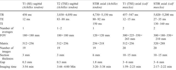
Table 1 Parameters of the standard sequences used for the examination of the Achilles tendon and the calf muscles T1 (SE) sagittal
(Achilles tendon)
T2 (TSE) sagittal (Achilles tendon)
STIR axial (Achilles tendon)
T1 (TSE) axial (calf muscles)
STIR axial (calf muscles) TR 450 ms 3,830–4,050 ms 4,730–5,150 ms 457–547 ms 5,020–5,200 ms TE 12 ms 83–88 ms 80–92 ms 12–15 ms 27–35 ms TI 150 ms 130–160 ms Number of averages 1 1–2 1 2 2 FOV 180×180 mm 180×180 mm 120×120 mm 300×225–350× 263 mm 300×180–350× 210 mm Matrix 512×256 512×256 256×218 512×256 320×288 Number of images 19 19 19 20 20 Section thickness 3 mm 3 mm 6 mm 10–15 mm 10–15 mm Gap 0.3 mm 0.3 mm 1.8 mm 3–6 mm 3–6 mm
hyperintense (compared with normal tendons) signal on T1-weighted images. A localized anterior bulge that shifted in a wavelike fashion from the lateral to the medial margin across the anterior surface of the tendon on axial images from proximal to distal was considered normal [18]. Partial tear was defined as tendon thickening with hyperintense signal on T1- and hyperintense signal on MR images with fluid-sensitive (T2-weighted and STIR) sequences. All of the criteria were required to diagnose tendinopathy or partial tear. Discontinuity of the Achilles tendon was rated as complete tear.
Muscles– qualitative Two additional observers (observer 1 [R1, F.M.B.] and observer 2 [R2, N.M.]) evaluated independently the soleus and gastrocnemius muscles, and subsequently a consensus was reached. Professional expe-rience in musculoskeletal radiology was 3 years for observer 1 (F.M.B.), and 4 years for observer 2 (N.M.). They determined if oedema was present or absent and they determined the degree of fatty degeneration. Oedema in the soleus and gastrocnemius muscles was diagnosed when diffuse increased signal was seen on the axial STIR images compared with the anterior muscle compartment. A grading system for fatty degeneration was used corresponding to a classification system for the rotator cuff muscles [12]: Grade 0 indicated no intramuscular fat; grade 1 some fatty streaks; grade 2 fat was evident but there was less fat than muscle; grade 3 equal amounts of fat and muscle tissue; and
grade 4 more fat than muscle tissue. Subsequently, fatty degeneration was defined as present if fatty degeneration of the muscle was equal to or higher than grade 2 [4,19]. The soleus muscle, the medial and the lateral head of the gastrocnemius muscle were assessed separately. The other lower leg muscles were not analysed in detail because oedema and fatty degeneration were not encountered in patients with Achilles tendon abnormalities during routine examination.
Muscle - quantitative On axial T1-weighted images the level was determined where the cross sectional area was maximal for the soleus muscle, the medial head of the gastrocnemius muscle, and the lateral head of the gastroc-nemius muscle, At these different levels, the maximal cross sectional area of the specific muscle was measured by the first author (professional experience in musculoskeletal radiology 1 year). Regions of interest were drawn around each muscle at magnification of 200% to minimise measurement errors. Cross sectional areas were measured by using the PACS workstation.
Statistical evaluation
Seven different statistical tests were performed in total. Differences in the frequencies of oedema among the three patient groups were tested for significance by usingχ2
Table 2 Qualitative and quantitative results of oedema, fatty degeneration and atrophy of the soleus and gastrocnemius muscles
Symptomatic patients (n=45) Normal (n=6) Tendinopathy (n=20) Partial tear/complete tear (n=19)
Mean age (range) (yr) 25 (10–40) 47 (13–81) 56 (28–78)
Gender (f/m) 3/3 11/9 6/13
Mean symptom stagesa IV III IV
Soleus muscle
Oedema (n) [%] 0 2 [10] 3 [16]
Fatty degeneration (n)b[%] 0 1 [5] 6 [32]
Goutallier 0/1/2/3/4 (n) 5/1/0/0/0 9/10/1/0/0 6/6/5/1/0
Maximal mean area (cm2) 26 27 28
Caput mediale
Oedema (n) [%] 0 6 [30] 8 [42]
Fatty degeneration (n)b[%] 0 1 [5] 4 [21]
Goutallier 0/1/2/3/4 (n) 5/1/0/0/0 10/9/1/0/0 5/9/2/2/0
Maximal mean area (cm2) 12 12 13
Caput laterale
Oedema (n) [%] 0/3 3/15 [20] 9/17 [53]
Fatty degeneration (n)b[%] 0 1 [6] 1 [6]
Goutallier 0/1/2/3/4 (n) (not available) 4/1/0/0/0 (1) 9/6/1/0/0 (4) 6/9/1/0/0 (3)
Maximal mean area (cm2) 6 5 7
a
stage III (early chronic) 6 weeks– 3 months of symptoms, stage IV (chronic) 3–12 months of symptoms
b
analysis and Fisher’s exact test. The Kruskal-Wallis test was used to assess significant differences in fat content according to Goutallier among the three parts of the soleus and gastrocnemius muscles in the three patient groups. P< 0.05 was considered to indicate a significant difference.
Differences in the frequencies of fatty degeneration in the three parts of the soleus and the gastrocnemius muscles among the three patient groups were tested for significance by the Mann–Whitney U test. P<0.05 was considered to indicate a significant difference.
Kendall’s tau test was used to assess a significant difference in the mean maximal area of the three parts of the soleus and gastrocnemius muscles in the three patient groups. Spearman’s rank test was used to assess a significant correlation among age, the condition of the Achilles tendon (normal Achilles tendon, tendinopathy, partial tear, complete tear), oedema and the fat content according to Goutallier of the three parts of the soleus and gastrocnemius muscles in the three patient groups.
κ Statistics were calculated for interobserver agreement. According to Landis and Koch [20], agreement was rated as follows: κ Values of 0–0.20 indicated slight agreement; values of 0.21–0.40, fair agreement; values of 0.41–0.60, moderate agreement; values of 0.61–0.80, substantial agreement; values of 0.81–0.99, excellent agreement. A κ value of 1.00 indicated absolute agreement. A computer
software package (IBM Statistics SPSS 18) was used to perform all statistical analysis.
Results
Achilles tendon
In 6 patients the Achilles tendon was normal (3 men, 3 women; mean 25 years; range 10–40 years) (Table 2). Tendinopathy was identified in 20 patients (9 men, 11 women; mean 47 years; range 13–81 years), partial tear was identified in 17 patients (Fig. 1), and two patients had a complete tear of the Achilles tendon (Fig. 2) (partial tear and complete tear: 13 men, 6 women; mean 56 years; range 28–78 years).
The soleus and gastrocnemius muscles
In patients with normal Achilles tendon no oedema was found in the soleus and gastrocnemius muscles. In patients with tendinopathy oedema was encountered in 35% (7/20) of the patients in the soleus and gastrocnemius muscles. Oedema in the soleus and gastrocnemius muscles was encountered in 47% (9/19) of the patients with partial or complete tears (Fig.2b).
Fig. 1 Sagittal T2-weighted (TR: 4,760 ms/TE: 85 ms) image of the right hindfoot (a) and axial T1-weighted (TR: 471 ms/11 ms) image of both calves (b) in a 75-year-old male patient with a partial tear of the Achilles tendon on the right side. The high signal at the site of the
partial tear of the Achilles tendon is marked (arrow) (a). Note the fatty degeneration of the soleus and gastrocnemius muscle on both sides, especially in the soleus muscle (arrows) (b)
Fig. 2 Sagittal T2-weighted (TR: 4,300 ms/TE: 93 ms) image of the left hindfoot (a) and axial STIR (TR: 5,200 ms/TE: 35 ms; TI: 160 ms) image of both calves (b) in a symptomatic 53-year-old female patient with complete tear of the Achilles tendon on the left side. The
gap between the two ends of the torn Achilles tendon is marked (arrows) (a). Note the oedema in the soleus and gastrocnemius muscle on the left side (arrowheads) (b)
In patients with normal Achilles tendons no fatty degeneration was found in the soleus and gastrocnemius muscles (Fig. 3). In patients with tendinopathy fatty degeneration was encountered in 10% (2/20) of the patients in the soleus and gastrocnemius muscles. In patients with partial tear or complete tear fatty degeneration was encountered in 32% (6/19) of the patients in the soleus and gastrocnemius muscles (Fig.1b).
In one patient with a normal Achilles tendon the lateral head of the gastrocnemius muscle had not been imaged. In four patients with tendinopathy the lateral head of the gastrocnemius muscle had not been imaged. In three patients with partial tear or complete tear the lateral head of the gastrocnemius muscle had not been imaged.
The three parts of the calf muscles
Qualitative
In patients with tendinopathy oedema was encountered in 10% (2/20) in the soleus muscle, in 30% (6/20) in the medial head of the gastrocnemius muscle and in 20% (3/15) in the lateral head of the gastrocnemius muscle. In five patients the lateral head of the gastrocnemius muscle was not completely visible on the STIR MR images. In patients with partial or complete tear of the Achilles tendon oedema was present in 16% (3/19) in the soleus muscle, in 42% (8/
19) in the medial head of the gastrocnemius muscle and in 53% (9/17) in the lateral head of the gastrocnemius muscle. In patients with tendinopathy fatty degeneration was encountered in 5% (1/20) in the soleus muscle, in 5% (1/ 20) in the medial head of the gastrocnemius muscle, and in 6% (1/16) in the lateral head of the gastrocnemius muscle. In patients with partial or complete tear fatty degeneration was present in 32% (6/19) in the soleus muscle, in 21% (4/ 19) in the medial head of the gastrocnemius muscle, and in 6% (1/16) in the lateral head of the gastrocnemius muscle.
Quantitative
In patients with normal Achilles tendons the mean maximal area of the soleus muscle was 26 ± 11 (standard devia-tion) cm2, of the medial head of the gastrocnemius muscle 12 ± 25 cm2, and of the lateral head of the gastrocnemius muscle 6 ± 2 cm2. In patients with tendinopathy of the Achilles tendon the mean maximal area of the soleus muscle was 27 ± 6 cm2, of the medial head of the gastrocnemius muscle 12 ± 5 cm2, and of the lateral head of the gastrocnemius muscle 5 ± 4 cm2. In patients with partial or complete tear of the Achilles tendon the mean maximal area of the soleus muscle was 28 ± 7 cm2, of the medial head of the gastrocnemius muscle 13 ± 5 cm2 and of the lateral head of the gastrocnemius muscle 7 ± 4 cm2.
Statistical evaluation
The soleus and gastrocnemius muscles
No significant difference (p>0.05) was observed in the prevalence of oedema among the three patient groups with a normal Achilles tendon, with tendinopathy, and with partial or complete tear (χ2analysis and Fisher’s exact test).
A significant difference was found among the three patient groups regarding the prevalence of fatty degeneration in the soleus muscle (p=0.048) and in the medial head of the gastrocnemius muscle (p=0.033; Kruskal-Wallis).
The three parts of the soleus and gastrocnemius muscles
Qualitative The prevalence of fatty degeneration in the soleus muscle and in the medial head of the gastrocnemius muscle was significantly more common in patients with a
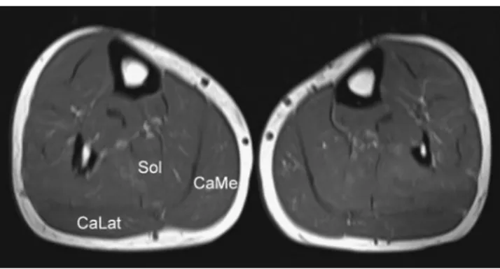
Fig. 3 Axial T1-weighted (TR: 547 ms/TE: 15 ms) image of the calves in a 55-year-old male patient with normal Achilles tendons. Both sides show normal muscle tissue without atrophy or fatty degeneration. (CaLat = Lateral head of the gastrocnemius muscle. Sol = Soleus muscle. CaMe = Medial head of the gastrocnemius muscle.)
Table 3 Interobserver agreement
Soleus muscle Gastrocnemius muscle, Caput mediale Gastrocnemius muscle, Caput laterale
Oedema 0.28a 0.53a 0.49a
Fatty degeneration 0.55a 0.48a 0.56a
aκ Statistics according to Landis and Koch [
partial or complete tear compared with the patients with a normal Achilles tendon (p=0.032 and p=0.021, respectively; Mann–Whitney U test).
Quantitative No significant difference (p > 0.05) of the mean maximal area of the soleus and gastrocnemius muscles between the three patient groups was observed (Kendall’s tau test).
Interobserver agreement
Interobserver agreement (Table 3) for assessment of oedema in the soleus muscle was fair (κ=0.28), in the medial head of the gastrocnemius muscle moderate (κ= 0.53), and in the lateral head of the gastrocnemius muscle moderate (κ=0.49).
Interobserver agreement for assessment of fatty degen-eration was moderate for the soleus and gastrocnemius muscles (soleus muscle [κ=0.55], medial head of the gastrocnemius muscle [κ=0.48], and lateral head of the gastrocnemius muscle [κ=0.56]).
Correlations
A significant correlation was found between the age of the patient and the fatty degeneration of the soleus muscle (r= 0.758, p<0.01), between the age of the patient and the fatty degeneration of the medial head of the gastrocnemius muscle (r=0.73, p<0.01) and between the age of the patient and the lateral head of the gastrocnemius muscle (r=0.588, p<0.01; Spearman’s rank test). A significant correlation between the Achilles tendon condition and the fatty degeneration of the soleus muscle (r=0.367, p=0.014) and the fatty degeneration of the medial head of the gastrocnemius muscle (r=0.395, p<0.01) was found. The correlation between the Achilles tendon condition and the fatty degeneration of the lateral head of the gastrocnemius
muscle was not significant (r=0.272, p=0.10; Spearman’s rank test) (Table4). No significant correlation (p>0.05) was found between the duration of symptoms and the preva-lence of oedema and fatty degeneration of the soleus and gastrocnemius muscles (Spearman’s rank test).
Discussion
The current study shows that oedema and fatty degenera-tion are common findings in patients with partial or complete tears of the Achilles tendon. To our knowledge, such muscle abnormalities have not been shown before in patients with Achilles tendons abnormalities. However, for the detection of these abnormalities a MR imaging protocol is required where the calf muscles are completely visible. Our routine MR protocol for the current study covered both sides of the lower leg. When the study was finished and analysed we changed the protocol to a unilateral imaging protocol of the calf to get higher spatial resolution for easier detection of changes in the muscles texture (Fig.4).
Table 4 Correlation among age, Achilles tendon condition and fatty muscle degeneration Condition Achilles
tendona
Fatty degeneration
Soleus Caput mediale
Gastrocnemius Caput laterale gastrocnemius Age Correlation coefficientb 0.523 0.758 0.730 0.588 p value <0.01 <0.01 <0.01 <0.01 Condition Achilles tendona Correlation coefficientb 1 0.367 0.395 0.272 p value <0.01 0.014 0.008 0.104 a
Condition = normal Achilles tendon, tendinopathy, partial tear, complete tear
bSpearman’s rank test
Fig. 4 Axial T1-weighted (TR: 610 ms/TE: 11 ms) image of the left calf in a 75-year-old male patient with tendinopathy of the Achilles tendon, which was seen after the study was finished and the image protocol was improved. Note the fatty degeneration of the soleus and gastrocnemius muscle (arrowheads), stage IV according to Goutallier
We assume that similar to the rotator cuff in the shoulder the quality of the soleus and gastrocnemius muscles may be an important factor for the functional results after surgical treatment of the Achilles tendon, and therefore imaging of the calf muscles may be essential. MR imaging has been shown to be a valuable method for the evaluation of atrophy and fatty degeneration of the rotator cuff muscles [21]. Ultrasound has also been used for the assessment of muscle atrophy and fatty degeneration of the rotator cuff of the shoulder [22]. As the Achilles tendon is commonly assessed by ultrasound this imaging technique would provide the most comfortable tool and an easily available method to evaluate the soleus and gastrocnemius muscles. However, ultrasound is only moderately accurate compared with MR imaging in the diagnosis of substantial fatty atrophy of the supraspinatus or infraspinatus muscle [23].
The interobserver agreement for the assessment of fatty degeneration was moderate for the soleus and gastrocne-mius muscles. The values are slightly lower than the values published for the shoulder [19,24]. In an assessment of the fatty degeneration of the muscles of the rotator cuff of the shoulder by CT versus MRI interobserver reproducibility was good to excellent for CT and for MRI [4,24].
Both age and the condition of the Achilles tendon were significantly correlated with the fatty degeneration of the soleus muscle and the medial head of the gastrocnemius muscle. Our data do not allow assessing if the Achilles tendon condition is more important for the fatty degener-ation of the gastrocnemius and soleus muscles than the age. The higher prevalence of fatty degeneration of the soleus muscles and medial head of the gastrocnemius muscles compared with the lateral head of the gastrocnemius muscle may be related to the microanatomy of the Achilles tendon. The fibres and fascicles of the Achilles tendon have a characteristic arrangement [25]. Proximally, they run parallel and then rotate distally. The range of rotation is variable. Therefore, the location of a tendinopathy or tear of the Achilles tendon might influence the site of oedema and fatty degeneration of the soleus and gastrocnemius muscles. If conservative treatment fails, a variety of surgical treatments can be utilised, such as debridement, direct suture of the torn tendon, local tissue transfer, augmentation and synthetic grafts. For the local tissue transfer either the flexor hallucis longus or the flexor digitorum longus tendon is used [26,27]. But the necessity for surgical repair is still a controversial topic. The decision between the different treatment options relies on many factors; including patient age, age of the tendon lesion, functional demand and the co-morbidities of the patient. Partial Achilles tendon tears are often difficult to treat [4,28]. In significant partial tears of the Achilles tendon the excision of the degenerated tissue can lead to complete pain relief and full restoration of function [29]. Several studies compared the different
treatment options of Achilles tendon disorders using MRI to document the size and the morphology of the tendon and the muscles [6, 7, 30]. Hahn et al. [31] evaluated the clinical outcome and MRI findings in 13 patients with chronic Achilles tendinopathy and tears treated with flexor hallucis tendon transfer. At an average follow-up of 46.5 months they found a fatty degeneration of the calf muscle in 10 out of 13 patients [31]. The soleus muscle was involved in nine patients; the gastrocnemius muscle was involved in one patient. These authors mentioned that their findings might have been present already preoperatively. Valderrabano et al. [32] emphasised that the knowledge of the recovery potential of the posterior tibial muscle after a longstanding posterior tibial tendon tear might be crucial for the surgeon deciding whether or not to reconstruct the torn tendon. However, to our knowledge no data exist on the condition of the calf muscles in patients with tendon abnormalities before surgical treatment.
Our study has limitations. Only two complete tears were included into our study. The time of symptoms could only be estimated based on review of the patient’s charts. Thus, the association between complete tears and the time of symptoms to the muscle abnormalities cannot be complete-ly answered. The fact that we had no patients older than 40 years and with normal Achilles tendons limits the evaluation of the influence of age on fatty degeneration of the calf muscles. Finally, not all soleus and gastrocnemius muscle parts were completely visible on all MR sequences. In summary, despite these limitations, our data demon-strate that oedema and fatty degeneration of the soleus and gastrocnemius muscles are common in patients with Achilles tendon abnormalities. The detection of soleus and gastrocnemius muscle abnormalities in patients with chronic Achilles tendon abnormalities may be of similar importance to rotator cuff therapy of the shoulder where the assessment of the muscle quality plays an important role in therapy decision-making among conservative treatment, tendon repair and tendon transfer [12–16].
Acknowledgement The authors thank Burkardt Seifert PhD, Zurich,
for his statistical support.
References
1. Clayton RA, Court-Brown CM (2008) The epidemiology of musculoskeletal tendinous and ligamentous injuries. Injury
39:1338–1344
2. Hess GW (2010) Achilles tendon rupture: a review of etiology, population, anatomy, risk factors, and injury prevention. Foot
Ankle Spec 3:29–32
3. Maffulli N (1995) Achilles tendon rupture. Br J Sports Med
29:279–280
4. Syed S, Bhatti A, Shah MM (2009) Spontaneous atraumatic Achilles tendon rupture in healthy individuals: biomechanical
5. Saltzman CL, Tearse DS (1998) Achilles tendon injuries. J Am
Acad Orthop Surg 6:316–325
6. Khan RJ, Fick D, Brammar TJ, Crawford J, Parker MJ (2004) Interventions for treating acute Achilles tendon ruptures. Cochrane Database Syst Rev CD003674
7. Metzl JA, Ahmad CS, Levine WN (2008) The ruptured Achilles tendon: operative and non-operative treatment options. Curr Rev Musculoskelet Med 1:161–164
8. Wapner KL, Pavlock GS, Hecht PJ, Naselli F, Walther R (1993) Repair of chronic Achilles tendon rupture with flexor hallucis
longus tendon transfer. Foot Ankle 14:443–449
9. Miskulin M, Miskulin A, Klobucar H, Kuvalja S (2005) Neglected rupture of the Achilles tendon treated with peroneus brevis transfer: a functional assessment of 5 cases. J Foot Ankle
Surg 44:49–56
10. Aktas S, Kocaoglu B (2009) Open versus minimal invasive repair
with Achillon device. Foot Ankle Int 30:391–397
11. Pajala A, Kangas J, Siira P, Ohtonen P, Leppilahti J (2009) Augmented compared with nonaugmented surgical repair of a fresh total Achilles tendon rupture. A prospective randomized
study. J Bone Joint Surg Am 91:1092–1100
12. Goutallier D, Postel JM, Bernageau J, Lavau L, Voisin MC (1994) Fatty muscle degeneration in cuff ruptures. Pre- and postoperative evaluation by CT scan. Clin Orthop Relat Res 78–83
13. Gerber C, Schneeberger AG, Hoppeler H, Meyer DC (2007) Correlation of atrophy and fatty infiltration on strength and integrity of rotator cuff repairs: a study in thirteen patients. J
Shoulder Elbow Surg 16:691–696
14. Goutallier D, Postel JM, Gleyze P, Leguilloux P, Van Driessche S (2003) Influence of cuff muscle fatty degeneration on anatomic and functional outcomes after simple suture of full-thickness tears.
J Shoulder Elbow Surg 12:550–554
15. Goutallier D, Postel JM, Radier C, Bernageau J, Zilber S (2009) Long-term functional and structural outcome in patients with intact repairs 1 year after open transosseous rotator cuff repair. J
Shoulder Elbow Surg 18:521–528
16. Gladstone JN, Bishop JY, Lo IK, Flatow EL (2007) Fatty infiltration and atrophy of the rotator cuff do not improve after rotator cuff repair and correlate with poor functional outcome. Am J Sports Med 35:719–728
17. Schweitzer ME, Karasick D (2000) MR imaging of disorders of the Achilles tendon. AJR Am J Roentgenol 175:613–625 18. Soila K, Karjalainen PT, Aronen HJ, Pihlajamaki HK, Tirman PJ
(1999) High-resolution MR imaging of the asymptomatic Achilles
tendon: new observations. AJR Am J Roentgenol 173:323–328
19. Saupe N, Pfirrmann CW, Schmid MR, Jost B, Werner CM, Zanetti M (2006) Association between rotator cuff abnormalities and reduced acromiohumeral distance. AJR Am J Roentgenol
187:376–382
20. Landis JR, Koch GG (1977) The measurement of observer agreement for categorical data. Biometrics 33:159–174
21. Thomazeau H, Boukobza E, Morcet N, Chaperon J, Langlais F (1997) Prediction of rotator cuff repair results by magnetic resonance imaging. Clin Orthop Relat Res 275–283
22. Khoury V, Cardinal E, Brassard P (2008) Atrophy and fatty infiltration of the supraspinatus muscle: sonography versus MRI.
AJR Am J Roentgenol 190:1105–1111
23. Strobel K, Hodler J, Meyer DC, Pfirrmann CW, Pirkl C, Zanetti M (2005) Fatty atrophy of supraspinatus and infraspinatus muscles:
accuracy of US. Radiology 237:584–589
24. Fuchs B, Weishaupt D, Zanetti M, Hodler J, Gerber C (1999) Fatty degeneration of the muscles of the rotator cuff: assessment by computed tomography versus magnetic resonance imaging. J
Shoulder Elbow Surg 8:599–605
25. Szaro P, Witkowski G, Smigielski R, Krajewski P, Ciszek B
(2009) Fascicles of the adult human Achilles tendon—an
anatomical study. Ann Anat 191:586–593
26. Qu JF, Cao LH, Zhao HB, Gao JH, Li SG, Du XJ, Sun Y, Peng Y, Wang L (2008) Flexor digitorum (hallucis) longus muscle tendon transfer in the repair of old rupture of the Achilles tendon. Zhongguo Gu Shang 21:297–299
27. Mahajan RH, Dalal RB (2009) Flexor hallucis longus tendon transfer for reconstruction of chronically ruptured Achilles
tendons. J Orthop Surg (Hong Kong) 17:194–198
28. Morberg P, Jerre R, Sward L, Karlsson J (1997) Long-term results after surgical management of partial Achilles tendon ruptures.
Scand J Med Sci Sports 7:299–303
29. Allenmark C (1992) Partial Achilles tendon tears. Clin Sports
Med 11:759–769
30. Fujikawa A, Kyoto Y, Kawaguchi M, Naoi Y, Ukegawa Y (2007) Achilles tendon after percutaneous surgical repair: serial MRI observation of uncomplicated healing. AJR Am J Roentgenol
189:1169–1174
31. Hahn F, Meyer P, Maiwald C, Zanetti M, Vienne P (2008) Treatment of chronic achilles tendinopathy and ruptures with flexor hallucis tendon transfer: clinical outcome and MRI findings. Foot Ankle Int 29:794–802
32. Valderrabano V, Hintermann B, Wischer T, Fuhr P, Dick W (2004) Recovery of the posterior tibial muscle after late reconstruction