*Corresponding author: Prof. Barbara Rothen-Rutishauser, BioNanomaterials, Adolphe Merkle Institute, University of Fribourg, Ch. de Verdiers 4, CH-1700 Fribourg, Switzerland,
Phone: +41 26 300 95 02, E-mail: barbara.rothen@unifr.ch Peter Wick and Matthias Roesslein: Empa, Swiss Federal Laboratories for Material Science and Technology, Particles-Biology Interactions Laboratory, Lerchenfeldstrasse 5, St. Gallen, Switzerland
Savvina Chortarea and Alke Petri-Fink: BioNanomaterials, Adolphe Merkle Institute, University of Fribourg, Ch. de Verdiers 4, Fribourg, Switzerland
Olivier T. Guenat: ARTORG Center, Lung Regeneration Technologies, Murtenstrasse 50, Bern, Switzerland; and Pulmonary Medicine and Thoracic Surgery Divisions, University Hospital of Berne, Bern, Switzerland
Janick D. Stucki: ARTORG Center, Lung Regeneration Technologies, Murtenstrasse 50, Bern, Switzerland
Stephanie Hirn: Walter Brendel Centre of Experimental Medicine, Klinikum der Universität München, Munich, Germany
Review
Peter Wick, Savvina Chortarea, Olivier T. Guenat, Matthias Roesslein, Janick D. Stucki,
Stephanie Hirn, Alke Petri-Fink and Barbara Rothen-Rutishauser*
In vitro-ex vivo model systems for nanosafety
assessment
Abstract: Engineered nanomaterials have unique and
novel properties enabling wide-ranging new applications in nearly all fields of research. As these new properties have raised concerns about potential adverse effects for the environment and human health, extensive efforts are underway to define reliable, cost- and time-effective, as well as mechanistic-based testing strategies to replace the current method of animal testing, which is still the most prevalent model used for the risk assessment of chemi-cals. Current approaches for nanomaterials follow this line. The aim of this review is to explore and qualify the relevance of new in vitro and ex vivo models in (nano) material safety assessment, a crucial prerequisite for translation into applications.
Keywords: alternative models; nanomaterials; risk
assessment.
DOI 10.1515/ejnm-2014-0049
Received December 19, 2014; accepted April 9, 2015; previously published online May 20, 2015
Introduction
Pressure on nanomaterial [for a definition see box and (1)] safety comes from society, consumer and regulatory bodies, but also from industry to identify potential adverse nanomaterials as early as possible during development of nanomaterial-based products in order to avoid economic and social drawbacks. Multifunctional, smart or adap-tive material concepts envisioned in nanomedicine, with an estimated worldwide market size of US$1 trillion (2), create new requirements for biological risk assessment. This in turn creates a demand for alternative test models which must be necessarily complex, but still as standard-ized as possible to allow high throughput, content and cost-effectiveness (3), allowing the acceleration of ‘faster to fail’ processes (4).
In general, risk assessment of chemicals is mainly based on animal testing strategies – an approach that has not changed over the last 40 years (5). Current approaches for the risk assessment of nanomaterials follow very similar lines. However, this strategy is both resource- and time-consuming, leading to a bottleneck and a back-log of materials requiring testing (6). In addition, the number of newly-developed nanomaterial-based material concepts is steadily increasing. A full assessment of the safety of such materials following traditional regulations would be extremely cost-intensive and time-consuming. More-over the outcome of animal testing regarding its predic-tive power for human beings poorly correlated, due to physiological and biochemical species dissimilarities (7). Furthermore, the principle of the 3Rs – replacement, reduction and refinement – became an increasing public
According to the European Commission, nanomaterials are defined as natural, incidental or manufactured (engineered) material-containing particles, in an unbound state or as an aggregate or agglomerate and where, for 50% or more of the particles in the number size distribution, one or more external dimensions is in the size range 1 nm–100 nm (1).
and legal demand which for ethical reasons supports the replacement of animal use with more human-relevant alternatives (8). Therefore new concepts for efficient, cheaper and evidence-based testing strategies were pro-posed, based on the use of human primary cells and cell lines (6).
In Switzerland more than 70 Mio CHF of public funds from the Swiss federation have been invested in research studies with animals, whereas < 500,000 CHF have been made available for the development of alterna-tive methods (9). A similar situation is reported for the EU. In the latest report, the use of 11.5 million animals was recorded for the year 2011, with rodents represent-ing more than 80% of the total animal number (10). Con-sequently, it is clearly time to realize a paradigm-shift towards the development of more complex and realistic in vitro alternatives.
Over the last 3–5 years, intensified efforts have been made towards a systematic development and evaluation of innovative and more reliable in vitro models in the hopes of improving R&D productivity in the pharmaceutical and biomedical industries. Thereby, the focus of this review is to explore and qualify the relevance of new human in vitro and ex vivo models in (nano)material safety assessment. Selected in vitro and ex vivo models are analyzed herein, and current challenges and perspectives associated with these approaches are further discussed, especially with regard to their ability to predict nanomaterial toxicity.
Human exposure to nanomaterials
Due to the enormous diversity of nanomaterials being pro-duced and used in a wide variety of consumer, industrial, and biomedical applications, the exposure routes to which humans may be potentially subjected to nanomaterials are numerous. These specific routes include inhalation, injection, ingestion and permeation through (diseased) skin (11). The availability and toxicity of any nanomate-rials to a biological organism is determined by both the toxicokinetics (TK) [administration, distribution, metabo-lism and transformation and excretion (ADME)] and toxi-codynamics (TD) (binding, interaction and induction of toxic effects) (12). As nanomaterials come into contact with the skin, the gastrointestinal or the respiratory tract, these biological compartments are “innately designed” to act as barriers to the passage of foreign materials into the organism (13). The epithelium provides a first interface between biological compartments, and after nanomate-rials have passed through the epithelial barrier they may
pass through the basement membrane and the subepi-thelial connective tissue layer and eventually come into contact with endothelial cells lining the capillaries. As endothelial cells play an important role in inflammation processes (14), these nanomaterials might affect endothe-lial cell function and viability, inducing pro-inflammatory stimuli. Biomedical application of nanomaterials requires most frequently the injection of these materials directly into the blood stream, bypassing the aforementioned clas-sical barrier tissues. Aspects of blood-compatibility, liver- or nephrotoxicity or interactions with internal barrier tissues are thus becoming more relevant in nanomaterial safety assessment.
Towards predictive cell culture
models of organs and barrier
systems
The human body includes more than 200 different cell types with distinct levels of differentiation, embedded in soft extracellular matrices, organized in different tissues and organs, regulated by complex signalling networks and cross-talk (15). Due to this complexity, predictive models should mimic the key parameters of the in vivo organ. To achieve this, the following approaches are in development [adapted from (3)]:
i) replacement of malignant or cancer-derived cell lines by primary or well-characterized human cell lines ii) movement from single cell type to multi-cellular
cultures
iii) movement from monolayer to organoid-like 3D models iv) tissue preparation from explants
What is still underestimated in the current cell-based models is the fact that living tissue in its correspond-ing microenvironment is a dynamic and movcorrespond-ing system (e.g., due to the bloodstream, lymph liquid or breathing) or alternatively represent a particular interface (e.g., a barrier between different compartments such as air- liquid). These models would facilitate both the fundamen-tal understanding of nanomaterial-biology interactions, elucidating specific mode-of-action mechanisms, as well as translational research aimed at accelerating the market readiness of nanomaterial-based innovations. These reflections are addressed and consistently emphasized in a number of reports and reviews on the subject (4, 16, 17) and are attracting increasing attention in the scientific community.
Human alternative models
Cell-based in vitro models: the bottom up
approach
Since the discovery of the possibility of maintaining animal tissues and cells in artificial media outside of the organism in glass dishes (in vitro) in the late 19th century (18), several key inventions have been made, such as the establishment of the first human carcinoma cell line HeLa (19), the production of monoclonal antibodies by cell fusion of mouse myeloma cells with lymphocytes origi-nating from the spleen (20), or the development of a com-plete cell culture medium such as Dulbecco’s modified Eagle medium developed by Harr Eagle and Renato Dul-becco (21). These achievements in cell culture technology significantly boosted not only the fields of virology and cell-transfection technology in recent decades, but also stem cell research, and have become indispensable tools in modern biomedical research.
Over the last 10 years, well-characterized cells either freshly isolated from tissue (primary cells) or cell lines were used as building blocks for co-culture systems or three-dimensional micro-tissues (22, 23). An impor-tant step forward in this field was the development of permeable supports that allow researchers to keep the culture medium on either side of the cultured epi-thelium separate, leading to increased differentiation of the cultured cells (24) or the growth of different cell types on two sides of the membranes (25, 26). Further-more, the medium can be removed from the upper side to expose the cells to (for example) air on one side, and
to allow them to be fed from the medium in the chamber underneath (27).
These advanced in vitro cultures close the obvious gap between monolayer cultures and animal models, combining the advantages for increased throughput capabilities and increased predictive power. However, most of these systems are still in their infancy and further validation is needed in terms of reliability, relevance and predictive power.
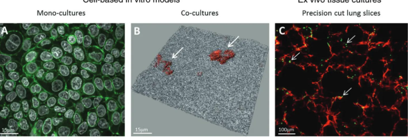
A number of promising examples have been published that carefully address physiological relevance, correlation and validation against established in vivo models. The respiratory tract, being the most sensitive entry port of nanomaterials, has been the focus of several hundred in vitro and in vivo studies [for reviews see (28–30)]. There-fore, it is not surprising that human advanced in vitro models have been developed in order to gain more insight into the mode of action of inhalable aerosols/(nano)parti-cles (Figure 1A, B).
Many monocultures exist that mimic the different compartments of lungs, i.e., conducting airways and lung parenchyma, however, the advantage of co-cultures in this research area was already recognized several years ago. The first two co-culture systems were described using epithelial and endothelial cells to study the impact of nan-oparticles (26, 31).
We recently reported on an in vitro triple-cell co-cul-ture model of the human airway wall composed of three main cell types: epithelial cells, human blood monocyte-derived macrophages and dendritic cells cultivated in a transwell system (32). A detailed characterization and val-idation of the system showed that the cell-cell interactions and communication in the culture system behave similar to that in vivo, indicating not only the proper functioning
Figure 1: Examples of conventional cell culture models and ex vivo models. Laser scanning micrographs representing epithelial monocul-tures (A549 lung epithelial type II cell line) labelled for F-actin (green) and cell nuclei (white) (xy projection) (A); co-culmonocul-tures of epithelial cells forming a tight monolayer (white) with macrophages on top (orange, arrows) (3D shadow projection with transparent renderings) (B), and a precision cut slice from rat lungs stained for F-actin (red) and macrophages (green, arrows) (C).
but also improved biological relevance of the system (29). Further improvements with four cell types were also described recently, showing that epithelial and endothe-lial cells, macrophages, and mast cells (33, 34) can be cul-tured together to study the impact of both engineered and environmental particles.
Liver- and nephrotoxicity profiles of nanomaterials used as carriers in nanobiomedical applications must be well defined since the materials have direct access to those organs when injected into the bloodstream. Nanomate-rial biodistribution studies elaborated in animal studies show a significantly high accumulation in the mononu-clear phagocyte system (MPS) (35). The liver, a multifunc-tional organ of the digestive system, plays a central role in homeostasis and detoxification of foreign substances (including nanomaterials) reaching the bloodstream (36). A number of in vitro approaches including two- (2D) and three-dimensional (3D) systems used to assess hepatotox-icity were summarized in great detail by Godoy and col-leagues in 2013 (37). In short, significant differences were observed in the response of hepatocytes after treatment with different drug substances depending on whether the cells were cultivated in monolayers or as 3D cultures, indicating the importance of the model system on the bio-logical effect assessment. Another recent report shows the potential of 3D liver microtissue models generated by primary hepatocytes for the toxicology assessment of nanomaterial exposures (38). Although promising 3D systems have been reported, the majority of the hazard assessments of nanomaterials to date have still involved hepatocyte monocultures, probably due to lack of clear guidance or validated advanced human in vitro systems.
The kidney, with its central role in metabolism and blood filtration, is continuously exposed to adverse metab-olites, drugs or nanoparticles, and therefore must be con-sidered in safety assessment. A recent study developed a 3D organoid kidney proximal tubule epithelial cell system based on isolated proximal tubules from male C57BL/6 mice cultivated in hyaluronic acid hydrogels (39, 40). Well-defined fluorescein isothiocyanate (FITC)-labelled carboxyl-terminated poly(amidoamine) (PAMAM) dendrim-ers < 6 nanometdendrim-ers in diameter were applied to consider an in-depth in vitro - in vivo correlation study. These dendrim-ers produced a set of toxicity indicators which accurately reflected the damage observed in vivo (41), indicating high predictive power of the system for nephrotoxicity. Further extension to disease models or human proximal tubules would help to strengthen this promising approach.
Despite all the advantages described in these exam-ples, advanced in vitro systems have become more and more time- and cost-intensive, and well-trained experts
are also needed to work with complex cell culture systems, all issues that should not be neglected. Therefore, ex vivo tissue preparation might present a viable alternative approach to obtain predictive model systems.
Ex vivo tissue preparation: bringing in vivo
tissue into Petri dishes
A promising alternative approach that provides a better in vivo-like environment is the use of precision-cut tissue slices (PCS), which represent an ex vivo model of the organ of study by maintaining the original architecture, i.e., taining all the cells types of the tissue in their natural con-formation (42). The advantage of this system is that slices from different species can be prepared and compared, such as from rodents as well as human biopsy material. Particu-larly impressive progress towards this end has recently been made in the field of lung research [for a review see (43)]. Although most studies have focused on aspects of pharma-toxicology (44, 45), some recent publications have proven that PCS-based approaches are highly relevant for the risk assessment of nanomaterials and xenobiotics in general, in terms of inflammation, organ injury and sensitization (46– 48). A recently published study showed, however, that the system might only be useful for ions released from nano-particles or soluble substances since it was observed that the silver nanoparticles did predominantly attach at the cut surfaces of the PCS from lung tissues (Figure 1C) but could hardly penetrate into deeper regions (49).
Fewer studies have been published using liver PCS to assess the interaction of nanomaterials with the organ slices (50, 51). The disadvantage of this technique is that by cutting the organ into slices the surface is covered with damaged cells, which might themselves induce an inflam-matory reaction. However, in general, this system has great potential and warrants further exploration.
Regardless of the chosen approach (bottom up or ex vivo), the balance between relevant output data and throughput capability has to be evaluated carefully. Not only throughput suitability but also the development of high content analysis applicable to advanced in vitro as well as ex vivo models should be considered to be able to gain further mechanistic understanding in (nano)material safety assessment [see also Ref (52) for more detailed insight].
Organs-on-chip approaches
Innovative in vitro platforms based on microfluidic tech-nologies and aimed at assessing nanomaterial safety
and efficacy are currently emerging [for a review see (53)]. These systems, called “organs-on-chip” (54, 55), intend to mimic the physiological conditions and archi-tecture of human tissues. 3D organoids made of human cells are created on bioengineered platforms in order to simulate key organ-level functions (56). These systems, which mimic the in vivo environment of specific organs in an unprecedented way, are widely seen as having the potential to improve in vitro model accuracy and experi-mental efficiency. Although most of the ongoing efforts target the development of more predictive preclinical in vitro models (57), the potential of these devices for toxi-cology evaluation of chemicals, such as nanomaterials, is undisputable.
One of the key assets of these novel technologies is their capability to accurately reproduce specific aspects of the cellular microenvironment of various tissues. They not only enable the creation of microstructures with dimen-sions that are similar to those of mammalian cells, but also allow control of the cellular microenvironment in space and time. The perfusion of cell cultures confined in such systems enables them to mimic the continuous transport of nutrients and oxygen, the dilution of the secreted cytokines and chemokines, in addition to the cel-lular waste products (58). Insoluble signals can also be reproduced by modifying the cell culture surface topogra-phy or stiffness, and/or by the addition of specific extra-cellular matrices (59). Mechanical stimuli, such as the cyclic mechanical strain of respiration movements, are additional and important factors that affect the cellular and organ homeostasis (60).
Microfluidic technologies offer further possibilities to recreate the cell microenvironment, including the integra-tion of cell culture membranes in microfluidic chambers, allowing for the creation of bioartificial barriers. Micro-fluidic barriers have recently been reported to recreate a number of in vivo barriers, such as the liver sinusoidal barrier (61), the blood-brain barrier (62, 63), and the gut (64), to name just a few.
Microfluidic air-liquid interfaces mimicking the alveolar barrier using porous membranes have also been described. Nalayanda and colleagues reported a perfused lung alveolar epithelial layer cultured on a PET porous membrane (65), whereas Zheng et al. (66) investigated the mechanical stresses induced by liquid plug propa-gation in a similar airway model. Recently, even more sophisticated lung alveolar models were reported that were able to mimic the mechanical processes of physi-ological breathing and the shear stress induced by the blood stream. Douville et al. (67) reported a microfluidic chip equipped with a 100 μm-thick poly(dimethysiloxane)
Figure 2: Example of an organ-on-chip: A lung-on-a-chip made of an array of three lung alveoli with a thin, alveolar barrier that can be cyclically stretched to mimic the respiration movements. Scale bar 5 mm.
(PDMS) membrane on which epithelial cells were cultured and cyclically stretched. This microfluidic alveolar model allowed the recreation of the fluid and solid mechanical stresses taking place in the alveoli during mechanical ventilation. In contrast to the latter model, Huh et al. (68) reported an innovative “breathing” lung-on-a-chip device in which the air-liquid interface was reproduced, with a 10 μm-thin, porous PDMS membrane on which epithelial and endothelial cells were cultured. In an experiment in which the epithelium was exposed to 12 nm silica nano-particles, evidence was obtained that “breathing” motions greatly accentuate the pro-inflammatory activities of silica nanoparticles. Furthermore, it was demonstrated that translocation of these nanoparticles across the alveolar-capillary interface was significantly increased in mechan-ically-stressed alveoli in comparison with static controls. Very recently, a novel lung alveolar barrier model was reported consisting of a 3.5 μm thin, porous and flexible membrane on which epithelial and endothelial cells were cultured and whose actuation mechanism is inspired by the lung diaphragm contraction (Figure 2). A permeability assay carried out with this advanced model revealed that the permeability of small hydrophilic molecules (FITC-Na+) was significantly increased if the cells were
mechani-cally and cyclimechani-cally stretched. In contrast, no significant effect was observed for a larger molecule [rhodamine B isothiocyanate-dextran (RITC-dextran)](69). These results are comparable to an in vivo study (70) that showed a higher clearance of hydrophilic solute upon increasing the human lung volume, suggesting that the novel lung alveolar barrier model better mimics the in vivo situation than existing in vitro models.
Although the expectations in the potential of such approaches are very high, their development is still in their infancy. More validation steps will be required to identify their unique possibilities and the benefits of reproduc-ing organ-level functions (e.g., breathreproduc-ing movements). One of the key factors that will need to be improved is the shift towards more relevant cells (e.g., primary cells) from the cell lines that are still broadly used in such models. Once these requirements are fulfilled such models may well provide low-cost alternatives to animal and clinical studies for faster and more efficient drug screening and material toxicological testing.
Qualification of reliable and
accurate in vitro models
The early results of 2D model systems assessing the toxic-ity of nanomaterials were often contradictory, for several reasons that were outlined in a number of publications over the last few years (71–75), and lacked in vitro - in vivo cor-relation. The reasons can be summarized into five groups: (i) no correlation between simple in vitro models and in
vivo measurements (39–41)
(ii) no informative assay controls documenting the per-formance characteristics of the model system (76) (iii) no adequate calculation of the applied dosages of
nanomaterials
(iv) absence of appropriate interference controls uncover-ing potential interference between nanomaterials and the detection system (73)
(v) absent or incomplete characterization of nano-materials (71)
The first reason (i) illustrates the poor relevance of common in vitro cell assays for the assessment of acute toxic effects of nanomaterials on full biological organisms. Furthermore, recent results of an interlaboratory compari-son underline that the last four aspects (ii–v) are impor-tant in obtaining an adequate comparability between the results of the different participating laboratories.
A number of recent studies illustrate that basic 2D cell culture models lack a reasonable correlation with in vivo studies (39–41). Therefore their predictability and relevance is also poor. At the same time the correla-tion between rodent models and humans is around 60% (77), which leads to some augmented risk in early clinical trials. Recently published results demonstrate some cor-relation between advanced 3D cell culture systems and in vivo models (39–41). This highlights the importance
of the adequacy of the selected model systems and their qualification with respect to reliability and accuracy. In a number of instances it proved difficult to determine the reasons for the poor comparability of common cell assays used for nanotoxicity assessments, as the controls were inadequate to document proper functioning of each step of an experimental procedure. A cause-and-effect analysis was an effective tool to categorize the various influences affecting the performance of common MTS cell prolifera-tion assays (78). Based on this outline, a number of con-trols that monitor the assay performance for the different steps of the procedure were integrated into the design of the standard 96-well plate. Results of a recent interlabora-tory comparison showed the combined strength of these controls to uncover potential shortcomings in the stand-ard operating procedure (unpublished observations, Dr. Matthias Roesslein). In addition, the full titration of the chemical positive control and the deduced EC50 value that is within tight specifications allows for documenta-tion of the proper funcdocumenta-tioning of the entire assay itself for each experiment. This enables comparability within and between laboratories over a longer period of time. The six major categories of a cause and effect analysis, such as cell maintenance, pipetting, instrument performance, toxic chemical positive control, assay protocol, and engi-neered nanomaterial handling and characterization, will also help in determining most influences of advanced 3D cell culture systems. This systematic summarization of effects facilitates the overview of functional principles of even complex cell culture systems. Furthermore, it allows the design of adequate controls that document their per-formance characteristics, in particular the EC50 value chemical positive control.
Until the publication of the in vitro sedimentation, diffusion and dosimetry (ISDD) model in 2010 (76), there was only a limited understanding of the actual dosing levels of nanomaterials during nanomaterial-cell inves-tigations using adhesive cell culture assays. With all rel-evant information stated in the publications, this allows conversion of the common weight/volume as the dosing unit of the nanomaterials to the actual number of particles interacting with the cells over the duration of the experi-ment. Even at the early stage of any advanced models, the calculation of the proper dosing of the investigated nano-materials is examined and potential scenarios have been outlined. The model has been extended by Rodriguez- Lorenzo and colleagues, who included heterogeneity, including polydispersity both in size and mass density to study the influence of heterogeneity on the particokinetics of nanoparticles and on the corresponding delivered dose (79). Therefore it will be essential that at least a full set of
dosing-relevant characteristics is given in a publication so that in the future a direct comparisons of results of the dif-ferent approaches is feasible.
Interference controls will require further optimiza-tions, as the readout system of any endpoint in a co-cul-ture or advanced 3D model system will be refined due to the more complex structure of these systems compared to a simple adhesive cell culture. In addition, basic materials characterization of any nanomaterial remains essential, regardless of the applied type of in vitro or ex vivo model system (71).
Regulatory environment
This review focuses on the scientific issues, the possibilities and limitations of advanced in vitro – and ex vivo model systems established to date for the risk assessments related to nanomaterial exposure. It summarizes the models that may one day have to qualify for the regulatory review and some unresolved questions about a “fit for purpose” solu-tion within the current regulatory framework. Two differ-ent regulatory frameworks address the specific objectives of industrial and medical applications. Industrial nano-materials are regulated under the REACH and OECD guide-lines following the tracks of chemicals with toxicity testing for product fillings. In addition, within the EU there are considerable efforts to replace the expensive and contro-versial discussed animal testing with in vitro alternatives (5–7). Despite the fact that changing this regulation is a long process, first efforts have been successful within the EU with a ban on animal testing for skin allergies of cosmet-ics (80). In contrast, the regulatory environment of medical related nanomaterials is highly fragmented with individual national regulations. The testing strategy of the pharma-ceutical industry focuses mainly on avoiding failure during clinical trials and their tremendous costs. Therefore they have a considerable interest in novel in vitro models able to better predict the clinical outcome.
Conclusions
Due to the inevitable exposure of nanomaterials to humans it is imperative to gain an understanding of how these materials interact with the human body, since there are increasing concerns as to the potential adverse effects on human health that the production of, and subsequent exposure to, such nanomaterials might pose. In the field of regulatory toxicology, animal testing is still the most
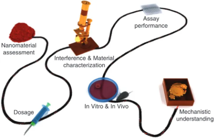
Assay performance
Interference & Material characterization
In Vitro & In Vivo
Mechanistic understanding Dosage
Nanomaterial assessment
Figure 3: Important components that allow to link nanomaterial assessment with mechanistic understanding in a biological system in a reliable way.
prevalent model used for risk assessment. It is, however, time to realize that a paradigm-shift in the understanding of toxicology towards a modern evidence-based research discipline can be supported by advanced in vitro and ex vivo models. It has already been recognized that in vitro and ex vivo models should be able to depict the complexity of an organ or tissue as far as possible, while maintaining the capability for standardization, high throughput and reproducibility. In addition the relevance of these models has to be validated towards animal models and especially towards human related epidemiological studies for nano-materials exposure. Furthermore clinical trials will be needed to assess the relevance of theses novel models for nanomaterials to be used for medical purpose.
Extensive efforts have been made to simulate differ-ent organs in a petri dish, and predictive cell culture or ex vivo models have been generated. It will require, however, a combined effort of many disciplines to focus more on the reliability of such systems by considering the thor-ough characterization of nanomaterials, but also the cell culture systems. In addition, validation with well-known toxic substances should be emphasized in order to gain a proof-of-concept.
Acknowledgments: This work was supported by the Swiss
National Science Foundation (Grants # 320030_138365; PP00P2_123373), the NRP64 program (Grants # 406440-131266/1; 4064-131232), the Adolphe Merkle Founda-tion, the Swiss National Science Foundation through the National Centre of Competence in Research Bio-Inspired Materials and by the Competence Centre for Materials Science and Technology (CCMX) Project Nano-screen. The Gebert-Rüf Stiftung (GRS-066/11) and the Swiss Com-mission for the Technology and Innovation (CTI 15794.1
PFLS-LS) are also acknowledged for their support. The authors thank Dr. Miguel Spuch Calvar for the design of Figure 3.
References
1. European Commission. Commission recommendation of 18 October 2011 on the definition of nanomaterial. OJL 2011;275:38–40.
2. Nel A, Xia T, Mädler L, Li N. Toxic potential of materials at the nanolevel. Science 2006;311:622–7.
3. Wick P, Grafmueller S, Petri-Fink A, Rothen-Rutishauser B. Advanced human in vitro models to assess metal oxide nanoparticle-cell interactions. MRS Bulletin 2014;39:984–9. 4. Astashkina A, Grainger DW. Critical analysis of 3-D organoid in
vitro cell culture models for high-throughput drug candidate toxicity assessments. Adv Drug Deliv Rev 2014;70:1–18. 5. Hartung T, Rovida C. Chemical regulators have overreached.
Nature 2009;460:1080–1.
6. National Research Council. Toxicity testing in the 21st century: a vision and a strategy. Washington, DC: The National Academies Press, 2007.
7. Rossini GP, Hartung T. Food for thought … Towards tailored assays for cell-based approaches to toxicity testing. ALTEX 2012;29:359–72.
8. Törnqvist E, Annas A, Granath B, Jalkesten E, Cotgreave I, Öberg M. Strategic focus on 3R principles reveals major reductions in the use of animals in pharmaceutical toxicity testing. PLoS One 2014;9:e101638.
9. www.lscv.ch/de/pages/tierversuche/alternativmethoden/ news/2013/ohne_tiere.html, 2014.
10. EU. Seventh report from the commission to the council and the European parliament on the statistics on the number of animals used for experimental and other scientific purposes in the mem-ber states of the European Union 2014.
11. Oberdorster G, Stone V, Donaldson K. Toxicology of nanoparti-cles: A historical perspective. Nanotoxicology 2007;1:2–25. 12. Niesink RJM, De Vries J, Holliger MA. Toxicology: principles and
applications. London: CRC Press, 1996;ISBN 10 0849392322. 13. Stern ST, McNeil SE. Nanotechnology safety concerns revisited.
Toxicol Sci 2008;101:4–21.
14. Michiels C. Endothelial cell functions. J Cell Physiol 2003;196:430–43.
15. Alberts B, Bray D, Johnson A, Johnson L, Raff M, Roberts K. Essential cell biology. An introduction to the molecular biology of the cell. New York: Garland Publishing, Inc., 1998.
16. Fadeel B, Garcia-Bennett AE. Better safe than sorry: understand-ing the toxicological properties of inorganic nanoparticles manufactured for biomedical applications. Adv Drug Deliv Rev 2010;62:362–74.
17. Johnston HJ, Hutchison G, Christensen FM, Peters S, Hankin S, Stone V. A review of the x and in vitro toxicity of silver and gold par-ticulates: particle attributes and biological mechanisms responsi-ble for the observed toxicity. Crit Rev Toxicol 2010;40:328–46. 18. Zurlo J, Rudacille D, Goldberg AM. Animals and alternatives in testing, history, science and ethics. New York: Mary Ann Liebert, Inc., 1993.
19. Puck TT, Marcus PI. A rapid method for viable cell titration and clone production with hela cells in tissue culture: the use of X-irradiated cells to supply conditioning factors. Proc Natl Acad Sci USA 1955;41:432–7.
20. Köhler G, Milstein C. Continuous cultures of fused cells secreting antibody of predefined specificity. Nature 1975;256:495–7. 21. Dulbecco R, Freeman G. Plaque production by the polyoma
virus. Virology 1959;8:396–7.
22. Lancaster MA, Renner M, Martin CA, Wenzel D, Bicknell LS. Cerebral organoids model human brain development and microcephaly. Nature 2013;501:373–79.
23. Kelm JM, Lorber V, Snedeker JG, Schmidt D, Broggini-Tenzer A, Weisstanner M, et al. A novel concept for scaffold-free vessel tissue engineering: self-assembly of microtissue building blocks. J Biotechnol 2010;148:46–55.
24. Handler JS, Green N, Steele RE. Cultures as epithelial models: porous-bottom culture dishes for studying transport and dif-ferentiation. Methods Enzymol 1989;171:736–44.
25. Bermudez LE, Sangari FJ, Kolonoski P, Petrofsky M, Goodman J. The efficiency of the translocation of Mycobacterium tuber-culosis across a bilayer of epithelial and endothelial cells as a model of the alveolar wall is a consequence of transport within mononuclear phagocytes and invasion of alveolar epithelial cells. Infect Immun 2002;70:140–6.
26. Birkness KA, Deslauriers M, Bartlett JH, White EH, King CH, Quinn FD. An in vitro tissue culture bilayer model to examine early events in mycobacterium tuberculosis infection. Infect Immun 1999;67:653–8.
27. Voisin C, Aerts C, Jakubczk E, Tonnel AB. La culture cellulaire en phase gazeuse. Un nouveau modele experimental d’etude in vitro des activites des macrophages alveolaires. Bull Eur Physi-opathol Respir 1977;13:69–82.
28. Nichols JE, Niles JA, Vega SP, Argueta LB, Eastaway A, Cortiella J. Modeling the lung: design and development of tissue
engineered macro- and micro-physiologic lung models for research use. Exp Biol Med (Maywood) 2014;239:1135–69. 29. Rothen-Rutishauser B, Blank F, Mühlfeld C, Gehr P. In vitro
models of the human epithelial airway barrier to study the toxic potential of particulate matter. Expert Opin Drug Metab Toxicol 2008;4:1075–89.
30. Hittinger M, Juntke J, Kletting S, Schneider-Daum N, de Souza Carvalho C, Lehr CM. Preclinical safety and efficacy models for pulmonary drug delivery of antimicrobials with focus on in vitro models. Adv Drug Deliv Rev 2014;S0169-409X:219–1.
31. Hermanns MI, Kasper J, Dubruel P, Pohl C, Uboldi C, Vermeersch V, et al. An impaired alveolar-capillary barrier in vitro: effect of proinflammatory cytokines and consequences on nanocarrier interaction. J R Soc Interface 2010;7:41–54. 32. Rothen-Rutishauser B, Kiama SG, Gehr P. A three-dimensional
cellular model of the human respiratory tract to study the inter-action with particles. Am J Respir Cell Mol Biol 2005;32:281–9. 33. Alfaro-Moreno E, Nawrot TS, Vanaudenaerde BM, Hoylaerts MF,
Vanoirbeek JA, Nemery B, et al. Co-cultures of multiple cell types mimic pulmonary cell communication in response to urban PM10. Eur Respir J 2008;32:1184–94.
34. Klein SG, Serchi T, Hoffmann L, Blömeke B, Gutleb AC. An improved 3D tetraculture system mimicking the cellular organi-zation at the alveolar barrier to study the potential toxic effects of particles on the lung. Part Fibre Toxicol 2013;10:31.
35. Kreyling WG, Hirn S, Schleh C. Nanoparticles in the lung. Nat Biotechnol 2010;28:1275–6.
36. Sasse D, Spornitz UM, Maly IP. Liver architecture. Enzyme 1992;46:8–32.
37. Godoy P, Hewitt NJ, Albrecht U, Andersen ME, Ansari N, Bhattacharya S, et al. Recent advances in 2D and 3D in vitro sys-tems using primary hepatocytes, alternative hepatocyte sources and non-parenchymal liver cells and their use in investigating mechanisms of hepatotoxicity, cell signaling and ADME. Arch Toxicol 2013;87:1315–530.
38. Kermanizadeh A, Løhr M, Roursgaard M, Messner S, Gunness P, Kelm JM, et al. Hepatic toxicology following single and multiple exposure of engineered nanomaterials utilising a novel primary human 3D liver microtissue model. Part Fibre Toxicol 2014;11:56.
39. Astashkina AI, Mann BK, Prestwich GD, Grainger DW. Comparing predictive drug nephrotoxicity biomarkers in kidney 3-D primary organoid culture and immortalized cell lines. Biomaterials 2012;33:4712–21.
40. Astashkina AI, Mann BK, Prestwich GD, Grainger DW. A 3-D organoid kidney culture model engineered for high-throughput nephrotoxicity assays. Biomaterials 2012;33:4700–11. 41. Astashkina AI, Jones CF, Thiagarajan G, Kurtzeborn K,
Ghandehari H, Brooks BD, et al. Nanoparticle toxicity assess-ment using an in vitro 3-D kidney organoid culture model. Biomaterials 2014;35:6323–31.
42. De Graaf IA, Olinga P, de Jager MH, Merema MT, de Kanter R, van de Kerkhof EG, et al. Preparation and incubation of precision-cut liver and intestinal slices for application in drug metabolism and toxicity studies. Nat Protoc 2010;5:1540–51. 43. Liberati TA, Randle MR, Toth LA. In vitro lung slices: a powerful
approach for assessment of lung pathophysiology. Expert Rev Mol Diagn 2010;10:501–8.
44. Morin JP, Baste JM, Gay A, Crochemore C, Corbière C, Monteil C. Precision cut lung slices as an efficient tool for in vitro lung physio-pharmacotoxicology studies. Xenobiotica 2013;43: 63–72.
45. Schlepütz M, Rieg AD, Seehase S, Spillner J, Perez-Bouza A, Braunschweig T, et al. Neurally mediated airway constriction in human and other species: a comparative study using precision-cut lung slices (PCLS). PLoS One 2012;7:e47344.
46. Kim YH, Tong H, Daniels M, Boykin E, Krantz QT, McGee J, et al. Cardiopulmonary toxicity of peat wildfire particulate matter and the predictive utility of precision cut lung slices. Part Fibre Toxicol 2014;11:29.
47. Lauenstein L, Switalla S, Prenzler F, Seehase S, Pfennig O, Förster C, et al. Assessment of immunotoxicity induced by chemicals in human precision-cut lung slices (PCLS). Toxicol In Vitro 2014;28:588–99.
48. Sauer UG, Vogel S, Aumann A, Hess A, Kolle SN, Ma-Hock L, et al. Applicability of rat precision-cut lung slices in evaluating nanomaterial cytotoxicity, apoptosis, oxidative stress, and inflammation. Toxicol Appl Pharmacol 2014;276:1–20. 49. Hirn S, Haberl N, Loza K, Epple M, Kreyling WG, Rothen-
Rutishauser B, et al. Pro-inflammatory and cytotoxic response to nanoparticles in precision-cut lung slices. Beilstein J Nanotechnol 2014;5:2440–9.
50. Dragoni S, Franco G, Regoli M, Bracciali M, Morandi V, Sgaragli G, et al. Gold nanoparticles uptake and cytotoxicity assessed on rat liver precision-cut slices. Toxicol Sci 2012;128:186–97.
51. Jetten MJ, Claessen SM, Dejong CH, Lahoz A, Castell JV, van Delft JH, et al. Interindividual variation in response to xenobiotic exposure established in precision-cut human liver slices. Toxicology 2014;2:61–9.
52. Giuliano KA, Haskins JR. High content screening: a powerful approach to systems cell biology and drug discovery. Totowa, NJ: Humana Press, 2010; ISBN 1-61737-746-5. 53. Mahto SK, Charwat V, Ertl P, Rothen-Rutishauser B, Rhee SW,
Sznitman J. Microfluidic platforms for advanced risk assess-ments of nanomaterials. Nanotoxicology 2014;22:1–15. 54. Ghaemmaghami AM, Hancock MJ, Harrington H, Kaji H,
Khademhosseini A. Biomimetic tissues on a chip for drug discovery. Drug Discovery Today 2012;17:173–81.
55. Marx U, Walles H, Hoffmann S, Lindner G, Horland R, Sonntag F, et al. Human-on-a-chip’ developments: a translational cutting-edge alternative to systemic safety assessment and efficiency evaluation of substances in laboratory animals and man? Altern Lab Anim 2012;40:235–57.
56. Bhatia SN, Ingber DE. Microfluidic organs-on-chips. Nat Biotechnol 2014;32:760–72.
57. http://www.nih.gov/news/health/sep2011/od-16.htm. 2014. 58. Young EW, Beebe D. Fundamentals of microfluidic cell culture in
controlled enviroments. Chem Soc Rev 2010;39:1036–48. 59. Choi CK, Breckenridge MT, Chen CS. Engineered materials
and the cellular microenvironment: a strengthening interface between cell biology and bioengineering. Trends Cell Biol 2010;20:705–14.
60. Polacheck WJ, Li R, Uzel SG, Kamm RD. Microfluidic platforms for mechanobiology. Lab Chip 2013;13:2252–67.
61. Lee PJ, Hung PJ, Lee LP. An artificial liver sinusoid with a micro-fluidic endothelial-like barrier for primary hepatocyte culture. Biotechnol Bioeng 2007;97:1340–6.
62. Booth R, Kim H. Characterization of a microfluidic in vitro model of the blood-brain barrier (muBBB). Lab Chip 2012;12:1784–92.
63. Griep LM, Wolbers F, de Wagenaar B, ter Braak PM, Weksler BB, Romero IA, et al. BBB on chip: microfluidic platform to mechanically and biochemically modulate blood-brain barrier function. Biomed Microdevices 2012;15:145–50.
64. Kim HJ, Ingber DE. Gut-on-a-Chip microenvironment induces human intestinal cells to undergo villus differentiation. Integr Biol (Camb) 2013;5:1130–40.
65. Nalayanda DD, Wang Q, Fulton WB, Wang TH, Abdullah F. Engineering an artificial alveolar-capillary membrane: a novel continuously perfused model within microchannels. J Pediatr Surg 2010;45:45–51.
66. Zheng Y, Fujioka H, Bian S, Torisawa Y, Huh D, Takayama S. Liquid plug propagation in flexible microchannels: a small airway model. Phys. Fluids (Woodbury, NY: 1994) 2009;21:71903.
67. Douville NJ, Zamankhan P, Tung YC, Li R, Vaughan BL, Tai CF. Combination of fluid and solid mechanical stresses contribute to cell death and detachment in a microfluidic alveolar model. Lab Chip 2011;11:609–19.
68. Huh D, Matthews BD, Mammoto A, Montoya-Zavala M, Hsin HY, Ingber DE. Reconstituting organ-level lung functions on a chip. Science (New York, N Y) 2010;328:1662–8.
69. Stucki A, Stucki JD, Hall S, Felder M, Mermoud Y, Schmid R, et al. A lung-on-chip array with an integrated bio-inspired respiration mechanism. Lab Chip 2015;15:1302–10.
70. Marks JD, Luce JM, Lazar NM, Wu JN, Lipavsky A, Murray JF. Effect of increases in lung volume on clearance of aerosolized solute from human lungs. J Appl Physiol 1985;59:1242–8.
71. Krug H, Wick P. Nanotoxicology: an interdisciplinary challenge. Angewandte Chemie (International Edition) 2011;50:1260–78. 72. Kaiser JP, Roesslein M, Buerki-Thurnherr T, Wick P. Carbon
nanotubes – curse or blessing. Curr Med Chem 2011;18: 2115–28.
73. Schrurs F, Lison D. Focusing the research efforts. Nat Nanotechnol 2012;7:546–48.
74. Cohen JM, Teeguarden JG, Demokritou P. An integrated approach for the in vitro dosimetry of engineered nanomateri-als. Part Fibre Toxicol 2014;11:20.
75. DeLoid G, Cohen JM, Darrah T, Derk R, Rojanasakul L, Pyrgiotakis G, et al. Estimating the effective density of engineered nanomaterials for in vitro dosimetry. Nat Comm 2014;5:3514.
76. Hinderliter PM, Minard KR, Orr G, Chrisler WB, Thrall BD, Pounds JG, et al. ISDD: a computational model of particle sedimentation, diffusion and target cell dosimetry for in vitro toxicity studies. Part Fibre Toxicol 2010;7:36.
77. Hartung T. Toxicology for the twenty-first century. Nature 2009;460:208–12.
78. Rösslein M, Elliott JT, Salit M, Petersen EJ, Hirsch C, Krug HF, et al. Use of Cause-and-Effect Analysis to Design a High-Quality Nanotoxicology Assay. Chemical Research Toxicol
2015;28:21–30.
79. Rodriguez-Lorenzo L, Rothen-Rutishauser B, Petri-Fink A, Balog S. Nanoparticle polydispersity can strongly affect in vitro dose. Part Part Syst Charact 2015;32:321–33.
80. The European Parliament and the Council of the European Union (2010). Directive 2010/63/EU, Directive 2010/63/EU of the Euro-pean parliament and of the council. Available at: http://eur-lex. europa.eu/LexUriServ/LexUriServ.do?uri=OJ:L:2010:276:0033: 0079:en:PDF (accessed March 10, 2015).
Bionotes
Peter Wick
Empa, Swiss Federal Laboratories for Material Science and Technology, Particles-Biology Interactions Laboratory, Lerchenfeldstrasse 5, St. Gallen, Switzerland
Peter Wick has, since 2014, headed the research laboratory for Particles-Biology Interactions at the Federal Laboratories on Materi-als Science and Technologies Empa in St Gallen. He studied and received his PhD in Cell and Molecular Biology at the University in Fribourg (Switzerland). Thereafter (2002) he moved to Empa and began his research in nanotoxicology among other topics with the national project NanoRisk, and is now active in further projects of the 7th Framework program of the EC, for example, NANOHOUSE,
MARINA, NanoSolutions & Flagship Graphene and is also founded by the SNF NFP64 research program. His general research interest is to study the interactions of nanomaterials with human barrier tissues in vitro and ex vivo with the purpose to obtain detailed mechanistic information about their uptake, accumulation, trans-port and effect on different types of cells or entire tissue. In addition he is interested in the improvement of currently used acute toxi-cological test methods to obtain reliable and robust results. He is author of around 100 publications, including over 60 peer-reviewed papers, in the field of Nanosafety. He is a member of the advisory board of the Swiss Action Plan on Nanomaterials and is an Editorial Board Member of Nanotoxicology.
Savvina Chortarea
BioNanomaterials, Adolphe Merkle Institute, University of Fribourg, Ch. de Verdiers 4, Fribourg, Switzerland
Savvina Chortarea obtained her BSc in Biology, with a specialization in Molecular Biology in 2010, at the Aristotle University of Thessa-loniki, Greece. She then graduated from the University of Edinburgh in the UK with an MSc in Medicinal and Biological Chemistry, in 2011. She is currently working towards her PhD degree in Nanotoxi-cology at the Adolphe Merkle Institute, University of Fribourg under the supervision of Prof. Barbara Rothen-Rutishauser and Prof. Alke Fink. Her research focuses on nanoparticle-lung cell interactions upon realistic, chronic exposures.
Olivier T. Guenat
ARTORG Center, Lung Regeneration Technologies, Murtenstrasse 50, Bern, Switzerland; and Pulmonary Medicine and Thoracic Surgery Divisions, University Hospital of Berne, Bern, Switzerland
Olivier T. Guenat is the Head of the Lung Regeneration Technologies Group at the Artificial Organs Center for Biomedical Engineering Research (ARTORG) at the University of Berne in Switzerland. He is associated with the Pulmonary Medicine and the Thoracic Surgery Divisions of the University Hospital of Berne. His research focuses on the development of organs-on-chip, in particular lung-on-chips that mimic the healthy and diseased in-vivo cellular microenviron-ments of the lung. Prior to his position at the University of Berne, he held a position at the Swiss Center for Electronics and Micro-electronics (CSEM), at the Ecole Polytechnique de Montréal (QC, Canada), before which he performed a post-doc at Harvard Medical School in Boston and at the University of Neuchâtel in Switzerland. He has received several awards such as a fellowship for advanced researchers from the Swiss National Foundation. He is also the founder of AlveoliX, a biotech start-up that aims at bringing organs-on-chip on the market.
Matthias Roesslein
Empa, Swiss Federal Laboratories for Material Science and Technology, Particles-Biology Interactions Laboratory, Lerchenfeldstrasse 5, St. Gallen, Switzerland
Matthias Roesslein studied Chemistry at the University of Basel from 1981 to 1985 and received his PhD degree with “summa cum laude” in 1989. Afterward he spent to 2 years as a postdoc at the University of Chicago and 4 years as an Assistant Professor at the Physical-Chemical Institute of the University Zurich, before joining Empa in 1996. From then he specialized as one of the world leading experts in “evaluation of measurement uncertainty and metrol-ogy” and wrote substantial parts of several guidelines in analytical chemistry. In 2006 he was appointed a position as Senior Scientist and joined the Empa laboratory for “Materials-Biology Interaction” focusing mainly on the standardization of in vitro assays to eluci-date the effect of engineered nanomaterials on different cell types.
Janick D. Stucki
ARTORG Center, Lung Regeneration Technologies, Murtenstrasse 50, Bern, Switzerland
Janick D. Stucki is a PhD student at the ARTORG Center for Biomedi-cal Engineering Research in the Lung Regeneration Technologies Lab (Bern, Switzerland). He studied Mechanical Engineering (BSc and MSc) at the Swiss Federal Institute of Technology (ETH Zurich, Switzerland). He conducted his Master’s thesis at IBM Research (Zurich) in the field of Microfluidics for Diagnostic Applications. His current research interests focus on the development of novel micro-fluidic organ-on-chip technologies (lung-on-chip) for biomedical and biological applications.
Stephanie Hirn
Walter Brendel Centre of Experimental Medicine, Klinikum der Universität München, Munich, Germany
Stephanie Hirn studied Biology at the Technische Universität München. In 2010, she joined the Institute of Lung Biology and Disease at the Helmholtz Zentrum München. There, she was involved in projects focusing on the biokinetics and lung toxicity of different nanomaterials. Since 2012, she has been working at the Walter Brendel Centre of Experimental Medicine of the Ludwig-Max-imilians-Universität München and received her Doctoral degree in 2014. Her main interests are the interactions of nanoparticles with cells of the lung and endothelial cells.
Alke Petri-Fink
BioNanomaterials, Adolphe Merkle Institute, University of Fribourg, Ch. de Verdiers 4, Fribourg, Switzerland
Alke Petri-Fink received her PhD in Chemistry from the University of Ulm, Germany in 1999. After a post-doctoral stay at the University of Gainesville, Florida, she joined the Institute of Materials Science at the École Polytechnique Fédérale de Lausanne (EPFL), first as a post-doctoral researcher, then as a Senior Scientist. She became an Associate Swiss National Science Foundation Professor in the Department of Chemistry at the University of Fribourg in 2009, and Full Professor in 2011 at the Adolphe Merkle Institute, Switzerland. Her research focuses on inorganic nanoparticles, their synthesis, surfaces, and interactions with biological cells.
Barbara Rothen-Rutishauser
BioNanomaterials, Adolphe Merkle Institute, University of Fribourg, Ch. de Verdiers 4, CH-1700 Fribourg, Switzerland, barbara.rothen@unifr.ch
Barbara Rothen-Rutishauser has received her PhD in 1996 in Cell Biology at the Swiss Federal Institute of Technology (ETH) in Zurich. From 1996 to 2000 she held a post-doctoral position in Biopharmacy at the Institute of Pharmaceutical Sciences at the ETH and in 2000 she joined Prof. Peter Gehr’s research group at the University of Bern, Switzerland. Barbara is an expert in the field of cell-nanoparticle interactions in the lung, with a special focus on 3D lung cell models and various microscopy techniques such as laser scanning and transmission electron microscopy. Since 2011 she is the new chair in BioNanomaterials at the Adolphe Merkle Institute, University of Fribourg, Switzerland, the position is shared equally with Prof. Alke Fink. The research group’s activities stretch over many fields from material synthesis and characterization to biologi-cal responses and risk assessment.