ORIGINAL ARTICLE
Effect of tin
–chloride pretreatment on bond strength
of two adhesive systems to dentin
Anne Peutzfeldt&Tamara Koch&Carolina Ganss&Simon Flury&Adrian Lussi
Received: 21 November 2012 / Accepted: 25 March 2013 / Published online: 17 April 2013 # Springer-Verlag Berlin Heidelberg 2013
Abstract
Objectives To determine the effect on resin composite-to-dentin bond strength of incorporation of an acidic tin–chlo-ride pretreatment in two adhesive systems.
Materials and methods Human molars were ground to ex-pose mid-coronal dentin. For microtensile bond strength (μTBS) testing, dentin was treated with Optibond FL or Clearfil SE according to one of six protocols (n=22/group). Group 1: Phosphoric acid etching, Optibond FL Prime, Optibond FL Adhesive (manufacturer’s instructions; con-trol); Group 2: Tin–chloride pretreatment, Optibond FL Prime, Optibond FL Adhesive; Group 3: Phosphoric acid etching, tin–chloride pretreatment, Optibond FL Prime, Optibond FL Adhesive; Group 4: Clearfil SE Primer, Clearfil SE Bond (manufacturer’s instructions; control); Group 5: Phosphoric acid etching, Clearfil SE Primer, Clearfil SE Bond; and Group 6: Tin–chloride pretreatment, Clearfil SE Primer, Clearfil SE Bond. The molars were then built up with resin composite (Clearfil Majesty Esthetic).
After storage (1 week, 100 % humidity, 37 °C) theμTBS was measured and failure mode was determined. Addition-ally, pretreated dentin surfaces were evaluated using SEM and EDX. TheμTBS results were analyzed statistically by a Welch Two Sample t-test and a Kruskal–Wallis test followed by exact Wilcoxon rank sum tests with Bonferroni–Holm adjustment for multiple testing (α=0.05).
Results When Optibond FL was used, partial or total re-placement of phosphoric acid with tin–chloride decreased μTBS significantly. In contrast, when Clearfil SE was used, inclusion of a tin–chloride pretreatment in the adhesive procedure increasedμTBS significantly.
Conclusions Tin–chloride pretreatment had a beneficial in-fluence on the bond promoting capacity of the MDP-containing adhesive system Clearfil SE.
Keywords Adhesion . Etch-and-rinse adhesive . Resin composite . Self-etch adhesive . Stannous chloride
Introduction
Tin-containing toothpastes have been on the market for de-cades because of its proven plaque- and gingivitis-reducing effect [1–4]. More recently, tin-containing toothpastes and mouth rinses have proved efficient in reducing erosive wear in enamel and dentin [5–9].
On sound enamel surfaces, tin is mainly deposited as a precipitation on the surface whereas on eroded surfaces, it is mainly incorporated into enamel [10]. Likewise for dentin, tin is deposited or incorporated to various extents in miner-alized and deminerminer-alized tissue [11]. A recent study inves-tigated the effect of a tin-containing fluoride mouth rinse on the bond of resin composite for erosively demineralized dentin mediated by the two-step self-etch adhesive system Clearfil
A. Peutzfeldt (*)
:
T. Koch:
S. Flury:
A. LussiDepartment of Preventive, Restorative and Pediatric Dentistry, School of Dental Medicine, University of Bern, Freiburgstrasse 7, CH-3010 Bern, Switzerland e-mail: [email protected] T. Koch e-mail: [email protected] S. Flury e-mail: [email protected] A. Lussi e-mail: [email protected] C. Ganss
Department of Conservative and Preventive Dentistry, Dental Clinic of the Justus Liebig University, Schlangenzahl 14, 35392 Giessen, Germany
SE Bond known to contain 10-methacryloyloxydecyl dihydrogen phosphate (MDP), an acidic phosphate mono-mer. Inclusion of the tin-containing fluoride mouth rinse in the cyclic de- and remineralization procedure increased the bond of the resin composite to the erosively demineralized dentin and did so to a significantly higher degree than did inclusion of a sodium fluoride solution [12]. This is of importance since erosive tooth wear is a condition that is becoming more and more common, and erosive wear is therefore becoming an increasingly significant factor to consider in the management of the dentition [13]. The superior efficacy of the tin-containing fluoride mouth rinse over the sodium fluoride solution was explained by the fact that tin incorporated in the fully demineralized organic ma-trix [11], via various reaction mechanisms, increases acces-sibility of the adhesive [12]. Provided that the demineralized organic matrix had been continually removed with collage-nase during the remineralization phase of the cyclic de- and remineralization procedure, as it happens in vivo, the study also found inclusion of the tin-containing fluoride mouth rinse to result in an overall increased mineral content and in a bond strength to erosively demineralized dentin that was even higher than the bond strength to sound dentin [12]. It remains to be clarified whether the high bond strength was caused by the amount of mineral, by adhesion to tin-supported hydroxyapatite being superior to adhesion to “normal” hydroxyapatite, or by modification of the collagen rendering it more amenable for the adhesive.
Encouraged by the positive effect of the tin-containing fluoride mouth rinse on the resin composite bond to erosively demineralized dentin, the aim of the present study was to incorporate an acidic tin-chloride pretreament into the adhe-sive procedure of two adheadhe-sive systems, one system that contained MDP and one system that did not contain MDP, and to determine the effect on the strength of the bond be-tween resin composite and sound, non-demineralized dentin. The null hypothesis was that there would be no effect on bond strength of the tin-chloride pretreatment procedure.
Additionally, dentin surfaces subjected to the various pretreatments were evaluated by scanning electron micros-copy (SEM) and energy dispersive X-ray spectrosmicros-copy (EDX).
Materials and methods Preparation of dentin specimens
Sound, extracted human third molars were cleaned under tap water and stored in 0.5 % chloramine solution at 4 °C until use. Before extraction, the patients had been informed about the use of their teeth for research purposes and verbal consent had been obtained. The molars were embedded in circular
molds with self-curing acrylic resin (Paladur, Heraeus Kulzer GmbH, Hanau, Germany) and ground on a grinding machine with silicon carbide (SiC) paper grit #320 (Struers LaboPol-21, Struers, Ballerup, Denmark) until the entire surface was in mid-coronal dentin. The dentin surfaces were air-dried and carefully checked for absence of enamel. Finally, all molars were ground with SiC paper grit #500 (Struers LaboPol-21, Struers), using a new SiC paper after the grinding of eight molars. A total of 150 extracted human third molars was used, i.e. 25 molars in each of six groups that underwent one of six pretreatment procedures: 22 molars for microtensile bond strength (μTBS) measurements and three molars for SEM and EDX evaluation. The materials used and the pretreatment procedures are listed in Table 1. For the SEM and EDX evaluation, the pretreatments did not involve application of Optibond FL Prime and Adhesive or Clearfil SE Bond and thus comprised the following steps from Table1: Group 1 and 2: steps 1–3, Group 3: steps 1–6, Group 4: steps 1–2, and Groups 5 and 6: steps 1–5.
μTBS measurement and failure mode determination Following the pretreatment procedure, the molars forμTBS testing were built up in two layers of 2 mm each with a resin composite (Clearfil Majesty Esthetic, Kuraray, Okayama, Japan; shade A4, Lot No. 0005HA). Each layer of resin composite was cured for 20 s with an LED light-curing unit (Bluephase Polywave in “High” power mode, Ivoclar Vivadent, Schaan, Liechtenstein). This light-curing unit was also used to cure the adhesive systems in accor-dance with Table1. Light power density was verified to be at least 1,000 mW/cm2at the beginning and end of each day of specimen preparation with a radiometer (Demetron Cur-ing Radiometer Model 1000, Demetron Research Corpora-tion, Danbury, CT, USA). The restored molars were kept for 1 week at 100 % humidity and 37 °C.
After storage, the molars were sectioned with an electron-ically programmable diamond saw under water-cooling (Struers Accutom-5, Struers) perpendicularly to the adhesive interface in both x and y directions to obtain nine beams from the most central part of each molar. Four beams per molar were randomly selected for measurement ofμTBS. In order to calculate the bonding surface (BSU (mm2)) of each beam, the width and breadth were measured using a digital caliper with an accuracy of 0.001 mm (Mitutoyo IP 65, Kawasaki, Japan). The beams were then fixed by their ends to notched Ciucchi's jigs mounted in a universal testing machine (Syndicad TC-550, Syndicad Dental Research, Munich, Germany) with a low viscosity resin (Optibond Adhesive). The beams were stressed in tension at a crosshead speed of 1.0 mm/min until fracture and the maximum force (Fmax(N)) was recorded. The
μTBS values (MPa) were calculated according to the formula μTBS=Fmax/ BSU.
The failure mode of each beam was stereomicroscopically determined at 45× magnification (Leica ZOOM 2000, Leica, Buffalo, NY, USA) and classified into one of the five follow-ing categories: (1) cohesive failure in dentin, (2) adhesive failure at the dentin– adhesive interface, (3) adhesive failure at the adhesive– resin composite interface, (4) mixed adhesive failure (failure modes 2 and 3), and (5) cohesive failure in resin composite.
SEM and EDX evaluation of dentin surfaces
The three molars from each group were dehydrated in a desic-cator for at least five days and then sputter-coated with gold (JFC-1200 fine coater, Jeol, Tokyo, Japan; 60 s, 40 mA). The dentin surfaces were evaluated under an SEM (JSM-6510, Jeol, Tokyo, Japan) equipped with a Silicon Drift Droplet Detector (X-Flash Detector 410-M, Bruker Nano GmbH, Ber-lin, Germany). For EDX, the acceleration voltage was set to 15 kV and EDX spectra were collected using count rates ~1 kcps. Count rates remained constant during the measurements, indicating that neither contamination nor loss of mass oc-curred. The magnification was 2,000× representing an area of 60 μm×45 μm for analysis. In addition, micrographs of 5,000× magnifications were made for the structural investiga-tion of the treated surfaces.
Statistical analysis ofμTBS values
Out of the four μTBS values obtained per molar, a mean μTBS value was calculated. Therefore, 22 μTBS values per group (one mean μTBS value per molar) were used for statistical analysis. A Welch Two Sample t-test was applied to test for an effect of adhesive system, and a Kruskal-Wallis test was applied to the results of each adhesive system separately to test for an effect of pretreatment. In case of a statistically signifi-cant effect, these tests were followed by exact Wilcoxon rank sum tests and Bonferroni-Holm adjustment for multiple testing. The main statistical analysis was performed with R version 2.14.1 (The R Foundation for Statistical Com-puting, Vienna, Austria; www.r-project.org). The level of significance was set atα=0.05.
Results
μTBS measurement and failure mode determination TheμTBS values are shown in Fig.1. There was a statistically significant effect of adhesive system and a statistically signif-icant effect of pretreatment for both adhesive systems. The
Table 1 Materials used and pretreatment procedures of dentin
Group 1 Group 2 Group 3 Group 4 Group 5 Group 6
Step1 35 % H3PO4a 15 s 35 % SnCl2b 15 s 35 % H3PO4a 7 s Clearfil SE Primere 15 s 35 % H3PO4a 15 s 35 % SnCl2b 15 s
Step 2 Water rinse 15 s Water rinse 15 s Water rinse 15 s Gently blow dry Water rinse 15 s Water rinse 15 s Step 3 Gently blow dry Gently blow dry Gently blow dry Clearfil SE Bondf Gently blow dry Gently blow dry Step 4 Optibond FL Primec
scrubbing 15 s
Optibond FL Primec scrubbing 15 s
35 % SnCl2b7 s Gently blow dry Clearfil SE
Primere15 s
Clearfil SE Primere15 s Step 5 Gently blow dry Gently blow dry Water rinse 15 s Light-cure 10 s Gently blow dry Gently blow dry Step 6 Optibond FL
Adhesived
Optibond FL Adhesived
Gently blow dry Clearfil SE Bondf Clearfil SE Bondf
Step 7 Gently blow dry Gently blow dry Optibond FL Primec scrubbing 15 s
Gently blow dry Gently blow dry
Step 8 Light-cure 10 s Light-cure 10 s Gently blow dry Light-cure 10 s Light-cure 10 s
Step 9 Optibond FL
Adhesived
Step 10 Gently blow dry
Step 11 Light-cure 10 s
a
35 % Phosphoric acid (H3PO4; pH<1) diluted from 85 % ortho-phosphoric acid (Lot No. 420613/1), Fluka Chemika, Buchs, Switzerland b
35 % Tin-chloride (SnCl2; pH<1), Gaba International, Therwil, Switzerland cOptibond FL Prime (Lot No. 3707007), Kerr, Bioggio, Switzerland d
Optibond FL Adhesive (Lot No. 3589423), Kerr, Bioggio, Switzerland
e
Clearfil SE Primer (Lot No. 01077A), Kuraray, Okayama, Japan. Please note that the application time was 15 s and not 20 s as recommended by the manufacturer.
f
results of the subsequent exact Wilcoxon rank sum tests are presented in Table2.
With regard to the effect of adhesive system, there was no significant difference inμTBS when the two adhesive sys-tems had been used according to the manufacturers’ instruc-tions (Groups 1: 33.3 (5.2) MPa and Group 4: 30.2 (5.4) MPa). Addition of either a phosphoric acid etching step (Group 5: 31.3 (8.7) MPa) or a tin-chloride application step (Group 6: 34.9 (7.2) MPa) prior to application of Clearfil SE resulted inμTBS values that also did not differ statistically from the Optibond FL control group (Group 1: 33.3 (5.2) MPa). When phosphoric acid etching had been replaced by tin-chloride application, Optibond FL (Group 2: 16.6 (6.8) MPa) resulted in aμTBS value that was significantly lower than theμTBS values obtained with any of the three Clearfil SE groups (Groups 4–6). Finally, a pretreatment with first phosphoric acid and then tin-chloride prior to application of Optibond FL (Group 3: 26.4 (6.1) MPa) resulted in aμTBS value that did not differ from that of the Clearfil SE control group (Group 4: 30.2 (5.4) MPa), but which was signifi-cantly lower that theμTBS value obtained when Clearfil SE had been preceeded by application of phosphoric acid (Group 5: 31.3 (8.7) MPa) or tin-chloride (Group 6: 34.9 (7.2) MPa).
With regard to the effect of pretreatment, all three μTBS values obtained with Optibond FL (Groups 1–3) varied from each other with statistical significance. The significantly highest μTBS value was obtained when the dentin had been etched with phosphoric acid according
to manufacturer’s instructions (Group 1: 33.3 (5.2) MPa), and the lowest μTBS value was obtained when phosphoric acid had been replaced by tin-chloride (Group 2: 16.6 (6.8) MPa). The combined phosphoric acid etching - tin-chloride pretreatment (Group 3: 26.4 (6.1) MPa) yielded an intermediary μTBS value. Where-as an additional pretreatment in the form of phosphoric acid (Group 5: 31.3 (8.7) MPa) did not significantly increase the μTBS value of Clearfil SE compared to the control (Group 4: 30.2 (5.4) MPa), pretreatment with tin-chloride (Group 6: 34.9 (7.2) MPa) did result in a significantly higher μTBS value.
The distribution of failure modes is shown in Table 3. The predominant failure mode was cohesive failure in the resin composite for all groups except Group 2 (SnCl2+
Optibond FL), which presented a majority of adhesive fail-ures at the dentin - adhesive interface. The second most common failure mode in all groups was mixed adhesive failure, and only very few cohesive failures in dentin were observed.
SEM and EDX evaluation of dentin surfaces
SEM evaluation revealed quite a similar dentin surface for all three molars in each group. Representative micrographs are shown in Fig.2. The dentin surface that had been ground but not subjected to any pretreatment (Fig. 2A) showed parallel grooves resulting from the grinding procedure and was covered entirely with a smear layer. Phosphoric acid
Fig. 1 Microtensile bond strength values (MPa) of the groups tested (median, lower and upper quartile, as well as lowest value still within 1.5× the interquartile range of the lower quartile and the highest value still within 1.5 x the interquartile range of the upper quartile)
etching (Group 1) led to complete removal of the smear layer and smear plugs, thus exposing the dentinal tubules (Fig.2B). Likewise, pretreatment with tin-chloride (Group 2) removed the smear layer and opened the tubules, but peritubular dentin was apparent, as were precipitations in the intertubular areas (Fig. 2C). Phosphoric acid etching followed by tin-chloride treatment (Group 3) resulted in an intermediary situation in that the peritubular dentin was now absent, but precipitations were present (Fig.2D). Pretreatment with the Clearfil SE Primer (Group 4) led to partial removal of the smear layer, leaving a very thin layer as well as remnants of smear plugs (Fig.2E). When Clearfil SE Primer application had been preceeded by phosphoric acid etching (Group 5), the results were similar to those in Group 1, i.e. complete removal of the smear layer and fully opened tubules (Fig.2F). Finally, when Clearfil SE Primer application had been preceeded by tin-chloride treatment (Group 6), the smear layer and smear plugs had been more effectively removed than following only application of Clearfil SE Primer and the dentin surface was covered with precipitates (Fig.2G).
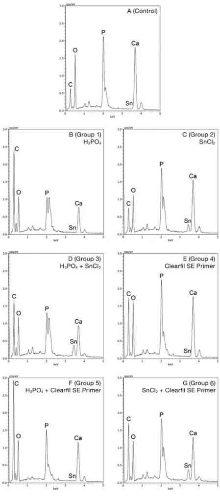
The EDX spectra revealed distinct differences between groups. Phosphoric acid etching (Group 1) led to demineral-ization of the dentin surface and showed only small peaks for Ca and P (Fig.3B). In contrast, pretreatment with tin-chloride (Group 2) removed much less Ca and P displaying distinct peaks for Ca and P (Fig.3C), and tin was clearly detectable. The short phosphoric acid etching followed by a short
tin-chloride pretreatment (Group 3) showed an intermediary result with respect to Ca and P, but led to a relatively high content of tin (Fig. 3D). Pretreatment with the Clearfil SE Primer (Group 4, Fig.3E) led to much less demineralization that did phosphoric acid etching and resulted in a mineral content similar to that of surfaces pretreated with tin-chloride (Fig.3C). When Clearfil SE Primer application had been preceeded by phosphoric acid etching (Group 5, Fig. 3F), the degree of demineralization resembled that of the combined phosphoric acid etching - tin-chloride pretreatment of Group 3. Finally, when Clearfil SE Primer application was preceeded by tin-chloride treatment (Group 6), distinct tin peaks were apparent (Fig.3G).
Discussion
Optibond FL, generally considered the gold standard of three-step etch-and-rinse adhesive systems, was used with three different pretreatments. The highest bond strength to dentin with this adhesive was obtained when pretreatment of dentin had been sought through a 15 s application of phosphoric acid i.e. when Optibond FL had been used according to the man-ufacturer’s instructions. As the SEM micrographs indicated and the EDX results proved, application of phosphoric acid led to a significantly higher degree of demineralization than did the 15 s application of tin-chloride, despite a pH similar to
Table 2 Comparison of microtensile bond strength values by Wilcoxon rank sum tests Group 2 (SnCl2+ Optibond FL) Group 3 (H3PO4+SnCl2+ Optibond FL) Group 4 (Clearfil SE) Group 5 (H3PO4+ Clearfil SE) Group 6 (SnCl2+ Clearfil SE) Group 1 (H3PO4+Optibond FL) P<0.001 P<0.005 n.s. n.s. n.s. Group 2 – P<0.001 P<0.001 P<0.001 P<0.001 Group 3 – – n.s. P<0.005 P<0.001 Group 4 – – – n.s. P<0.05 Group 5 – –- – – n.s. n.s. = not significant
Table 3 Failure mode
Groups (n=22 molars per group) Failure mode (4 beams per molar = 88 beams per group) Cohesive failure in dentin (%) Adhesive failure at dentin-adhesive interface (%) Adhesive failure at adhesive-resin composite interface (%) Mixed adhesive failure (%) Cohesive failure in resin composite (%) Group 1 (H3PO4+Optibond FL) 0 1 4 27 68 Group 2 (SnCl2+Optibond FL) 0 53 9 31 7 Group 3 (H3PO4+SnCl2+Optibond FL) 0 7 0 42 51
Group 4 (Clearfil SE) 3 5 14 36 42
Group 5 (H3PO4+Clearfil SE) 0 2 12 31 55
that of phosphoric acid. A scaffold of collagen fibrils nearly totally depleted of hydroxyapatite allowing for effective pen-etration of first the primer (Optibond FL Prime) and then the adhesive/bond (Optibond FL Adhesive) into the interfibrillar spaces of the collagen network and into the dentin tubules most probably account for the superior bonding performance obtained via phosphoric acid etching. These bond strength results are corroborated by the failure mode observations: Whereas cohesive failure in resin composite was the most common failure mode when the dentin had been pretreated with phosphoric acid, adhesive failure was the most com-mon failure mode when the dentin had been pretreated with tin-chloride.
The fact that the pretreatment involving a 7 s application of phosphoric acid followed by a 7 s application of tin-chloride (Table1) gave a bond strength result with Optibond FL that lay in between those of the two other groups implies that dentin demineralization was of paramount importance and that presence of tin precipitations had a negative effect on the bond promoting capacity of Optibond FL.
As all self-etch adhesive systems, Clearfil SE, the so-called gold standard of two-step self-etch adhesive systems, simul-taneously etches and primes the dentin upon application of the primer component leading to partial dissolution of the smear layer and demineralization of the underlying dentin and to micro-mechanical bonding via a relatively thin hybrid layer
Fig. 2 Micrographs of the scanning electron microscopy (SEM) for qualitative
evaluation of dentin surfaces of the groups tested
Fig. 3 Spectra of the energy dispersive X-ray spectroscopy (EDX) for qualitative
evaluation of dentin surfaces of the groups tested
(0.5– 1 μm) [14,15]. In corroboration with previously pub-lished results [15–17], the SEM micrographs and the EDX results showed that application of the Clearfil SE Primer led to less demineralization of the dentin than did phosphoric acid etching. The incomplete demineralization, resulting from the relative mildness (pH≈2) of the primer [18], leaves substantial amounts of hydroxyapatite crystals around the collagen fibrils. It has been shown that calcium ions released during the partial dissolution of hydroxyapatite diffuse within the hybrid layer and join together MDP molecules in nano-layers [19,20], the process being driven by the formation of MDP-calcium salts. The low solubility of these salts and the hydrophobicity of the nano-layered structure is believed to render the hybrid layer less prone to hydrolytic degradation and thus to increase the stability of the bond [16,20,21].
The introduction of a phosphoric acid etching step prior to application of the Clearfil SE Primer had no significant effect on the bond strength. This finding is at odds with the results of previously published studies in which bond strength was reduced as a consequence of phosphoric acid etching [15,
22]. The negative effect was explained by inferior quality of the hybrid layer caused by incomplete infiltration of the demineralized collagen network by the bonding resin [22,
23] and lack of chemical interaction between MDP and hy-droxyapatite [15].
In contrast, the introduction of a tin-chloride pretreatment step prior to application of the Clearfil SE Primer led to an increase in bond strength as compared to the standard proce-dure despite more pronounced removal of smear layer and more pronounced demineralization as revealed by SEM and EDX. Several possible modes of interaction of the tin ions present in the tin precipitates have been proposed [12]. The fact that the tin-chloride pretreament had a negative effect on the performance of Optibond FL, but a favorable effect on the performance of Clearfil SE leads to rejection of the null hypothesis and also suggests that the bond-promoting effect is linked to the presence of MDP. This lends support to the theory that tin ions function in the same manner as does calcium in the MDP-calcium salt to promote “docking” of MDP to collagen. Tin ions linked to free collagen sites are obviously bound to MDP thereby enhancing adhesion. Such an action mode would also explain the superior adhesion performance of Clearfil SE on erosively altered dentin with no calcium present in the hybrid layer [12]. Our hypothesis that tin binds to collagen and with MDP forms an MDP-tin salt leading to additional docking sites for MDP should be the aim of further research.
Conclusion
Whereas partial or total replacement of phosphoric acid etch-ing with tin-chloride pretreatment had a negative effect on the
bond-promoting capacity of a three-step etch-and-rinse adhe-sive, inclusion of a tin-chloride pretreatment had a beneficial influence on the resin composite to dentin bond strength mediated by an MDP-containing adhesive system.
Acknowledgments The authors would like to thank Gaba Interna-tional AG, Therwil, Switzerland and Kuraray Europe, Germany for providing the materials needed. Furthermore, we thank J. Wandel and Prof. Dr. J. Hüsler, Institute of Mathematical Statistics and Actuarial Science, University of Bern for statistical analyses.
Conflicts of interest The authors declare no conflicts of interest, real or perceived, financial or non-financial.
References
1. Van Loveren C (1990) The antimicrobial action of fluoride and its role in caries inhibition. J Dent Res 69:676–683, Special No 2. Van Loveren C (2001) Antimicrobial activity of fluoride and its in
vivo importance: identification of research questions. Caries Res 35(Suppl. 1):65–70
3. Miller S, Truong T, Heu R, Stranick M, Bouchard D, Gaffar A (1994) Recent advances in stannous fluoride technology: antibacterial efficacy and mechanism of action towards hypersen-sitivity. Int Dent J 44:83–94
4. Paraskevas S, van der Weijden GA (2006) A review of the effects of stannous fluoride on gingivitis. J Clin Periodontol 33:1–13 5. Ganss C, Neutard L, von Hinckeldey J, Kilmek J, Schlueter N
(2010) Efficacy of a tin/fluoride rinse: a randomized in situ trial on erosion. J Dent Res 89:1214–1218
6. Ganss C, Schlueter N, Hardt M, Schattenberg P, Klimek J (2008) Effect of fluoride compounds on enamel erosion in vitro: a comparison of amine, sodium and stannous fluoride. Caries Res 42:2–7
7. Ganss C, Lussi A, Sommer N, Klimik J, Schlueter N (2010) Efficacy of fluoride compounds and stannous chloride as erosion inhibitors in dentine. Caries Res 44:248–252
8. Ganss C, Lussi A, Grunau O, Klimek J, Schlueter N (2011) Conventional and anti-erosion fluoride toothpastes: effect on enamel erosion and erosion-abrasion. Caries Res 45:581–589 9. Huysmans MCDNJM, Jager DHJ, Ruben JL, Unk DEMF,
Klijn CPAH, Vieira AM (2011) Reduction of erosive wear in situ by stannous fluoride-containing toothpaste. Caries Res 45:518–523
10. Schlueter N, Klimek J, Ganss C (2009) Efficacy of an experimen-tal tin-F-containing solution in erosive tissue loss in enamel and dentine in situ. Caries Res 43:415–421
11. Ganss C, Hardt M, Lussi A, Cocks AK, Klimek J, Schlueter N (2010) Mechanism of action of tin-containing fluoride solutions as anti-erosive agents in dentine - an in vitro tin-uptake, tissue loss, and scanning electron microscopy study. Eur J Oral Sci 118:376– 384
12. Flury S, Koch T, Peutzfeldt A, Lussi A, Ganss C (2013) The effect of a tin-containing fluoride mouth rinse on the bond between resin composite and erosively demineralised dentin. Clin Oral Invest 17:217–225
13. Lussi A (2006) Erosive tooth wear– a multifactorial condition of growing concern and increasing knowledge. Monogr Oral Sci 20:1–8
14. Tay FR, Sano H, Carvalho R, Pashley EL, Pashley DH (2000) An ultrastructural study of the influence of acidity of self-etching
primers and smear layer thickness on bonding to intact dentin. J Adhes Dent 2:83–98
15. Van Landuyt KL, Kanumilli P, de Munck J, Peumans M, Lambrechts P, van Meerbeek B (2006) Bond strength of a mild self-etch adhesive with and without prior acid-etching. J Dent 34:77–85
16. Yoshida Y, Nagakane K, Fukuda R, Nakayama Y, Okazaki M, Shintani H, Inoue S, Tagawa Y, Suzuki K, de Munck J, van Meerbeek B (2004) Comparative study on adhesive performance of functional monomers. J Dent Res 83:454–458
17. van Meerbeek B, de Munck J, Yoshida Y, Inoue S, Vargas M, Vijay P, van Landuyt K, Lambrechts P, Vanherle G (2003) Buonocore memorial lecture. Adhesion to enamel and dentin: current status and future challenges. Oper Dent 28:215–235
18. de Munck J, Vargas M, Iracki J, van Landuyt K, Poitevin A, Lambrechts P, van Meerbeek B (2005) One-day bonding effective-ness of new self-etch adhesives to bur-cut enamel and dentin. Oper Dent 30:39–49
19. Fukegawa D, Hayakawa S, Yoshida Y, Suzuki K, Osaka A, van Meerbeek B (2006) Chemical interaction of phosphoric acid ester with hydroxyapatite. J Dent Res 85:941–944
20. Yoshiraha K, Yoshida Y, Nagaoka N, Fukegawa D, Hayakawa S, Mine A, Nakamura M, Minagi S, Osaka A, Suzuki K, van Meerbeek B (2010) Nano-controlled molecular interaction at ad-hesive interfaces for hard tissue reconstruction. Acta Biomater 6:3573–3582
21. Yoshida Y, Yoshihara K, Nagaoka N, Hayakawa S, Torii Y, Ogawa T, Osaka A, van Meerbeek B (2012) Self-assembled nano-layering at the adhesive interface. J Dent Res 91:376–381
22. Torii Y, Itou K, Nishitani Y, Ishikawa K, Suzuki K (2002) Effect of phosphoric acid etching prior to self-etching primer application on adhesion of resin composite to enamel and dentin. Am J Dent 15:305–308
23. Walker MP, Wang Y, Swafford J, Evans A, Spencer P (2000) Influence of additional acid etch treatment on resin cement dentin infiltration. J Prosthod 9:77–81