The Second International Conference on Sentinel Node Biopsy
in Mucosal Head and Neck Cancer
Sandro J. Stoeckli, MD,
1Madeleine Pfaltz, MD,
2Gary L. Ross, MD,
3Hans C. Steinert, MD,
4D. G. MacDonald, FRCPath,
5Christian Wittekind, MD,
6and
David S. Soutar, MD
31Department of Otorhinolaryngology, Head and Neck Surgery, University Hospital, Frauenklinikstrasse 24, CH-8091 Zurich,
Switzerland
2Department of Pathology, University Hospital, Zurich, Switzerland 3
Plastic Surgery Unit, Canniesburn Hospital, Glasgow, United Kingdom
4
Division of Nuclear Medicine, University Hospital, Zurich, Switzerland
5
Oral Pathology Unit, Glasgow Dental Hospital and School, Glasgow, United Kingdom
6
Institute of Pathology, University of Leipzig, Leipzig, Germany
Background: The Second International Conference on Sentinel Node Biopsy in Mucosal Head and Neck Cancer was hosted by the Department of Otorhinolaryngology, Head and Neck Surgery of the University Hospital in Zurich, Switzerland, from September 12 to 13, 2003. The aims of this conference were to present the results of validation studies and to achieve a consensus on methodological requirements.
Methods: More than 80 delegates from 20 countries attended the conference. The presented validation studies were summarized and compared with the literature. Consensus was achieved concerning requirements for lymphatic mapping and histopathologic work-up.
Results: Twenty centers presented results on 379 patients with cN0 disease. Sentinel nodes were identified in 366 (97%) of 379. Of these 366, 103 (29%) were positive for occult metastasis, and 263 (71%) were negative. Of those 263 patients, 11 patients (4%) showed nodal disease not revealed by the sentinel lymph node biopsy (SNB). The negative predictive value of a negative sentinel node for the remaining neck was 96%. The consensus conference resulted in the use of a radiotracer, lymphoscintigraphy, and a handheld gamma probe for lymphatic mapping as minimal requirements. The use of conventional hematoxylin and eosin staining and immu-nohistochemistry for cytokeratin is mandatory. Step-sectioning of the entire node at intervals of 150 lm is recommended.
Conclusions: The conference attracted delegates from all over the world, thus underscoring the high interest in the topic. With regard to the presented data and the data from the literature, SNB for early oral and oropharyngeal cancer is sufficiently validated. The con-sensus conference resulted in the definition of minimal methodological requirements for accurate SNB.
Key Words: Sentinel node biopsy—Elective neck dissection—Micrometastasis—Oral carci-noma—Head and neck carcinoma.
Since its introduction for malignant melanoma1 and breast cancer,2 sentinel lymph node biopsy (SNB) has gained popularity in head and neck can-cer, especially for early lesions of the oral cavity and the oropharynx.3–8 The First International Confer-ence on Sentinel Node Biopsy in Mucosal Head and Neck Cancer, held in Glasgow in 2001, brought
Received November 19, 2004; accepted June 3, 2005; published online September 19, 2005.
Address correspondence and reprint requests to: Sandro J. Stoeckli, MD; E-mail: [email protected].
Published by Springer Science+Business Media, Inc.Ó 2005 The Society of Surgical Oncology, Inc.
together more than 80 participants from throughout the world and summarized promising initial results from 22 centers.9 Pioneering work has continued in this field, with an increasing number of centers adopting the technique. Multicenter trials have commenced to determine the reliability and repro-ducibility of the technique.10SNB has been shown to improve staging in the clinically N0 neck for patients with early squamous cell carcinoma (SCC) of the oral cavity and oropharynx.11–13 Other sites, such as the supraglottic larynx and the hypopharynx, are also under investigation.14
The Second International Conference on Sentinel Node Biopsy in Mucosal Head and Neck Cancer was hosted by the Department of Otorhinolaryngology, Head and Neck Surgery of the University Hospital in Zurich, Switzerland, from September 12 to 13, 2003. More than 80 delegates from 20 different countries attended the conference, thus confirming the contin-uing high interest in this topic. The aims of this conference were to present updated results and technical innovations during the free paper session and to achieve a consensus on most methodological aspects of SNB by means of keynote lectures and case discussions. This article summarizes the results and provides a consensus review regarding the future of SNB in the management of the clinically N0 neck in head and neck SCC.
RESULTS OFTHE CONFERENCE The following results are an analysis of all 20 centers that contributed to the second SNB confer-ence. Most centers performed SNB followed by elective neck dissection irrespective of the histological evaluation of the sentinel lymph node (SLN). The pathologic characteristics of the SLNs were subse-quently compared with those of the neck dissection. Three centers had performed SNB only to stage the neck. In these cases, a therapeutic neck dissection was performed only if the SLN was positive, and no further treatment to the neck was performed if the SLN was negative. Only centers that performed de-tailed analysis of the SLN, including immunohisto-chemistry and step serial sectioning, were included in the data analysis.
Three hundred seventy-nine patients with clinically N0 disease were included. SLNs were identified in 366 (97%) of 379. Of these 366 patients, 103 (29%) were staged positive for occult lymph node metastasis with SNB, and 263 (71%) were staged negative. Four pa-tients had bilateral positive SLNs. Of the 263 papa-tients
staged SLN negative, 11 patients (4%) showed nodal disease in the neck dissection specimen or, in cases in which the neck was not further explored after nega-tive SNB, developed subsequent nodal disease. The negative predictive value of a negative SLN for the remaining necks was therefore 96%. Of the 103 pa-tients staged positive with SNB, 85 (83%) of 103 were upstaged because of routine hematoxylin and eosin (H&E) staining, whereas 18 (17%) of 103 were up-staged because of the additional use of immunohis-tochemical staining.
DISCUSSION
Results of the Conference and Overview of the Literature
The principles of SNB are universally accepted as follows. According to the definition by the Interna-tional Union Against Cancer,15 the SLN is the first lymph node to receive lymphatic drainage from a primary tumor. There might be more than one SLN for a specific tumor. If lymphatic spread occurs, the SLNs are the first involved. Other nodes should be involved only subsequently. The SLNs can be iden-tified by peritumoral injection of a radioactive tracer. The tracer mimics lymphatic spread and accumulates in the first-echelon node. With the help of lympho-scintigraphy and a gamma probe, the SLNs can be localized and selectively excised. Histological evalu-ation of the SLN detects possible clinically occult metastasis and allows for histological staging of the neck. In head and neck SCC, most centers have performed SNB within the context of an elective neck dissection; this has resulted in upstaging of the N0 neck in a considerable number of cases. Several cen-ters have now adopted SNB alone as a means of staging the clinically N0 neck. In these cases, only
patients with a pN+ neck, as assessed by SNB,
warrant further treatment by formal neck dissection. Patients with negative SNB findings would receive no further treatment to the neck. It is recommended that centers should perform at least 10 cases of SNB in the context of an elective neck dissection before using SNB alone as a staging tool.9
The cumulative results of all those who contributed to the conference confirm the high accuracy of SNB for early oral and oropharyngeal SCC, as previously reported in the literature. With an SLN detection rate of 97%, the technical feasibility has been well dem-onstrated. The 96% negative predictive value of a negative SLN underlines the high reliability of the
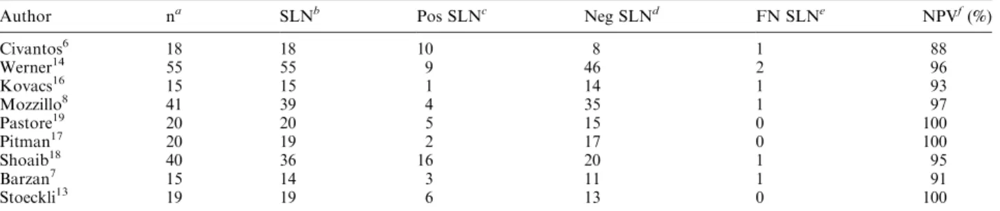
technique. As with any other novel technique, SNB has to be validated for its technical feasibility and the accuracy of the results. Technical feasibility means the likelihood of detecting an SLN by means of lymphoscintigraphy and intraoperative use of a handheld gamma probe. Validation for accuracy of the results implies comparison with a standard of reference—in case of SNB, elective neck dissection. Most published studies so far are validation studies. For a summary of the literature on SNB validation in conjunction with an elective neck dissection, we per-formed a MEDLINE search. With respect to the well-known learning curve,9 only studies including more than 10 patients with SCC of the oral cavity, oropharynx, or both were selected. Nodal disease had to be excluded by computed tomography, magnetic resonance imaging, or ultrasonography. Lympho-scintigraphy with a radiotracer for lymphatic map-ping was mandatory; the use of blue dye, optional. We collected 9 articles5–8,14,16–19 that met these inclusion criteria with a total of 243 N0 patients. The results are listed in Table 1. The mean SLN detection rate was 97% (range, 90%–100%), and the negative predictive value of a negative SLN for the remainder of the neck was 96% (range, 88%–100%). This liter-ature overview, in conjunction with the reported re-sults of the conference, proves the high reliability and accuracy of SNB and suggests that SNB can be used as a staging tool in the future.
To answer the question of validation with a mul-ticenter prospective study, the American College of Surgeons Oncology Group in 2003 opened a patho-logic validation trial for patients with T1 and T2 clinically and radiologically N0 SCCs of the oral cavity. The evolution of management of oral cancer
in North America has been in such a manner that the ‘‘watchful waiting’’ approach to the N0 neck has gradually fallen into disfavor, particularly for tongue and floor-of-mouth cancers, and there is a strong standard favoring elective neck dissection for the clinically negative neck. This trial, therefore, was designed with the introduction of radiological lym-phoscintigraphy followed by resection of the oral cancer, sentinel lymphadenectomy, and immediate selective neck dissection of levels I through IV. The end point of the trial is the pathologic comparison of the SLN with the completion neck dissection speci-men. Multiple sections and immunohistochemistry for cytokeratin are performed on the SLN and on the largest node from each level of the neck dissection specimen. Surgeon education and credentialing, to ensure technical standardization of the procedure, is a strong component of the trial. Quality control with independent audits and oversight, according to the standards of the National Cancer Institute, is an important component as well. The goal is to produce statistically significant data regarding the false-nega-tive rate of a negafalse-nega-tive SNB and the overall accuracy of the SNB procedure and to set the stage for a future randomized survival trial of SNB (without further dissection if the SLN is negative) versus planned initial elective neck dissection. The number of pa-tients estimated necessary to achieve a statistically significant result was estimated at 160 to achieve a 90% confidence interval. The trial is accruing well, and the termination of accrual is expected for the end of the year 2005. Results are not available until the end of the study to prevent the introduction of bias. To date there have been no significant complications related to the SLN procedure.
TABLE 1. Summary of the literature review
Author na SLNb Pos SLNc Neg SLNd FN SLNe NPVf(%)
Civantos6 18 18 10 8 1 88 Werner14 55 55 9 46 2 96 Kovacs16 15 15 1 14 1 93 Mozzillo8 41 39 4 35 1 97 Pastore19 20 20 5 15 0 100 Pitman17 20 19 2 17 0 100 Shoaib18 40 36 16 20 1 95 Barzan7 15 14 3 11 1 91 Stoeckli13 19 19 6 13 0 100
SLN, sentinel lymph node; Pos, positive; Neg, negative; FN, false negative; NPV, negative predictive value.
a
Number of patients.
bNumber of patients with successful detection of an SLN. cNumber of patients with occult disease in the SLN. dNumber of patients without occult disease in the SLN.
eNumber of patients with false-negative sentinel lymph nodes, i.e., negative SLNs but occult disease in the neck dissection specimen. f
In Europe, where most of the published validation data come from, the protocol for a prospective observational trial has been developed in Cannies-burn. In this trial, patients are not randomized to receive either SNB or elective neck dissection because of the high number of patients necessary in such a study to reach significant statistical power. The aim of the study is to prove that SNB does not achieve results worse than those expected with elective neck dissection. Patients with oral and oropharyngeal SCC of the categories T1 and T2 are enrolled. All patients undergo SNB, followed by neck dissection only in case of occult metastasis in the SLN. Preliminary results included 134 T1/2 tumors of the oral cavity or oropharynx in clinically N0 patients. In 93% of pa-tients, an SLN was identified. Upstaging of disease occurred in 34%. The negative predictive value of the technique with a mean follow-up of 24 months was 97%. The preliminary results of this multicenter trial concluded that SNB can be successfully applied to early T1/2 tumors of the oral cavity or oropharynx in a standardized fashion by centers worldwide. Consensus Discussion on Indications for and Techniques of SNB
Indications for SNB
Most validation studies so far have published re-sults from SNB for early oral and oropharyngeal SCC. These two sites are commonly accessible for tracer injection with the patient awake, thus allowing for preoperative lymphoscintigraphy. Larger tumors are difficult to completely surround by the tracer injection and are more likely to drain to more than one lymphatic basin. Furthermore, formal neck dis-section is often required to obtain adequate access for resection or reconstruction of larger tumors.
Sites other than the oral cavity and oropharynx and tumor categories other than T1 and T2 are cur-rently under investigation in different institutions but generally lack enough validation of data to be uni-versally recommended. Currently, there are three accepted indications for SNB in early oral/oropha-ryngeal SCC: (1) staging of the ipsilateral neck in unilateral cT1/2 cN0 tumors, (2) staging of the ipsi-lateral and contraipsi-lateral neck in midline tumors or tumors crossing the midline (cT1/2 cN0), and (3) staging of the contralateral neck in midline tumors or tumors crossing the midline (cT1/2 cN+; ipsilateral). Radiological evaluation with computed tomogra-phy, magnetic resonance imaging, or ultrasonogra-phy may become mandatory for clinical staging of the neck before SNB to exclude patients with a high
probability of nodal disease. SNB in an unrecognized N+ neck is likely to result in a false-negative result because replacement of the SLN stroma by tumor infiltration may block and reroute the lymphatic drainage to a non-SLN.
Lymphatic Mapping
The technique of lymphatic mapping may vary among institutions as a result of differences in radiotracer availability and scanning equipment. It is interesting to note that the published results with regard to the SLN detection rate are comparable between different techniques once the learning curve has been overcome. Minimal requirements for lym-phatic mapping are the use of a radiotracer for pe-ritumoral injection and the availability of a handheld gamma probe for intraoperative localization of the SLN. Preoperative lymphoscintigraphy is highly recommended because it facilitates SLN localization and increases the probability of SLN detection. The experience with blue dye as an adjunct to radiotracer is controversial, and its use is optional. The sole use of a blue dye without radiotracer is discouraged be-cause of very poor SLN identification rates. Selection of the radiotracer, performance of dynamic or static lymphoscintigraphy, and type of gamma probe are dependent on every institutionÕs possibilities. It is of paramount importance that any institution starting with SNB perform a validation study of the technique in conjunction with elective neck dissection. Only if the results are comparable to those in the literature can the learning curve be considered as overcome and the technique of lymphatic mapping as reliable. Histological Work-Up
Thorough histological work-up of the SLNs is crucial and surpasses by far what is routine for lymph node assessment in most pathology units. The fixed node is dissected free of fat and bisected through the largest axis. If the slices of the halved nodes are thicker than approximately 2.5 mm, then they should be further divided into slices of 2.5 mm. Step serial sectioning of the entire SLN and combined conven-tional H&E and immunohistochemical staining are mandatory. The current recommendation is step se-rial sectioning of the entire SLN at 150-lm intervals. At each step, four adjacent sections are mounted. At each level, one section is stained by H&E and one by immunohistochemistry for cytokeratin. The addi-tional two serial sections are retained for repeat or further studies. If no cytokeratin positivity is found, then the node is declared tumor free. If cytokeratin
positivity is found, then the positive section is com-pared with the immediately adjacent serial section previously stained with H&E to determine whether the positivity is due to the presence of viable tumor cells. By using the recommended technique, all metastases and micrometastases should be identified. Intraoperative assessment by frozen section is controversial. Although immediate analysis, offering the possibility of performing a neck dissection during the same procedure in case of a positive SLN, is desirable, many pathologists are afraid that the fro-zen-section procedure will result in a considerable loss of tissue, thus making definitive assessment of the remaining lymph node less reliable. Our preliminary unpublished experience in Zurich and the published data from a few studies20,21 suggest that frozen-sec-tion analysis of the SLN is accurate and can be reli-ably used for immediate intraoperative treatment decisions. Nevertheless, further data are needed in this field, and, therefore, no recommendation as to the intraoperative frozen-section examination of SLNs can be provided to date.
Reporting
The more precisely the lymph nodes from a neck dissection are examined, the more metastases will be found.22–24Because SLNs have to be worked up very thoroughly, a fairly high number of clinically inap-parent metastases are expected to be revealed. Many authors use the term micrometastasis for any metas-tasis detected by histological work-up of a clinically N0 neck. However, histologically detected metastases in a cN0 neck are by definition occult metastases. According to Hermanek et al.,25occult metastases are further subdivided into isolated tumor cells (ITCs), micrometastases, and (macro)metastases. Differenti-ation between micrometastases and metastases is performed by size. Micrometastases are by definition smaller than 2 mm; macrometastases are larger than 2 mm. ITCs have been defined in the 6th edition of the tumor-node-metastasis classification of malignant tumors as
[S]ingle tumor cells or small clusters of cells not more than 0.2 mm in greatest dimension that are usually detected by immunohistochemistry or molecular methods, but which may be verified with H&E stains. ITC do not typically show evidence of metastatic activity (e.g., proliferation or stromal reaction) or penetration of vascular or lymphatic sinus walls.15
Because these three subtypes of occult metastases might have different prognostic value and/or effects
on treatment, they have to be differentiated and separately reported by the pathologist, as indicated by the International Union Against Cancer tumor-node-metastasis classification (6th edition).
In the 6th edition of the tumor-node-metastasis classification of malignant tumors released in 2002, a definition of the SLN and proposals for classification were introduced. Classification of tumors evaluated by SNB has to be performed according to these cri-teria. When SNB is attempted, it has to be indicated by the addition of the designation (sn) after the N stage. Cases with ITC are classified as pN0 (i+) (sn); those with a micrometastasis are classified as pN1 (mi) (sn). Exact classification is pivotal to avoid stage migration when comparing the results of treatment with the results of historical cohorts.
Effecton Treatment
Formal neck dissection according to the primary tumor site is mandatory in all cases of positive SNB, irrespective of the type and size of occult metastases, because the probability of further metastasis is high.5 Adjuvant radiation should be discussed in cases of multinodal macrometastatic disease or extracapsular spread according to each institutionÕs guidelines. The benefit of adjuvant radiation for micrometastatic disease and ITCs is questionable because of a lack of evidence. Therefore, no recommendations are possi-ble.
CONCLUSIONS
Delegates from 20 different countries discussed technical aspects and clinical results at the Second International Conference on Sentinel Node Biopsy in Mucosal Head and Neck Cancer in Zurich, Switzer-land, from September 12 to 13, 2003. The reported results confirmed the high accuracy and reliability of SNB for early oral and oropharyngeal SCC. The conference participants agreed on a consensus that suggested regulations for the future use of SNB.
REFERENCES
1. Morton DL, Duan-Ren W, Wong JH, et al. Technical details of intraoperative lymphatic mapping for early stage melanoma. Arch Surg1992;127:392–9.
2. Krag DN, Weaver DL, Alex JC, Fairbank JT. Surgical resec-tion and radiolocalizaresec-tion of the sentinel lymph node in breast cancer using a gamma probe. Surg Oncol 1993;2:335–40. 3. Alex JC, Sasaki CT, Krag DN, Wenig B, Pyle PB. Sentinel
lymph node radiolocalization in head and neck squamous cell carcinoma. Laryngoscope 2000;110:198–203.
4. Shoaib T, Soutar DS, Prosser JE, et al. A suggested method for sentinel node biopsy in squamous cell carcinoma of the head and neck. Head Neck 1999;21:728–33.
5. Stoeckli SJ, Steinert H, PfaltzM, Schmid S. Sentinel lymph node evaluation in squamous cell carcinoma of the head and neck. Otolaryngol Head Neck Surg 2001;125:221–6.
6. Civantos FJ, GomezC, Duque C, et al. Sentinel node biopsy in oral cavity cancer: correlation with PET scan and immuno-histochemistry. Head Neck 2003;25:1–9.
7. Barzan L, Sulfaro S, Alberti F, et al. Gamma probe accuracy in detecting the sentinel lymph node in clinically N0 squamous cell carcinoma of the head and neck. Ann Otol Rhinol Laryngol 2002;111:794–8.
8. Mozzillo N, Chiesa F, Botti G, et al. Sentinel node biopsy in head and neck cancer. Ann Surg Oncol 2001;8(9 Suppl):103–5. 9. Ross GL, Shoaib T, Soutar DS, et al. The first international conference of sentinel node biopsy in mucosal head and neck cancer and adoption of a multicenter trial protocol. Ann Surg Oncol2002;9:406–10.
10. Ross GL, Soutar DS, MacDonald G, et al. Sentinel node biopsy in head and neck cancer: preliminary results of a mul-ticenter trial. Ann Surg Oncol 2004;11:690–6.
11. Ross G, Shoaib T, Soutar D, et al. The use of sentinel node biopsy to upstage the clinically N0 neck in head and neck cancer. Arch Otolaryngol Head Neck Surg 2002;128:1287–91. 12. Ross GL, Soutar D, MacDonald DG, Shoaib T, Camilleri IG.
Improved staging of cervical metastasis in clinically negative patients with head and neck squamous cell carcinoma. Ann Surg Oncol2004;11:213–8.
13. Stoeckli SJ, PfaltzM, Steinert H, Schmid S. Histopathological features of occult metastasis detected by sentinel lymph node biopsy in oral and oropharyngeal squamous cell carcinoma. Laryngoscope2002;112:111–5.
14. Werner JA, Dunne AA, Ramaswamy A, et al. The sentinel node concept in head and neck cancer: solution for the con-troversies in the N0 neck? Head Neck 2004;26:603–11.
15. Sobin LH, Wittekind CH, eds. TNM Classification of Malig-nantTumours. 6th ed. New York: Wiley, 2002.
16. Kovacs AF, Acker P, Berner U, Risse JH. Sentinel-Lymphk-notenexstirpation. HNO 2001;49:646–53.
17. Pitman KT, Johnson JT, Brown ML, Myers EN. Sentinel lymph node biopsy in head and neck squamous cell carcinoma. Laryngoscope2002;112:2101–13.
18. Shoaib T, Soutar DS, MacDonald DG, et al. The accuracy of head and neck carcinoma sentinel lymph node biopsy in the clinically N0 neck. Cancer 2001;91:2077–83.
19. Pastore A, Turetta GD, Tarabini A, Turetta D, Feggi L, Pelucchi S. Sentinel lymph node analysis in squamous carci-noma of the oral cavity and oropharynx. Tumori 2002;88:S58– 60.
20. Tschopp L, Nuyens M, Stauffer E, Krause T, Zbaeren P. The value of frozen section analysis of the sentinel lymph node in clinically N0 squamous cell carcinoma of the oral cavity and oropharynx. Otolaryngol Head Neck Surg 2005;132:99–102. 21. Asthana S, Deo SV, Shukla NK, Jain P, Anand M, Kumar R.
Intraoperative neck staging using sentinel node biopsy and imprint cytology in oral cancer. Head Neck 2003;25:368– 72.
22. Ambrosch P, Brinck U. Detection of nodal micrometastases in head and neck cancer by serial sectioning and immunostaining. Oncology1996;10:1221–6.
23. Woolgar JA. Micrometastasis in oral/oropharyngeal squamous cell carcinoma: incidence, histopathological features and clin-ical implications. Br J Oral Maxillofac Surg 1999;37:181– 6.
24. van den Brekel MW, van der Waal I, Meijer CJ, Freeman JL, Castelijns JA, Snow GB. The incidence of micrometastases in neck dissection specimens obtained from elective neck dissec-tions. Laryngoscope 1996;106:987–91.
25. Hermanek P, Hutter RVP, Sobin LH, Wittekind C. Classifi-cation of isolated tumor cells and micrometastasis. Cancer 1999;86:2668–73.