Emergence of Nanoplastic in the Environment and Possible Impact
on Human Health
Roman Lehner,
†Christoph Weder,
†Alke Petri-Fink,
†,‡and Barbara Rothen-Rutishauser
*
,††Adolphe Merkle Institute, University of Fribourg, Chemin des Verdiers 4, 1700 Fribourg, Switzerland
‡Chemistry Department, University of Fribourg, Chemin du Musée 9, 1700 Fribourg, Switzerland
ABSTRACT: On account of environmental concerns, the fate and
adverse effects of plastics have attracted considerable interest in the
past few years. Recent studies have indicated the potential for
fragmentation of plastic materials into nanoparticles, i.e.,
“nano-plastics,” and their possible accumulation in the environment.
Nanoparticles can show markedly different chemical and physical
properties than their bulk material form. Therefore possible risks and hazards to the environment need to be considered and addressed.
However, the fate and effect of nanoplastics in the (aquatic)
environment has so far been little explored. In this review, we aim to provide an overview of the literature on this emerging topic, with an emphasis on the reported impacts of nanoplastics on human health,
including the challenges involved in detecting plastics in a biological environment. Wefirst discuss the possible sources of
nanoplastics and their fates and effects in the environment and then describe the possible entry routes of these particles into the
human body, as well as their uptake mechanisms at the cellular level. Since the potential risks of environmental nanoplastics to humans have not yet been extensively studied, we focus on studies demonstrating cell responses induced by polystyrene
nanoparticles. In particular, the influence of particle size and surface chemistry are discussed, in order to understand the possible
risks of nanoplastics for humans and provide recommendations for future studies.
1. INTRODUCTION
Synthetic polymers are one of the most important classes of materials of the 21st century and impact our society and daily
life in ways that cannot be overestimated.1 The properties of
polymers are directly linked to their macromolecular architectures, which can readily be varied in a broad parameter space. As a result, a plethora of synthetic polymers have been developed that cover a broad range of attractive mechanical and other characteristics, including many property combina-tions that cannot be accessed with naturally occurring
polymers such as proteins and cellulose.2 The large family of
synthetic polymers has traditionally been classified into four
groups, namely, (i) thermoplastic polymers or “plastics”, i.e.,
polymers that melt above a specific temperature and can be
shaped before solidification upon cooling; (ii) thermosetting
polymers or “thermosets”, which are provided as (normally
liquid) precursors and are irreversibly cured after shaping through a chemical reaction into an infusible, insoluble
polymer network; (iii) elastomers; and (iv) synthetic fibers.3
Strictly speaking, only group (i) and subsets of groups (iii) and (iv) display thermoplastic character and should thus be
referred to as“plastics”. However, as an overwhelming majority
of polymer particles in the environment stem indeed from thermoplastic materials, the environmental science community has adopted the practice of referring to all polymers as “plastics”; we follow this convention herein as well. Polymers are still mainly derived from petroleum-based raw materials
(although there is an increasing trend toward using biobased
feedstocks),4 and their immense utility is based on their vast
range of chemical and physical properties that can be accessed and their low production and processing costs. This makes polymeric materials useful for manifold applications that range from packaging materials to toys and sporting goods to medical
devices and construction materials.5−7 Commercially used
polymers are always formulated with additives such as
stabilizers, flame retardants, plasticizers, fillers, and pigments,
which contribute to the overall materials properties.8Although
additives are typically present in much smaller amounts than the polymer, they are not necessarily benign and can have an
adverse effect on the environment.9
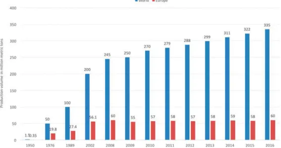
The production of polymers, including thermoplastics, thermosets, and elasto-mers, as well as biodegradable and biobased plastics, has
increased significantly over the past 60 years (Figure 1). In
2016, around 335 million metric tons of polymers used as plastics were produced worldwide, representing trillions of dollars in terms of global economic returns, with China being
the leading producer.10
Growing production also leads to an increase of plastic waste and, unfortunately, also promotes widespread accumulation in
http://doc.rero.ch
Published in "Environmental Science & Technology 53(4): 1748–1765, 2019"
which should be cited to refer to this work.
the environment, where the material can become brittle and start to fragment. In the environment the degradation of macroscopic plastic objects (>5 mm) into smaller pieces, often referred to as microplastics, typically occurs by a combination of chemical and physical processes that notably can involve photodegradation, oxidation, hydrolytic degradation, and mechanical disintegration. These processes are collectively referred to as weathering and, depending on the polymer type
and morphology, can differ significantly.12,13Photodegradation
caused by the ultraviolet (UV) portion of sunlight generally initiates the weathering process; this radiation is capable of
breaking chemical bonds in synthetic polymers.14The process
affects not only the polymers themselves but also additives
implemented within the materials, resulting in changes in their chemical structure and physical properties. Oxygen can increase the absorption of UV radiation by the formation of a complex with conjugated unsaturated hydrocarbons, thereby
accelerating the degradation process.15 In addition, polymers
such as polyesters or polyamides can also be degraded by hydrolysis, i.e., the cleavage of ester or amide bonds by
reaction with water.16,17These chemical degradation processes
cause a reduction of the polymer molecules’ molecular weight.
Eventually this leads to a reduction of mechanical strength and in many cases an increased absorption of water, which causes a further reduction of the mechanical properties and creates bulk and surface stress gradients, which eventually lead to cracking of the material. External mechanical forces also contribute to the fragmentation into smaller pieces, whose dimensions gradually diminish over time. The degraded plastic pieces form a highly heterogeneous collection, varying in size, shape, and
density as well as the chemical composition of the specific
material.
Newest observations studying the degradation of poly-ethylene microplastic pellets have shown that already an
exposure time of 8 weeks in artificial seawater induces severe
microcracking of the pellets surfaces, which, in combination with mechanical forces, can break down the material into
smaller sized particles.18 Further studies also revealed the
formation of nanosized polystyrene particles after 4 weeks of
exposure in a weathering chamber.19
Several studies have been published in the past few years estimating the mass of the overall global plastic waste as well as
the mass of plastic waste entering our oceans (Figure 2), which
are considered to be the global ecosystem that is mostly
affected by plastic waste. In 2009, Barnes et al.20estimated that
plastic materials constitute 10% of the total discarded waste.
Jambeck et al.21 presented numbers in the range of
Figure 1.Polymer production worldwide and in Europe, including thermoplastics, polyurethanes, and other polymers (thermosets, adhesives, coatings, and sealants) but not polyethylene terephthalate (PET), polyamide (PA), polypropylene (PP), or polyacrylicfibers. The growth rate of polymer production in Europe has stabilized in the past decade, which is related to the effects of the economic crisis of 2007−2008, as well as higher energy and raw material prices leading to higher product prices and decreasing competitiveness with theflourishing Middle Eastern plastics industry. Numbers adopted from PlasticEurope.11
Figure 2.Plastic sample taken from an expedition in the Ligurian Sea in May 2018 by the “Sail and Explore Association” using a manta trawl during 30 min with a mesh size of 0.3 mm. The Mediterranean Sea represents one of the most polluted seas worldwide with nearly four times higher concentrations of microplastic fragments compared to the North Pacific Gyre and a record-breaking number of 1.25 million microplastic fragments per square kilometer floating.53 Reprinted with permission from Sail and Explore Association, Copyright 2018.
4.8−12.7 million tons of mismanaged plastic waste that was generated in 192 coastal countries and discarded into the oceans in the year 2010. In addition, based on data collected in
24 expeditions conducted during 2007−2013, Eriksen et al.22
reported that a minimum of 5.25 trillion plastic particles with a
weight of up to 270,000 tons arefloating in the world’s oceans.
Another study reported the number of small plastic particles
(nominally <200 mm) to be in the range of 15−51 trillion,
with a collective weight of 93,000 to 236,000 tons, which corresponds to approximately 1% of the global plastic waste
entering the oceans in the year 2010.23 A recent study from
2018 predicted, using data received from multivessel and
aircraft surveys in the so-called“Great Pacific Garbage Patch”,
that at least 79 thousand metric tons of ocean plastic are
floating inside an area of 1.6 million km2, which correlates to
numbers 4−16 times higher than previously reported.24Other
studies have also proven the occurrence and accumulation of
large plastic items in the deep sea,25−27and recently published
data show that plastic objects have been ingested by animals of the benthic zone, assuming a sedimentation behavior of the
plastic particles.28,29Sedimentation of plastic particles is only
possible if the density of plastics is higher than that of seawater (which is the case for roughly a third of all polymers produced) or if they become negatively buoyant by the formation of
biofilms and adherence to other particles.30−33The smaller the
particles are, the faster they can reach their critical sinking
density.34 Plastic particles are also deliberately added to
personal care products.35 A threat that has thus far been
underestimated is the impact of microplastics in soils,
sediments, and freshwater on the terrestrial ecosystem.36
Research has only recently begun in this direction, as an estimated 80% of microplastic pollution in the oceans
originates from land.37,38
In the past decade, much attention has been paid to the
formation of microplastics. The exact definition of these
particles differs but generally includes particles with
dimensions between 1μm and 5 mm.39Thefirst small plastic
particles (<5 mm) were detected in open waters in the
1970s.40,41Recent studies confirmed the expectation that the
degradation process of plastic particles does not stop at the micrometer level and that plastic microparticles continue to
disintegrate to form plastic nanoparticles.19,42,43 Such
nano-particles usually exhibit different chemical and physical
properties than macroscopic objects based on the same
material.44In addition, their interactions with living organisms
can also greatly differ.45Thus, the differences between
micro-and nanoplastics are not trivial, micro-and the interactions of
nanoplastics with the environment and organisms are a specific
concern.46Like plastic microparticles, nanoplastics can adsorb
and carry hydrophobic chemicals that have a potential biological and toxicological impact on the environment, such as polychlorinated biphenyls (PCBs) released from objects like electrical devices, inks, paints, or the pesticide
dichlorodiphe-nyltrichloroethane (DDT).47−49A clear understanding of the
interaction of nanoplastics with the environment, especially with living organisms, is important to assess possible health
hazards, because nanoplastic particles can react differently than
their micronsized counterparts. However, current research
aimed at addressing the effects of plastic is focusing mainly on
aquatic systems and only limited data are available regarding
the impact of nanosized plastic particles on human health,50,51
although their formation in the environment is increasing and
thus also the possible transfer to humans via the food chain.52
It is apparent that the pollution of oceans with plastic has become a major environmental issue. Although the long-term impact of micro- and nanoplastics in the (aquatic)
environ-ment is still difficult to predict, this aspect might prove to be a
significant challenge to our society.54 The purpose of this
review is to provide an overview of current knowledge specific
to nanoplastics and their impact on the environment and
human health. We briefly discuss sources and nanoplastic
formation in the aquatic environment and the impact of nanoplastics on aquatic species and human exposure which is most likely via seafood. Furthermore, we will focus on possible entry routes of nanoplastics into the human body focusing on the cellular level.
2. SOURCES AND FATE OF PLASTIC IN THE ENVIRONMENT
The accumulation of plastics and their degradation products in the environment has continuously increased in recent decades, which is unsurprising given that 50% of plastic products are
produced for single-use applications and are soon discarded.55
Discarded packaging plastics account for a significant part of
the total solid waste that ends up in landfills, where, depending
on their composition, they remain unchanged or may degrade into fragments upon microbial heat production and further
biodegrade to carbon dioxide and water.56 Macroplastics
represent the main source of plastic litter. Once in the environment they can lead to entanglement and, despite their size, being ingested and retained by various species, including
seabirds,fish, and cetaceans, which then often die from related
causes such as starvation.57,58 Microplastic particles affect an
even greater number of organisms, including primary consumers of the food chain such as zooplankton, bivalves,
and small fish.59−61 It has been shown that (fragments of)
textilefibers with dimensions in the micrometer range, which
are disengaged from clothes during washing, are not removed
by the filter systems of wastewater treatment plants and
therefore end up in the environment.62 In addition, personal
care products such as toothpastes and facial scrubs often contain polyethylene-based microplastic particles which then
end up in the wastewater.63The presence of microplastics has
been reported all around the globe, from polar regions frozen in arctic ice to the open water around the equator, and from
coastal areas down to the deep sea.30,64The process of plastic
degradation progresses with increased time in the environment
and may eventually lead to the formation of nanoparticles.65
For example, a recent study by Lambert et al. demonstrated the formation of plastic nanoparticles under laboratory conditions
when disposable polystyrene coffee cup lids were exposed to
UV light.19This observation supports the notion that wherever
polymeric objects are released in the environment, the degradation to nanoplastic should a priori be considered. At the same time, it is clear that the degradation pathways of
different polymer types can vary significantlywhile some
polymers degrade into nanoparticles, others are known or can be expected to form water-soluble fragments. From exper-imental observations made so far, it can be estimated that plastic nanoparticles can be released into the environment as “primary”, i.e., intentionally manufactured particles for industrial purposes, e.g., paints, adhesives, electronics, and
cosmetics, and/or emerge as“secondary particles” as a result of
the degradation of larger plastic objects.66−68 Primary
nanoplastics used in households and industry are most likely
not being collected by the wastewater treatment facilities and are thus discharged as sewage into the aquatic environment.
However, our current knowledge of nanoplastics in the
environment is still very limited and only a few scientific
publications are available. In addition, the literature remains
inconsistent about the categorization of the particle sizes.39A
number of studies have set the upper size limit of nanoplastics at either 100 or 1000 nm. A recent publication by Gigault et al.
proposes an appropriate definition of nanoplastics on the basis
of physical and chemical properties. According to the authors,
a nanoplastic is defined as “particles within a size ranging from
1 nm up to 1000 nm resulting from the degradation of industrial plastic objects and can exhibit a colloidal
behaviour“.69
One could, however, recommend the use of
the official EU recommendation on the definition for
nanomaterials, i.e., “A natural, incidental or manufactured
material containing particles, in an unbound state or as an aggregate or as an agglomerate and where, for 50% or more of the particles in the number size distribution, one or more
external dimensions is in the size range of 1 nm−100 nm”.70
Another difficulty is that currently no detection method can
confirm the presence of nanoplastic components in the
environment.71
2.1. Effects of Nanoplastics on the Aquatic
Environ-ment. Numerous studies have shown that macro- and
microplastics can have a significant adverse impact on the
aquatic environment and have been reported elsewhere in
detail.72−74But what are the effects of nanoplastic particles on
the aquatic environment?
In recent years several experimental studies using model polystyrene nanoparticles have emphasized that various organisms such as daphnia, mussels, zooplankton, and algae can actively ingest nanoplastic particles or adsorb them to their
surfaces.75−78
In a laboratory setting, the effects of amino-functionalized polystyrene nanoparticles with a diameter of 50 nm and
concentrations between 1−50 μg/mL have been investigated
in the hemocytes of the marine bivalve Mytilus galloprovincialis
by Canesi et al.79 The hemocytes were exposed to different
concentrations of amino-functionalized polystyrene particles, which stimulated an increase in extracellular reactive oxygen species and lead to apoptosis within 1 h of exposure. This rapid response is in line with the physiological role of hemocytes in cell-mediated immunity. Sun et al. showed that nanoplastic particles at a size of 50 nm and a concentration of 80 mg/L induce toxicity related to oxidative stress toward marine bacterium Halomonas alkaliphila compared to microplastic
particles at a size of 1μm. Furthermore, amine-modified 50 nm
polystyrene nanoparticles induced higher oxidative stress
toward bacteria than that induced by nonmodified particles.80
The toxicity of amine-modified polystyrene particles was
further proven by Tallec et al. Exposure of 50 nm polystyrene
nanoparticles at concentrations between 0.1 and 25μg/mL to
Pacific oysters (Crassostrea gigas) showed an increase in
toxicity for both gametes and embryos in comparison to nonmodified particles. Overall, their data highlight that exposures to polystyrene nanoparticles may have deleterious
effects on planktonic stages of oysters.81 Brandts et al.
investigated the effects of polystyrene nanoparticles,
individ-ually or combined with carbamazepine, on the mediterranean mussel Mytilus galloprovincialis. Carbamazepine is among the most commonly detected drugs in the environment and may adsorb onto nanoplastics. For this purpose, mussels were
exposed for 96 h to a range of concentrations of 110 nm polystyrene particles (from 0.05 up to 50 mg/L), to
carbamazepine (6.3 μg/L) alone and to a mixture of
polystyrene + carbamazepine (0.05 mg/L + 6.3 μg/L).
Observations of different biomarkers from the digestive glands,
gills, and hemolymph were conducted. After exposure to polystyrene, carbamazepine, and their mixture, clear evidence
for genotoxicity was found in hemocytes as well as significant
downregulation in gene expression after combined exposure of polystyrene and carbamazepine. Moreover, it could be shown that even at the lowest concentrations tested, polystyrene
nanoparticles can induce oxidative damage.82Della Torre et al.
demonstrated that the accumulation of 50 nm amino-modified
polystyrene nanoparticles at tested concentrations of 1−50 μg/
mL in sea urchin embryos induced changes in gene expression
and embryotoxicity.83A recent study investigated the effects of
75 nm polystyrene nanoparticles at concentrations between 10 and 400 mg/L on the physiological changes (i.e., survival) and expression levels of stress defense genes of Daphnia pulex. The expression of the gene encoding the energy-sensing enzyme AMPK (adenosine monophosphate-activated protein kinase)
was influenced by the nanoplastic in different age groups.
Thus, age must be considered as a factor of great relevance
when predicting toxic effects.84
Effects of nanoplastic on the innate immune system of fish
have been reported by Greven et al., indicating that the stress response to polystyrene and polycarbonate nanoparticles could stimulate the degranulation of primary granules, oxidative burst
activity, and neutrophil extracellular trap release.85
Experi-ments with zebrafish revealed that after 7 days of exposure to
fluorescent polystyrene nanoparticles with a diameter of 70 nm
and at concentrations between 0.025 and 0.2 μg/μL,
inflammation and lipid accumulation in the liver occurred.86
Marques-Santos et al. investigated the effect of 50 nm
amino-modified polystyrene particles at concentrations of 1−25 μg/
mL on sea urchin Paracentrotus lividus immune system cells
(coelomocytes) in the presence of celomic fluid and at
different particle concentrations. Amino-modified polystyrene
particles acquired a protein corona once incubated with
celomic fluid, dominated by the toposome precursor protein,
which triggers particle−coelomocytes interactions and
tox-icity.87
Wegner et al. showed that 30 nm polystyrene nanoparticles,
that were tested at concentrations of 0.1−0.3 g/L, have an
adverse effect on the feeding behavior of the blue mussel
Mytilus edulis because of a reduced filtering activity in the
presence of the particles.88Pitt et al. investigated the potential
toxicity of polystyrene nanoparticles in developing zebrafish
(Danio rerio) and characterized the uptake and distribution of the particles within embryos and larvae. Embryos at 6 h postfertilization were exposed to 51 nm polystyrene
nano-particles (approximately 1 mg/(g of fish)) up to 120 h
postfertilization. At 24 h postfertilization the particles could be found in the yolk sac and had migrated to the gastrointestinal tract, gallbladder, liver, pancreas, heart, and brain between 48 and 120 h postfertilization. In addition, it could be seen that the particles altered larval behavior as evidenced by swimming
hypoactivity in exposed larvae.89An initial study of the effect of
nanoplastic particles delivered through the food chain (algae−
zooplankton−fish) on the brain tissue of fish was conducted by
Mattsson et al. They showed that 52 nm positively charged
amino-modified polystyrene nanoparticles above 0.075 g/L are
toxic to Daphnia magna and that fish eating nanoplastic
contaminated Daphnia showed behavioral disorders. Further-more, polystyrene nanoparticles were detected in the brain
tissue of all fedfish using hyperspectral imaging. This finding
proves for the first time that plastics nanoparticles can be
transported across the blood−brain barrier in fish.90
The majority of the mentioned studies showing an adverse
effect on different organisms were conducted using
amino-modified polystyrene nanoparticles and concentrations which
are not environmentally relevant as they are all too high. The
amino modification induces a positive surface charge to the
particles, which is experimentally interesting to test charge
effects. Nevertheless, it is well-known from the field of
nanomedicine that aminated surfaces are mainly used for coupling to, e.g., proteins or antibodies and that the positive charge of cationic polymers can facilitate cellular uptake and endolysosomal escape, thereby overcoming major cellular barriers and consequently increasing the possibility for further
intracellular reactions.91 The most important class of plastic
materials containing nitrogen in the form of amides is
polyamides. These can be hydrolyzed to afford amines, but it
is not entirely clear to what extent the amines remain intact (depending on the type, they may be protonated and oxidized). However, polyamides are not widely used as packaging materials, and therefore amine-containing nano-plastic particles stemming from polyamides are not expected to be a major environmental factor.
Additionally, nanoplastic particles in the environment will most probably interact with their surroundings, leading to
homo- or heteroagglomerates composed of different
constit-uents, due to the size and hydrophobic properties of the
materials.92,93 This might lead to the formation of a surface
corona of various adsorbed molecules evoking a change of the physical and chemical parameters as well as a reduced surface-area-to-volume ratio, thus changing their biological activity and
maybe affecting their sedimentation properties.94 These facts
have to be considered for experimental approaches regarding
influences on aquatic systems, in order to mimic natural
conditions as closely as possible.
For nanoplastics, uptake by several aquatic species, the adsorption of hydrophobic persistent organic pollutants (POPs), and the leaching of chemicals and POPs have been demonstrated, highlighting their potential biological and
toxicological impacts on the environment.95
However, a number of studies showing adverse effects such
as reactive oxygen species production and reproductive malfunctioning upon exposing aquatic organisms to nano-plastics have used concentrations several orders of magnitude higher than concentrations predicted to be environmentally
relevant such as 1 pg L−1to 15μg L−1for nanoplastics at sizes
of about 50 nm.96Reflecting this fact will help to understand
the impact of environmentally relevant nanoplastic concen-trations. In addition, there is a lack of knowledge regarding how nanoplastics (and hitchhikers) are transferred up the food chain and how they accumulate and interact with the environment, especially with living organisms. Thus, the impact of a contaminated food chain on humans and the resulting health hazards are not at all clear.
3. IMPACT OF NANOPLASTICS ON HUMAN HEALTH
Exposure to nanoplastic might occur via oral inhalation, ingestion, or absorption by the skin in connection with the use
of plastic products or unintentional means (Figure 3).
Inhalation is likely to be relevant in occupational exposure
scenarios that involve nanoplastic-containing aerosols,97
whereas potential contact with the skin can occur through the use of personal care products such as nanoplastics-containing skin care and cleansing products, or contaminated water or air. According to the current knowledge, ingestion of nanoplastic particles is likely to represent the main route of entry, since nanoplastic particles can be ingested by eating seafood or drinking contaminated water. In addition, nano-plastic uptake and accumulation, as well as trophic transfer of nanoplastic within aquatic organisms has been demonstrated under experimental conditions, thus fortifying the possibility that nanoplastics might accumulate in the food chain, and thus
result in human exposure.98,99 Microplastic particles have
already been found in several seafood species, such as fish,
shrimps, and bivalves, but also in other foods, such as honey,
beer, salt, and sugar.60,100−104 Recent studies using FTIR
spectroscopy have also shown the existence of microplastics in tap water and bottled water as well as drinking water from groundwater sources. Out of 159 samples of globally sourced tap water, 81% were found to contain microplastic particles,
mainlyfibers smaller than 5 mm with an overall mean of 5.45
particles/L.105 From a total of 259 individual bottles of water
from 11 different brands and 27 different lots, 93% showed
signs of microplastic contamination with an average of 10.4
particles/L.106Analysis of groundwater from the northwestern
part of Germany revealed that an overall mean of 0.7
microplastics/m−3 can be found.107 These studies reiterate
that the occurrence of nanoplastics in various food products cannot be ruled out. Unfortunately, there are currently no routine methods available that permit detection of nanoplastics in foods, and as a result, no data are available that go beyond the above-mentioned research works.
Human health might also be affected due to the transfer or
leaching of chemical additives from the plastic material itself. Within the plastic manufacturing process, chemicals such as plasticizers, pigments, or stabilizers are added to give the
desired properties of the final product, e.g., their flexibility,
color, and stability. Nowadays, thousands of different
chemicals are currently used for these purposes, and it is known that some of these chemicals can leach out during the product life cycle into the environment, leading to endocrine disruption or acute toxicity when exposure to organisms
occur.108 The same considerations apply for the monomers
(i.e., the chemical building blocks) used to manufacture the
polymers in the first place (of which small amounts may
Figure 3.Schematic illustration showing the three major pathways of human exposure to nanoplastics, i.e. via (A) the lung, (B) the gastrointestinal (GI) tract, and (C) the skin.
remain in the polymers) and the products formed by the chemical degradation of polymers. The most prominent example of a leaching monomer is bisphenol A (BPA), which is used for the preparation of polycarbonate and certain
epoxy resins. It has been shown that BPA causes adverse effects
in humans due to its estrogenic activity, including several metabolic diseases as well as reproductive and developmental
effects.109−111 In particular, polycarbonate drinking bottles
used for newborns showed high leaching of BPA. Newborns have a much higher risk than adults since a higher internal body burden is expected, expressed as concentration in blood/ plasma, due to increased absorption or decreased elimination
compared to the internal body burden of adults.112
3.1. Entry Routes of Nanoplastics into the Human Body. Three major exposure pathways to nanoplastics are possible, i.e., via the lung, the gastrointestinal (GI) tract, and the skin. The lung has a very large alveolar surface area of ca.
150 m2, with a very thin tissue barrier of less than 1μm,113
allowing nanosized particles to penetrate into the capillary blood system and to distribute throughout the human
body.114,115 Several studies have shown that exposure to
synthetic polymers can have an adverse effect on the health if
absorbed by the respiratory system.116−118 Studies applying
polystyrene particles to alveolar epithelial cells in vitro have shown that nanoplastic particles are taken up and that the
uptake rate is size-dependent (Figure 4).119−122 Recently,
syntheticfibers in atmospheric fallout in urban areas have been
highlighted as a possible source for human exposure to
microplastics by inhalation.123 Atmospheric fallout of
micro-plastics, collected with a stainless steel funnel, was investigated
in two different urban and suburban sites in Paris. About 30%
of particles observed were synthetic fibers with diameters
between 7 and 15μm and predominant sizes of 200−600 μm.
Between 2 and 355 particles/m2/day, with an average of 110±
96 particles/m2/day were observed. Fluxes were found to be
systemically higher at the urban site than at the suburban site.
Rainfall was also shown to have a clear effect on the
concentrations observed.123 Dris et al. investigated
micro-plastic fibers in indoor and outdoor air. Two private
apartments and one office were chosen as indoor sites,
where a concentration between 1.0 and 60.0 fibers/m3 was
measured, which was significantly higher than the outdoor
concentration (between 0.3 and 1.5fibers/m3). Around 33% of
thefibers they collected indoors were of petrochemicals origin
with polypropylene being predominant, while the resting 67%
consisted mainly of cellulose.124However, there are thus far no
data available concerning the number or concentration of aerosolized nanoplastics. The numbers presented by the studies of Dris et al. are more than 1000 times lower than
the permissible exposure maximum of 5 mg/m3 respirable
nuisance dust/(8 h working day) established by the U.S.
Occupational Safety and Health Administration (OSHA).125
This suggests that the concentrations of microplastics found in the air in these studies might be too low to have an adverse
effect on human health. Nevertheless, more data are needed, in
particular taking into account the physicochemical character-istics of the material as well as their sizes.
Furthermore, Wright and Kelly suggest that human exposure to micro- and nanoplastics through inhalation might occur by the transportation of sea salt aerosols. These aerosols can arise from wave action containing polymer particles of appropriate sizes, which then can be transported by the wind to coastal environments. Another scenario involves the application of wastewater treatment sludge containing plastic particles to agricultural land as a fertilizer. Once the sludge is dried, wind can further transport the plastic particles and distribute them. For further information regarding inhalation exposure of
microplastics, we refer readers to Wright and Kelly,50Gasperi
et al.,126 and Prata,97 who provide detailed overviews of the
topic.
Although the concentration of synthetic polymer particles in the air is usually very low, the GI tract, with its large surface
area of ca. 200 m2, represents the primary exposure site for
plastic particle uptake. Ingestion and accumulation of nano-plastic particles in the GI tract has been demonstrated in a
wide range of aquatic organisms, such asfish, mussels, and also
birds.89,98,127−129 Human uptake of plastics might occur by
intentional swallowing, leading in the worst case to a plastic
bezoar,130a rare cause of GI obstruction occurring mainly in
people with psychiatric ailments or, more likely, unintention-ally via the food chain by the consumption of plastic-contaminated food and drinks or possibly through migration of nanoplastic particles from the packaging materials into food products.
It has been shown that microplastics ingested by fish are
poorly absorbed via the GI barrier into the tissue, and gutting
of the fish reduces the possibility of consuming microplastic
particles. In contrast, polystyrene nanoparticles have been shown to overcome the GI barrier and to translocate into the
underlying tissue, as demonstrated by several groups withfilter
feeders and shellfish.76,88,128 Data on the release of
nano-plastics from packaging released into food products are currently unavailable; therefore, the exposure to humans
cannot be estimated for this method of intake.131
Most of our current knowledge regarding nanoplastic and intestine interaction comes from cell culture studies using intestine cells. Several in vitro studies have investigated the internalization and translocation of polystyrene nanoparticles in intestine cell monocultures or even more complex human intestinal cell models. Forte et al. showed that polystyrene nanoparticles with a diameter of 44 nm accumulate faster and
Figure 4.Human lung epithelial cancer cells (A549) labeled for F-actin (purple) and the nuclei (blue) showing uptake of 200 nm amino-modified polystyrene particles labeled with FITC (yellow) after exposure for 24 h.
more efficiently in the cytoplasm of human gastric adenocarcinoma cells than otherwise identical particles with a diameter of 100 nm. In addition, the 44 nm particles showed a strong upregulation of the interleukin (IL)-6 and IL-8 genes,
which are involved in gastric pathologies.132 Walczak et al.
determined the translocation of polystyrene particles with diameters of 50 and 100 nm, having positively and negatively charged as well as uncharged surfaces, in three in vitro intestine cell models of increasing complexity using Caco-2, HT29-MTX, and M-cells. Their results showed that both the size and the chemical composition of the particle surface impacts their
translocation. The size clearly affected the translocation of the
nanoparticles, ranging up to 7.8% for the 50 nm and 0.8% for the 100 nm particles. However, the surface chemistry seems more important, as the two types of negatively charged 50 nm
NPs had a greater than 30-fold difference in translocation
rates.133 Mahler et al. suggested that acute oral exposure of
positively charged polystyrene nanoparticles can disrupt
intestinal iron transport and cellular uptake.134
The cellular uptake of nanoparticles is strongly influenced by
their interactions with surrounding biological components, such as proteins, phospholipids, or carbohydrates, due to their
size, surface chemistry, and charge.135 Adsorption of proteins
from body fluids on nanoparticle surfaces results in the
formation of a so-called “protein corona” around the
particle.136 Hence, interactions between organs and tissue
generally occur with protein-coatedrather than bare
nanoparticles, likely causing changes in the characteristics and properties of the nanoparticle. Polystyrene nanoparticles have been shown to form protein corona complexes in in vitro
GI studies, resulting in increased translocation,137the change
of the protein corona composition over time depending on the
local environment,138 and increased cellular uptake and
toxicity.139 Furthermore, the protein corona caused the
polystyrene nanoparticles to be retained in the gut.
In conclusion, it has been shown that, in contrast to microplastic particles, nanoplastic particles overcome the GI barrier in aquatic organisms and can translocate into the underlying tissue. In addition, polystyrene nanoparticles can translocate the human intestine barrier in vitro. However, the reported studies rely solely on model polystyrene nanoparticles
and are not from environmental samples (Figure 5). Other
polymers such as PP, PE, and PET are, however, the main polymer material present in the environment. Thus,
extrap-olations of findings from polystyrene materials to other
polymers should be made with caution, and new model studies should be introduced including PP, PE, and PET.
The skin forms a protective barrier against external influences such as physical injuries, chemical agents, or bacteria. Contact of plastics with the skin might occur via the use of cosmetic products containing nanoplastic particles or with nanoplastic-contaminated water. The stratum corneum is the physical barrier of the skin, and due to the hydrophobic
properties of the plastic particles, significant nanoplastic uptake
through human skin in water is not to be expected. However, possible entry routes include hair follicles, exits of sweat glands,
or via injured skin areas.140 Skin penetration and resulting
tissue distribution of plastics has been studied by
Alvarez-Roman et al., who applied fluorescent polystyrene particles
with diameters of 20 and 200 nm to a porcine skin tissue
model.141Confocal laser scanning microscopy images revealed
a higher accumulation of the 20 nm polystyrene nanoparticles than with the 200 nm sized particles in the follicular openings,
whereas neither particle type was found to penetrate into deeper skin tissue, possibly due to the barrier properties of the
stratum corneum. These results were confirmed by an
additional study conducted by Campbell et al. a few years later, who observed that polystyrene particles with diameters of
20−200 nm penetrate only into the surface layers,
approx-imately 2−3 μm, of the stratum corneum.142 A study
conducted by Vogt et al. identified fluorescent polystyrene
nanoparticles of a diameter of 40 nm in the perifollicular tissue of human skin explants pretreated with cyanoacrylate follicular
stripping and confirmed uptake by Langerhans cells after
transcutaneous application to human skin.143Microbeads used
in cosmetics such as scrubs and shampoos are processed by mechanical means that may lead to their fragmentation into potentially more hazardous nanoplastics. In a recent publication, Hernandez et al. tested facial scrubs containing
polyethylene microbeads with diameters of 200 μm for the
presence of nanoplastics. Scanning electron microscopy data
revealed nanoparticles ranging in size from 24± 6 to 52 ± 14
nm. The material composition was tested by X-ray photo-electron spectroscopy and Fourier transform infrared
spec-troscopy, which confirmed that the identified nanoparticles
consisted of polyethylene.67 The further potential ability of
nanoplastics to overcome the skin barrier can be extrapolated from data available from studies using nanoparticles. Skin
stress evoked by UV radiation has a widespread effect on the
integrity of the skin barrier. An initial in vivo study conducted by Mortensen and colleagues investigated nanoparticle skin penetration of carboxylated quantum dots (QDs) with and without UV exposure. Their results showed qualitatively higher levels of penetration of QD nanoparticles in the UV exposed mice, due to the perturbed expression of tight-junction-related proteins (Zonula occludens-1, claudin-1, and occludin), which
promote intercellular adhesion.144 The effect that common
commercial skin care lotions may have on the penetration of QD nanoparticles into the skin was determined by Jatana et al.
In their study, QDs were added tofive commercial skin lotions
and applied to freshly excised human skin as well as C57BL/6
hairless mice. Their findings suggest that certain ingredients
Figure 5. Transmission electron microscopy image showing commercially available spherically shaped polystyrene nanoparticles used for in vitro studies.
(e.g., urea, glycerol, andα-hydroxyl acids) found in common commercial skin care lotions can enhance NP penetration into
the skin.145In addition, several chemical penetration enhancers
have shown their potential to support skin penetration by
nanoparticles.146Kuo et al. illustrated the differences between
oleic acid, ethanol, and oleic acid−ethanol enhancers for the
transport of zinc oxide nanoparticles. The results showed that all three were capable of enhancing the transdermal delivery of
zinc oxide nanoparticles.147 However, there is currently little
evidence that polymeric nanoparticles larger than 100 nm can
penetrate into intact skin.148
Once the plastic particles have entered the human body, they may overcome the primary tissue barriers and be transported through the bloodstream to secondary organs. In vitro studies have shown that carboxylated polystyrene nanoparticles can adsorb to, and penetrate into, red blood cells (RBCs) as a result of van der Waals, electrostatic, hydrogen bonding, and hydrophobic forces between
poly-styrene and the RBCs.149,150This so-called cellular hitchhiking
mechanism allows the polystyrene nanoparticles to avoid rapid clearance by the liver and spleen and, therefore, increase their circulation time.
Secondary barriers able to be reached via the bloodstream
include the placental barrier and the blood−brain barrier
(BBB). Grafmueller and colleagues showed with an ex vivo human placental perfusion model that polystyrene particles with diameters of up to 240 nm can be taken up by cells of the placental barrier and transported from the fetal to the maternal
blood circulation.151 The operating translocation mechanism
seems likely to be an energy-dependent transport pathway. The BBB is a highly selective barrier regulating the uncontrolled
diffusion of molecules into the brain. However, Yang et al.
demonstrated BBB permeability with polystyrene nanoparticles having a diameter of 20 nm using an in vivo rat model by implantation of a microdialysis probe into the cerebral cortex
of anesthetized rats injected with fluorescent polystyrene
nanoparticles.152A recent study by Rafiee et al. attempted to
assess the neurobehavior of rats exposed to pristine polystyrene
nanoparticles upon oral exposure. They investigated different
concentrations of 25 and 50 nm polystyrene particles (1, 3, 6, and 10 mg of polystyrene nanoparticles/(kg of body weight)/ day) administrated orally with adult male Wistar rats for 5
weeks. No statistically significant behavioral effects were
observed in any of the tests performed.153
In conclusion, it has been shown by in vitro and in vivo studies that micro- as well as nanoplastic particles might be taken up into the human body and can overcome tissue barriers, thus also allowing interaction with single cells. However, real-world plastics sources (rather than commercially available perfectly spherical polystyrene particles) have not yet
been used; therefore, further studies applying different plastic
materials with significant size and/or shape polydispersity as
well as environmentally relevant concentrations need to be
conducted tofill the existing knowledge gaps.
3.2. Cellular Uptake Routes and the Intracellular Fate of Nanoplastic Particles. There are several pathways by
which nanoparticles can be taken up by cells.154 Although
passive diffusion through the cell membrane (also referred to
as adhesive interaction), channel- or transport-protein-mediated uptake have been described, endocytotic nano-particle uptake is the major mechanism of cells. A range of endocytotic pathways, including phagocytosis and macro-pinocytosis, as well as clathrin- and caveolae-mediated
endocytosis, have been described.155−157Not all of the studies
we present here have been performed with intestine cells; however, our aim is to provide an overview of the current knowledge of polystyrene nanoparticles interaction with human cells.
The first interaction of nanoparticles with cells is the
interaction with the outer cell membrane. Rossi et al. investigated the interaction of polystyrene particles with biological membranes by coarse-grained molecular simulations, which revealed that nanosized polystyrene particles can permeate easily into lipid bilayer membranes, alter the
membrane structure, and reduce molecular diffusion, thereby
affecting possible cell functions.158 Fiorentino et al.
inves-tigated cellular uptake of 44 nm polystyrene particles on
human colon fibroblasts and bovine oviductal epithelial
cells.159By way of uptake inhibition studies, they demonstrated
that the polystyrene nanoparticles were internalized mainly via a clathrin-independent uptake mechanism. dos Santos et al.
studied the effects of transport inhibitors on the cellular uptake
of 40 and 200 nm carboxylated polystyrene nanoparticles in human cervical HeLa cells, human glial astrocytoma 1321N1,
and human lung epithelial A549 cell lines.160 The results
clearly indicated that in all cases polystyrene nanoparticles entered the cells via active energy-dependent processes. However, since none of the transport inhibitors could fully inhibit polystyrene uptake, the possibility remains that one cell line uses multiple uptake pathways simultaneously.
RBCs are highly specialized cells distinguished by their lack of a cell nucleus and endocytotic uptake mechanism. Studies have been conducted in order to demonstrate cellular uptake
of different nanoparticles by RBCs and to better understand
the operative uptake mechanism.161,162Rothen-Rutishauser et
al. showed uptake of nanosized polystyrene particles (diameter
< 200 nm) by RBCs.163 This result indicated neither an
endocytotic nor an actin-based uptake mechanism, therefore
suggesting a passive diffusion mechanism for this specific cell
type.
However, it is important to mention that the uptake routes are not only size- and surface-chemistry-dependent but also
cell-type-specific. The same material can show different cellular
uptake pathways: for example, polystyrene nanoparticles with a diameter of 120 nm, which surface had been modified to feature amidine groups, have been shown to be taken up by rat alveolar epithelial monolayers via non-endocytic mechanisms, in contrast to MDCK-II cells, which show particle uptake in an
energy-dependent manner.121,164 Kuhn et al. showed that a
combination of several distinguishable endocytotic uptake mechanisms is involved in the uptake of 40 nm carboxylated polystyrene nanoparticles by macrophages and epithelial cells. Selected endocytotic pathway inhibitors were applied, and it was revealed in the case of J774A.1 macrophages that macropinocytosis, phagocytosis, and clathrin-mediated endo-cytosis pathways are involved, whereas for human alveolar epithelial cells (A549), the uptake was dependent on
caveolin-and clathrin-mediated endocytosis pathways.165 Nonvesicular
transport through the membrane might allow the nanoplastic particles to interact directly with intracellular molecules or to release their payload, such as POPs, directly into the cytoplasm. This might lead to accumulation of POPs in the
cell and potential toxicological effects on the human body.166
Endocytosis of plastic particles will follow the intracellular endocytotic pathway involving early and late endosomes
followed by fusion with lysosomes. Salvati et al.122
strated the intracellular lysosomal localization of 40−50 nm
polystyrene nanoparticles in A549 cells. Thesefindings are in
agreement with other published data, which show accumu-lation of nanosized polystyrene particles in the lysosome, whereas no lysosomal escape or even particle degradation was
observed under acidic conditions.167
In another study, the importance of the physicochemical properties of mesoporous silica and polystyrene nanoparticles
were compared; the results revealed clear differences in cellular
uptake mechanisms using ovarian cancer cells.168It was shown
that the two types of nanoparticles never showed an overlap in their endocytotic uptake routes. Mesoporous silica particles entered the cells via caveola-mediated endocytosis and, depending on their size, resided within the lysosome (50 nm) or were relocated in the cytoplasm (10 nm). Cellular uptake of polystyrene nanoparticle occurred via a
caveola-independent route. Amine-modified 50 nm polystyrene
particles were localized within the lysosome and showed
toxicity after 4−8 h in comparison to 30 nm carboxyl-modified
polystyrene particles, which did not follow the classic acidic endocytotic pathway and were not toxic. It is well-known from the literature that a positive surface charge of nanoparticles often results in increased cytotoxicity and cellular uptake by
unspecific binding to negatively charged sugar moieties on the
cell surface, whereas negatively charged particles impair
endocytosis due to repulsive interactions.169,170 A variety of
cellular uptake routes and intracellular localizations of polystyrene nanoparticles have been observed depending on the physicochemical properties; however, there is a lack of
specific quantitative data regarding cellular entry and the fate
of nanoplastics.
It is obvious that size matters and plastic nanoparticles
interact differently with human cells in comparison with larger
ones; however, the uptake is also dependent on the particle
charge which might influence the behavior in the cell culture
media. Since most of the in vitro studies described here have
used polystyrene particles, it is of course difficult to extrapolate
the results to other kind of plastic particles and more research with other plastics is highly recommended.
3.3. Adverse Effects of Nanoplastics. Nanosized
materials in general have raised concerns regarding possible hazards and risks for the environment and especially for human health. An understanding of possible human exposure is critical
to determine health hazards. Adverse effects of nanoparticles in
vivo include cytotoxicity, (pro-)inflammation, or production of
reactive oxygen species (ROS).171A number of in vitro studies
using human cell lines (Table 1) have revealed that polymer
nanoparticles have the potential to activate the innate immune system, inducing inflammatory responses, or mediating oxidative stress. In in vitro experiments, Brown et al. showed
pro-inflammatory responses of polystyrene particles with
diameters of 202 and 535 nm upon exposure to human A549 lung cells, observing an increased IL-8 expression relative to that elucidated by polystyrene nanoparticles with a diameter
of 64 nm.172 As seen by Forte et al., unmodified polystyrene
nanoparticles with a diameter of 44 nm induced a strong upregulation of IL-6 and IL-8 genes in experiments using human gastric adenocarcinoma cells, indicating that the
induction of pro-inflammatory responses by polystyrene does
not necessarily have to be charge-driven (as suggested by many studies) but may instead be a material-based or simple particle
occurrence issue.132McCarthy et al. identified the activation of
ion channels in human lung cells after exposure to polystyrene
nanoparticles. Application of 20 nm carboxylated polystyrene particles caused continuous short-circuit currents by the
activation of basolateral K+ channels and the stimulation of
Cl− and HCO3− secretion. These findings underline that
polystyrene nanoparticles may have the ability to affect the
epithelial cell function and physiological processes.173Fuchs et
al. investigated the effect of 120 nm carboxylates and
amino-modified polystyrene particles on the polarization of human
macrophages into M1 or M2 phenotype. Their observations revealed that the expression of the M1 markers CD86, NOS2,
TNF-a, and IL-1b was not affected, while for M2 types both
nanoparticles impaired expression of scavenger receptors CD163 and CD200R, and the release of IL-10. The amino-modified particles also inhibited the phagocytosis of Escherichia coli by both, M1 and M2 macrophages, while the carboxylated
particles did not affect the phagocytosis of the bacteria by M2,
but increased protein mass in M1 and M2, TGF-b1 release by
M1, and ATP levels in M2.174
A new strategy to develop efficient nanoparticle-based drug
delivery systems involves the use of RBCs as a carrier system, which can increase the circulation of nanoparticles in vivo. In this context, Barshtein et al. investigated RBCs with attached polystyrene nanoparticles having diameters of 50, 108, and 243 nm, probing in particular the aggregation of RBCs and their adhesion to endothelial cells. The authors showed that the formation of RBC aggregates increased with decreasing particle size and in parallel a distinctly elevated adhesion to endothelial
cells was observed.175 In addition, Barbul et al. showed that
polystyrene particles with sizes of 27, 45, and 100 nm adsorbed to RBCs led to shape transformations and reduced cell deformability, with the 27 nm particles showing the strongest
effect.176
Furthermore, Xia et al. showed the influence of 30 nm
polystyrene nanoparticles on the endocytotic pathway in macrophages and human cancer cell lines A549, HePG-2, and HCT116. The particles induced the formation of large vesicle-like structures, which blocked the vesicle transport in the endocytic system and the distributions of regular proteins required in cytokinesis, which led to the formation of
binucleated cells.177 A recent study by Inkielewicz-Stepniak
et al. investigated the role of mucin in the toxicological impact
of polystyrene nanoparticles. Their findings showed that
amine-modified 57 nm sized polystyrene particles have the
ability to strongly interact and aggregate mucin and induced apoptosis equally on mucin- and non-mucin-secreting
intestinal epithelial cells.178 Further studies using cationic
polystyrene nanoparticles indicated the increase in ROS production and endoplasmatic reticulum stress caused by misfolded protein aggregation in macrophage (RAW 264.7) and lung epithelial (BEAS-2B) cells, leading to autophagic cell
death.179,180 A further study confirming that polystyrene
particles are able to induce ROS production was conducted by Liu et al. In their study they showed that polystyrene particles 500 nm in size could stimulate ROS generation in
human liver cells.181
Aside from polystyrene, an in vitro study using unmodified
polyethylene particles with sizes ranging from 0.3 to 10 μm
showed unequivocally that the plastic particles stimulated
mice-derived macrophages to produce significant levels of
cytokines such as IL-6, IL-1β, and TNF-α.182 Furthermore,
several studies of human patients have shown that normal usage of implants such as total joint replacements made of
polyethylene can lead to the production of wear debris.183−185
Table 1. Overview of in Vitro Studies Using Human Cell Lines Demonstrating Cellular Uptake and E ffects upon Exposure to Polystyrene Nanoparticles material, modification size, nm major findings target/cell line ref polystyrene, unmodified 44 induced strong up-regulation of IL-6 and IL-8 genes human gastric adenocarcinoma cells (AGS) Forte et al. 132 polystyrene, unmodified, carboxylated, amino-functionalized 50, 100 size dependency regarding particle translocation human colon carcinoma cells (Caco-2), human colon adenocarcinoma cells (HT29-MTX), human intestine microfold cells Walczak et al. 133 polystyrene, carboxylated 44 maximum particle uptake after 30 min human colon fibroblasts (HCF) Fiorentino et al. 159 polystyrene, carboxylated, amino-functionalized 50, 200 disruption of iron transport due to polystyrene particle exposure human colon carcinoma cells (Caco-2), human colon adenocarcinoma cells (HT29-MTX) Mahler et al. 134 polystyrene, carboxylated, amino-functionalized 20, 40 higher uptake efficiency of carboxylated particles compared to amino-funtionalized particles, induction of apoptosis human colon carcinoma cells (Caco-2) Thubagerre and Reinhard 190 polystyrene, amino-functionalized 57 binding of mucin and induction of adoptosis human colon carcinoma cells (Caco-2, LS174T, HAT-29) Inkielewic z-Stepniak et al. 178 polystyrene, carboxylated 20, 40, 100 40 nm particles internalized faster than 20 or 100 nm particles in both cell lines human lung carcinoma cells (A549), human astrocytoma 1321N1 Varela et al. 119 polystyrene, carboxylated 116 cellular uptake human lung carcinoma cells (A549) Deville et al. 120 polystyrene, carboxylated 40 − 50 cellular uptake irreversible, intracellular concen-tration increased linearly human lung carcinoma cells (A549) Salvati et al. 122 polystyrene, n.d.a. 64, 202, 535 increased IL-8 expression for 202 and 535 nm particles compared to 64 nm human lung adenocarcinoma cells (A549) Brown et al. 172 polystyrene, carboxylated 20 activation of ion transport human lung adenocarcinoma (Calu-3) McCarthy et al. 173 polystyrene, unmodified, carboxylated, amino-functionalized 60 amino-functionalized polystyrene particles induce autophagic cell death through the induction of endoplasmic reticulum stress human bronchial epithelium (BEAS-2B), mouse monocyte macrophage (RAW 264.7) Chiu et al. 179 polystyrene, unmodified, carboxylated, amino-functionalized 60 amino-functionalized particles induced mitochon-drial changes leading to cell death human bronchial epithelium (BEAS-2B), mouse monocyte macrophage (RAW 264.7) Xia et al. 180 polystyrene, unmodified, carboxylated, amino-functionalized 50, 100 amine-modified particle-induced oxidative stress, mitochondrial disruption and release of cytochrome C, indicating apoptotic cell death human immortalized alveolar epithelial type 1 cells (TT1), primary human alveolar macrophages (MAC), primary human alveolar type 2 (AT2) epithelial cells Ruenraroengsak and Tetley 191 polystyrene, unmodified, carboxylated, amino-functionalized 50 particles partly adsorbed and internalized, then released by Calu-3 cells, while THP-1 macrophages quickly incorporated all particles, aminated polystyrene nanobeads were more cytotoxic and genotoxic than unmodified and carboxylated ones human Calu-3 epithelial cells, human monocytic leukemia cell line THP-1 Paget et al. 192 polystyrene, carboxylated, amino-functionalized 100 concentration of internalized nanoparticles, their uptake kinetics, and mechanism may differ considerably between primary cells and a related tumor cell line human monocytic leukemia cell line THP-1, macrophages Lunov et al. 193 polystyrene, carboxylated 20, 100, 200, 500, 1000 20 nm particles were taken up passively, 100 − 1000 nm actively and passively, 20 nm stimulated IL-8 secretion in human monocytes and induced oxidative burst, 500 and 1000 nm particles stimulated IL-6 and IL-8 secretion in monocytes and macrophages human monocytic leukemia cell line THP-1, human histocytic lymphoma cells U937 Prietl et al. 194 polystyrene, carboxylated, amino-functionalized 120 both particle types impaired expression of scav-enger receptor CD163 and CD200R, and the release of IL-10 human monocyte macrophages Fuchs et al. 174 polystyrene, unmodified n.d.a. 20, 100, 200, 500, 1000, 2000 cellular uptake pathway of polystyrene particles is cell-line-dependent bone-marrow-derived macrophages (BMDM), fibroblast L929, kidney epithelial cells 293T Firdessa et al. 195 polystyrene, carboxylated 44 maximum particle uptake after 1 h human renal epithelial cells (HRCE) Monti et al. 196 polystyrene, carboxylated, amino-functionalized 110 cellular uptake human microvascular endothelial cells (ISO-HAS1), human umbilical vein endothelial cells Tenzer et al. 138
http://doc.rero.ch
Wear debris polyethylene particles seem to trigger the release
of pro-inflammatory factors, such as TNF-α, IL-1, and
pro-osteoclastic factors such as receptor activator of NF-κB ligand
(RANKL), resulting in periprosthetic bone resorption (osteolysis) and, in the worst case, eventual loss of the
implant.186 As illustrated by Devane et al., the tissue around
ultrahigh molecular weight polyethylene-based prostheses often contains large numbers of small particles with sizes
ranging from 0.2 to 10 μm, and macrophages have been
frequently observed, confirming the recruitment of the innate
immune system.187 Shanbhag et al. analyzed the composition
and morphology of wear debris from interfacial membranes of
failed total hip replacements. Theirfindings revealed that most
of the particles found were polyethylene and had a mean size
of around 530 nm.188 To overcome the observed negative
effects, surgeons are now increasingly using metal-on-metal
joint replacements.
Furthermore, the production of fluorescein labeled
poly-ethylene terephthalate (PET) nanoparticles was recently
shown by Magri et al.189 In their study, they present a
top-down approach based on laser ablation of polymers to form PET nanoparticles with weak acid groups on their surface, which should mimic real environmental nanopollutants. In vitro studies using human intestinal epithelial cells (Caco-2) showed internalization and biopersistance as well as long-term stability in a simulated lysosomal environment. In addition, cells treated with nano-PET particles did not increase ROS generation over 96 h, and no cytotoxicity was detectable. However, as suggested by the authors, possible long-term
effects in cells or tissue cannot be excluded and need to be
evaluated further.
In conclusion, it has been shown by in vitro and in vivo data that nanosized plastic particles can induce the activation of cell
responses, especially effects on the immune system. However,
long-term studies with repeated exposures are absent, as well as thorough material testing, including plastics such as PE, PET, and PP, since most of the current studies are performed with
polystyrene particles. Furthermore, it is important to fill the
knowledge gap between environmental relevant concentrations and the concentrations found in the organisms/cells to carry out relevant studies on the impact on human health.
4. DETECTION OF NANOPLASTICS
The reliable detection of polymer-based nanoparticles in environmental samples or complex biological matrices is highly challenging, due to their small size and the fact that their
overall chemical composition is not all too different from that
of organic matter. The detection of nanoplastic particles
according to their size is not sufficient, as additional chemical
characterization is needed for identification of the particles. To
the best of our knowledge, only one study has so far been published showing the detection and chemical analysis of nanoplastics in an environmental sample. Ter Halle et al. collected water samples from the North Atlantic Subtropical
Gyre, and after ultrafiltration of the water, they used dynamic
light scattering to prove the presence of nanoparticles. To evaluate the chemical identity of the particles, they conducted
pyrolysis coupled with gas chromatography−mass
spectrome-try. They estimated that the colloidal fraction signal of seawater could be attributed on average to a mixture of 73%
PVC (±18%), 18% PET (±16%), and 9% PS (±10%). In
addition, they observed changes in the pyrolytic signals of polyethylene with decreasing debris size, concluding that this
Table 1. continued material, modification size, nm major findings target/cell line ref polystyrene, carboxylated, amino-functionalized 30, 50, 1000 50 nm positively charged PS particles accumulated within lysosome in comparison to 30 nm negatively charged particles, cellular uptake caveolae-independent human ovarian carcinoma cells (SKOV-3), human ovarian carcinoma cells (NIH-OVCAR-3) Maneerat et al. 168 polystyrene n.d.a. 20, 200 uptake of particles by hepatocytes was size-, time-, and serum-dependent, uptake of 200 nm particles was limited, but 20 nm NPs were internalized by both cell types human hepatocyte carcinoma cells (HepG2), human hepatoblastoma cells (C3A) Johnston et al. 197 polystyrene, carboxylated 500 polystyrene particles could stimulate O2 ̇ generation human liver cells (Huh 7) Liu et al. 181 polystyrene, unmodified 20, 93, 220, 560, 1010 20 nm particles taken up avidly by all cell lines; HUVEC, ECV 304, and HNX 14C show particle uptake up to 1010 nm; HepG2 did not take up particles larger than 93 nm human bladder carcinoma cells (ECV 304), human hepatocyte carcinoma cells (HepG2), human head and neck squamous cancer cells (HNX 14C), human vein endothelial cells (HUVEC) Zauner et al. 198 polystyrene, carboxylated 110 carboxylated particles internalized more rapidly and showed higher accumulation mesenchymal stem cells Jiang et al. 199 polystyrene, amino-functionalized 100 increased cellular uptake of particles under fluidic shear stress human embryonic kidney epithelial cells (HEK293T), human pancreatic adenocarcinoma cells (PANC-1), human colorectal adenocarcinoma cells (HT29), human lung adenocarcinoma cells (A549) Kang et al. 200 n.d.a. = n o data available