RESEARCH ARTICLE
OMA and OPA—Software-Supported Mass Spectra
Analysis of Native and Modified Nucleic Acids
Adrien Nyakas,
2Lorenz C. Blum,
3Silvan R. Stucki,
1Jean-Louis Reymond,
1Stefan Schürch
11
Department of Chemistry and Biochemistry, University of Bern, Bern, Switzerland
2
Genome BC Proteomics Centre, University of Victoria, Victoria, BC, Canada
3
Institute of Molecular Systems Biology, ETH Zurich, Zürich, Switzerland
Abstract.The platform-independent software package consisting of the oligonucle-otide mass assembler (OMA) and the oligonucleoligonucle-otide peak analyzer (OPA) was created to support the analysis of oligonucleotide mass spectra. It calculates all theoretically possible fragments of a given input sequence and annotates it to an experimental spectrum, thus, saving a large amount of manual processing time. The software performs analysis of precursor and product ion spectra of oligonucleotides and their analogues comprising user-defined modifications of the backbone, the nucleobases, or the sugar moiety, as well as adducts with metal ions or drugs. The ability to expand the library of building blocks and to implement individual structural variations makes it extremely useful for supporting the analysis of therapeutically active compounds. The functionality of the software tool is demonstrated on the examples of a platinated double-stranded oligonucleotide and a modified RNA sequence. Experiments also reveal the unique dissociation behavior of platinated higher-order DNA structures.
Key words: DNA, RNA, Oligonucleotides, Tandem mass spectrometry, Software, Fragmentation, Double-stranded DNA, Nucleic acids, Cisplatin
Received: 4 September 2012/Revised: 26 October 2012/Accepted: 30 October 2012/Published online: 21 December 2012
Introduction
T
he implementation of structural alterations to nucleic acids is of major interest in the field of antisense RNA (asRNA) research as well as RNA interference (RNAi) strategies. Modifications are tailored to substantially enhance the cellular stability of oligonucleotide-based therapeutic agents. Tandem mass spectrometry (MS/MS) has been shown to be a versatile tool for oligonucleotide sequencing, irrespective of limited sample volume or modifications. The general fragmentation pathways of oligonucleotides have been investigated exten-sively in the past decades and it was shown that the product-ions formed upon collision induced dissociation (CID) of DNA and RNA are well predictable on the basis of the underlying dissociation mechanisms [1–5]. Upon fragmentation, DNAprimarily forms a-B- and w-ions, whereas RNA preferentially generates c- and y-ions (nomenclature by McLuckey et al. [6]). It has to be taken into account that the types of fragment ions formed strongly depend on structural modifications present.
Oligonucleotides have gained attention as a target for anticancer drugs. Formation of covalent bonds with oligonu-cleotides was observed for transition metal complexes, such as cisplatin. Cisplatin is one of the most widely used chemother-apeutic drugs and was found to form adducts with DNA, RNA, and proteins in vivo. The impact of cisplatin on the gas-phase dissociation of single-stranded DNA and RNA has been investigated extensively in the past few years [7, 8].
In vivo nucleic acids mainly occur in their double-stranded form. Duplexes can be transferred into the gas phase by soft ionization techniques, such as electrospray ionization (ESI) and matrix-assisted laser desorption/ionization (MALDI) without dissociation of the double strands occurring [9–12]. Soon after successful ionization of duplex DNA, first tandem mass spectrometric (MS/MS) studies were published [13,14]. Dyson and co-workers observed strand separation upon ESI-MS of platinated DNA duplexes and subsequently subjected the dissociated single strands to collision-induced dissociation
Electronic supplementary material The online version of this article (doi:10.1007/s13361-012-0529-1) contains supplementary material, which is available to authorized users.
Adrien Nyakas, Lorenz C. Blum and Silvan R. Stucki contributed equally to this project.
(CID) to identify the adduct sites [15, 16]. These studies demonstrated the potential of tandem mass spectrometry for investigating the binding of therapeutically active compounds to their target, but also pointed out the high complexity of the product ion spectra, which renders data interpretation very time-consuming. High-resolution tandem mass spectrometry is a valuable tool to ensure structure confirmation and evaluation of oligonucleotides for therapeutic purposes. However, pro-cessing of large data sets without software support is challenging and extremely laborious.
Software to support the interpretation of oligonucleotide mass spectra has been reported earlier. Most of the available tools focus on the a-B- and w-ions, which are known to be most prevalently formed upon CID of DNA. Ni et al. presented an algorithm that recognizes the peaks of 5'- and 3'-terminal fragments in the product ion spectra and builds a mass ladder starting from the a2-B- and w1-ions [17]. The
authors stated that correlation of mass spectra might only work up to the 15-mer level.
The web-tool MONGO [18] is able to calculate the m/z values of the classic DNA fragments, as well as the , M-B-, y-, d-H2O-ions, and internal fragments for a given
oligonucleotide sequence.
Oberacher et al. developed a comparative sequencing (COMPAS) algorithm that compares in silico and experimental m/z values of DNA fragments [19]. It calculates a fitting value, which expresses the closeness of matching between the measured and theoretical data. Sequence mutations are detected readily by the COMPAS algorithm and it also works for longer oligonucleotides (9 50 nucleobases).
Rozenski and McCloskey presented the simple oligonucle-otide sequencer (SOS), which, for the first time, allowed considering selected backbone and nucleoside modifications [20].
With MS2links, a comprehensive tool for processing biological macromolecules was developed as an integrated part of the MS3D software suite [21,22].
The software tool presented by Kretschmer et al. is specialized in sequence confirmation of modified oligonucleo-tides and also considers next neighbor position switches [23]. It allows the implementation of user-defined modifications and provides the opportunity to customize the output to obtain only data on specific fragment-ions, such as c- and y-ions. Hence, analysis of RNA product ion spectra is feasible as well. The maximum number of nucleobases is limited, though, and it can handle deconvoluted product ion spectra only.
Generally, the above mentioned software tools do not support the interpretation of spectra of adducts formed between (modified) oligonucleotides and metal ions or drugs, nor do they perform simultaneous processing of two hybridized strands.
Hence, an easy to use Java application was developed to facilitate the interpretation of product ion spectra of native and modified nucleic acids and their adducts. The oligonucleotide mass assembler (OMA) and oligonucleotide peak analyzer (OPA) combine many of the features of previous software and
provide additional functions that simplify the interpretation of product ion spectra of oligonucleotides and of higher-order nucleic acids in their native and modified forms. The software is available for download fromhttp://www.schuerch.dcb.unibe. ch/omaopa/.
Experimental
Oligonucleotides, Chemicals, and Solvents
The single-stranded complementary DNA 15-mers (ss-1: 5' ATTAGGTTGGTTATA 3'; ss-2: 5' TATAACCAACCTAAT 3') were custom-synthesized as triethylammonium salts on a 1.0μmol scale by TriLink Biotechnologies (San Diego, CA, USA) and used without further purification. Oligonucleotide stock solutions were prepared by rediluting the lyophilized sample with HPLC-grade water (Sigma-Aldrich Inc., Buchs, Switzerland) to a concentration of 1 nmol/μL.
Annealing and Platination
Prior to annealing, the complementary single-strands were diluted with a 300 mM NH4OAc (Sigma-Aldrich Inc., St.
Louis, MO, USA) buffer to yield a concentration of 500 pmol/μL. The oligonucleotide solutions were heated to 90 °C and cooled slowly over night to a final temperature of 20 °C. The formed duplexes were stored at −20 °C. The single- and double-strands were incubated in a cisplatin (Sigma-Aldrich Inc., St. Louis, MO, USA) solution in a molar ratio of 1:1 (ss:cisplatin; ds:cisplatin) for 24 h at 37 °C. The platinated oligonucleotides were stored at −20 °C. Prior to mass spectrometry, the unplatinated and platinated duplexes were diluted with water to give a final concentration of 100 pmol/μL. All samples were analyzed immediately to reduce the time the duplex is exposed to low buffer concentration, which could result in dissociation of the double-strand.
Mass Spectrometry
All experiments were performed on a LTQ Orbitrap XL mass spectrometer (Thermo Fisher Scientific, Bremen, Germany) equipped with a nanoESI source. Oligonucleotides were analyzed in negative ion mode with a potential of −850 V applied to the nanospray needle and a source temperature of 130 °C. Tandem mass spectrometric experiments were performed with the precursor-ions selected within a window of ±3m/z. CID was performed with helium as the collision gas. Spectra were acquired over a m/z range from 200 to 2000 and relative collision energies in the range from 20 % to 40 % were applied. Calibration of the instrument was performed with ProteoMass LTQ/FT Hybrid ESI Negative Mode Calibration Mix solution (Supelco Analytical, Bellefonte, PA, USA). The Xcalibur software package (Xcalibur 2.0.7, Thermo Fisher Scien-tific) was used for data processing.
Software Development
OMA and OPA were written in Java and, thus, run on all platforms supporting the Java Runtime Environment 6 or later (Windows, Mac OS X, Linux). The software consists of two main parts. The first tool creates a reference peak list for a given input sequence, OMA. A single or a double strand can be entered, consisting of native or modified nucleotides. User-defined modifications can be added easily. OMA calculates the m/z values of all possible double- and single-strand molecular-ions as well as all the corresponding a/a-B/b/c/d, w/x/y/z, and internal fragments. The user can also choose to generate sodium, potassium, and cisplatin adducts. The second software part compares one or more generated reference lists to experimental spectra, OPA. It allows to select the ions of interest and also to specify a tolerance (in ppm) to account for the individual mass accuracy of different mass spectrometers. All spectra can be loaded from file or directly from the clipboard and are rapidly annotated with matching peaks. The format of the output is tab-separated, can be further processed by any spreadsheet program (such as Microsoft Excel or OpenOffice Calc).
Results and Discussion
Software-Supported Spectrum Interpretation:
Data Handling and Features
The capabilities of the OMA and OPA software are demonstrated on MS and MS/MS data of the platinated 15-mer DNA duplex 5'-ATTAGGTTGGTTATA-3'_3'-TAATCCAACCAATAT-5'. All data were acquired on a LTQ Orbitrap XL, processed with Xcalibur 2.0.7, extracted with the built-in“export” option, and subsequently analyzed with the new Java-based software tools. A significant benefit of Java applications is their platform independence, as they work on all major operating systems. All related programs available so far were mainly programmed for Windows or for specific application software such as Excel. Since only tab-separated values are required for the OPA, any instru-ment software that exports data in this manner can be used with the new software tool. If the instrument software does not generate tab-separated files, they can be easily created in any spreadsheet program.
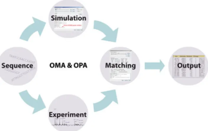
The software facilitates standardized analysis of oligonu-cleotide product ion spectra by providing the opportunity to search for specific fragment-ions and to consider peaks within a certain error window only. The general procedure of data evaluation is depicted in Figure1.
Oligonucleotide Mass Assembler
—OMA
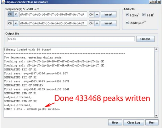
The OMA allows to define one or two DNA/RNA or mixed sequences and generates thereof a list of all theoretical product ions, resulting from either backbone cleavage, base-loss, formation of covalent and noncovalent adducts, or a combination thereof (Figure2). In contrast to other software
tools, the OMA also considers series of multiply charged ions of single- and double-stranded sequences. Thus, not only interpretation of product ion spectra is supported but also processing of single-stage mass spectra. Peak lists generated by the mass assembler are automatically written as text files into the chosen directory.
Oligonucleotide Peak Analyzer
—OPA
The peak analyzer imports the data as a text file in a tab-separated manner from the given source and subsequently compares the theoretical m/z values with the experimental data. The file needs to contain the column “m/z” for successful matching. Optional data columns for the charge state or absolute and relative peak intensities can be processed as well. If the instrumental software exports the peak lists with the annotated columns “m/z” and “Charge” right away, the m/z values can be loaded directly from clipboard and do not have to be saved as a text file in advance. The generated peak list is either saved in a file or written back into the clipboard.
OPA settings are easily adjusted according to the needs of the operator, as the data output can be customized by selecting the types of fragment ions to be matched. Options are provided for all product-ions generated by backbone cleavage (a-B/a/w; b/x; c/y; d/z) as well as base loss from the precursor ion (Figure 3). By default, the OPA is able to consider the presence of internal fragments, sodium and potassium adducts, as well as platination by cisplatin.
To account for instruments that generate low resolution data, the software is able to calculate and match the average masses of fragment-ions, if the corresponding box is checked. The tolerance window for peak recognition can be set individually. However, it has to be taken into account that a wider window will result in a larger number of false positive results, which renders manual re-evaluation of the assignments necessary.
Figure 1. General workflow of the OMA and OPA software. Oligonucleotide MS/MS data are acquired experimentally. The m/z values of theoretically possible fragment ions are calculated by the OMA and, finally, experimental and simulated data are matched by the OPA, which exports the result in a spreadsheet compatible form
For unambiguous peak assignment, the charge states of the ions are considered as well. Results are written into a spreadsheet providing the specifics of the peaks in a tabulated manner. If desired, only findings that match both, the m/z value and the charge state, will be inserted into the final peak list and charge mismatches will be excluded. Otherwise, the corresponding fragments are marked by a question mark in the result. The final output is given either as a full peak list, also including the peaks that have not been annotated, or as a truncated list containing the assigned peaks only. To support re-evaluation of the results, the theoretical reference mass and the mass error between calculated and experimental masses can be added to the output.
Example 1: Platinated Double-Stranded DNA
Successful transfer of a platinated duplex was achieved by carefully optimizing the ionization conditions. The resulting mass spectrum shows the peaks of the platinated and unplatinated duplex as well as of the corresponding unannealed single-strands ss1 and ss2 (inset in Figure4).
Tandem mass spectrometry was performed with the intact sevenfold negatively charged platinated duplex as the precursor ion. Hence, fragments of both strands are present in the product ion spectrum. For data processing, the sequences of the two strands are entered into the OMA and the types of adduct ions to be considered are selected. The software creates a file containing the theoretical m/z values of the fragments of both single strands, which serves as input for the OPA.
After matching of the data, the fragment ion output is individualized with the corresponding prefix ss1 or ss2, according to the respective strand. That the sequences are entered into the OMA individually has the benefit that the single strands do not have to be complementary. Hence, spectra of oligonucleotides that inherently do not hybridize perfectly are analyzed correctly by the software.
Table S1 gives an overview of the results obtained by processing the MS/MS data of the platinated double-stranded DNA with different settings. The comparison illustrates that efficiency of the evaluation and clarity of the output are increased by restricting the types of fragment ions and limiting the number of peaks to be matched. The results obtained by applying more stringent settings are summarized in Supplemental Figure S1. Matching was based on the 250 most abundant ions and the selection of fragment ion types was limited to a-B- and w-ions, base losses, and cisplatin adducts. Also included are the multiply charged ions of the single- and double-strands.
The product ion spectra of the unplatinated and platinated double-strands were found to differ considerably. Collisional activation of the unplatinated duplex (m/z 1304.23, z0−7) yielded almost exclusively the corresponding dissociated single-strands (Figure4a). Figure 4bshows the product ion spectrum of the platinated duplex (m/z 1336.66, z0−7). It reveals that cleavage of covalent bonds is considerably more pronounced after addition of cisplatin, and the formation of internal fragment ions is increased as well. Internal
frag-Figure 2. Screenshot of the OMA generating the peak list for a platinated duplex. For the platinated double-stranded DNA 15-mer more than 430,000 potential fragment ions are calculated if all options are selected
ments of w-/a-B-character constitute the majority amongst all dissociation products. The nomenclature of internal fragments is explained in Supplemental FigureS2.
The potential cleavage sites from which an internal fragment can result are also given in the spreadsheet output. For the sequence AGTTACT, the internal fragment
[w-{--TTddd}a-B-]-1 (Supplementary FigureS2) would addi-tionally be assigned as [w5 09 a5-B] fragment. Since short
internal fragments can often originate from various positions within a sequence, all of them are listed (e.g., [w509 a13-B],
[w909 a9-B], and [w1409 a4-B] for ss1 (ATTAGGTTGGT
TATA)); ss1 was mainly detected in its platinated form, whereas ss2 showed a highly abundant peak referring to the unplatinated single-strand. This observation is in good agree-ment with the expected preference of platination according to the sequences. The fact that the platinated double-strand did not yield any platinated fragment was unexpected, since despite the
two positive charges introduced by cisplatin, the number of charges on the backbone should be sufficiently high for compensation. However, smaller internal fragments compris-ing two or three phosphate groups only can be precluded from detection in the mass spectrometer due to platination.
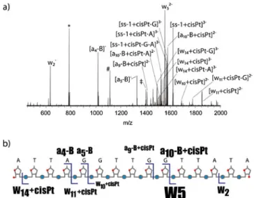
In order to obtain more information on the precise platination site, the triply negatively charged [ss1 + cisPt]-ion (m/z 1619.61), which was generated upon dissociation of the platinated double-strand, was subjected to a further dissociation step in a MS3experiment (Figure5). Two major pathways were observed. The first one is extensive neutral loss of purine bases,
Figure 4. Product ion spectra of (a) the unplatinated and (b) the platinated double-stranded DNA. The sevenfold negatively charged ions were selected as the precursors for CID. The relative collision energy was set to 25 %. Inset: Electrospray mass spectrum demonstrating successful gas-phase transfer of the platinated double-stranded DNA. ss1 and ss2 refer to the unhybridized single-strands, whereas DS indicates the double-stranded DNA
and the second one is the preferred cleavage of the 3'-C–O bond adjacent to the vicinal nucleobases G9G10 (formation of the w5-ion), indicating that cisplatin-binding
predomi-nantly occurred at this position [7]. However, the presence of an a9-B + cisPt-ion gives evidence for G5G6 at the beginning of the
sequence representing an alternative binding site for cisplatin. The example of double-stranded DNA, which generates a large number of product-ions, shows that the software strongly facilitates fast and reliable data interpretation. It further demonstrates that assignment of cisplatin adduct sites within DNA duplexes is feasible, if MS3data can be acquired.
User-Defined Modifications
Besides the processing of product ion spectra of DNA and its adducts, the OMA and OPA software package was programmed to support the analysis of single- and double-stranded RNA and modified nucleic acids as well. Various types of nucleobases, sugar moieties, and backbone linkers are provided by the user interface (Supplementary FiguresS2andS3), which appears by clicking on the “insert” button of the initial OMA window (Figure2). Each modification is illustrated by a small icon.
In order to account for the steadily increasing number of structural modifications introduced into therapeutic
oligonu-Figure 5. (a) MS3 spectrum of triply negatively charged [ss1 + cisPt], which was generated by strand separation of the platinated duplex in the first activation stage. Internal fragments are represented by the symbols *: [w-{−−TTddd}a-B], #: [w-{−−−TTGdddd}a-B], ‡: [w-{−− −ATTdddd}a-B]. (b) Overview of the cleavage sites, with the font size of the fragment labels indicating the relative intensity. Pt(NH3)2 is abbreviated by cisPt
Figure 6. (a) Product ion spectrum and sequence of the platinated 2'-methoxy-methylphosphonate RNA. (b) OPA output for the modified sequence
cleotides, the library on which calculation of fragment ions by the OMA is based is expandable. Expansion of the oma.lib is straightforward, since solely the elemental composition and the desired abbreviation of the modification have to be entered in a new line (Supplementary FigureS4).
Example 2: Modified RNA
In Figure 6a, the product ion spectrum of a platinated 2'-methoxy-methylphosphonate oligoribonucleotide, together with its sequence and the most abundant product ions, are shown. The strength of the OMA software is that it also accepts oligonucleotides bearing multiple modifications at different positions. In the example given, the sequence was entered into the OMA interface as mC-mC-mG_mG_mU-mU (with _ indicating the backbone modification). The output obtained by considering all possible types of fragment ions is shown in Figure 6b. In contrast to unmodified RNA, abundant fragment ions were assigned as members of the d- and z-ion series, which is in good agreement with the dissociation mechanisms of methoxy-methylphosphonate [8].
Conclusions
The OMA/OPA package represents a new generation of software to support the interpretation of oligonucleotide product ion spectra. Since it is based on Java, it is independent of the operating system. It combines features of previous programs and exhibits additional functions, such as free expandability of the building block library with modifications, direct analysis of the double-strand MS/MS data, and inclusion of sodium, potassium, and cisplatin adducts. The functionality of the software is demonstrated on the example of a platinated double-stranded 15 bp DNA and a highly modified oligor-ibonucleotide. It is demonstrated that processing of product ion spectra of the unplatinated and the platinated duplex is considerably simplified and that the time for thorough interpretation of the analytical data of is reduced considerably. Furthermore, results reveal the influence of platination on the dissociation behavior of the platinated double-stranded DNA.
Acknowledgments
The authors gratefully acknowledge financial support of this work by the Swiss National Science Foundation (grant no. 200020_121843).
References
1. Wang, Z., Wan, K.X., Ramanathan, R., Taylor, J.S., Gross, M.L.: Structure and fragmentation mechanisms of isomeric T-rich oligodeox-ynucleotides: A comparison of four tandem mass spectrometric methods. J. Am. Soc. Mass Spectrom.9(7), 683–691 (1998)
2. Schürch, S., Bernal-Mendez, E., Leumann, C.J.: Electrospray tandem mass spectrometry of mixed-sequence RNA/DNA oligonucleotides. J. Am. Soc. Mass Spectrom.13(8), 936–945 (2002)
3. Tromp, J.M., Schürch, S.: Gas-phase dissociation of oligoribonucleo-tides and their analogs studied by electrospray ionization tandem mass spectrometry. J. Am. Soc. Mass Spectrom.16, 1262–1268 (2005) 4. Andersen, T.E., Kirpekar, F., Haselmann, K.F.: RNA fragmentation in
MALDI mass spectrometry studied by H/D-exchange: mechanisms of general applicability to nucleic acids. J. Am. Soc. Mass Spectrom.17 (10), 1353–1368 (2006)
5. Wu, J., McLuckey, S.A.: Gas-phase fragmentation of oligonucleotide ions. Mass Spectrom. Rev.237, 197–241 (2004)
6. McLuckey, S.A., VanBerkel, G.J., Glish, G.L.: Tandem mass-spec-trometry of small, multiply charged oligonucleotides. J. Am. Soc. Mass Spectrom.3(1), 60–70 (1992)
7. Nyakas, A., Eymann, M., Schürch, S.: The influence of cisplatin on the gas-phase dissociation of oligonucleotides studied by electrospray ionization tandem mass spectrometry. J. Am. Soc. Mass Spectrom.20 (5), 792–804 (2009)
8. Nyakas, A., Stucki, S.R., Schürch, S.: Tandem Mass Spectrometry of modified and platinated oligoribonucleotides. J. Am. Soc. Mass Spectrom.22(5), 875–887 (2011)
9. Ganem, B., Li, Y.T., Henion, J.D.: Detection of oligonucleotide duplex forms by ion-spray mass-spectrometry. Tetrahedron Lett.34(9), 1445– 1448 (1993)
10. Light-Wahl, K.J., Springer, D.L., Winger, B.E., Edmonds, C.G., Camp, D.G., Thrall, B.D., Smith, R.D.: Observation of a small oligonucleotide duplex by electrospray ionization mass-spectrometry. J. Am. Chem. Soc. 115(2), 803–804 (1993)
11. Aaserud, D.J., Kelleher, N.L., Little, D.P., McLafferty, F.W.: Accurate base composition of double-strand DNA by mass spectrometry. J. Am. Soc. Mass Spectrom.7(12), 1266–1269 (1996)
12. Bayer, E., Bauer, T., Schmeer, K., Bleicher, K., Maler, M., Gaus, H.J.: Analysis of double-stranded oligonucleotides by electrospray mass-spectrometry. Anal. Chem.66(22), 3858–3863 (1994)
13. Doktycz, M.J., Habibi-Goudarzi, S., McLuckey, S.A.: Accumulation and storage of ionized duplex DNA-molecules in a quadrupole ion-trap. Anal. Chem.66(20), 3416–3422 (1994)
14. Wan, K.X., Gross, M.L., Shibue, T.: Gas-phase stability of double-stranded oligodeoxynucleotides and their noncovalent complexes with DNA-binding drugs as revealed by collisional activation in an ion trap. J. Am. Soc. Mass Spectrom.11(5), 450–457 (2000)
15. Egger, A.E., Hartinger, C.G., Ben Hamidane, H., Tsybin, Y.O., Keppler, B.K., Dyson, P.J.: High resolution mass spectrometry for studying the interactions of cisplatin with oligonucleotides. Inorg. Chem.47(22), 10626–10633 (2008)
16. Groessl, M., Tsybin, Y.O., Hartinger, C.G., Keppler, B.K., Dyson, P.J.: Ruthenium versus platinum: interactions of anticancer metal-lodrugs with duplex oligonucleotides characterised by electrospray ionisation mass spectrometry. J. Biol. Inorg. Chem.15(5), 677–688 (2010)
17. Ni, J.S., Pomerantz, S.C., Rozenski, J., Zhang, Y.H., McCloskey, J.A.: Interpretation of oligonucleotide mass spectra for determination of sequence using electrospray ionization and tandem mass spectrometry. Anal. Chem.68(13), 1989–1999 (1996)
18. Rozenski, J.: Mongo oligo mass calculator. Available at: URLhttp:// library.med.utah.edu/masspec/mongo.htm. Accessed February 5, 2011 19. Oberacher, H., Wellenzohn, B., Huber, C.G.: Comparative sequencing
of nucleic acids by liquid chromatography tandem mass spectrometry. Anal. Chem.74(1), 211–218 (2002)
20. Rozenski, J., McCloskey, J.A.: SOS: A simple interactive program for ab initio oligonucleotide sequencing by mass spectrometry. J. Am. Soc. Mass Spectrom.13(3), 200–203 (2002)
21. Yu, E.T., Hawkins, A., Kuntz, I.D., Ran, L.A., Rothfuss, A., Sale, K., Young, M.M., Yang, C.L., Pancerella, C.M., Fabris, D.: The collaboratory for MS3D: a new cyberinfrastructure for the structural elucidation of biological macromolecules and their assemblies using mass spectrometry-based approaches. J Proteome Res.7(11), 4848– 4857 (2008)
22. Kellersberger, K.A., Yu, E.T., Kruppa, G.H., Young, M.M., Fabris, D.: Top-down characterization of nucleic acids modified by structural probes using high-resolution tandem mass spectrometry and automated data interpretation. Anal. Chem.76(9), 2438 (2004)
23. Kretschmer, M., Lavine, G., McArdle, J., Kuchimanchi, S., Murugaiah, V., Manoharan, M.: An automated algorithm for sequence confirmation of chemically modified oligonucleotides by tandem mass spectrometry. Anal. Biochem.405(2), 213–223 (2010)