ISOLATION AND IDENTIFICATION OF ENDOPHYTIC FUNGI ON
PLANTS GROWING IN SALT ENVIRONMENTS, USING ITS AND 18S
MOLECULAR METHODS
Rabiaa KOUADRIAa,b*, Mohammed BOUZOUINAa and Brahim LOTMANIa
aPlant Protection Laboratory, Abdelhamid Ibn Badis University, Mostaganem, BP300, 27000, Algeria. bMolecular Genetic Laboratory, Ain Shams University, Cairo, Egypt.
Abstract
Endophytic fungi seem to play particle roles for the survival of plants inhabiting stressful habitats. This study focused on the identification of fungal endophytic community, associated with roots plants growing in salt environments by sequencing ITS and 18S rDNA regions. Based on the culture characteristics and growth morphology of colonies, 6 fungi species obtained from roots were collected from three plots with different salinities (< 4 dS/m, 4 to 8 dS/m and 8 to 16 dS/m). ITS sequences and 18S rDNA gene were compared with those available in the GenBank databases, to identify the following species: Alternaria chlamydospora and Chaetomium coarctatum (salinity < 4dS/m), Alternaria chlamydospora, Embellisia phragmospora, Phoma betae, Fusarium equseti, Chaetomium coarctatum and Fusarium graminearum (4 to 8dS/m); and Chaetomium coarctatum (8 to 16dS/m). Results indicate that Chaetomium coarctatum was considered as the most dominant fungus in studied plots. The fungal root endophytic community in natural vegetation under abiotic salt stress opens up possibilities for further investigations on the role of endophytes.
Key words : Endophytic fungi, salinity, rDNA, molecular identification.
Introduction
Soil salinity has become a global problem. In Algeria, a large part of agricultural regions characterized by an arid and semi-arid climate is affected by salinity process. Nearly 3.2 million hectares are threatened by salinity problem (Benmahioul et al., 2009); two million square kilometers are desert (arid) over the total area of the country (2.381.740 km2), while the rest (381.740 km2) is semi-arid (semi-desert) and sub-humid (Nedjraoui, 2001). Salt stress affects physiological processes of plants, causing a nutrient imbalance, altering levels of growth regulators, inhibiting photosynthesis and protein synthesis, all of which lead to reduced plant growth (Abeer et al., 2015).
Search for biological means such as endophytic fungi is a cheaper alternative and certainly more effective than the use of conventional means of desalination such as drainagefor the tolerance of cultivated plants to salt stress.
Fungal endophytes can colonize plants and help their partner to survive under extreme environmental conditions by secreting beneficial secondary metabolites. Among the >300.000 plants on Earth, almost all the vegetal species had diverse endophytes within their tissues (Strobel and Strobel, 2007). Endophytes are microorganisms that live within the intracellular or intercellular spaces of host plants (Strobel, 2003). They play crucial roles as decomposers, mutualists and parasites in ecological processes on earth (Liu et al., 2015). Endophytes live and grow in roots, stems and/or leaves, without causing any apparent disease symptoms (Petrini, 1991). Plant roots can be associated with a large variety of endophyte microorganisms (Dighton et al., 2005).
Identification of filamentous fungi, from plants is very complex (Souza et al., 2004). Because of their difficult morphological identification, genetic methods can be used for classifying microbial strains in diverse hierarchical taxonomic levels. Information contained in the 18S and 28S ribosomal genes and the internal transcribed spacer *Author for correspondence : E-mail: [email protected]
Study site and biological material
Sampling areas are located in Relizane; Algerian West. Spontaneous plant species were collected from three plots with different degrees of salinity (Plot1: < 4dS/m, Plot2: from 4 to 8dS/m and Plot3: from 8 to 16dS/m), to isolate endophytic fungi (Lat. 35° 47’ 46"N, Long. 0° 33’ 11", Alt. 50m). In this study, as the soil is plowed, only one plant species was collected from the low-salt plot (< 4dS/ m).
Plant sterilization and endophytes isolation
Endophytes were isolated from plant roots using Huang et al. (2007) method. Surface sterilization of roots followed the procedure of Larran et al. (2007). Root segments (5mm) were washed thoroughly in running tap water, sterilized with sodium hypochlorite (NaOCl, 5%) for 3min, rinsed three times in sterile distilled water, and then dried on a sterile filter paper. Segments were placed in a 90mm diameter Petri dish containing a mixture of potato dextrose agar (PDA) medium and amoxiallin (15mL/l), and incubated at 25° C with darkness. After 7 days, hyphae emerging were transferred to fresh PDA for purification and identification.
Molecular identification DNA extraction
DNA was extracted using the acid washed beads extraction method(Moller et al., 1992). Fungal mycelia (2-3mm) was immersed in 2mL tube with glass beads, containing 500µL of CTAB extraction buffer (20mM EDTA, 0.1M Tris-HCL pH 8, 1.4M NaCl, 2% CTAB and 0.1% -mercaptoethanol) and 20µL of solution of Proteinase K. Solution was incubated at 56°C. After centrifugation (3min on 12000 rpm), supernatant was transferred to a fresh tube following the addition of 400µL of chloroform-isoamyl alcohol (4:1). Samples were gently mixed and centrifuged at 12000 rpm for 5min. After
consisted of 50-100ng genomic DNA, 25µL of 2x PCR buffer: MyTaq DNA polymerase, 1 µL (10mM) of primer forward (ITS1/NS1) and 1 µL of primer reverse (ITS2/ NS7) and 22µL H2O. All amplification products were electrophoresed in Agarose gel 1.8%.
PCR products were purified using the DNA fragment purification kit and sequenced in forward and reverse directions by Macrogen (Holland).
Sequence analysis
Sequence analysis of the ITS and 18S sequences was carried out using Bio Edit Sequence Alignment Editor v.7.2.3(Hall, 1999). Phylogenetic trees were built using the neighbor-joining (NJ) methods and bootstrap test (Kumar et al., 2004). Bootstrap tests were performed using 1000 replicates. The search for homologous sequences was done using Basic Local Alignment Search Tools (BLAST) at the National Center for Biotechnology Information (www.ncbi.nlm.nih.gov/BLAST). All fungal sequences considered were at least 98% identical to the best hit in the NCBI database (Varanda et al., 2016).
Results
Plants species and isolated endophytic fungi Based on 63 root segments of plant species seeded, 31 showed infection with endophytic fungi (table 1), representing an infection rate of 49.21%. A total of 06 fungi were isolated from plant roots collected from salt environments.
Molecular identification PCR amplification
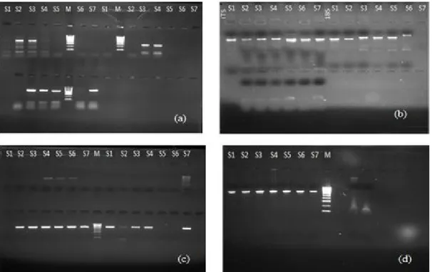
Amplification of ITS and 18S regions were successfully carried out with the primers ITS1/ITS2 and NS1/NS7 respectively, amplification by PCR showed a single band of about 500 bp for all isolates (fig. 1). Sequences were trimmed and assembled to its pair (forward+reverse of each sample/locus).
Similarity of the sequences
Sequences were compared with GenBank database, and results are shown in table 2. According to molecular data, strains S1 and S2 were identified to Alternaria genus with Pairwise identity 99.80%. Results of BLAST showed that ITS rDNA sequences of isolate S5 shared high homology with Fusarium equiseti with high bootstrap support value of 100%. Sequence identity ranged from 99 to 100% in ITS rDNA sequences and from 98 to 100% in 18S rDNA sequence.
Phylogenetic analysis using NJ method and bootstrap
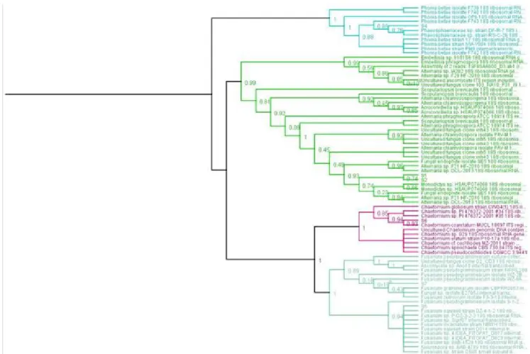
Phylogenetic tree is shown in fig. 2. Use of ITS and 18S sequencing and phylogenetic analysis identified 6 of endophytic fungi strains belonging to Dothidiomycetes
and Sordariomycetes: Alternaria chlamydospora and
Chaetomium coarctatum (salinity<4dS/m), Alternaria chlamydospora, Embellisia phragmospora, Phoma betae, Fusarium equseti, Chaetomium coarctatum and Fusarium graminearum (4 to 8dS/m) and Chaetomium coarctatum (8 to 16dS/m).
Discussion
Fungal endophytes have been found within all plants from diverse habitats (Rodriguez et al., 2009). Isolation results showed that spontaneous collected plants hosted one or two endophytic fungi. Debbab et al. (2012) explained that all plant species host at least one or more type of endophyte.
Internal transcribed spacer (ITS) region and 18S rRNA genes are applied in fungi molecular studies
Fig. 1 : Electrophoresis gel photos for (a) DNA extraction, (b) PCR amplification of ITS and 18S sequences, (c) purification of the
PCR product for ITS sequences and (d) purification of the PCR product for 18S sequences. S1-S7: samples (endophytic fungi). M: DNA Marker.
Table 1 : Plants species and isolated endophytic fungi.
Plots Number of collected species Name of plant species Number of isolated endophytic fungi
Plot 01 01 Avena fatua 02
Plot 02 05 Beta macrocarpa 01
Anabasis prostraba 02
Avena fatua 01
Sueda fructosa 01
Lolium perenne 01
(Curlevski et al., 2010). In this study, various primer were used for examining the fungal community on roots by isolating, culturing, and molecularly identifying in ITS region (Mello et al., 2011) and 18S rRNA genes (Hoshino and Morimoto, 2010). ITS locus was successfully assembled for all the 7 isolates. ITS sequences are hypervariable and used particularly for fungi identification at species or lower levels (Toju et al., 2012). While in 18S, only Reverse direction passed the HQ% (high quality) for samples 2 to 7 except for sample 1. 18S rRNA sequences may not always allow fungi identification on genus or species level (Anderson and Parkin, 2007). ITS region revealed higher richness, diversity, and more
dynamic than 18S rRNA. Comparing with GenBank, results showed that sequences had more than 97% similarity. Comparison between ITS and 18S-rRNA sequences with those available in GeneBank databases allowed us to analyze the phylogenetic affiliation of these fungi. Sequence identity of ITS rDNA of strains S1 to S7 ranged from 99 to 100%, strains S2 to S7 were located with high bootstrap support 98% in their 18S rRNA sequence. All isolates described belong to Ascomycota phylum (Porras-Alfaro and Bayman, 2011), where the seven isolated fungi represented two classes: Dothidiomycetes (S1 and S2: Alternaria, S3: Embellisia, S4: Phoma) and Sordariomycetes (S5 and S7: Fusarium,
Fig. 2 : Phylogenetic relationships among 7 endophytic fungi derived from Dothidiomycetes and Sordariomycetes (Ascomycota).
Table 2 : Summary of the fungal endophytic community associated with roots plants growing in salt environments. Sample Loci Accession n° Description Similarity
S1 ITS KF993329 Alternaria chlamydospora 99.5%
S2 ITS KF993329 Alternaria chlamydospora 99.5%
S3 ITS JQ796758 Embellisia phragmospora 99.6%
S4 ITS KM249077 Phoma betae 99.6%
S5 ITS KU377478 Fusarium equiseti 100.0%
S6 ITS NR_144822 Chaetomium coarctatum 99.4%
S6: Chaetomium).
Endophytic fungi associated with plant roots can provide potential benefits to their host plants (Urcelay et
al., 2011). It is assumed that most plant–fungus
mutualistic interactions take place in roots (Rodriguez et
al., 2009). Data showed a low diversity of endophytic
fungi, there are evidences that mycorrhizal populations become reduced or absent in habitats subjected to certain environmental stresses (Porras-Alfaro et al., 2008).
Fusarium and Phoma were the most frequent root
endophytic genera, followed by Alternaria, Embellisia and Chaetomium (Jose et al., 2008). Phoma and
Fusarium might play potential roles on plant host tolerance
to high-stress environmental conditions (González-Teuber
et al., 2017).
Only a few halophytic plants were studied (Anita and Sridhar, 2009), Alternaria and Phoma isolated from
Beta macrocarpa and Salicornia sp., respectively, are
considered to play an important ecological role for halophytes stress resistance (Barrow et al., 2007).
Alternaria chlamydospora and Fusarium equiseti were
more common in salt environments (Jose et al., 2008). Fungi belonging to Alternaria genera were clearly predominant under desert and salty environments (Smolyanyuk and Bilanenko, 2011). Alternaria,
Embellisia, Phoma and Fusarium, pigmented endophytes
and common in halophytes, play an important ecological role for plant survival and stress resistance (Sun et al., 2012). Alternaria spp. was noted for its resistance to UV radiation through its dark pigmentation (English and Gerhardt, 1946) by producing high levels of pigments, which protect fungi inhabiting plants growing in high-salinity. Embellisia and Phoma, showing pigmented spores, are likely predominant on Salicornia (Wong and Hyde, 2001).
Conclusion
This study provided information that all studied species harbored fungal endophytes, which seem to play potential roles on plant host tolerance to stressful conditions. The five fungal genera detected: Alternaria, Embellisia,
Phoma, Fusarium and Chaetomium, dominated by
Dothideomycetes and Sordariomycetes classes were belonging to Ascomycetes. Chaetomium coarctatum was common endophyte all plots.
The fungal root endophytic community in natural vegetation under abiotic salt stress opens up possibilities for further investigations on the role of endophytes. Moreover, it is imperative to conserve the endophytes of these plants ex situ, to discover their roles on plant host tolerance to stressful conditions.
Acknowledgments
The authors are grateful to the members of National Institute of Soils, Irrigation and Drainage, El-Matmer, Relizane, Algeria (INSID), for their help.
We extend our profound gratitude to Mahmoud Magdy El Mosalamy, Samah Mohamed Rizk, and Mohamed Abdel-Salam Rached, for their valuable help in the Molecular Genetic Laboratory, Ain Shams University, Cairo, Egypt.fxQ1.
References
Abeer, H., A. A. Alqarawi and D. Egamberdieva (2015) Arbuscular mycorrhizal fungi mitigates NaCl induced adverse effects on Solanum lycopersicum L. Pakistan Journal of Botany, 47 : 327–340.
Anderson, I. C., C. D. Campbell and J. I. Prosser (2003). Potential bias of fungal 18S rDNA and internal transcribed spacer polymerase chain reaction primers for estimating fungal biodiversity in soil. Environ Microbiol., 5 : 36–47. Anderson, I. C. and P. I. Parkin (2007). Detection of active soil
fungi by RTPCR amplification of precursor rRNA molecules. J. Microbiol Methods, 68 : 248–253.
Anita, D. D. and K. R. Sridhar (2009). Assemblage and diversity of fungi associated with mangrove wild legume Canavalia cathartica. Tropical and Subtropical Agroecosystems,
10 : 225-235.
Barrow, J., M. Lucero, I. Reyes-Vera and K. Havstad (2007). Endosymbiosis fungi structurally integrated with leaves reveals a lichenous condition of C4 grasses. In vitro Cell Dev Biol., 1 : 65-70.
Benmahioul, B., F. Daguin and M. Kaid-Harche (2009). Effect of salt stress on germination and in vitro growth of pistachio (Pistacia vera L.). C. R. Biologies, 332 : 164-170.
Curlevski, N. J. A., Z. H. Xu, I. C. Anderson and J. W. G. Cairney (2010). Diversity of soil and rhizosphere fungi under Araucaria bidwillii (Bunya pine) at an Australian tropical montane rainforest site. Fungal Divers, 40 : 12–22. Debbab, A., A. H. Aly and P. Proksch (2012). Endophytes and
associated marine derived fungi ecological and chemical perspectives. Fungal Divers, 57 : 45–83.
Dighton, J., J. J. White and P. Oudemans (2005). The Fungal Community: Its Organization and Role in the Ecosystem. Taylor & Francis Group, CRC Press, Boca Raton, FL. English, H. and F. Gerhardt (1946). The effect of ultraviolet
radiation on the viability of fungus spores and on the development of decay in sweet cherries. Phytopathology,
36 : 100-111.
González-Teuber, M., C. Vilo and L. Bascuñán-Godoy (2017). Molecular characterization of endophytic fungi associated with the roots of Chenopodium quinoa inhabiting the Atacama Desert, Chile. Genomics Data, 11 : 109–112.
vegetation in Mediterrnean environments with special reference to Fusarium spp. FEMS Microbiol Ecol., 64 : 90–105.
Kumar, S., K. Tamura and M. Nei (2004). MEGA3: integrated software for molecular evolutionary genetics analysis and sequence alignment. Brief Bioinform., 5 : 150–163. Larran, S., A. Perelló, M. R. Simón and V. Moreno (2007). The
endophytic fungi from wheat (Triticum aestivum L.). World J Microbiol Biotech., ZS : 565-572.
Liu, J., J. Wang, G. Gao, G. M. Bartlam and Y. Wang (2015). Distribution and diversity of fungi in freshwater sediments on a river catchment scale. Front Microbiol., 6 : 329. Mello, A., C. Napoli, C. Murat, E. Morin, G. Marceddu and P.
Bonfante (2011). ITS-1 versus ITS-2 pyrosequencing: a comparison of fungal populations in truffle grounds. Mycologia, 103 : 1184-1193.
Moller, E. M., G. Bahnweg, H. Sandermann and H. H. Geiger (1992). A simple and efficient protocol for isolation of high molecular weight DNA from filamentous fungi, fruit bodies and infected plant tissues. Nucleic Acids Res., 22 : 6115– 6116.
Nedjraoui, D. (2001). Country pasture / forage resource profiles. Algeria.
Petrini, O. (1991). Fungal Endophytes of Tree Leaves. In: J.H. Andrews, S. S. Hirano (eds.) Microbial Ecology of Leaves. Brock/Springer Series in Contemporary Bioscience. Springer, New York, NY.
Porras-Alfaro, A. and P. Bayman (2011). Hidden fungi, emergent properties: endophytes and microbiomes. Annu Rev Phytopathol., 49 : 291–315.
Strychnos cogens bentham. Acta Amaz., 34(2) :185-195. Sterflinger, K. and H. Prillinger (2001). Molecular taxonomy
and biodiversity of rock fungal communities in an urban environment (Vienna, Austria). Antonie van Leeuwenhoek,
80 : 275-286.
Strobel, G. A. (2003). Endophytes as sources of bioactive products. Micro Infect., 5 : 535–544.
Strobel, S. A. and G. A. Strobel (2007). Plant endophytes as a platform for discovery-based undergraduate science education. Nat Chem Biol., 3 : 356–359.
Sun, Y., Q. Wang, X. D. Lu, I. Okane and M. Kakishima (2012). Endophytic fungi associated with plants collected from desert areas in China. Mycol Prog., 11 : 781–790. Toju, H., A. S. Tanabe, S. Yamamoto and H. Sato (2012).
High-coverage ITS primers for the DNA-based identification of Ascomycetes and Basidiomycetes in environmental samples. PLoS ONE, 7(7) : e40863.
Urcelay, C., J. Acho and R. Joffre (2011). Fungal root symbionts and their relationship with fine root proportion in native plants from the Bolivian Andean highlands above 3.700 m elevation. Mycorrhiza, 21 : 323–330.
Varanda, C. M. R., M. Oiveira, P. Materatski, M. Landum, M.I.E. Clara and M. D. O. Félix (2016). Fungal endophytic communities associated to the phyllosphere of grapevine cultivars under different types of management. Fungal Biol., 120(12) : 1525-1536.
Wong, K. M. and D. Hyde (2001). Diversity of fungi on six species of Gramineae and one species of Cyperaceae in Hong Kong. Mycol Res., 105 : 1485–1491.
![[PDF] Apprendre et enseigner Matlab tutoriel pdf | Cours informatique](data:image/gif;base64,R0lGODlhAQABAIAAAP///wAAACH5BAEAAAAALAAAAAABAAEAAAICRAEAOw==)