HAL Id: hal-02559387
https://hal.archives-ouvertes.fr/hal-02559387
Submitted on 26 May 2021
HAL is a multi-disciplinary open access
archive for the deposit and dissemination of sci-entific research documents, whether they are pub-lished or not. The documents may come from teaching and research institutions in France or abroad, or from public or private research centers.
L’archive ouverte pluridisciplinaire HAL, est destinée au dépôt et à la diffusion de documents scientifiques de niveau recherche, publiés ou non, émanant des établissements d’enseignement et de recherche français ou étrangers, des laboratoires publics ou privés.
Distributed under a Creative Commons Attribution - NonCommercial - NoDerivatives| 4.0 International License
Acute Kidney Injury in Hospitalized Patients: A
Prospective Observational Study
Cedric Aglae, Laurent Muller, Pascal Reboul, Sylvain Cariou, Barbar Saber
Davide, Remi Trusson, Ziyad Messikh, David-Paul de Brauwere, Jean-Yves
Lefrant, Olivier Moranne
To cite this version:
Cedric Aglae, Laurent Muller, Pascal Reboul, Sylvain Cariou, Barbar Saber Davide, et al.. Hetero-geneity of Cause, Care, and Prognosis in Severe Acute Kidney Injury in Hospitalized Patients: A Prospective Observational Study. Canadian Journal of Kidney Health and Disease, BioMed Central, 2019, 6, pp.205435811989217. �10.1177/2054358119892174�. �hal-02559387�
Creative Commons Non Commercial CC BY-NC: This article is distributed under the terms of the Creative Commons Attribution-NonCommercial 4.0 License (http://www.creativecommons.org/licenses/by-nc/4.0/) which permits non-commercial use, reproduction and distribution of the work without further permission provided the original work is attributed as specified on the SAGE and Open Access pages (https://us.sagepub.com/en-us/nam/open-access-at-sage).
Journal canadien de la santé et de la maladie rénale
https://doi.org/10.1177/2054358119892174
Canadian Journal of Kidney Health and Disease
Volume 6: 1 –10 © The Author(s) 2019 Article reuse guidelines: sagepub.com/journals-permissions DOI: 10.1177/2054358119892174 journals.sagepub.com/home/cjk
Original Clinical Research
892174CJKXXX10.1177/2054358119892174Canadian Journal of Kidney Health and DiseaseAglae et al research-article20192019
Heterogeneity of Cause, Care, and
Prognosis in Severe Acute Kidney Injury
in Hospitalized Patients: A Prospective
Observational Study
Cedric Aglae
1, Laurent Muller
2, Pascal Reboul
1,
Sylvain Cariou
1, Barbar Saber Davide
2, Remi Trusson
2,
Ziyad Messikh
1, David-Paul De Brauwere
3, Jean-Yves Lefrant
2,
and Olivier Moranne
1Abstract
Background: KDIGO (Kidney Disease: Improving Global Outcomes) defines acute kidney injury (AKI) solely by serum
creatinine (SCr) and urine output variation. Severe AKI is a syndrome covering various clinical situations.
Objective: To describe severe AKI heterogeneity by department of hospitalization. Design: This is a prospective observational single-center study.
Setting: Adult patients hospitalized in a French tertiary hospital from August 2016 to December 2017. Patients: All adults with severe AKI, defined by dialysis for AKI or an increase in SCr above 354 μmol/L.
Measurements: Patient characteristics, clinical and laboratory presentation, AKI cause, medical indication for renal
replacement therapy (RRT), planned palliative care, and vital status 30 days after severe AKI.
Methods: A global description of patient characteristics, care, and prognosis and comparison by department of hospitalization:
intensive care unit (ICU), nephrology, and others.
Results: The study included 480 patients (73% men, median age: 72 years, range: 64-83), with medical histories including
cardiovascular disease, diabetes, cancer, and chronic kidney disease. Principal causes were sepsis (104; 22%), hypovolemia (98; 20%), obstructive AKI (84; 18%), acute tubular necrosis (ATN; 74; 15%), and cardiorenal syndrome (51; 11%). Severe AKI was diagnosed in the ICU for 188 (39%) patients, the nephrology department for 130 (27%), and in other wards for 162 (34%). Patient characteristics differed by department for age, comorbidity, cause, and RRT use and indications. Palliative care was planned for 72 (15%) patients, most frequently in other wards.
Limitations: We studied a subgroup of stage 3 KDIGO AKI patients in a single center without cardiac surgery.
Conclusion: Patients hospitalized for severe AKI have frequent and various comorbidities, different clinical presentations,
care, hospitalization in various departments, and different prognosis. The heterogeneity of this severe AKI implies the need for personalized care, which requires prognostic tools that include information besides SCr and diuresis.
Abrégé
Contexte: Le KDIGO définit l’insuffisance rénale aigüe (IRA) uniquement par une variation de la créatinine sérique (SCr) et
de la diurèse. L’IRA grave est un syndrome couvrant diverses situations cliniques.
Objectif: Décrire l’hétérogénéité de l’IRA grave selon l’unité d’hospitalisation. Type d’étude: Étude observationnelle prospective menée dans un seul centre.
Sujets: Des adultes hospitalisés entre août 2016 et décembre 2017 dans un centre de soins tertiaires en France.
Participants: Tous les adultes atteints d’IRA grave, définie par un traitement de dialyse ou un taux de SCr au-delà de 354
µmol/l.
Mesures: Les caractéristiques du patient, le tableau clinique et de laboratoire, l’étiologie de l’IRA, l’indication médicale pour
une thérapie de remplacement rénal (TRR), le plan de soins palliatifs et le statut vital 30 jours après l’épisode d’IRA grave.
Méthodologie: Une description globale des caractéristiques des patients, des soins et du pronostic, ainsi qu’une comparaison
selon l’unité d’hospitalisation: unité de soins intensifs (USI), néphrologie et autres.
Résultats: L’étude portait sur 480 patients (73 % d’hommes) âgés de 64 à 83 ans (âge médian: 72 ans) avec des antécédents
incluant maladies cardiovasculaires, diabète, cancer ou insuffisance rénale chronique. Les principales causes de l’IRA grave étaient une septicémie (104, 22 %), une hypovolémie (98, 20 %), une IRA obstructive (84, 18 %), une nécrose tubulaire
aigüe (74, 15 %) ou un syndrome cardio-rénal (51, 11 %). Le diagnostic avait été posé à l’USI pour 188 patients (39 %), en néphrologie pour 130 patients (27 %) et dans d’autres unités pour 162 patients (34 %). Les caractéristiques des patients différaient entre les unités de soins en ce qui concerne l’âge, les comorbidités, l’étiologie et les indications de TRR. Un plan de soins palliatifs existait pour 72 patients (15 %), le plus souvent dans les autres unités.
Limites: Nous avons étudié un sous-groupe de patients atteints d’IRA de stade 3 (classification KDIGO) dans un seul centre
sans chirurgie cardiaque.
Conclusion: Les patients hospitalisés pour une IRA grave présentent des comorbidités, des tableaux cliniques, des soins
et des pronostics variés et sont admis dans différentes unités d’hospitalisation. Cette hétérogénéité de l’IRA grave met en relief le besoin de soins personnalisés qui nécessitent des outils pronostics basés sur des informations autres que la SCr et la diurèse.
Keywords
AKI, clinical epidemiology, care, prognosis, renal replacement therapy, dialysis
Received July 28, 2019. Accepted for publication October 24, 2019.
1Service Nephrologie-Dialyse-Aphérèse, CHU Carémeau, Université de Montpellier-Nîmes, France 2Service des Réanimation, CHU Carémeau, Université de Montpellier-Nîmes, France
3Service de Biochimie et Biologie Moléculaire, CHU Carémeau, Université de Montpellier-Nîmes, France
Corresponding Author:
Olivier Moranne, Service Nephrologie-Dialyse-Aphérèse, CHU Carémeau, Université de Montpellier-Nîmes, Place du Pr Debré, Nimes 30000, France. Email: olivier.moranne@chu-nimes.fr
What was known before
Acute kidney injury (AKI) is defined according to variations of SCr and diuresis, with a 3-stage prognostic classification. Most descriptions of AKI come from intensive care units (ICUs). Severe AKI could be defined as a homogenous prog-nostic stage.
What this adds
The clinical presentation, care, and prognosis of patients hospitalized with severe AKI in a tertiary hospital are highly heterogeneous.
Introduction
Acute kidney injury (AKI) is a syndrome characterized by a rapid reduction in kidney function that sharply decreases clearance of excess water, electrolytes, and toxins. It is highly prevalent among hospitalized patients and is associ-ated with poor outcomes including increased length of stay
and mortality.1 In 2012, KDIGO (Kidney Disease:
Improving Global Outcomes), an international nonprofit organization that develops and implements clinical practice guidelines for kidney disease, issued a clinical practice guideline that proposed a new classification of AKI based on variations in serum creatinine (SCr), urine volume, and
dialysis treatment.2 This definition facilitates homogeneous
description and has resulted in better knowledge of AKI
epidemiology worldwide.3
However, no other clinical information, including
clini-cal presentation, is recorded,1 even though AKI is not a
single homogeneous disease but a “loose collection of syn-dromes characterized by an abrupt decrease in glomerular
filtration rate (GFR) with multiple potential causes.”4 The
KDIGO definition and classification thus appear too simple for several of its patterns of clinical presentation. Recent articles have debated the definition and classification of
AKI, offering conflicting opinions.5 Barasch et al5 argue
that focus on creatinine-based staging has de-emphasized the traditional clinical approach of determining the cause of AKI and prevents progress in the personalized medicine needed to treat these various syndromes. Kellum and
Lameire6 counter that, in the absence of current alternatives
to SCr, its significant advantages in clinical and research environments include the facilitation of efforts to address variations in the delivery of AKI care.
Moreover, most descriptive data for AKI come from severely ill patients treated in intensive care units (ICUs).
Few data from non-ICU patients are available.4,7 A French
study that recently reported a difference in AKI prognosis according to whether it is coded in the French discharge data-base as the principal or an associated diagnosis indicates that AKI covers a wide range of clinical situations and types of care that differ according to the department in which the
patient is hospitalized.8
Acute kidney injury occurs across a wide range of dis-eases treated by a variety of specialists: Most episodes of
AKI are managed by nonnephrologists.9 In 2009, the UK
Aglae et al 3
Death reported that this heterogeneity of patient manage-ment led to “good care” for only 50% of AKI patients, and
only 31% were referred to nephrologists.10 Descriptive data
about AKI nonetheless come mainly from administrative data or from ICU patient cohorts; specific information is missing about its cause, its clinical features, and the care pro-vided. Outcomes for severe AKI are reported mainly from ICUs and not for hospitals as a whole. Moreover, few studies describe the indications for dialysis according to the unit of hospitalization (ICU or nephrology). More precise descrip-tive information is needed to improve individualized patient-centered care for this inpatient population with severe AKI.
In this context, the single-center prospective study we report here aimed to describe the incidence and patterns of severe AKI diagnosed in a tertiary university hospital, from its clinical presentation to its global care, according to the department of hospitalization: ICU, nephrology, and other wards.
Methods
Study Population
All adults (≥18 years) presenting severe AKI at or after admission to our university hospital between August 2016 and December 2017 were eligible to participate. They were prospectively identified by physicians in the ICU and the nephrology department and by the biochemistry laboratory for the hospital overall. Inclusion criteria were severe AKI, defined according to 2 of the 3 criterion definitions of KDIGO stage 3 AKI: an increase in SCr above 354 µmol/L or renal replacement therapy (RRT) indication, but not the
3-fold increase in SCr increase within 7 days with SCr below 354 µmol/L. Exclusion criteria were chronic kidney disease (CKD) KDIGO stage 5, patients living with a kidney trans-plant, planned dialysis for bilateral surgical nephrectomy, patients under guardianship, patients who refused, and patients with no health insurance. Exhaustiveness of inclu-sions in the hospital during the study period was verified regularly by the biochemistry department, which sent us information about all patients with SCr above 354 µmol/L. The patients requiring RRT for AKI were also screened weekly by the investigators throughout the study. Figure 1 presents a flowchart of the study population.
Patient characteristics. The following information was
recorded at inclusion by the hospital staff physician respon-sible for the patient care and during the hospitalization by the study data team: sociodemographic information (age, sex, height, weight, and body mass index [BMI]) and prespeci-fied chronic illnesses (coronary heart disease, treated hyper-tension, heart failure, cardiac arrhythmia, cerebrovascular disease, peripheral vascular disease, chronic obstructive pul-monary disease or asthma, noninvasive ventilation except for supplemental oxygen, diabetes mellitus, chronic liver dys-function, solid organ malignancies, and hemopathy). The comorbidities were defined according to the “dictionary used
with the REIN registry.”11 A Charlson comorbidity score was
calculated at inclusion, and the Simplified Acute Physiology
score (SAPS II)12 within 24 hours after admission to the
“inclusion ward.” The presence of CKD according to the KDIGO definition and care by a nephrologist at least 3 months before inclusion was also recorded. In the absence of information about the kidney function before hospitalization,
Figure 1. Flowchart of the study.
the GFR was estimated at a value of 75 mL/min/1.73 m2, as
recommended.1 At inclusion, we also recorded laboratory
values for SCr (enzymatic method), urea, lactates, hemoglo-bin, potassium, phosphates, bicarbonates, albumin, and for urine sample values, including the protein/creatinine ratio.
Acute kidney injury cause was defined according to
stan-dard nephrology definitions13 and the clinical context, as
suggested by Kellum and Prowle.4 If a kidney biopsy was
done for AKI, we used the results to specify the precise cause. The different causes considered were prerenal kidney injury with a clinical context of real hypovolemia and rapid reversibility of AKI after fluid administration; obstructive kidney failure with documented urinary tract obstruction; and renal kidney injury including acute tubular necrosis (ATN), glomerulopathy, vascular or acute interstitial nephri-tis, as assessed by a nephrologist based on medical history, clinical presentation, kidney imaging, and urine analysis. Acute tubular necrosis was diagnosed in cases of sustained renal ischemia, drug direct tubular injury, rhabdomyolysis, cast nephropathy, or persistent AKI more than 72 hours after hemodynamic correction. Acute kidney injury in the context of type I cardiorenal syndrome was defined by acute heart
failure complicated by AKI,14 and AKI with liver-renal
syn-drome by AKI with cirrhosis or liver failure.15 AKI
associ-ated with sepsis was defined according to the 2016
definition16 and AKI with multiorgan failure by the failure of
at least 3 organs. Finally, hospital-acquired AKI was defined by its occurrence ≥48 hours after hospital admission, sur-gery-associated AKI by its occurrence within 72 hours after surgery, and community-acquired AKI as that not acquired in a hospital. Two investigators (C.A. and O.M.) reached a con-sensus about each probable cause of AKI. As Kellum et al point out, this diagnosis is sometimes complex; therefore, we first describe causes according to clinical syndrome, as in sepsis or multiorgan failure, and then as a cause as hypovole-mic, renal, or obstructive.
Trajectory and care. We recorded patient trajectory during
hospitalization with information about different depart-ments of hospitalization for patients. The “inclusion ward” was defined as the ward where the patient presented severe AKI requiring inclusion. In this context, we defined 3 groups of inclusion wards; the ICU and the nephrology ward (NW) both had RRT available, whereas the third group was “all other wards, without RRT care.” The “ICU department” contains 2 units, 1 surgical and 1 medical, with different patient profiles. Other departments recorded included the ward, if any, before the “inclusion ward” and the last ward of stay before discharge, transfer to another facility, or inpatient death.
The reasons for patient admission were separated into 9 main groups according to the principal reason: sepsis, cardiovascular, neurologic, digestive disease, respiratory distress, hypovolemia or hemorrhage, AKI, or other diag-noses. More than 1 condition could be selected in each
case. During hospitalization, dates of hospital admission, inclusion, discharge or death, and need for RRT were recorded.
At inclusion, that is, at the diagnosis of AKI, physicians reported any palliative care plan. When RRT was indicated, the attending physician specified the reason from the follow-ing items selected in advance: oligoanuria, hyperkalemia, acute pulmonary edema, metabolic acidosis, hypercalcemia volume overload with diuretic resistance, toxin clearance, or refractory shock. Several causes could be selected. Similarly, when dialysis was not considered to be indicated, physicians were asked to select the reason from a list of situations including the absence of clinical and laboratory indications and patient refusal or preference for palliative care. The fol-lowing specific treatments were also recorded during hospi-talization: need for mechanical ventilation and intravenous infusion of inotropes or vasopressors.
Statistical Analysis
We reported the incidence of hospitalization for severe AKI in adults over the study period according to the overall num-ber of stays in this tertiary university hospital for adults hospitalized in medical, surgical, and obstetric wards. The population characteristics, clinical presentation at inclusion, trajectory, and care are reported for the overall population and by inclusion ward. Quantitative values with Gaussian distribution are expressed as means with their standard devi-ations (SDs) and compared with the analysis of variance (ANOVA) test; those with non-Gaussian distribution are expressed as medians and their interquartile ranges (IQRs) and compared by inclusion ward with the Kruskal-Wallis nonparametric test. Qualitative values are expressed by numbers (with percentages) for qualitative values and com-pared by inclusion ward with the chi-square test. Significance was defined as P < .05 with a bilateral test. Statistical anal-yses were performed with SAS software version 9.3 (SAS Institute Inc). The statistical comparison of the variables according to group is a global comparison between the ICU (surgical and medical combined), the NW, and other wards (OW).
Ethics
This study was approved by the Ethics Committee (REB) and the French Data Protection Authority (Commission Nationale Informatique & Libertés; CNIL number: 1963867v0). All patients or their substitute decision-makers received clear information that they could object to the collection of their health records, and none expressed any opposition.
Results
During the study period, 480 (mean age: 72 years, IQR: [64, 83], 73% male) patients were included among the 507 who
Aglae et al 5
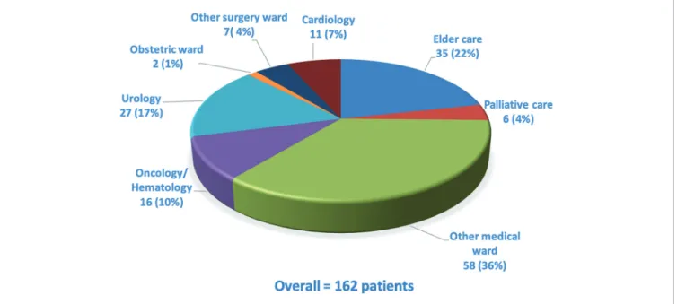
were eligible (Figure 1). Overall, 188 (39%) were included from the ICU: 91 (19%) from the surgical ICU and 97 (20%) from the medical ICU. The NW was the inclusion ward for 130 (27%) and the OW for 162 (34%). Figure 2 lists the OW, together with the number of patients included in each; they included elder care, oncology/hematology, palliative care, urology, obstetrics, cardiology, and other medical and surgi-cal wards. Of the overall hospital patient admissions (N = 21 312) during the study period, 2.3% were of patients diag-nosed with severe AKI at admission or during their stay. The prespecified chronic illnesses and sociodemographic infor-mation are presented for the overall population and accord-ing to the inclusion ward in Table 1. Patients included in the ICU were younger (mean age: 66 years) than those from the NW (73) or OW (83) (P < .001). Comorbidities were numer-ous, especially cardiovascular and neoplastic. Nearly half the patients (n = 234; 49%) were known to have had CKD before this hospitalization; only 82 (35%) had previous nephrology care. A history of CKD was most frequent in patients included in the NW.
Clinical Characteristics and Laboratory Values
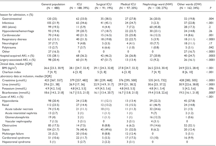
Other characteristics including reason for hospitalization, community- vs hospital-acquired AKI, surgery-associated AKI, clinical data, and AKI cause are reported in Table 2. The principal reasons for admission were gastrointestinal disease (24.8%), sepsis (21.9%), hypovolemia (19.5%), and AKI (19.5%). Acute kidney injury was hospital acquired for 28% of the study population and surgery associated for 20.3%, including 78% of the cases regarded as emergency surgery. At inclusion, the median Charlson index was 7
(IQR: [4, 9]) and was highest in the OW inclusion group. The median BMI was 26.6 (IQR: [23.3, 30.9]) with significant differences according to inclusion ward, and the values were lower for patients included in OW than in the ICU and NW. The median SCr (423 μmol/L, IQR: [367, 557]) also differed between these groups and was lower in the ICU, as were bicarbonate levels.
AKI Causes
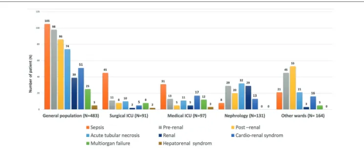
The main causes of AKI were sepsis (22%), hypovolemia AKI (20%), obstructive AKI (18%), ATN (15%), and cardio-renal syndrome (11%) (Figure 3). These causes differed sig-nificantly between wards: septic AKI, multiorgan failure, and cardiorenal and hepatorenal syndromes were most fre-quent in the ICU, whereas hypovolemia and obstructive AKI were predominant in the OW inclusion group. Finally, renal AKI, which includes ATN, acute glomerular injury, acute vascular nephritis, and acute interstitial nephritis, was pre-dominant in the NW.
Trajectory and Care
Table 3 summarizes the data about care plans, indications for RRT, and specific treatment during hospitalization. At inclusion, treatment limitations (such as “do not resusci-tate” orders) were in effect for 72 patients (15%), with the largest proportion of them being included in OW. During hospitalization, 194 patients (40%) required RRT, with more of them being included in the ICU (73%) than in the NW (43%); none of the OW inclusions received RRT. The most common indications for RRT reported by physicians
Table 1. Sociodemographic Data, Medical History, and Kidney Information.
General
(N = 480)(N = 188; 39%)ICU (N = 91; 19%)Surgical ICU Medical ICU (N = 97; 20%)
Nephrology ward (NW)
(N = 130; 27%) Other wards (OW) (N = 162; 34%) P (general)
Sociodemographic data
Men, n (%) 351 (73.1) 145 (77.1) 70 (76.9) 75 (77.3) 88 (67.7) 118 (72.8) .32 Age (years), median [IQR] 72 [64, 83] 68 [60, 76] 70 [64, 78] 66 [57, 74] 73 [63, 82] 83 [71, 89] <.0001 Past medical history, n (%)
Cardiovascular disease 440 (91.7) 174 (93) 82 (90.1) 92 (94.9) 115 (88.5) 151 (93) .28 Coronary heart disease 116 (24.3) 46 (24.5) 21 (23.1) 25 (25.8) 29 (22.3) 41 (25.8) .88 Heart failure 128 (26.7) 42 (22.3) 15 (16.5) 27 (27.8) 37 (28.5) 49 (30.3) .10 Peripheral vascular disease 86 (17.9) 35 (18.6) 19 (20.9) 16 (16.5) 23 (17.7) 28 (17.3) .87 Cardiac arrhythmia 108 (22.6) 30 (16.0) 15 (16.5) 15 (15.5) 29 (22.3) 49 (30.8) .012 Cerebrovascular disease 61 (12.8) 17 (9.0) 7 (7.7) 10 (10.3) 11 (8.5) 33 (20.8) .003 Treated hypertension 320 (67.1) 127 (67.6) 58 (63.7) 69 (71.1) 93 (71.5) 100 (62.9) .31 Diabetes mellitus 176 (36.7) 72 (38.3) 33 (36.3) 39 (40.2) 49 (37.7) 55 (34.0) .77 Chronic liver dysfunction 29 (6) 17 (9.0) 9 (9.9) 8 (8.3) 7 (5.4) 5 (3.1) .11 Solid organ malignancies 130 (27.1) 43 (22.9) 21 (23.1) 22 (22.7) 27 (20.8) 60 (37.0) .006 Hemopathy 40 (8.4) 13 (7.0) 6 (6.7) 7 (7.4) 11 (8.5) 16 (9.9) .85 Asthma or COPD 66 (13.8) 31 (16.6) 17 (18.9) 14 (14.4) 16 (12.4) 19 (11.7) .42 NIV or supplemental oxygen 37 (7.7) 20 (10.6) 13 (14.3) 7 (7.2) 8 (6.2) 9 (5.6) .069 Renal information (previous 6 months)
CKD, n (%) 235 (48.9) 77 (41.0) 33 (36.3) 44 (45.4) 83 (63.9) 74 (45.7) <.001 GFR (mL/min/1.73 m2), median [IQR] 56 [37, 81] 71 [46, 90] 73 [47, 90] 68 [46, 91] 54 [30, 71] 46 [32, 75] <.0001
Note. “P” for the global comparison of values between ICU, NW, and OW. ICU = intensive care unit; IQR = interquartile range; COPD = chronic obstructive pulmonary disease; NIV = noninvasive ventilation; CKD = chronic kidney disease; GFR = glomerular filtration rate.
Table 2. Reason for Admission, Clinical Data, Laboratory Findings, and Causes of AKI.
General population
(N = 480) (N = 188; 39%)ICU (N = 91; 19%)Surgical ICU (N = 97; 20%)Medical ICU Nephrology ward (NW) (N = 130; 27%) Other wards (OW) (N = 162; 34%) P
Reason for admission, n (%)
Gastrointestinal 120 (25) 62 (33.0) 35 (38.5) 27 (27.8) 26 (20.0) 32 (19.8) .004 Infectious 105 (21.9) 65 (34.6) 41 (45.1) 24 (24.7) 3 (2.3) 37 (22.8) <.001 AKI (alone) 99 (19.5) 11 (5.9) 4 (4.4) 7 (7.2) 60 (45.8) 28 (17.3) .2 Hypovolemia/hemorrhage 93 (19.4) 39 (20.7) 17 (18.7) 22 (22.7) 30 (23.1) 24 (14.8) .26 Cardiovascular 70 (14.6) 40 (21.3) 15 (16.5) 25 (25.8) 16 (12.3) 14 (8.6) .001 Respiratory distress 63 (13.1) 40 (21.3) 18 (19.8) 22 (22.7) 5 (3.9) 18 (11.1) <.0001 Neurological 37 (7.7) 9 (4.8) 5 (5.5) 4 (4.1) 10 (7.7) 18 (11.1) .17 Polytrauma 13 (2.7) 7 (3.7) 6 (6.6) 1 (1.0) 1 (0.8) 5 (3.1) .042 Other 27 (16.3) 0 0 0 0 27 (16.7) <.0001 Hospital-acquired AKI, n (%) 135 (28.1) 68 (36.2) 42 (46.2) 26 (26.8) 27 (20.8) 40 (24.7) .0003 Surgery-associated AKI, n (%) 98 (20.4) 60 (31.9) 47 (51.7) 13 (13.4) 12 (9.2) 26 (16.1) <.0001 Clinical data, median [IQR]
BMI (kg/m2) 26.6 [23.3, 30.9] 28.1 [24.7, 32.4] 29.1 [24.3, 32.8] 27.8 [24.7, 32.3] 26.5 [23.0, 30.4] 24.9 [22.5, 28.4] <.001
Charlson index 7 [4, 9] 6 [3, 8] 5 [3, 8] 6 [3, 8] 7 [4, 9] 8 [6, 10] <.001 Laboratory data at inclusion, median [IQR]
Creatinine (μmol/L) 423 [367, 557] 379 [257, 482] 381 [239, 468] 376 [295, 500] 535 [415, 732] 428 [382, 505] <.0001 Urea (mmol/L) 29.6 [21, 38] 26.9 [17, 36] 22.9 [14.9, 31.7] 29.9 [21, 38.2] 30.6 [23, 37.2] 30.9 [22.6, 38.8] <.0001 Potassium (mmol/L) 4.9 [4.2, 5.6] 4.8 [4.2, 5.5] 4.9 [4.3, 5.6] 4.8 [4.0, 5.5] 4.8 [4.1, 5.4] 5 [4.2, 5.6] .096 Bicarbonates (mmol/L) 18.6 [14.5, 21.8] 16.7 [13.5, 21.0] 16.1 [13.4, 20.7] 16.7 [13.8, 21.0] 19.4 [15.8, 22.6] 19.5 [16.1, 21.8] .0007 Cause of AKI, n (%) <.001 Hypovolemia 98 (20.4) 24 (12.8) 11 (12.1) 13 (13.4) 29 (22.3) 45 (27.8) Renal 112 (23.5) 27 (14.4) 12 (13.2) 15 (15.5) 61 (46.9) 24 (14.8) Acute tubular necrosis 74 (15.4) 21 (11.1) 10 (11) 11 (11.3) 32 (24.6) 21 (13) Acute interstitial nephritis 13 (2.7) 2 (1) 1 (1.1) 1 (1) 9 (7) 2 (1.2) Glomerulonephritis 19 (4) 2 (1) 1 (1.1) 1 (1) 16 (12.3) 1 (0.6) Vascular nephropathy 7 (1.5) 3 (3.1) 0 3 (3.1) 4 (3.1) 0 Obstructive 85 (17.5) 14 (7.5) 8 (8.8) 6 (6.2) 19 (14.6) 52 (32.1) Sepsis 104 (21.7) 76 (40.4) 45 (49.6) 31 (32.0) 8 (6.2) 20 (12.4) Multiorgan failure 25 (5.2) 20 (10.6) 8 (8.8) 12 (12.4) 0 5 (3.1) Cardiorenal syndrome 51 (10.6) 22 (11.7) 5 (5.5) 17 (17.5) 13 (10) 16 (9.9) Hepatorenal syndrome 5 (1) 5 (2.7) 2 (2.2) 3 (3.1) 0 0
Note. “P” for the global comparison of values between ICU, NW, and OW. AKI = acute kidney injury; ICU = intensive care unit; BMI = body mass index; IQR = interquartile range.
Aglae et al 7
Figure 3. Cause of acute kidney injury with differences between inclusion groups. Note. ICU = intensive care unit.
Table 3. Trajectory and Care.
General population
(N = 480) (N = 188; 39%)ICU (N = 91; 19%)Surgical ICU (N = 97; 20%)Medical ICU Nephrology ward (NW) (N = 130; 27%) Other wards (OW) (N = 162; 34%) P
Palliative care, n (%) 72 (14.9) 10 (5.3) 3 (3.3) 7 (7.2) 4 (3.1) 58 (35.4) <.0001 RRT delivery 194 (40.4) 139 (73.9) 67 (73.6) 72 (74.2) 55 (42.3) 0 <.0001 Reason for RRT, n (%) Anuria 112 (23.3) 85 (45.2) 46 (68.7) 39 (40.2) 27 (20.8) — .07 Metabolic acidosis 106 (22.1) 86 (45.7) 43 (64.2) 43 (44.3) 20 (15.4) — .005 Hyperkalemia 62 (12.9) 48 (25.5) 24 (35.8) 24 (24.7) 14 (10.8) — .45 Acute pulmonary edema 33 (6.9) 17 (9.0) 7 (10.5) 10 (10.3) 16 (12.3) — .02 Volume overload with diuretic resistance 30 (6.3) 17 (9.0) 8 (8.8) 9 (9.3) 13 (10.0) — .16 Toxic clearance 23 (4.8) 13 (6.9) 4 (4.4) 9 (9.3) 10 (7.7) — .10 Refractory shock 5 (1.0) 5 (2.7) 3 (3.3) 2 (2.1) 0 — .32 Hypercalcemia 4 (0.8) 2 (1.1) 0 2 (2.9) 2 (3.6) — .32 Specific treatment, n (%) Vasopressor use 143 (29.8) 67 (35.6) 71 (78.0) 67 (69.1) 1 (0.8) 4 (2.4) <.0001 Mechanical ventilation 115 (23.9) 54 (47) 61 (67.0) 54 (55.7) 0 0 <.0001 Trajectory, n (%)
Ward before inclusion <.0001
Medicine 67 (13.9) 24 (12.8) 8 (8.8) 16 (16.5) 31 (23.9) 12 (7.4) Surgery 23 (4.8) 17 (9.0) 12 (13.2) 5 (5.2) 6 (4.6) 0 Emergency 245 (51.0) 62 (32.9) 33 (36.3) 29 (29.9) 58 (44.6) 125 (77.2)
Inhospital death, n (%) 141 (29.4) 80 (42.6) 34 (37.4) 46 (47.4) 12 (9.3) 49 (30.3) <.0001 30-day mortality, n (%) 141 (29.4) 70 (37.2) 30 (33.0) 40 (41.2) 12 (9.2) 59 (36.4) <.0001
Note. “P” for global comparison between ICU, NW, and OW. ICU = intensive care unit; RRT = renal replacement therapy.
were anuria (57%), metabolic acidosis (54%), hyperkale-mia (32%), and diuretic-resistant volume overload (15%). Acute pulmonary edema was most frequent in patients included in the NW, whereas metabolic acidosis and anuria were the principal indications in the ICU (Figure 4). In OW, none of them were treated by RRT because of treatment limitations in 58 (36%) and because of the causes of AKI that can be treated by specific treatment other than RRT such as intravenous (IV) infusion for hypovolemia (45; 28%), urine derivation for obstructive
(52; 32%), and AKI or cardiac treatment for cardiorenal syndrome (16; 16%). Finally, 143 patients (30%) needed vasopressors and 115 (24%) mechanical ventilation, mostly in the ICU group. Slightly more than half of the patients (n = 245; 51%) were admitted via the emergency department to their inclusion ward. A total of 141 (29%) of these patients died during this hospitalization. The rates of death in the hospital and at 30 days differed according to department of inclusion and were highest among those included from the ICU.
Discussion
Our prospective study, conducted over an 18-month period in a tertiary teaching hospital, included 480 patients with severe AKI, that is, 2.3% of overall hospital admissions. Our study showed that this population was relatively old and had frequent comorbidities, which were cardiovascular (92%), diabetes mellitus (37%), cancer (35%), and CKD (50%) before hospitalization. These patients were managed in sev-eral different wards, not only in the ICU and NW, and these wards varied according to patients’ clinical presentation and laboratory results. The causes of severe AKI varied, with sepsis being predominant in the ICU and renal AKI in the NW. Indications for RRT for AKI also differed between the ICU and the NW, and no patients initially diagnosed in OW had RRT. Finally, the prognosis of our study population with severe AKI and a high rate of comorbidities was poor, with a high inhospital mortality of almost one third. It nonetheless differed according to inclusion ward and was associated with different patterns and care. In this population comprising a subgroup of patients with KDIGO stage 3, the high inhospi-tal morinhospi-tality rate was probably due not only to AKI but also to the causes of AKI and the high rate of comorbidities. This study underlines that severe AKI is not limited to the ICU and that its clinical presentation and care are extremely heterogeneous.
The demographics of the study population were consis-tent with previous epidemiologic studies, in particular with 2 French cohort studies based on the national discharge
database.8,17 We observed that most patients in our cohort
had major comorbidities, including but not limited to dia-betes, cardiovascular disease, and cancer. The median Charlson index score was 7. Moreover, the median patient age was 72 years. Patients included in the ICU were sig-nificantly younger than those included in the NW and OW,
as in a nationwide French study.17 A novelty for our cohort
is information about CKD history, which was frequent in this population: Around 50% of the patients had previously met the definition for CKD, but only 17% of them were managed by nephrologists. This CKD history rate is higher than that in French administrative database studies or in other studies of AKI, which report rates from 10% to 35%.18-23 The high frequency of CKD history may be
explained in part by the quality of information collected, due to the study’s prospective design and its use of the KDIGO definition of CKD, rather than estimated GFR or administrative codes. It may also be explained by the age profile of our cohort.
Gastrointestinal diseases, sepsis, hypovolemia, AKI, and cardiovascular disorders were the leading reasons for hospi-talization. These reasons can vary in populations by hospital type, especially in relation to both solid organ transplantation and cardiac surgery, the latter being strongly associated with AKI occurrence. Reports indicate that AKI complicates 18% of hospitalizations for cardiac surgery, with 2% to 6%
requir-ing RRT.24 Otherwise, we observed that 28% of AKI cases
were hospital acquired and 72% community acquired. The same ratio was observed in a UK study in 2 tertiary centers (6% of ICU stays and 25% AKI stage 3): 27% hospital
acquired vs 73% community acquired.25
In our study, the incidence of severe adult AKI was 2.3% of overall hospitalizations. This finding is consistent with the UK data from a multicentre randomized trial performed in 5 UK hospitals that showed an incidence of 2.5% of
severe AKI.26
Sepsis was the leading cause of AKI in our study popula-tion, followed by prerenal, postrenal, and cardiorenal AKI. In a Scottish population study of all hospitalized patients (including 8.5% patients in ICU and 35% in the failure stage of the RIFLE classification), sepsis and hypovolemia were
Figure 4. Indications for renal replacement therapy and differences between inclusion groups for 194 patients (40.4%). Note. ICU = intensive care unit.
Aglae et al 9
the leading causes of AKI.27 In comparison, the study of all
hospitals in Madrid (and with 27% of patients in the ICU) by
Liano and Pascual22 found the 3 leading causes of AKI to be
ATN (45%), hypovolemic AKI (21%), and obstructive AKI (10%). Nonetheless, the definition of AKI was different then, and the cases of ATN probably include some cases of septic
AKI, which has only become understood more recently.4 The
main indications for RRT in our study were oligoanuria, met-abolic acidosis, hyperkalemia, and volume overload—causes similar to those from a Canadian study of a cohort of elderly
ICU patients.28 RRT in our hospital is performed by a
con-tinuous venovenous hemodiafiltration technique in the ICU (medical and surgical) and by intermittent hemodialysis in the NW. Because the study design was observational, we described the frequency of RRT indication according to the ward of inclusion. The frequency of RRT differed according to not only inclusion ward but also the causes of RRT, patient comorbidities, and AKI causes. We described the population and practices according to the ward and observed different rates of RRT according to the ward but also for indication or no indication of RRT showing heterogeneity of this popula-tion with severe AKI.
For the overall population, we report that hospital mortal-ity and 30-day mortalmortal-ity were similar, around 29%. Hospital mortality for RRT patients was 40.2%. A multicentre, stepped-wedge cluster randomized trial performed in 5 UK hospitals included patients with AKI aged ≥18 years and
observed an overall 30-day mortality rate of 24.5%.26 In the
French cohort studies based on the national discharge data-base for patients with AKI requiring RRT, the inhospital
mortality of 47% was similar to ours.17
The strength of this study is its prospective design and the quality of information collected for medical history. We used the entire KDIGO definition of CKD, because it is frequently underestimated by focusing only on GFR in studies based on medical administrative databases. Thus, this prospective analysis of all adults with severe AKI in a university hospital allows us to show the frequent comor-bidities and heterogeneity of severe AKI in clinical presen-tation, severity, and care in different hospital departments. Its results emphasize that the care of patients with severe AKI depends on the presence of treatment limitations and that the indications for RRT differ between intensivists and nephrologists.
Limitations
Our study has several limitations that must be considered in interpreting our data. First, we studied a subgroup of patients with KDIGO stage 3 AKI with a restricted defini-tion that does not include the urine output criteria or the tripling of baseline SCr with a maximum SCr below 354 µmol/L. The single-center design in a tertiary hospital with no cardiac surgery ward or solid organ transplantation
limits the generalizability of this result without limiting the extent of heterogeneity in the population.
This study indicates the substantial heterogeneity of severe AKI and reinforces the fact that AKI is not a single disease and is not managed only in ICUs. This probably implies the need for an individualized patient-centered approach and the wide implementation of the KDIGO AKI classification to discuss the planning of care.
Conclusion
This study reports 3 hospitalization patterns in patients with severe AKI. Only 39% of these patients are hospitalized in the ICU, 27% are in the NW, and 34% in OW. Clinical pre-sentation, AKI cause, and thus its care, including reasons for RRT, differ according to the department of hospitalization with broad heterogeneity for the same AKI stage. The het-erogeneity of severe AKI and of its treatment in this popula-tion of a subgroup of patients with stage 3 KDIGO AKI implies the need for more individualized and patient-cen-tered care and for improvement of prognostic tools to include more information than simply SCr and diuresis.
Ethics Approval and Consent to Participate
This study was approved by the Ethics Committee and the French Data Protection Authority (Commission Nationale Informatique & Libertés; CNIL number: 1963867v0).
Consent for Publication
All authors consent to the publication of this study.
Availability of Data and Materials
The data and materials are not available for this study.
Acknowledgments
The authors thank the University Hospital of Nîmes for its struc-tural, human, and financial support through the award obtained by our team during the internal call for tenders “Thématiques émer-gentes.” The authors also thank Jo Ann Cahn for reviewing the English.
Author Contributions
O.M. conceived and designed the study. C.A., L.M., S.D.B., R.T., Z.M., J.-Y.L., and O.M. analyzed and interpreted the data. C.A., L.M., S.D.B., J.-Y.L., D.-P.D.B., and O.M. drafted and revised the article. C.A., L.M., P.R., S.C., S.D.B., R.T., Z.M., J.-Y.L., and O.M. provided intellectual content of critical importance to the work described. C.A., L.M., P.R., S.C., S.D.B., R.T., Z.M., D.-P.D.B., J.-Y.L., and O.M. approved the final version to be published.
Declaration of Conflicting Interests
The author(s) declared no potential conflicts of interest with respect to the research, authorship, and/or publication of this article.
Funding
The author(s) disclosed receipt of the following financial support for the research, authorship, and/or publication of this article: The cur-rent study is part of the thematic portfolio for clinical research stud-ies which were at least partly supported through the University Hospital of Nîmes call for internal bids “Thématiques émergentes.”
ORCID iD
Olivier Moranne https://orcid.org/0000-0002-3127-1415
References
1. Kellum JA, Lameire N. Diagnosis, evaluation, and manage-ment of acute kidney injury: a KDIGO summary (Part 1). Crit
Care. 2013;17:204.
2. Kellum JA, Lameire N, Aspelin P, et al. Work group member-ship. Kidney Int. 2012;2:1.
3. Mehta RL, Burdmann EA, Cerda J, et al. Recognition and management of acute kidney injury in the International Society of Nephrology 0by25 Global Snapshot: a multinational cross-sectional study. Lancet. 2016;387:2017-2025.
4. Kellum JA, Prowle JR. Paradigms of acute kidney injury in the intensive care setting. Nat Rev Nephrol. 2018;14:217-230. 5. Barasch J, Zager R, Bonventre JV. Acute kidney injury: a
prob-lem of definition. Lancet. 2017;389:779-781.
6. Kellum JA, Lameire N. The definition of acute kidney injury.
Lancet. 2018;391:202-203.
7. Selby NM, Crowley L, Fluck RJ, et al. Use of electronic results reporting to diagnose and monitor AKI in hospitalized patients.
Clin J Am Soc Nephrol. 2012;7:533-540.
8. Riffaut N, Moranne O, Hertig A, Hannedouche T, Couchoud C. Outcomes of acute kidney injury depend on initial clinical fea-tures: a national French cohort study. Nephrol Dial Transplant. 2018;33:2218-2227.
9. Sykes L, Sinha S, Hegarty J, et al. Reducing acute kidney injury incidence and progression in a large teaching hospital.
BMJ Open Qual. 2018;7:e000308.
10. NCEPOD—Acute Kidney Injury: Adding Insult to Injury Report (2009). www.ncepod.org.uk/2009report1/Downloads/ AKI_report.pdf
11. Couchoud C, Stengel B, Landais P, et al. The Renal Epidemiology and Information Network (REIN): a new registry for end-stage renal disease in France. Nephrol Dial Transplant. 2006;21:411-418.
12. Le Gall JR, Lemeshow S, Saulnier F. A new Simplified Acute Physiology Score (SAPS II) based on a European/North American multicenter study. JAMA. 1993;270:2957-2963. 13. Lameire N, Biesen WV, Vanholder R. Acute renal failure.
Lancet. 2005;365:417-430.
14. Ronco C, Di Lullo L. Cardiorenal syndrome. Heart Fail Clin. 2014;10:251-280.
15. Ginès P, Guevara M, Arroyo V, et al. Hepatorenal syndrome.
Lancet. 2003;362:1819-1827.
16. Singer M, Deutschman CS, Seymour CW, et al. The Third International Consensus Definitions for Sepsis and Septic Shock (Sepsis-3). JAMA. 2016;315:801-810.
17. Garnier F, Couchoud C, Landais P, Moranne O. Increased inci-dence of acute kidney injury requiring dialysis in metropolitan France. PLoS ONE. 2019;14:e0211541.
18. Carlson N, Hommel K, Olesen JB, et al. Trends in one-year outcomes of dialysis-requiring acute kidney injury in Denmark 2005-2012: a population-based nationwide study. PLoS ONE. 2016;11(7):e0159944.
19. Kolhe NV, Muirhead AW, Wilkes SR, et al. The epide-miology of hospitalised acute kidney injury not requiring dialysis in England from 1998 to 2013: retrospective analy-sis of hospital episode statistics. Int J Clin Pract. 2016;70: 330-339.
20. Nisula S, Kaukonen K-M, Vaara ST, et al. Incidence, risk fac-tors and 90-day mortality of patients with acute kidney injury in Finnish intensive care units: the FINNAKI study. Intensive
Care Med. 2013;39:420-428.
21. Wonnacott A, Meran S, Amphlett B, Talabani B, Phillips A. Epidemiology and outcomes in community-acquired ver-sus hospital-acquired AKI. Clin J Am Soc Nephrol. 2014;9: 1007-1014.
22. Liano F, Pascual J. Epidemiology of acute renal failure: a prospective, multicenter, community-based study. Kidney Int. 1996;50:811-818.
23. Uchino S, Kellum JA, Bellomo R, et al. Acute renal failure in critically ill patients: a multinational, multicenter study. JAMA. 2005;294:813-818.
24. Thiele RH, Isbell JM, Rosner MH. AKI associated with cardiac surgery. Clin J Am Soc Nephrol. 2015;10:500-514.
25. Wonnacott A, Meran S, Amphlett B, Talabani B, Phillips A. Epidemiology and outcomes in community-acquired ver-sus hospital-acquired AKI. Clin J Am Soc Nephrol. 2014;9: 1007-1014.
26. Selby NM, Casula A, Lamming L, et al. An organiza-tional-level program of intervention for AKI: A pragmatic stepped wedge cluster randomized trial. J Am Soc Nephrol. 2019;30:505-515.
27. Ali T, Khan I, Simpson W, et al. Incidence and outcomes in acute kidney injury: a comprehensive population-based study.
J Am Soc Nephrol. 2007;18:1292-1298.
28. Bagshaw SM, Adhikari NKJ, Burns KEA, et al. Selection and receipt of kidney replacement in critically ill older patients with AKI. Clin J Am Soc Nephrol. 2019;14:496-505.