HAL Id: hal-01231270
https://hal.archives-ouvertes.fr/hal-01231270
Submitted on 19 Nov 2015
HAL is a multi-disciplinary open access
archive for the deposit and dissemination of sci-entific research documents, whether they are pub-lished or not. The documents may come from teaching and research institutions in France or abroad, or from public or private research centers.
L’archive ouverte pluridisciplinaire HAL, est destinée au dépôt et à la diffusion de documents scientifiques de niveau recherche, publiés ou non, émanant des établissements d’enseignement et de recherche français ou étrangers, des laboratoires publics ou privés.
phagocytophilum[/i] proteins involved in infection of the
tick vector, [i]Ixodes scapularis[/i]
Margarita Villar, Nieves Ayllon, Katherine M. Kocan, Elena
Bonzon-Kulichenko, Pilar Alberdi, Edmour F. Blouin, Sabine Weisheit,
Lourdes Mateos-Hernandez, Alejandro Cabezas-Cruz, Lesley Bell-Sakyi, et al.
To cite this version:
Margarita Villar, Nieves Ayllon, Katherine M. Kocan, Elena Bonzon-Kulichenko, Pilar Alberdi, et al.. Identification and characterization of [i]Anaplasma phagocytophilum[/i] proteins involved in infection of the tick vector, [i]Ixodes scapularis[/i]. PLoS ONE, Public Library of Science, 2015, 10 (9), 26 p. �10.1371/journal.pone.0137237�. �hal-01231270�
Identification and Characterization of
Anaplasma phagocytophilum Proteins
Involved in Infection of the Tick Vector,
Ixodes scapularis
Margarita Villar1☯, Nieves Ayllón1☯, Katherine M. Kocan2, Elena Bonzón-Kulichenko3, Pilar Alberdi1, Edmour F. Blouin2, Sabine Weisheit4,5, Lourdes Mateos-Hernández1,
Alejandro Cabezas-Cruz1,6, Lesley Bell-Sakyi4,5, Marie Vancová7,8, Tomáš Bílý7,8, Damien F. Meyer9,10, Jan Sterba7, Marinela Contreras1, Nataliia Rudenko7, Libor Grubhoffer7,8,
Jesús Vázquez3, José de la Fuente1,2*
1 SaBio, Instituto de Investigación en Recursos Cinegéticos IREC-CSIC-UCLM-JCCM, Ronda de Toledo s/ n, 13005, Ciudad Real, Spain, 2 Department of Veterinary Pathobiology, Center for Veterinary Health Sciences, Oklahoma State University, Stillwater, Oklahoma, 74078, United States of America, 3 Centro Nacional de Investigaciones Cardiovasculares, Melchor Fernández Almagro 3, 28029, Madrid, Spain, 4 The Roslin Institute and Royal (Dick) School of Veterinary Studies, University of Edinburgh, Easter Bush, Midlothian, EH25 9RG, United Kingdom, 5 The Pirbright Institute, Ash Road, Pirbright, Woking, GU24 0NF, United Kingdom, 6 Center for Infection and Immunity of Lille (CIIL), INSERM U1019– CNRS UMR 8204, Université Lille Nord de France, Institut Pasteur de Lille, Lille, France, 7 Institute of Parasitology, Biology Centre of the Academy of Sciences of the Czech Republic, Branišovská 31, České Budějovice, CZ-37005, Czech Republic, 8 Faculty of Science, University of South Bohemia, Branišovská 31, České Budějovice, CZ-37005, Czech Republic, 9 CIRAD, UMR CMAEE, Site de Duclos, Prise d’eau, F-97170, Petit-Bourg, Guadeloupe, France, 10 INRA, UMR1309 CMAEE, F-34398, Montpellier, France
☯ These authors contributed equally to this work. *jose_delafuente@yahoo.com
Abstract
Anaplasma phagocytophilumis an emerging zoonotic pathogen transmitted by Ixodes sca-pularisthat causes human granulocytic anaplasmosis. Here, a high throughput quantitative proteomics approach was used to characterize A. phagocytophilum proteome during rick-ettsial multiplication and identify proteins involved in infection of the tick vector, I. scapularis. The first step in this research was focused on tick cells infected with A. phagocytophilum and sampled at two time points containing 10–15% and 65–71% infected cells, respectively to identify key bacterial proteins over-represented in high percentage infected cells. The second step was focused on adult female tick guts and salivary glands infected with A. pha-gocytophilumto compare in vitro results with those occurring during bacterial infection in vivo. The results showed differences in the proteome of A. phagocytophilum in infected ticks with higher impact on protein synthesis and processing than on bacterial replication in tick salivary glands. These results correlated well with the developmental cycle of A. phago-cytophilum, in which cells convert from an intracellular reticulated, replicative form to the nondividing infectious dense-core form. The analysis of A. phagocytophilum differentially represented proteins identified stress response (GroEL, HSP70) and surface (MSP4) pro-teins that were over-represented in high percentage infected tick cells and salivary glands
OPEN ACCESS
Citation: Villar M, Ayllón N, Kocan KM, Bonzón-Kulichenko E, Alberdi P, Blouin EF, et al. (2015) Identification and Characterization of Anaplasma phagocytophilum Proteins Involved in Infection of the Tick Vector, Ixodes scapularis. PLoS ONE 10(9): e0137237. doi:10.1371/journal.pone.0137237
Editor: Kelly A. Brayton, Washington State University, UNITED STATES
Received: April 13, 2015 Accepted: August 13, 2015
Published: September 4, 2015
Copyright: © 2015 Villar et al. This is an open access article distributed under the terms of the
Creative Commons Attribution License, which permits unrestricted use, distribution, and reproduction in any medium, provided the original author and source are credited.
Data Availability Statement: All relevant data are within the paper and its Supporting Information files. Funding: This research was supported by MEC, Spain, grant BFU2011-23896 (JF), European Union FP7 ANTIGONE project number 278976 (JF) and Postdok BIOGLOBE, Czech Republic CZ.1.07/ 2.3.00/30.0032 co-financed by the European Social Fund and the state budget of the Czech Republic (JS). N. Ayllón was funded by Ministerio de Educacion, Cultura y Deporte, Spain. S. Weisheit was supported by the POSTICK ITN (Post-graduate training network for capacity building to control ticks
when compared to low percentage infected cells and guts, respectively. The results demon-strated that MSP4, GroEL and HSP70 interact and bind to tick cells, thus playing a role in rickettsia-tick interactions. The most important finding of these studies is the increase in the level of certain bacterial stress response and surface proteins in A. phagocytophilum-infected tick cells and salivary glands with functional implication in tick-pathogen interac-tions. These results gave a new dimension to the role of these stress response and surface proteins during A. phagocytophilum infection in ticks. Characterization of Anaplasma prote-ome contributes information on host-pathogen interactions and provides targets for devel-opment of novel control strategies for pathogen infection and transmission.
Introduction
Anaplasma phagocytophilum (Rickettsiales: Anaplasmataceae) is a tick-borne pathogen that is the etiologic agent of human, canine and equine granulocytic anaplasmosis and tick-borne fever of ruminants [1–3]. Despite this organism being an emerging zoonotic pathogen in many regions of the world, vaccines are not available for prevention of transmission and infection of humans and animals [4]. While A. phagocytophilum infection may resolve without therapy, the pathogen has been shown to be susceptible to tetracycline antibiotics [4].
A. phagocytophilum is an intracellular bacterium that infects tick tissues such as gut and sali-vary glands and vertebrate host neutrophils [5–11]. While transcriptomics and proteomics analyses have contributed to our understanding of the mechanisms by which A. phagocytophi-lum infection affects host and vector gene expression and protein content [8–17], less informa-tion is available on bacterial molecular mechanisms involved in pathogen infecinforma-tion and multiplication [18–21]. Proteomics characterization of Anaplasma spp. provides information on host-pathogen interactions and suggests possible targets for the control of pathogen infec-tion and transmission [17–26].
The transcriptome and/or proteome of A. phagocytophilum have been characterized in tick salivary glands during transmission feeding [21] and in human HL-60 cells [18–20]. The prote-ome of the closely related pathogen A. marginale was characterized in IDE8 and ISE6 tick cells [23,24,26]. These experiments demonstrated the existence of host-specific Anaplasma pro-teins that may be involved in bacterial infection and multiplication. However, the Anaplasma proteome has not been characterized in low and high percentage infected tick cells to identify proteins functionally important during bacterial multiplication in the tick vector.
The proteome is dynamic with each developmental stage presenting an ensemble of proteins that give rise to substantial diversity and thus the need to characterize changes as infection pro-ceeds from low to high percentage infected tick cells. Although in vivo data might be more rele-vant to understand bacterial infection [21], in vitro studies allow for monitoring A.
phagocytophilum infection for a better comparison between low and high percentage infected cells. Nevertheless, the experimental approach using tick cell cultures should be complemented with in vivo studies to identify bacterial proteins playing a relevant role during infection and multiplication in the tick vector.
The aim of this research was to identify A. phagocytophilum proteins involved in infection of the tick vector, I. scapularis. Our hypothesis was that A. phagocytophilum proteins that increase as infection proceeds in cultured tick cells and ticks may be important for infection. To address this hypothesis, we characterized A. phagocytophilum proteome during rickettsial multiplication in I. scapularis cultured tick cells, guts and salivary glands and demonstrated
and tick-borne diseases) within the FP7-PEOPLE-ITN programme (EU Grant No. 238511). The funders had no role in study design, data collection and analysis, decision to publish, or preparation of the manuscript.
Competing Interests: The authors have declared that no competing interests exist.
that this bacterium uses certain stress response and surface proteins to favor pathogen infection and multiplication. Characterization of Anaplasma proteome contributes information on host-pathogen interactions and also provides targets for development of novel control strategies for pathogen infection and transmission.
Results and Discussion
Changes in A. phagocytophilum proteome correlate with bacterial
infection cycle in adult female tick guts and salivary glands
The transcriptome and proteome of A. phagocytophilum were previously characterized in I. scapularis tick salivary glands during transmission feeding with similar protein functions iden-tified by both approaches [21]. In vivo data is more relevant to understand bacterial infection. However, the proteome is dynamic with each developmental stage presenting an ensemble of proteins that give rise to substantial diversity. Additionally, the limited amount of material that can be obtained in vivo may affect the detection of proteins present at low levels but playing an important role during bacterial life cycle. Therefore, it is important to characterize changes as infection increases from low to high percentage infected cells in vitro and in vivo in both guts and salivary glands. These tick tissues were selected for this study because guts and salivary glands play a major role during pathogen acquisition, multiplication and transmission [17].
The first step in this research was focused on I. scapularis embryonic ISE6 cells in which it is easy to select for low and high percentage A. phagocytophilum-infected cells, thus allowing for the identification of key bacterial proteins over-represented in high percentage infected cells when compared to cells infected at low percentage. A high throughput iTRAQ-based quantita-tive proteomics approach was used to characterize A. phagocytophilum differential protein representation in high percentage (65–71% infected cells) when compared to low percentage (10–15% infected cells) infected ISE6 tick cells. Infected tick cell cultures are asynchronous and therefore both late and early-infected cells are present in high percentage infected cells, but likely with more late than early-infected cells when compared to the low percentage infected cells. The statistical analysis of proteomics data revealed a total of 765 Anaplasma proteins at a false discovery rate (FDR) of 5% (S1 Table). Of them, 88 were differentially represented in high percentage infected cells when compared to low percentage infected cells (S1 Table). Analysis of protein ontology for bacterial differentially represented proteins showed that biological pro-cesses (BP) cell division, infection, metabolic process, stress response and translation were affected as infection proceeded in tick cells (Fig 1A).
The second step in this research was focused on adult female tick guts and salivary glands infected with A. phagocytophilum to compare in vitro results obtained under controlled condi-tions with those occurring during bacterial infection in vivo. The results of quantitative proteo-mics showed the identification of 1295 unique Anaplasma proteins in infected tick guts (N = 988) and/or salivary glands (N = 1248) (S2 Table). These proteins were identified with 2906 and 6696 peptide-spectrum matches (PSM) in tick guts and salivary glands, respectively (S2 Table). After grouping proteins with the same description, the analysis of protein ontology showed that while proteins in cell division, infection, metabolic process and protein folding BPs were over-represented in infected guts (Fig 1B), in infected salivary glands proteins in the proteolysis, stress response, transcription and translation BPs were over-represented (Fig 1C). Of the proteins over-represented in high percentage infected ISE6 cells, major surface protein 4 (MSP4), 60 kDa chaperonin (GroEL) and chaperone protein DnaK (HSP70) were also over-represented in infected tick salivary glands when compared to tick guts (S1andS2Tables).
Previous characterization of the A. phagocytophilum proteome has shown that chaperones (GroEL), MSP2 (P44) surface, translation and stress response proteins are among the most
abundant proteins found in I. scapularis tick salivary glands [21] while in human HL-60 cells, DNA replication proteins are more abundant [19,20]. In the closely related species A. margin-ale grown in cultured tick cells, surface proteins including MSP4 are abundant in the bacterial proteome [23,26], together with proteins involved in stress response (GroEL), transcription and translation [23]. These results supported previous reports that A. phagocytophilum tran-scription and translation are more active than replication in tick salivary glands during tick transmission feeding at both mRNA and protein levels [21]. Herein, the characterization of A. phagocytophilum proteome in infected tick guts and salivary glands agreed with these previous findings and showed that infection, cells division and metabolic proteins are more abundant in tick guts while proteins in the stress response, transcription and translation pathways are more abundant in tick salivary glands (Fig 1B and 1C). The more abundant proteins in infected cul-tured tick cells represented a combination of those observed in infected tick guts and salivary
Fig 1. A. phagocytophilum protein ontology in infected tick cells and adult female guts and salivary glands. (A) Rickettsia cell protein ontology for biological process of differentially represented proteins in high percentage infected cells when compared to low percentage infected cells. (B) Rickettsia protein ontology for biological process of proteins identified in infected adult female guts. (C) Rickettsia protein ontology for biological process of proteins identified in infected adult female salivary glands. Only proteins identified with a FDR< 0.05 and at least 2 peptides per protein were included in the analysis. After discarding tick proteins, proteins with the same description in the Anaplasmataceae were grouped and the total number of PSM for each protein were normalized against the total number of PSM on each infected tick cell or tissue and compared between low and high percentage infected cells or between salivary glands and guts by Chi2-test (P = 0.05; N = 3 for tick cells and N = 2 for ticks). Biological processes with over-represented proteins in high percentage infected cells and in infected tick guts or salivary glands are indicated with red arrows (P<0.05).
glands (Fig 1A). These results reflected the fact that infected tick cell cultures are asynchronous and correlated well with the developmental cycle of A. phagocytophilum, in which cells convert from an intracellular reticulated, replicative form to the nondividing infectious dense-core form [7,20,21].
The A. phagocytophilum proteins over-represented in high percentage
infected tick cells and salivary glands are surface-exposed
The A. phagocytophilum proteins GroEL, MSP4 and HSP70 over-represented in high percent-age infected tick cells and infected salivary glands were selected for further characterization. These proteins are major surface proteins (MSP4) or members of the stress response pathway (GroEL and HSP70).
Recombinant A. phagocytophilum GroEL, MSP4 and HSP70 proteins were produced in Escherichia coli to produce antibodies in rabbits. Antibodies against all recombinant proteins produced good results when used in Western blot and immunofluorescence analyses. The specificity of the antibodies was demonstrated by Western blot against Anaplasma and recom-binant proteins (Fig 2A and 2B) (S1 Fig) and by immunofluorescence in which antibodies
Fig 2. Specificity of antibodies produced in rabbits against A. phagocytophilum recombinant proteins. (A) Western blot analysis of 10μg of recombinant A. phagocytophilum proteins produced in E. coli (red dots) demonstrates the specificity of the antibodies produced in rabbits. E. coli cell proteins were included as negative control. (B) A. phagocytophilum (NY18) purified from infected ISE6 tick cells and recombinant E. coli were mock treated (-) or surface digested with trypsin (+) and 10μg protein loaded onto polyacrylamide gels for Western blot analysis using rabbit antibodies produced against recombinant proteins. (C) Immunofluorescence assay of E. coli producing recombinant A. phagocytophilum MSP4, HSP70 and GroEL proteins and reacted with (a) control pre-immune IgGs which gave similar results for all recombinant E. colior purified antibodies against (c) MSP4, (e) HSP70, and (g) GroEL (green, FITC) or (b, d, f, h) stained with DAPI (blue). Bars, 10μm. (D) Immunofluorescence assay of control E. coli producing recombinant tick Porin and reacted with purified antibodies against (a) MSP4, (b) GroEL, and (c) HSP70 (green, FITC) or (d-f) stained with DAPI to rule out cross-reaction of antibodies against A. phagocytophilum proteins with E. coli proteins. Bars, 10μm. The E.coli induced for the production of recombinant A. phagocytophilum proteins were fixed with 4% paraformaldehyde and used for immunofluorescence. E. coli cells producing recombinant tick Porin were used as control.
against recombinant A. phagocytophilum proteins recognized recombinant E. coli (Fig 2C) but did not react with E. coli cells expressing the recombinant tick mitochondrial Porin, thus ruling out cross-reaction with E. coli proteins (Fig 2D).
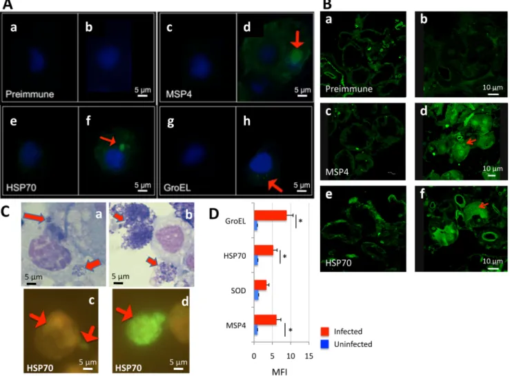
Antibodies against all recombinant proteins recognized A. phagocytophilum in high per-centage infected I. scapularis ISE6 tick cells (Fig 3A) and female tick salivary glands (Fig 3B). However, GroEL was detected by immunofluorescence at very low levels in infected tick cells (Fig 3A and 3h) while MSP4 and HSP70 were clearly detected in bacterial morulae in infected cells (Fig 3A, 3d and 3f). To corroborate proteomics results, A. phagocytophilum HSP70 protein levels were characterized in low and high percentage infected tick cells by immunofluorescence. In infected IDE8 tick cells, A. phagocytophilum HSP70 levels increased as bacterial morulae matured and were present on many extracellular rickettsia after exiting the tick cell (Fig 3C), thus corroborating proteomics results for this protein. Flow cytometry was also used to charac-terize A. phagocytophilum MSP4, HSP70 and GroEL protein levels in high percentage infected and uninfected ISE6 tick cells and in comparison with superoxide dismutase (SOD) that was not over-represented in high percentage infected tick cells when compare to low percentage infected cells (Fig 3D). The results showed that MSP4, HSP70 and GroEL were recognized at higher levels in infected than in uninfected tick cells while for SOD differences were not statisti-cally significant between samples, thus corroborating that MSP4, HSP70 and GroEL proteins were produced at higher levels in high percentage infected tick cells (Fig 3D).
The subcellular localization of selected A. phagocytophilum over-represented proteins was characterized in high percentage infected tick cells and salivary glands. Bacteria purified from infected ISE6 tick cells and recombinant E. coli were mock treated or surface digested with trypsin and proteins loaded onto polyacrylamide gels for Western blot analysis using rabbit antibodies produced against recombinant proteins. All recombinant proteins were sensitive to protease treatment after purification but the results showed that while MSP4 was resistant to trypsin digestion in both A. phagocytophilum and E. coli, HSP70 and GroEL were extracellular and exposed to protease digestion (Fig 2B). Furthermore, as previously shown for A. marginale major surface proteins [27], all three proteins were localized by immunofluorescence on the membrane of recombinant E. coli (Fig 2C). These results suggested that MSP4 may be a trans-membrane protein and showed that HSP70 and GroEL are surface-exposed in A. phagocytophi-lum and recombinant E. coli.
A. phagocytophilum over-represented and surface-exposed proteins
are involved in rickettsia-tick interactions
GroEL, which belongs to the chaperonin/HSP60 family, prevents misfolding and promotes the refolding and proper assembly of unfolded polypeptides generated under stress conditions and has been used for genetic characterization of tick-borne bacteria [28]. HSP70 is also a chapero-nin involved in protein folding and stress response [29]. Recently, GroEL and HSP70 were shown to relocate to the Bacillus subtilis membrane to restore membrane structure and func-tion after ethanol stress [30] and to function in the molecular processing of Borrelia burgdorferi flagellin [31]. GroEL was also shown to function in Caulobacter crescentus multiplication and response to oxidative stress [32]. These results suggested that bacterial heat shock proteins function as molecular chaperones to protect cells from stress-induced lethal damage but also have important roles under physiological growth conditions by acting as carriers for immuno-genic peptides, assisting in protein export or mediating adherence to host cells and may play an essential role during cell division [30–34]. The role of major surface proteins in adhesion to tick cells for bacterial infection has been demonstrated in A. marginale [24,35–37] and MSP1a levels have been associated with bacterial differential adhesion to host cells [27,38]. The GroEL
and HSP70 proteins may be functionally relevant at the tick-A. phagocytophilum interface because as shown here they are localized on the cell membrane in A. phagocytophilum as in other bacteria [24,30,31,33,34,39] and may interact with other membrane proteins [32].
The characterization of protein-protein direct and/or functional interactions using STRING suggested that GroEL possibly interacts with HSP70 (Fig 4A), suggesting a physical and/or functional connection between these proteins. These proteins have been shown to interact directly (physically) or indirectly (functionally) in other tick-borne bacteria but data were not
Fig 3. Antibodies against recombinant proteins recognize A. phagocytophilum in infected tick cells and ticks by immunofluorescence. (A) Uninfected and A. phagocytophilum (NY18)-infected ISE6 tick cells were characterized by immunofluorescence in (a, c, e, g) uninfected and (b, d, f, h) infected cells. Representative immunofluorescence images are shown for tick cells stained with rabbit preimmune (control) or anti-A. phagocytophilum protein antibodies (green, FITC; blue, DAPI). Arrows show the localization of A. phagocytophilum proteins in infected cells. Bars, 5μm. (B) Sections were made from I. scapularis female ticks after feeding on an uninfected (a, c, e) or A. phagocytophilum (NY18)-infected (b, d, f) sheep. Representative immunofluorescence images are shown for salivary gland sections stained with rabbit preimmune (control) or anti-A. phagocytophilum protein antibodies (green, FITC). Arrows show the localization of A. phagocytophilum proteins in infected cells. Bars, 10μm. (C) IDE8 tick cells were collected in low and high percentage A. phagocytophilum (L610)-infected cells and representative immunofluorescence images are shown. (a, b) Bright-field images of Giemsa-stained (a) low percentage and (b) high percentage infected tick cells. Bacteria stain purple (arrows) and host nuclei stain pink. (c) Low percentage and (d) high percentage infected tick cells were stained with rabbit anti-A. phagocytophilum HSP70 protein antibodies (green, FITC). Arrows show the localization of A. phagocytophilum proteins in infected cells. Bar, 5μm. (D) Flow cytometry profile showing MFI values determined using a FITC-conjugated secondary antibody. A. phagocytophilum (NY18)-infected and uninfected control ISE6 tick cells were washed, fixed, permeabilized and incubated with primary unlabeled antibody (preimmune IgG isotype control, MSP4, SOD, HSP70 and GroEL), washed in PBS and incubated with FITC-goat anti-rabbit IgG. MFI was calculated as the MFI of the test-labeled sample minus the MFI of the isotype control, shown as Ave+SD and compared between infected and uninfected tick cells by Student's t-test (*P<0.05) (N = 3).
available for A. phagocytophilum [31]. The interaction between recombinant A. phagocytophi-lum HSP70 and GroEL proteins was confirmed using immunoprecipiation where GroEL was co-purified with HSP70 using anti-HSP70 or anti-GroEL antibodies (Fig 4B). Furthermore, the interaction between recombinant proteins with A. phagocytophilum protein extracts corrobo-rated the interaction between GroEL and HSP70 after immunoprecipitation and Western blot analysis (Fig 4C). The interaction between MSP4 and GroEL or HSP70 was confirmed using histidine-tagged MSP4 and nickel beads (Fig 4D).
Fig 4. Characterization of A. phagocytophilum protein-protein interactions. (A) Protein-protein interactions were characterized in silico using STRING 8.3 (http://string-db.org). The STRING score value is shown, defined as threshold of significance to include the interaction (maximum value = 1) computed by combining the probabilities from the different evidence channels, correcting for the probability of randomly observing an interaction. (B) Protein-protein interactions were characterized in vitro using A. phagocytophilum HSP70 (red arrow) and GroEL (blue arrow) recombinant proteins and tick Porin as control [15]. The proteins were mixed in equimolar amounts and immunoprecipitated using anti-GroEL or anti-HSP70 antibodies and Protein G Dynabeads. The purified proteins were eluted using Laemmli sample buffer and loaded onto a 12% SDS-PAGE gel for Western blot analysis using anti-HSP70, anti-GroEL or anti-Porin antibodies. (C) Protein-protein interactions were characterized in vitro using A. phagocytophilum protein extracts, recombinant HSP70 (red arrow) and GroEL (blue arrow) proteins and tick Porin as control [15]. Protein G Dynabeads were incubated with purified anti-HSP70, anti-GroEL or anti-Porin antibodies and then 130μg of A. phagocytophilum proteins were added. Unbound proteins were removed and the beads were washed three times with PBS with addition of 0.1% Triton X-100, resuspended in Laemmli sample buffer and loaded onto a 12% SDS-PAGE gel for Western blot analysis using anti-HSP70 or anti-GroEL antibodies. (D) Protein-protein interactions were characterized in vitro using A. phagocytophilum anti-HSP70 (red arrow), GroEL (blue arrow) and MSP4 (green arrows) recombinant proteins and tick Porin as control [15]. Nickel beads were covered with histidine-tagged MSP4, washed and incubated with GroEL or HSP70, MSP4 or Porin as control. After incubation, beads were washed and proteins eluted in Laemmli sample buffer and loaded onto a 15% SDS-PAGE gel.
To characterize the possible role of these proteins in Anaplasma-tick interactions, the bind-ing of GroEL, HSP70 and MSP4 to tick cells was assessed usbind-ing the recombinant E. coli model [27] in which E. coli producing surface-exposed A. phagocytophilum proteins were used for binding to tick cells (Table 1). The results demonstrated that GroEL, HSP70 and MSP4 were involved in binding to tick cells (Table 1). Furthermore, E. coli producing recombinant GroEL and HSP70 with truncated peptide-binding domains that are involved in protein-protein inter-actions (Fig 5A) did not bind to tick cells, providing additional support for the role of these proteins in A. phagocytophilum-tick interactions (Fig 5B). Remarkably, binding to tick cells was more pronounced in bacteria producing recombinant A. phagocytophilum MSP4 than the A. marginale MSP1a positive control [27,38] (Fig 5B).
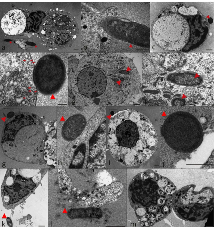
The interaction of recombinant E. coli producing A. marginale MSP1a (positive control;Fig 6a and 6b) and A. phagocytophilum MSP4 (Fig 6c–6f), HSP70 (Fig 6g–6k) and GroEL (Fig 6l) proteins was also characterized by electron microscopy in comparison to Thioredoxin-produc-ing cells (Fig 6m) to provide additional evidence for the role of these proteins in rickettsia-tick interactions. The results showed not only interaction with tick cells for bacteria with surface exposed A. phagocytophilum MSP4, HSP70 and GroEL (Fig 6c–6l) but also internalization of recombinant bacteria producing MSP4 (Fig 6e and 6f). Bacterial internalization was observed for MSP1a and MSP4 only (Fig 6e and 6f) and the presence of microtubules close to tick cell plasma membrane suggested the beginning of the process of phagosome formation (Fig 6d). An insight into the three-dimensional cell morphology during the interaction between MSP4-producing E. coli and tick cells revealed the presence of small vesicles inside a tick cell phagocytic cup (S1 Video). Interestingly, one vesicle was in direct contact with the bacterial outer membrane suggesting a bacterial origin for these vesicles (S1 Video). These small vesicles were clearly distinguished only by electron tomography (S1 Video) and measured from 12 to 34 nm in diameter (mean value ± SD = 20.4 ± 5.5 nm; N = 37).
To provide additional support for the role of A. phagocytophilum GroEL, HSP70 and MSP4 proteins in rickettsia-tick interactions, antibodies against these proteins were used to inhibit infection of tick cells. Antibodies against these proteins could affect the interaction between bacterial ligands and tick receptors to block infection or affect the interaction with proteins
Table 1. Adhesion to cultured tick cells by recombinant E. coli producing surface-exposed A. phago-cytophilum proteins.
Protein No. CFU (mean± SE) recovered from ISE6 tick cells (number of replicates)
Thioredoxin (expression vector alone)– Negative control
7±3 (N = 6) A. phagocytophilum GroEL short-length 6±1 (N = 2) A. phagocytophilum GroEL full-length 17±6 (N = 4)* A. phagocytophilum HSP70 short-length 5±2 (N = 2) A. phagocytophilum HSP70 full-length 41±28 (N = 4)* A. phagocytophilum MSP4 152±50 (N = 4)* A. marginale MSP1a—Positive control 65±20 (N = 4)*
E. coli strains were grown and induced for the production of recombinant proteins. E. coli strains with expression vector alone producing recombinant Thioredoxin and A. marginale MSP1a were used as negative and positive control, respectively. Adhesive bacteria were quantitated as the number of colony forming units (CFU) recovered from each test and compared to the Thioredoxin control values by Student’s t-test for paired samples (*P<0.05). GroEL and HSP70 were produced as full-length and short-length (amino acids 275–475 and 262–460 for GroEL and HSP70, respectively) proteins.
Fig 5. Characterization of A. phagocytophilum protein adhesion to tick cells. (A) Schematic representation of GroEL and HSP70 functional domains in full-length and short-length proteins produced in E. coli and used for binding experiments to tick cells. (B) E. coli strains were grown and induced for the production of recombinant proteins. E. coli strains with expression vector alone producing recombinant Thioredoxin and A. marginale MSP1a were used as negative and positive control, respectively. Adhesive bacteria were quantitated as the number of colony forming units (CFU) recovered from each test, shown as fold increase and compared to the Thioredoxin (Trx) control values by Student’s t-test for paired samples (*P<0.05;Table 1).
functionally important for bacterial infection and/or multiplication in tick cells. The results showed an inhibitory effect of anti-GroEL, anti-HSP70 and anti-MSP4 but not anti-tick Porin antibodies on A. phagocytophilum infection of tick cells when compared to cells treated with pre-immune serum (Fig 7), thus corroborating the role of these proteins in rickettsial adhe-sion/invasion of tick cells.
Fig 6. Electron micrographs of ISE6 tick cells reacted with recombinant E. coli producing A. phagocytophilum recombinant proteins. Representative images of recombinant E. coli shown by arrowheads adhered to tick cells. (a, b) A. marginale MSP1a positive control; (c-f) MSP4; (g-k) HSP70; (l) GroEL; (m) Thioredoxin negative control. Microtubules are shown by arrows in (d). Scale bars, 1μm (a, c, e, g, i, k-m), 500 nm (b, d, f, h, j). doi:10.1371/journal.pone.0137237.g006
It has been shown that bacterial GroEL interacts with lectin-like oxidized low-density lipo-protein receptor-1 (LOX-1) to mediate bacterial adherence to vertebrate host cells [34]. How-ever, the LOX-1 homolog was not identified in the I. scapularis genome, thus suggesting that the adhesion of A. phagocytophilum to tick cells may not be through interactions between GroEL and LOX-1.
The increase in protein levels in high percentage infected tick cells and tick salivary glands (S1andS2Tables) and the inhibition of bacterial infection in tick cells treated with protein-specific antibodies (Fig 7) supported the hypothesis that higher GroEL, HSP70 and MSP4 levels facilitate A. phagocytophilum infection of tick cells through interaction with a still unknown receptor or interacting molecule. The origin of the vesicles observed in tick cells interacting with recombinant E. coli with membrane-exposed MSP4 in the process of phagosome forma-tion is unknown. However, it can be hypothesized that the vesicles arising from the outer mem-brane of MSP4-expressing E. coli were released as a response to phagocytosis. As described in other Gram-negative bacteria, the production of these possible outer membrane vesicles can be stress-induced and/or involved in pathogenesis through secretion of virulence factors, adhesins and degradative enzymes [40–42]. Additionally, protein-protein interactions between HSP70, GroEL and MSP4 suggested that these proteins might form a complex on the rickettsial mem-brane to facilitate the interaction with tick cells.
Fig 7. Inhibition of A. phagocytophilum infection by antibodies against over-represented proteins. The antibodies against surface-exposed proteins, GroEL, HSP70 and MSP4, were used to characterize the inhibition of pathogen infection in ISE6 tick cells. Tick cells were treated with different rabbit antibodies and then infected with A. phagocytophilum (NY18). Treatments included rabbit pre-immune serum, anti-A. phagocytophilum GroEL, HSP70 and MSP4 protein antibodies and anti-tick Porin antibodies. Untreated cells were left uninfected or infected with A. phagocytophilum (NY18). A. phagocytophilum infection levels were determined by 16S rDNA and msp4 PCR and normalized against tick 16S mitochondrial rDNA with similar results. Normalized msp4 levels are shown in arbitrary units as Ave+S.D and were compared between infected and uninfected untreated cells and between infected cells treated with the pre-immune serum and antigen-specific antibodies by Student’s t-test with unequal variance (P<0.05; N = 4 replicates per treatment).
Fig 8. Characterization of the mRNA levels for selected genes encoding for A. phagocytophilum over-represented proteins. The mRNA levels for groEL, msp4 and hsp70 were determined by real-time RT-PCR in low and high percentage infected ISE6 tick cells. Amplification efficiencies were
normalized against tick 16S rRNA and mRNA levels expressed in arbitrary units. The ratio of normalized mRNA levels in high to low percentage-infected cells was represented as Ave+SD. Normalized Ct values were compared between low and high percentage infected tick cells by Student's t-test (*P<0.05) (N = 5).
doi:10.1371/journal.pone.0137237.g008
Fig 9. Proposed mechanisms of how stress response and surface proteins facilitate A. phagocytophilum infection in high percentage infected tick cells and tick salivary glands. The levels of certain bacterial stress response and surface proteins are higher in high percentage A. phagocytophilum-infected tick cells and tick salivary glands. MSP4, GroEL and HSP70 interact and bind to tick cells, thus facilitating rickettsia-tick interactions and infection. In high percentage infected tick cells and tick salivary glands, bacteria reduce multiplication once they infect the cells but infection is required to complete the life cycle and get ready for transmission. The activation of stress response proteins in A. phagocytophilum may represent a mechanism by which rickettsiae increase infection by facilitating interaction with tick cells and protecting bacteria against stress. The T4SS may be associated with the secretion of HSP70 and other stress response proteins. Abbreviations: T4SS, Type IV Secretion System; Question mark indicates that secretion of HSP70 in a T4SS-dependent manner remains to be proved.
The A. phagocytophilum type IV secretion system may be involved in
the secretion of stress response proteins during rickettsial infection and
multiplication in ticks
The mechanism by which A. phagocytophilum HSP70 and GroEL are processed to appear as surface-exposed proteins is unknown. As other Gram-negative bacteria [43], A. phagocytophi-lum has a type IV secretion system (T4SS) that translocates effector molecules to host cells to exert their activity on transcription and apoptosis and favor rickettsial infection [44–46]. These effector molecules have not been fully characterized but may be responsible for some of the changes occurring in tick transcriptome and proteome in response to rickettsial infection. Herein, we speculate that some of the A. phagocytophilum stress response proteins over-repre-sented during rickettsial infection may constitute T4SS effectors.
Preliminary results showed that A. phagocytophilum VirB2, VirB6, VirB10 and VirD4 were over-represented in tick salivary glands (S2A Fig), and their homologs played a central role in the structure and function of the E. coli T4SS [43]. Additionally, the preliminary analysis of A. phagocytophilum proteins present in tick salivary glands and/or high percentage infected tick cells using the S4TE software [47] to identify candidate T4SS effectors suggested that Iron-binding protein (APH_0051), Peptidase (APH_1159) and HSP70 (APH_0346) harbor several characteristic features of proteins that may be secreted by the T4SS (S2B Fig). S4TE is an easy-to-use and customizable algorithm that do not use a machine learning approach for the predic-tion of candidate effector proteins secreted by T4SS in genomes of any size [47]. Even though further functional validation is needed to confirm this hypothesis, the A. phagocytophilum T4SS may be involved in the secretion of stress response proteins such as HSP70 during rickett-sial infection and multiplication in ticks.
A. phagocytophilum over-represented proteins are regulated at the
transcriptional level
The mRNA levels were characterized in low and high percentage infected tick cells for A. pha-gocytophilum groEL, msp4 and hsp70 encoding proteins over-represented in high percentage infected tick cells and salivary glands when compared to low percentage infected tick cells and guts, respectively. The results of the RT-PCR demonstrated that mRNA levels were signifi-cantly higher in high percentage infected cells (Fig 8). These results suggested that A. phagocy-tophilum GroEL, MSP4 and HSP70 over-represented proteins in high percentage infected cells are regulated at the transcriptional level. However, as described in other bacteria, post-tran-scriptional mechanisms such as enhancement of mRNA stability and/or translation [48] may also affect GroEL, MSP4 and HSP70 protein levels. In A. phagocytophilum, the activation of p44 has been linked to post-transcriptional bacterial RNA splicing [49] which may account for the increase in protein levels observed in high percentage infected tick cells.
Conclusions
In this study we have characterized A. phagocytophilum proteome during rickettsial multiplica-tion in I. scapularis cultured tick cells, guts and salivary glands and provided new evidences on the behavior of this pathogen in ticks. Cultured tick cells are a good model for the study of tick-pathogen interactions and the tick tissues selected for this study play a major role during path-ogen acquisition, multiplication and transmission. The results of these studies showed that in agreement with previous findings at the mRNA and protein levels [21], the proteome of A. pha-gocytophilum in tick salivary glands had a higher impact on protein synthesis and processing than on bacterial replication. These results correlated well with the developmental cycle of A.
phagocytophilum, in which rickettsia convert from an intracellular reticulated, replicative form to the nondividing infectious dense-core form. Additionally, some of the proteins identified here were predicted based on genomics information [50] and proteomics results corroborated the existence of these proteins, thus expanding linkage of the genome annotation with the pro-teome. The most important finding of these studies was the increase in the level of bacterial stress response and surface proteins in high percentage A. phagocytophilum-infected tick cells and salivary glands with implications for tick-pathogen interactions (Fig 9). These results sup-ported our hypothesis that A. phagocytophilum proteins that increase as infection proceeds in cultured tick cells and ticks are important for pathogen infection and gave a new dimension to the role of certain stress response and surface proteins during A. phagocytophilum infection in ticks. These results also demonstrated that MSP4, GroEL and HSP70 could interact and bind to tick cells, thus playing a role in rickettsia-tick interactions (Fig 9). The MSP4 interaction with tick cells may induce the secretion of vesicles at the phagocytic cup to aid in adhesin secre-tion for rickettsial infecsecre-tion. The T4SS may be involved in the secresecre-tion of some stress response proteins (Fig 9). As suggested previously for A. marginale, these proteins may be targeted as antigens for use in vaccine development for control of Anaplasma infection in tick vectors by reducing or blocking transmission to vertebrate hosts [37]. Additionally, these proteins could be used to develop novel therapeutic interventions for A. phagocytophilum and future experi-ments should evaluate these proteins as antigens for vaccination against HGA.
Methods
I. scapularis tick cells and sample preparation
The I. scapularis embryo-derived tick cell line ISE6, provided by Ulrike Munderloh, University of Minnesota, USA, was cultured in L15B300 medium as described previously [37], except that the osmotic pressure was lowered by the addition of one-fourth sterile water by volume. The ISE6 cells were first inoculated with A. phagocytophilum (human NY18 isolate)-infected HL-60 cells [51] and maintained according to Munderloh et al. [52] until infection was established and routinely passaged. Infected ISE6 cells were frozen in liquid nitrogen and served as inocu-lum for uninfected cells. Uninfected and infected cultures (N = 3 independent cultures each) were sampled at 3 days post-infection (dpi) (low percentage infected cells, 10–15% (Ave±SD, 12±2)) and 8 dpi (high percentage infected cells, 65–71% (Ave±SD, 68±3)). The percentage of cells infected with A. phagocytophilum was calculated by examining at least 200 cells using a 100x oil immersion objective. The cells were centrifuged at 10,000 g for 3 min, and cell pellets were frozen in liquid nitrogen until used for protein and RNA extraction. Approximately 107 cells were pooled from each condition and homogenized with a needle (27G) in 500μl lysis buffer (phosphate buffered saline (PBS), 1% Triton X-100, supplemented with Complete prote-ase inhibitor cocktail (Roche, Bprote-asel, Switzerland). Samples were sonicated for 1 min in an ultra-sonic cooled bath followed by 10 sec of vortexing. After 3 cycles of ultra-sonication-vortexing, total cell extracts were centrifuged at 200 x g for 5 min to remove cell debris. The supernatants were collected and protein concentration was determined using the Bradford Protein Assay (Bio-Rad, Hercules, CA, USA) with BSA as standard. Total RNA was extracted from the same cell cultures (N = 5) using TriReagent (Sigma, St. Louis, MO, USA) following manufacturer’s recommendations.
I. scapularis ticks and sample preparation
I. scapularis ticks were obtained from the laboratory colony maintained at the Oklahoma State University Tick Rearing Facility. Larvae and nymphs were fed on rabbits and adults were fed on sheep. Off-host ticks were maintained in a 12 hr light: 12 hr dark photoperiod at 22–25°C
and 95% relative humidity. Adult female I. scapularis were infected with A. phagocytophilum (NY18) by feeding on a sheep inoculated intravenously with approximately 1x10⁷A. phagocy-tophilum-infected HL-60 cells (90–100% infected cells) [53]. In this model, over 85% of ticks become infected with A. phagocytophilum in guts and salivary glands [53]. One hundred female adult ticks were removed from the sheep 7 days after infestation, held in the humidity chamber for 4 days and dissected for protein extraction from guts and salivary glands. Uninfected ticks were prepared in a similar way but feeding on an uninfected sheep. Two independent samples were collected and processed for each tick tissue. Total proteins were extracted from uninfected and A. phagocytophilum-infected gut and salivary gland samples using the AllPrep DNA/RNA/ Protein Mini Kit (Qiagen, Valencia, CA, USA). Animals were housed and experiments con-ducted with the approval and supervision of the Oklahoma State University Institutional Ani-mal Care and Use Committee (AniAni-mal Care and Use Protocol, ACUP No. VM1026).
Proteomics data collection and analysis for A. phagocytophilum
proteome in ISE6 tick cells
Protein extracts (150μg) from the four experimental conditions, low percentage infected tick cells and matching uninfected control at 3 dpi and high percentage infected tick cells and matching uninfected control at 8 dpi were on-gel concentrated by SDS-PAGE and trypsin digested as described previously [54]. The desalted protein digest was resuspended in 0.1% for-mic acid and analyzed by RP-LC-MS/MS using an Easy-nLC II system coupled to an ion trap LTQ mass spectrometer (Thermo Scientific). The peptides were concentrated (on-line) by reverse phase chromatography using a 0.1×20 mm C18 RP precolumn (Thermo Scientific), and then separated using a 0.075×100 mm C18 RP column (Thermo Scientific) operating at 0.3 ml/min. Peptides were eluted using a 180-min gradient from 5 to 40% solvent B (Solvent A: 0,1% formic acid in water, solvent B: 0,1% formic acid in acetonitrile). ESI ionization was done using a Fused-silica PicoTip Emitter ID 10 mm (New Objective, Woburn, MA, USA) interface. Peptides were detected in survey scans from 400 to 1600 amu (1 mscan), followed by fifteen data dependent MS/MS scans (Top 15), using an isolation width of 2 mass-to-charge ratio units, normalized collision energy of 35%, and dynamic exclusion applied during 30 sec periods.
The MS/MS raw files were searched against a compiled database containing all sequences from Ixodida (77,195 Uniprot entries in March 2015) and Anaplasmataceae (64,677 Uniprot entries in March 2015) (http://www.uniprot.org) using the SEQUEST algorithm (Proteome Discoverer 1.4, Thermo Scientific). The following constraints were used for the searches: tryp-tic cleavage after Arg and Lys, up to two missed cleavage sites, and tolerances of 1 Da for pre-cursor ions and 0.8 Da for MS/MS fragment ions and the searches were performed allowing optional Met oxidation and Cys carbamidomethylation.
A false discovery rate (FDR)< 0.05 was considered as condition for successful peptide assignments and at least 2 peptides per protein were the necessary condition for protein identi-fication (S1 Table). After discarding tick proteins, proteins with the same description in the Anaplasmataceae were grouped and the total number of PSM for each protein were normalized against the total number of PSM on each infected tick cells and compared between low and high percentage infected tick cells by Chi2-test (P = 0.05) to select over-represented proteins in high percentage infected cells when compared to low percentage infected cells. Results are the mean of three replicates (S1 Table). Protein ontology analysis for BP was done using the STRAP software (Software for Researching Annotations of Proteins; [http://www.bumc.bu. edu/cardiovascularproteomics/cpctools/strap/] developed at the Cardiovascular Proteomics Center of Boston University School of Medicine (Boston, MA, USA). Protein ontology
annotations were done with a probability cutoff value of 0.5 for importing protXML files and using the UniProtKB database.
Proteomics data collection and analysis for A. phagocytophilum
proteome in adult female tick guts and salivary glands
Proteins were digested using the filter aided sample preparation (FASP) protocol [55]. Briefly, samples were dissolved in 50 mM Tris-HCl pH8.5, 4% SDS and 50 mM DTT, boiled for 10 min and centrifuged. Protein concentration in the supernatant was measured by the Direct Detect system (Millipore, Billerica, MA, USA). About 150μg of protein were diluted in 8 M urea in 0.1 M Tris-HCl (pH 8.5) (UA), and loaded onto 30 kDa centrifugal filter devices (FASP Protein Digestion Kit, Expedeon, TN, USA). The denaturation buffer was replaced by washing three times with UA. Proteins were later alkylated using 50 mM iodoacetamide in UA for 20 min in the dark, and the excess of alkylation reagents were eliminated by washing three times with UA and three additional times with 50 mM ammonium bicarbonate. Proteins were digested overnight at 37°C with modified trypsin (Promega, Madison, WI, USA) in 50 mM ammonium bicarbonate at 40:1 protein:trypsin (w/w) ratio. The resulting peptides were eluted by centrifugation with 50 mM ammonium bicarbonate (twice) and 0.5 M sodium chloride. Tri-fluoroacetic acid (TFA) was added to a final concentration of 1% and the peptides were finally desalted onto C18 Oasis-HLB cartridges and analyzing by LC-MS/MS using a C-18 reversed phase nano-column (75μm I.D. x 50 cm, 3 μm particle size, Acclaim PepMap 100 C18; Thermo Fisher Scientific, Waltham, MA, USA) in a continuous acetonitrile gradient consisting of 0–30% B in 145 min, 30–43% B in 5 min and 43–90% B in 1 min (A = 0.5% formic acid; B = 90% acetonitrile, 0.5% formic acid). A flow rate of ca. 300 nl/min was used to elute peptides from the reverse phase nano-column to an emitter nanospray needle for real time ionization and peptide fragmentation on orbital ion trap mass spectrometer model Orbitrap Elite (Thermo Fisher Scientific).
The MS/MS raw files were searched as described previously for the ISE6 tick cells allowing, in this case, precursor and fragment mass tolerances of 600 ppm and 1200 mmu, respectively. Peptide identification was validated using the probability ratio method [56] and false discovery rate (FDR) was calculated using inverted databases and the refined method [57] with an addi-tional filtering for precursor mass tolerance of 12 ppm. A false discovery rate (FDR)< 0.05 was considered as condition for successful peptide assignments and at least 2 peptides per pro-tein were the necessary condition for propro-tein identification (S2 Table). Proteins with the same description in the Anaplasmataceae were grouped and the total number of PSM for each pro-tein were normalized against the total number of PSM on each infected adult female tick tissue and compared between salivary glands and guts by Chi2-test (P = 0.05) to select over-repre-sented proteins in salivary glands when compared to tick guts. Results are the mean of two rep-licates. This experimental approach was previously used for the characterization of the tick proteome in response to A. phagocytophilum infection [58]. Protein ontology analysis for BP was done using the STRAP software (Software for Researching Annotations of Proteins; [http://www.bumc.bu.edu/cardiovascularproteomics/cpctools/strap/] developed at the Cardio-vascular Proteomics Center of Boston University School of Medicine (Boston, MA, USA). Pro-tein ontology annotations were done with a probability cutoff value of 0.5 for importing protXML files and using the UniProtKB database.
Production of recombinant proteins and antibody preparation
The recombinant A. phagocytophilum (NY18) proteins GroEL, MSP4 (AFD54597), HSP70, and SOD were produced in E. coli BL21 cells (Champion pET101 Directional TOPO
Expression kit, Carlsbad, CA, USA), induced with IPTG and purified using the Ni-NTA affin-ity column chromatography system (Qiagen Inc., Valencia, CA, USA) following manufactur-er’s recommendations. Tick mitochondrial Porin was obtained as previously described [15]. Purified proteins were used to immunize rabbits and IgGs from preimmune and immunized animals were purified (Montage Antibody Purification Kit and Spin Columns with PROSEP-A Media, Millipore, Billerica, MA, USA) and used for analysis.
Flow cytometry of tick cells incubated with antibodies
A. phagocytophilum (NY18)-infected and uninfected control ISE6 tick cells were washed in phosphate buffered saline (PBS), fixed and permeabilized with Intracell fixation and permeabi-lization kit (Inmunostep, Salamanca, Spain) following manufacturer recommendations. After permeabilization, the cells were washed in PBS and incubated with primary unlabeled antibody (preimmune IgG isotype control, MSP4, SOD, HSP70 and GroEL; 50μg ml-1), washed in PBS and incubated in 100μl of PBS with FITC-goat anti-rabbit IgG (Sigma, Madrid, Spain) labeled antibody (diluted 1/500) for 15 min at 4°C. Finally, the cells were washed with PBS and resus-pended in 500μl of PBS. All samples were analyzed on a FACScalibur Flow Cytometer, equipped with the CellQuest Pro software (BD-Biosciences, Madrid, Spain). The viable cell population was gated according to forward scatter and side scatter parameters. The level of MSP4, SOD, HSP70 and GroEL in the viable A. phagocytophilum-infected and uninfected tick cells was determined as the geometric median fluorescence intensity (MFI) of the test-labeled sample minus the MFI of the isotype control [15] and compared between infected and unin-fected cells by Student's t-test (P = 0.05).
Immunofluorescence in cultured tick cells
Antibodies against A. phagocytophilum GroEL, MSP4 and HSP70 were used for immunofluo-rescence studies in I. scapularis IDE8 or ISE6 cells. Uninfected and high percentage (80% infected cells) A. phagocytophilum (NY18)-infected or uninfected cells and low percentage (20% infected cells) and high percentage (80% infected cells) A. phagocytophilum (canine L610 isolate)-infected I. scapularis tick cells were used. The L610 isolate was provided by Erich Zweygarth, Lehrstuhl für Vergleichende Tropenmedizin und Parasitologie, Ludwig-Maximi-lians University, Munich, Germany. Cells were fixed with 10% neutral buffered formaldehyde. After 2 washes with PBS, 40μl aliquots of cell suspension were placed on microscope slides and air-dried. The cells were permeabilized with 0.3% Triton-X100 in PBS for 30 min and 0.1% sodium dodecyl sulphate in PBS for 10 min, and washed 1x with PBS. After blocking with the CAS blocking agent (Invitrogen, Paisley, UK) for 1h, purified rabbit IgG (diluted 1: 100 to 1:300 in CAS blocking agent) was added and incubated overnight, followed by 3 washes in PBS. Incubation for 1 h with goat-anti-rabbit IgG conjugated with FITC (Abcam, Cambridge, UK) (diluted 1:1000 in CAS blocking agent) was followed by 3 washes in PBS. The slides were mounted in mounting medium containing DAPI (Vector Laboratories, Peterborough, UK). Images were acquired on an Axioskop 2 fluorescence microscope with an AxioCam MRc cam-era (Carl Zeiss, Cambridge, UK). Cytocentrifuge smears of A. phagocytophilum-infected and uninfected IDE8 cells were fixed for 3 min with technical methanol before staining for 20 min in Giemsa stain (10% v/v in deionised water buffered to pH 7.2). The percentage of cells infected with A. phagocytophilum was calculated by examining at least 200 cells using a 100x oil immersion objective. Images were acquired as described above.
Immunofluorescence in adult female ticks
Adult female I. scapularis were infected with A. phagocytophilum (NY18) as described above. Female ticks were removed from the sheep 10 days after infestation, held in the humidity chamber for 4 days and fixed with 4% paraformaldehyde in 0.2M sodium cacodylate buffer, dehydrated in a graded series of ethanol and embedded in paraffin. Sections (4μm) were pre-pared and mounted on glass slides. The paraffin was removed from the sections with xylene and the sections were hydrated by successive 2 min washes with a graded series of 100, 95, 80, 75 and 50% ethanol. The slides were treated with Proteinase K (Dako, Barcelona, Spain) for 7 min, washed with PBS and incubated with 3% bovine serum albumin (BSA; Sigma-Aldrich) in PBS for 1 h at room temperature. The slides were then incubated for 14 h at 4°C with primary antibodies diluted 1:100 to 1:300 in 3% BSA/PBS and after 3 washes in PBS developed for 1 h with goat-anti-rabbit IgG conjugated with FITC (Sigma-Aldrich) (diluted 1:160 in 3% BSA/ PBS). The slides were washed twice with TBS and mounted in ProLong Antifade reagent (Molecular Probes, Eugene, OR, USA) or in mounting medium containing DAPI (Vector Lab-oratories, Peterborough, UK). The sections were examined using a Leica SP2 laser scanning confocal microscope (Leica, Wetzlar, Germany). Sections of uninfected ticks and IgGs from preimmune serum were used as controls.
Immunofluorescence in recombinant E. coli
The E.coli induced for the production of recombinant A. phagocytophilum proteins as
described before were fixed with 4% paraformaldehyde in PBS for 20 min at room temperature. E. coli cells producing recombinant tick Porin [15] were used as control. Bacterial smears were prepared using a cytocentrifuge. The cells were treated with 0.3% Triton X-100 in PBS for 30 min, blocked with 3% BSA in PBS for 1h at room temperature and incubated overnight at 4°C with purified antibodies (Pre-immune control, 35μg ml-1; HSP70 and MSP4, 22μg ml-1; GroEL, 30μg ml-1) and developed as described previously for tick cells using goat-anti-rabbit IgG conjugated with FITC (Sigma; 1/160 dilution). The slides were washed twice with TBS and mounted in ProLong Antifade reagent (Molecular Probes). Images were acquired on a Nikon Eclipse Ti-U microscope with a 100x oil immersion objective and a Nikon Digital Sight DS Vi1 camera.
Surface trypsin digestion of A. phagocytophilum and recombinant E. coli
The NY18 isolate of A.phagocytophilum was propagated in cultured ISE6 tick cells as described above. The A phagocytophilum-infected cells (approximately 1x10⁷cells) were collected when 80%-90% of the cells were infected as determined by detection of intracellular morulae in stained cytospin cell smears. Host cell-free bacteria were isolated from cell lysates after five pas-sages through a 27-gauge syringe, followed by differential centrifugation in Percoll gradients as previously described for A. marginale to separate bacteria from host cell debris [59]. The pellet of purified A. phagocytophilum was resuspended in 200μl of SPG buffer (0.25 mM sucrose, 10 mM sodium phosphate, 5 mM L-glutamic acid, pH 7.2), and 5μl of sequencing-grade trypsin (Promega) was added to half of the cell reaction mixture. Bacteria were incubated at 37°C for 30 min and then centrifuged at 10,000×g for 15 min and resuspended in Laemmli protein load-ing buffer, boiled for 5 min and loaded onto a 12% SDS-PAGE [21]. The E.coli induced for the production of recombinant A. phagocytophilum proteins (GroEL, MSP4, HSP70) were treated as described above for A. phagocytophilum and 10μg protein were resuspended in Laemmli sample buffer and applied on 12% SDS-PAGE.
Characterization of protein-protein interactions
Protein-protein interactions were characterized in silico using STRING 8.3 (http://string-db. org) and in vitro using recombinant MSP4, HSP70, GroEL proteins produced as described above. Tick mitochondrial Porin [15] was used as a control. Equimolar amounts of the recom-binant HSP70, GroEL or Porin proteins were mixed in PBS and incubated overnight at 4°C. At the same time, 10μl of Dynabeads Protein G slurry (Life Technologies/Thermo Fisher Scien-tific, Rocckford, IL, USA) were incubated with 10μl of purified HSP70 or GroEL anti-bodies. The magnetic beads were then washed three times with PBS. The HSP70:GroEL and HSP70:Porin protein mixture was added and incubated for 2 h. Unbound proteins were removed and the beads were washed three times with PBS with addition of 0.1% Triton X-100 (Sigma). The beads were resuspended in Laemmli sample buffer, boiled for 5 min and loaded onto a 12% SDS-PAGE gel. After electrophoresis, proteins were transferred to a 0.2μm nitro-cellulose membrane (Bio-Rad). The membrane was blocked with 1% bovine serum albumin (BSA) for two hours at room temperature and incubated overnight at 4°C with primary anti-bodies (anti-HSP70, anti-GroEL or anti-Porin rabbit antianti-bodies) at 1:200 dilution in 100 mM Tris HCL Buffer Saline (TBS). After incubation with the secondary anti-rabbit IgG peroxidase conjugated antibody (Sigma), protein bands were visualized with TMB stabilized substrate for horseradish peroxidase (Promega).
To characterize the interaction of recombinant HSP70 and GroEL with A. phagocytophilum proteins, a pellet of A. phagocytophilum purified as described above was used. Tenμl of Dyna-beads Protein G slurry (Life Technologies/Thermo Fisher Scientific) were incubated with 10μl of purified anti-HSP70, anti-GroEL or anti-Porin antibodies. The magnetic beads were then washed three times with PBS and 130μg of A. phagocytophilum proteins were added and incu-bated for 2 hours. Unbound proteins were removed and the beads were washed three times with PBS with addition of 0.1% Triton X-100 (Sigma). The beads were resuspended in Laemmli sample buffer and loaded onto a 12% SDS-PAGE gel for Western blot analysis as described above.
For the characterization of MSP4 interactions with GroEL and HSP70, 3 aliquots of 100μl of nickel beads slurry (Maxwell polyhistidine protein purification Kit, Promega, Madison, WI, USA) in 10 mM PBS (50:50) were incubated each with 120μg of histidine-tagged MSP4 during 8 h at 4°C. After centrifugation, beads were washed with 1 ml of 10 mM PBS and incubated overnight with 30μg of GroEL, HSP70 or Porin. After incubation and centrifugation, beads were washed with 1 ml of 10 mM PBS and proteins were eluted in Laemmli sample buffer, boiled for 5 min and loaded onto a 15% SDS-PAGE gel. After electrophoresis, proteins were stained with SyproRubi (BioRad, Hercules, CA, USA) and visualized after fluorescence scanning.
Adhesion of recombinant E. coli strains to ISE6 tick cells
Adhesion of recombinant E. coli strains to ISE6 tick cells was tested as previously reported [27]. Briefly, E. coli strains were grown and induced as described before. E. coli strains with expression vector alone producing recombinant Thioredoxin and A. marginale MSP1a [27] were used as negative and positive control, respectively. Cell densities were determined and adjusted to 108cells ml-1in LB. One hundred microlitres (107bacteria) culture were added to 900 ml of 106cells ml-1suspensions of ISE6 tick cells in LB. Tick cells and bacteria were incu-bated for 30 min at 37°C with occasional agitation. Cells were then collected by centrifugation, washed two times in PBS and resuspended in 100 ml of PBS. Elimination of unbound bacteria from tick cells with bound bacteria was performed by Percoll (Sigma) gradient separation [27]. The band containing tick cells was removed with a pipette and washed in PBS. The final cell
pellet was lysed in 1 ml of sterile water and 5μl plated onto LB agar plates containing 100 μg of ampicillin per ml. Adhesive bacteria were quantitated as the number of colony forming units (CFU) recovered from each test and compared to the Thioredoxin control values by Student’s t-test for paired samples (P = 0.05).
Electron microscopy
Cells were fixed in 2.5% glutaraldehyde in 0.1 M cacodylate buffer. Cells were rinsed in the cacodylate buffer, centrifuged and transferred into specimen carriers (1.2 mm in diameter, Leica). Then, 20% BSA was added and cells were immediately frozen using a Leica EM PACT2 high-pressure freezer. Freeze substitution (Leica EM ASF2) was carried in 2% osmium tetrox-ide diluted in 100% acetone at -90°C. After 96 h, material was warmed up to -20°C at a rate of 5°C/h and left for 24 h. Finally, the cells were warmed up again at the rate of 5°C/h to 4°C and left for 24 h. Pellets were removed from the carriers, rinsed three times in 100% acetone for 15 min each. Samples were infiltrated in graded series of SPI-pon resin (SPI) solutions (25%, 50%, 75%) diluted in acetone, 1 h at each step. After overnight incubation in pure resin, the samples were embedded in fresh resin solution and polymerized at 60°C for 48 h. Ultrathin sections were cut using an ultramicrotome Leica UCT, transferred to grids, double stained in ethanolic uranyl acetate for 30 min and lead citrate for 20 min. The samples were observed using a JEOL 1010 transmission electron microscope at an accelerating voltage of 80 kV. Images were cap-tured with MegaView III camera (SIS GmbH).
For electron tomography, 10 nm gold nanoparticles coupled to Protein A (Aurion) were adsorbed to both sides of each section as fiducial markers and sections were carbon coated. Images were obtained using JEOL 2100F transmission electron microscope equipped with a high-tilt stage, Gatan camera (Orius SC 1000) and SerialEM automated acquisition software [60]. Tilt series images were collected as dual-axis tilt series over -60° to 60° tilt range along X-axis and -47° to 47° along Y-X-axis (increments 0.6 and 0.4, respectively) at pixel size of 0.554 nm. The IMOD software package was used for tomogram reconstruction.
Antibody inhibition assay
The inhibitory effect of antibodies against the differentially represented surface-exposed pro-teins, GroEL, MSP4 and HSP70, on A. phagocytophilum (NY18) infection of ISE6 tick cells was conducted as described previously for A. marginale [37]. Confluent monolayers of ISE6 tick cells were pooled and used to seed 24-well plates for each assay. Each well received 1x10⁶tick cells in L-15B medium 48 h prior to inoculation with A. phagocytophilum. Infected cultures for inoculum were harvested when monolayers were detaching (90% infected cells) and host cells were mechanically disrupted with a syringe and 26-gauge needle. Rabbit IgGs (2.2–2.4 mg/ml) were mixed with inoculum (1:1) for 60 min before being placed on the cell monolayers. Each monolayer then received 100μl of the inoculum plus IgG mix and plates were incubated at 34°C for 30 min. The inoculum was removed from the wells and monolayers washed three times with PBS. Complete medium (1 ml) was added to each well and the plates were incubated at 34°C. Controls for each trial included (a) inoculum incubated with pre-immune IgG, (b) inoculum incubated with medium only (untreated infected control), (c) uninfected tick cells that served as background control, and (d) incubation with anti-tick Porin IgG. Four replicates were done for each treatment. After 7 days, monolayers from all wells were harvested, resus-pended in 1 ml PBS and frozen at -70°C. Samples were thawed and solubilized with 1% Triton-X100 and processed for A. phagocytophilum detection by PCR after DNA extraction using TriReagent (Sigma) according to the manufacturer’s recommendations. A. phagocytophilum infection levels were determined by Anaplasma 16S rDNA and msp4 real-time PCR
normalizing against tick 16S mitochondrial rDNA as described previously [7,61] but using oli-gonucleotide primers MSP4-L (5’-CCTTGGCTGCAGCACCACCTG-3’) and MSP4-R (5’-TGCTGTGGGTCGTGACGCG3’) and PCR conditions of 5 min at 95°C and 35 cycles of 10 sec at 95°C, 30 sec at 55°C and 30 sec at 60°C. Results were compared between treatments by the Student’s t-test with unequal variance (P = 0.05).
Analysis of mRNA levels by real-time RT-PCR
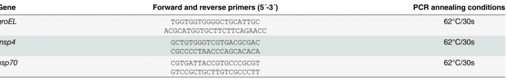
Real-time RT-PCR was performed on tick RNA samples obtained from low and high percent-age A. phagocytophilum (NY18)-infected ISE6 tick cells (N = 5 for each time point) with gene-specific primers (Table 2) using the iScript One-Step RT-PCR Kit with SYBR Green and the iQ5 thermal cycler (Bio-Rad, Hercules, CA, USA) following manufacturer's recommendations. A dissociation curve was run at the end of the reaction to ensure that only one amplicon was formed and that the amplicons denatured consistently in the same temperature range for every sample [62]. The mRNA levels were normalized against tick 16S rRNA [13] using the genNorm method (ddCT method as implemented by Bio-Rad iQ5 Standard Edition, Version 2.0) [63]. Normalized Ct values were compared between low and high percentage infected tick cells by Student's t-test with unequal variance (P = 0.05).
Prediction of T4SS effectors
The A. phagocytophilum proteins identified as over-represented in tick salivary glands and/or high percentage infected tick cells were subjected to S4TE software analysis to evaluate their potential as T4SS substrates. This software screens proteins for thirteen characteristic features of T4SS effectors [47]. S4TE also serves to visualize the distribution of predicted T4SS effectors relative to whole genome architecture [47].
Supporting Information
S1 Fig. Western blot analysis of the specificity of antibodies produced in rabbits against A. phagocytophilum recombinant proteins. Western blot analysis of 10 μg of recombinant A. phagocytophilum MSP4, GroEL and HSP70 proteins produced in E. coli (red arrows) demon-strates the specificity of the antibodies produced in rabbits. E. coli cell proteins were included in two lanes (x2) as negative control. Abbreviation: MW, molecular weight markers (Spectra multicolor broad range protein ladder; Thermo Scientific).
(PDF)
S2 Fig. Characterization of the possible role of A. phagocytophilum type IV secretion sys-tem during pathogen infection.(A) Characterization of the T4SS proteins in infected tick guts and salivary glands. Only peptides with a confidence of at least 99% and identified with two or more PSM in at least one of the samples were considered to perform protein quantifica-tion. Proteins with the same description in the Anaplasmataceae were grouped and the total
Table 2. Primer sequences and PCR conditions used for real-time RT-PCR.
Gene Forward and reverse primers (5´-3´) PCR annealing conditions
groEL TGGTGGTGGGGCTGCATTGC ACGCATGGTGCTTCTTCAGAACC 62°C/30s msp4 GCTGTGGGTCGTGACGCGAC CGCCCCTAACCCAGCACACA 62°C/30s hsp70 CGTGATTACCGTGCCCGCGT GTCCGCTGCTTGTCGCCCTT 62°C/30s doi:10.1371/journal.pone.0137237.t002