HAL Id: hal-01257895
https://hal.sorbonne-universite.fr/hal-01257895
Submitted on 18 Jan 2016
HAL is a multi-disciplinary open access
archive for the deposit and dissemination of
sci-entific research documents, whether they are
pub-lished or not. The documents may come from
teaching and research institutions in France or
abroad, or from public or private research centers.
L’archive ouverte pluridisciplinaire HAL, est
destinée au dépôt et à la diffusion de documents
scientifiques de niveau recherche, publiés ou non,
émanant des établissements d’enseignement et de
recherche français ou étrangers, des laboratoires
publics ou privés.
Distributed under a Creative Commons Attribution| 4.0 International License
Anoctamin 6 is localized in the primary cilium of renal
tubular cells and is involved in apoptosis-dependent cyst
lumen formation
V. Forschbach, M. Goppelt-Struebe, K. Kunzelmann, R. Schreiber, R.
Piedagnel, A. Kraus, K.-U. Eckardt, B. Buchholz
To cite this version:
V. Forschbach, M. Goppelt-Struebe, K. Kunzelmann, R. Schreiber, R. Piedagnel, et al.. Anoctamin
6 is localized in the primary cilium of renal tubular cells and is involved in apoptosis-dependent
cyst lumen formation. Cell Death and Disease, Nature Publishing Group, 2015, 6 (10), pp.e1899.
�10.1038/cddis.2015.273�. �hal-01257895�
Anoctamin 6 is localized in the primary cilium of renal
tubular cells and is involved in apoptosis-dependent
cyst lumen formation
V Forschbach
1, M Goppelt-Struebe
1, K Kunzelmann
2, R Schreiber
2, R Piedagnel
3,4, A Kraus
1, K-U Eckardt
1and B Buchholz*
,1Primary cilia are antenna-like structures projected from the apical surface of various mammalian cells including renal tubular
cells. Functional or structural defects of the cilium lead to systemic disorders comprising polycystic kidneys as a key feature.
Here we show that anoctamin 6 (ANO6), a member of the anoctamin chloride channel family, is localized in the primary cilium of
renal epithelial cells in vitro and in vivo. ANO6 was not essential for cilia formation and had no effect on in vitro cyst expansion.
However, knockdown of ANO6 impaired cyst lumen formation of MDCK cells in three-dimensional culture. In the absence of
ANO6, apoptosis was reduced and epithelial cells were incompletely removed from the center of cell aggregates, which form in
the early phase of cystogenesis. In line with these data, we show that ANO6 is highly expressed in apoptotic cyst epithelial cells
of human polycystic kidneys. These data identify ANO6 as a cilium-associated protein and suggest its functional relevance in
cyst formation.
Cell Death and Disease (2015) 6, e1899; doi:10.1038/cddis.2015.273; published online 8 October 2015
Primary cilia are non-motile protrusions of the apical
mem-brane of various cell types.
1The ciliary membrane contains
receptors and ion channels that link mechanical or chemical
stimuli including fluid flow, sonic hedgehog and growth factors
to intracellular signaling cascades regulating cell
differentia-tion, migration and growth.
2Mutations in genes encoding for
proteins that are necessary either for the function or the
structure of the primary cilium lead to ciliopathies, systemic
disorders that are typically characterized by the development
of polycystic kidneys.
3Anoctamins (ANO1-ANO10, TMEM16A-K) form a family of
10 proteins that are supposed to act as Ca
2+-activated
chloride channels with no homology to other known ion
channels.
4,5In contrast to the other paralogues, the function of
ANO1 and ANO2 as Ca
2+-activated chloride channels has
been confirmed
in vivo and in vitro.
6–8Besides its ability to
conduct ions ANO6 has also been shown to act as a
phospholipid scramblase.
9,10ANO3, 4, 7, 9 may also act as
Ca
2+-dependent phospholipid scramblases.
11However, data
about their functional roles are very limited so far. ANO6 is the
most widely expressed paralogue.
12Mutations in ANO6 cause
the Scott syndrome, which is characterized by a defect
in Ca
2+-dependent phospholipid scrambling of plasma
membrane phospholipids.
10In addition, ANO6 is involved in
bone mineralization, cell volume regulation, cell proliferation
and apoptosis.
12Despite the broad expression and function of
ANO6, there is only sparse data about the subcellular
localization of ANO6. Recently, we have shown that ANO6 is
expressed in cyst-forming epithelial cells together with ANO1,
which is widely expressed in epithelial cells.
13Knockdown of
ANO1 but not ANO6 significantly reduced
secretion-dependent cyst growth pointing towards distinct functions of
ANO1 and ANO6 in the cyst epithelium.
13Lumen formation of different epithelial cell types
repre-sents a fundamental step for the proper development of
several organs including lungs, pancreas, intestine and
kidneys.
14It comprises complex cell–cell and cell–matrix
recognition, establishment of apical–basal polarity as
well as cavitation, which depends on apoptosis of cells
situated within the lumen.
14The mechanisms involved in
apoptosis-dependent cavitation are incompletely
under-stood. Interestingly, in polycystic kidneys, a prime example
of misled lumen formation leading to cystogenesis and
subsequent cyst growth, cyst epithelial cells show increased
levels of apoptosis.
15This study was conducted to determine the subcellular
localization of ANO6 in renal tubular cells and a possible role
of this protein in cyst formation.
1Department of Nephrology and Hypertension, Friedrich-Alexander-University Erlangen-Nuernberg, 91054 Erlangen, Germany;2Department of Physiology, University of
Regensburg, 93053 Regensburg, Germany;3Sorbonne Universités, UPMC Univ Paris 06, UMR_S 1155, F-75005 Paris, France and4INSERM, UMR_S 1155, F-75005
Paris, France
*Corresponding author: B Buchholz, Department of Nephrology and Hypertension, Friedrich-Alexander-University Erlangen-Nürnberg, Ulmenweg 18, D-91054 Erlangen, Germany. Tel: +49 9131 8539002; Fax: +49 9131 8539209; E-mail: Bjoern.Buchholz@uk-erlangen.de
Received 30.4.15; revised 11.8.15; accepted 27.8.15; Edited by A Oberst
Abbreviations: ANO, anoctamin; MDCK, Madin-Darby Canine Kidney; TMEM, transmembrane protein; sh, short hairpin; Ctrl, control; HCD, human collecting duct; ATP, adenosine triphosphate; IBMX, 3-isobutyl-1-methylxanthine; TUNEL, TdT-mediated dUTP-biotin nick end labeling; BNIP3, BCL2/adenovirus E1B 19 kDa interacting protein 3; UTP, uridine triphosphate; Kif3a, kinesin family member 3A; HIF-1α, hypoxia-inducible factor 1, alpha subunit; DMEM, Dulbecco’s modified Eagle’s medium; BSS, balanced salt solution; FCS, fetal calf serum; hPTECs, human primary tubular epithelial cells; ADPKD, autosomal dominant polycystic kidney disease; FITC, fluorescein isothiocyanate; Rte, transepithelial resistance; Vte, transepithelial voltage; Isc, short-circuit current; PBS, phosphate-buffered saline; PBS+, PBS supplemented with 0.9 mM calcium chloride and 0.49 mM magnesium chloride; S.E.M., standard error of the mean; Fsk, forskolin; DIC, digital interference contrast
Results
Anoctamin 6 is localized in the primary cilium of renal
tubular cells. ANO6 localization was analyzed in canine and
human renal tubular cells. For this purpose, we developed
and characterized three different antibodies as described in
Materials and Methods. First, we analyzed the subcellular
localization of endogenous ANO6 in polarized Madin-Darby
Canine Kidney (MDCK) cells, which originate from collecting
duct cells. We found distinct signals in dense, polarized cells
grown on permeable supports that colocalized with
acety-lated tubulin, a marker for the primary cilium (Figures 1a–h).
Comparable results were obtained with each of the three
antibodies (Supplementary Figures 1A–F). In addition, ANO6
also seemed to be expressed in the plasma membrane
(Supplementary Figures 1G–I). To confirm the specificity of
these findings, we next generated MDCK cell clones stably
expressing one of two shRNAs directed against ANO6
(shANO6#1 and shANO6#2) or control shRNA (shCtrl),
respectively. Both, shANO6#1 and shANO6#2 provided a
significant reduction of ANO6 expression of more than 80%
(Figure 2a). Of note, both cell clones still formed cilia
(Figures 2c and d) but showed a marked reduction of ciliary
ANO6 signal (Figures 2b–d). This finding confirmed ciliary
localization of ANO6, but indicated that ANO6 is not essential
for cilium formation. Next, we tested for the localization in
human tubular cells to exclude cell line- or species-specific
localization of ANO6. In Human Collecting Duct (HCD)
cells,
16we found identical membranous and ciliary staining
patterns as in MDCK cells (Figures 3a–d). In addition, we
also analyzed primary human tubular cells isolated from
nephrectomized kidneys comprising proximal, distal and
collecting duct cells
17(Figures 3e–h and Supplementary
Figure 2). To distinguish cells originating from different
tubular segments, we took advantage of the fact that human
proximal
epithelial
cells
uniquely
express
N-cadherin
instead of E-cadherin as major cell–cell adhesion molecule.
17Ciliary localization of ANO6 was found irrespective of the
tubular origin (Figures 3i–l) indicating ciliary expression of
ANO6 in different tubular segments.
ANO6 is involved in apoptosis-dependent lumen
forma-tion of MDCK cysts. To test for a funcforma-tional role of ANO6 in
cyst formation, we used the MDCK cyst model.
18Both,
control-transfected MDCK cells as well as MDCK cells stably
deficient for ANO6 formed cysts within a collagen matrix and
showed comparable cyst sizes in the presence of forskolin
(Figures 4a and b). This suggests that ANO6 is not essential
for fluid secretion into the cyst lumen, the main mechanism of
cyst expansion, although forskolin led to a significant
translocation of ANO6 from the cytosol towards the apical
membrane (Supplementary Figures 3A and B). This was
further confirmed by Ussing chamber experiments where
MDCK cells stably deficient for ANO6 showed unaffected
transepithelial chloride secretion upon treatment with either
ATP or IBMX/forskolin compared with control-transfected
cells (Supplementary Figures 3C–F). However, in cysts
derived from MDCK cells lacking ANO6, lumen formation
was incomplete. This was caused by an increased number of
cells situated within the cyst lumen, and reflected by a
significant reduction of the lumen-to-cyst ratio (Figures 4c–e).
In accordance with our previous findings,
13the lumen-to-cyst
ratio was augmented in the presence of forskolin in both
ANO6-competent and ANO6-deficient cells (Figures 4c–e),
again reflecting preserved lumen expansion owing to
transe-pithelial chloride secretion.
Next, we were interested to determine the mechanisms
underlying the increased number of cells within the cyst lumen
in ANO6-deficient cells. We tested whether intracystic cell
accumulation was owing to increased cell proliferation of
deficient cells. However, cell proliferation in
ANO6-deficient MDCK cells was reduced (Supplementary Figure 3G),
which is in line with a previous report, where ANO6 has been
shown to be involved in cell proliferation of myoblasts.
19X Y Z X Y Z
DAPI
X Y Zac. Tubulin
X Y ZANO6
ANO6
ac. Tubulin
merge
20µm 20µm 20µm 5µm
colocalized signals
colocalized signals
Figure 1 Anoctamin 6 is localized in the plasma membrane and the primary cilium of MDCK cells. (a) MDCK cells grown on permeable supports were stained for ANO6 providing round distinct signals; (b) cells in a were stained for the ciliary marker acetylated tubulin; (c) merged photo of a and b are shown; (d) calculated colocalization in white of the magnified section marked in c are shown as described in the Materials and Methods section; (e–h) three-dimensional illustration of MDCK cells stained for nuclei (DAPI, e), ANO6 (f) and primary cilia (acetylated tubulin, g); (h) calculated colocalization of ANO6 and acetylated tubulin highlighted in white
Anoctamin 6 promotes renal cyst lumen formation
V Forschbachet al
2
As ANO6 has been reported to be pro-apoptotic in
lymphocytes, macrophages and platelets,
20–22and
in vitro
lumen
formation
is
based
on
apoptosis-dependent
cavitation,
14,23we next examined apoptosis in
ANO6-deficient cells. During apoptosis, ANO6 is necessary for
calcium-dependent phospholipid scrambling resulting in an
increased presentation of negatively charged
phosphatidyl-serines at the outer membrane leaflet.
20This can be
visualized by binding of annexin V protein to negatively
charged phosphatidylserines.
18Indeed and in accordance
with previous findings, lack of ANO6 significantly reduced
calcium-dependent binding of annexin V in MDCK cells
shANO6#1 shANO6#2 ac. Tubulin ANO6 ac. Tubulin ANO6 ac. Tubulin ANO6 shControl X Y Z X Y Z X Y Z 0 25 50 75 100 125
*
*
ANO6 mRNA [% control]
shANO6#1shANO6#2 shControl
Figure 2 MDCK cells stably deficient for ANO6 show reduced signal of ANO6 within the cilium. (a) Downregulation of ANO6 mRNA in two MDCK cell clones (clone 3 (cl.3) and clone 7 (cl.7)) stably transfected with two distinct shRNAs directed against ANO6 (shANO6#1 and shANO6#2) compared with MDCK cells stably transfected with scrambled shRNA (shControl) serving as control, *Po0.05; (b–d) three-dimensional illustration of MDCK cells described in a stained for acetylated tubulin (upper panel) and ANO6 (lower panel)
50µm 10µm
ANO6 ac. Tubulin merge colocalized signals
10µm
ANO6 ac. Tubulin merge colocalized signals
50µm 50µm
ANO6 E-Cadherin merge merge
50µm 10µm
50µm 50µm
50µm
50µm 50µm
Figure 3 ANO6 localizes in the primary cilium of a human collecting duct cell line (HCD) and human primary tubule cells. HCD cells were grown on permeable supports and stained for ANO6 (a) and acetylated tubulin (b); (c) merged photo of a and b is shown; (d) calculated colocalization highlighted in white of the magnified section marked in c is shown; primary human renal tubule cells were stained for ANO6 (e) and acetylated tubulin (f); (g) merged photo of e and f is shown; (h) calculated colocalization highlighted in white of the magnified section marked in g is shown; primary human renal tubule cells were stained for ANO6 (i) and the distal tubular cell marker E-cadherin (j); (k) merged photo of i and j is shown; (l) magnified section marked in k is shown. Arrowheads mark ANO6-positive cells stained positive for E-cadherin. Arrows mark ANO6-positive cells stained negative for E-cadherin
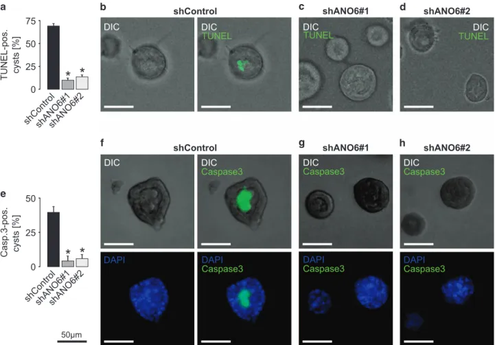
(Figure 5). We then compared the expression of apoptotic
markers during the early stages of MDCK lumen formation of
ANO6-competent and ANO6-deficient cells. Strikingly, luminal
cells in ANO6-deficient MDCK cysts were largely negative for
TUNEL staining as well as for activated caspase 3, whereas
luminal cells of ANO6-competent cysts were highly positive for
these apoptotic markers (Figure 6). These data depict ANO6
as a pro-apoptotic protein that is involved in MDCK cyst lumen
formation.
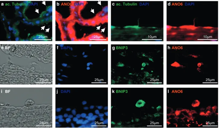
ANO6 is expressed in cyst-lining epithelial cells of
polycystic kidneys. To determine the relevance of these
findings for polycystic kidney disease in humans, we
analyzed sections of human polycystic kidneys. In polycystic
kidneys, increased apoptosis is a key feature of the cyst
epithelium and highly correlates with the degree of cyst
formation.
24ANO6 could be detected in human cyst-lining
cells and—in line with the in vitro data—colocalized
with the ciliary marker acetylated tubulin (Figures 7a–d).
shANO6#2
50µmshANO6
#1
50µmshControl
50µm 0 25 50 75 100 125cyst size [%]
shANO6#1 shANO6#2 shControl n.s. 0 10 20 30 40lumen/cyst ratio [%]
shANO6#1shANO6#2shControl shControlshANO6#1shANO6#2
* *
*
*
+Fsk +Fsk +Fsk
+Fsk +Fsk +Fsk
shANO6#1 shANO6#2
shControl shControl shANO6#1 shANO6#2
50µm
*
*
*
*
*
*
*
*
Figure 4 MDCK cells deficient for ANO6 show disturbed lumen formation within a collagen matrix. Non-transfected MDCK cells as well as stable control-transfected MDCK cells (shControl) and MDCK cells stably deficient for ANO6 (shANO6#1 and shANO6#2) were grown within a collagen I matrix in the presence and absence of 10μM forskolin (Fsk) to form cysts for 5 days. (a) Cyst sizes of control-transfected MDCK cells and ANO6-deficient cells± S.E.M. in the presence of 10 μM forskolin relative to non-tranfected cells (set 100%) from three individual experiments comprising the analysis of ~ 75–125 cysts per condition; (b) representative cysts within the collagen matrix at day 5 are shown; (c) ratio of luminal area and cyst area± S.E.M. in the presence and absence of 10 μM forskolin (Fsk) was determined in cysts described in a from three individual experiments comprising the analysis of ~ 75–125 cysts per condition, *Po0.05; (d) representative cysts in the absence and (e) presence of 10 μM forskolin (Fsk) stained for F-actin (upper panel) and DAPI (lower panel) are shown. * represents proper cyst lumen, arrows point at cells situated within the cyst lumen
0 1 2 3 4 5 6 0 0.1 1 shControl shANO6#2 shANO6#1
Annexin V [% pos. cells]
ATP [mM]
*
*
50µm 50µm 50µm
50µm 50µm 50µm
shControl shANO6#1 shANO6#2 0.1 mM
ATP
1 mM ATP
Figure 5 ANO6 is involved in calcium-dependent phospholipid scrambling of MDCK cells. (a) Stably control-transfected MDCK cells (shControl) and MDCK cells stably deficient for ANO6 (shANO6#1 and shANO6#2) were grown on cell culture dishes. After incubation with either 0, 0.1 or 1 mM ATP, FITC-labeled annexin V was added and the fraction of annexin V-positive cells was analyzed, *Po0.05. (b) representative photos of the cells described in a (green: annexin V-FITC) are shown
Anoctamin 6 promotes renal cyst lumen formation
V Forschbachet al
4
In addition, ANO6 was highly expressed in rounded apoptotic
epithelial cells characterized by strong expression of the
pro-apoptotic protein BNIP3 (Figures 7e–l), supporting
the hypothesis of a functional role of ANO6 in apoptosis of
cyst-lining epithelial cells.
Discussion
The important function of the primary cilium is reflected by a
strong compartmentalization. Thus, several proteins have been
found to be exclusively or at least preferentially localized in the
ciliary membrane or the ciliary lumen.
3In addition, primary cilia
are characterized by distinct calcium concentrations compared
with the cytosol regulated by calcium-permeable non-selective
cation channels within the ciliary membrane.
25Here we show
that ANO6, a calcium-activated chloride channel, is also
preferentially localized in the cilium. In addition, we show that
ANO6 is involved in cyst formation by mediating
apoptosis-dependent cavitation, a prerequisite for proper lumen formation.
Ciliary localization of proteins depends on the recognition of
a ciliary targeting sequence. The most common one is the VxP
motif, which has been found in many ciliary transmembrane
proteins including the polycystins.
26This sequence is also
present in ANO6 and several other anoctamins, as shown in
Supplementary Table 1. In line with our data showing a ciliary
localization in canine and human tubular cells, the VxP motif of
ANO6 is conserved among different species (Supplementary
Table 2), emphasizing its functional relevance and suggesting
that additional anoctamins may have a role in ciliary function.
In fact, ANO1, another member of the anoctamin chloride
channel family, has recently also been shown to be localized in
the cilium and to be involved in ciliogenesis.
27Unlike reported
for ANO1, we found no evidence for a role of ANO6 in cilia
formation, as cilia were well expressed in ANO6-knockdown
cells. However, as our knockdown efficiency was about 80%
we cannot rule out that complete knockout might affect
ciliogenesis. In a previous report, we showed that ANO1 is
involved in apical chloride secretion of cyst-forming renal cells,
whereas knockdown of ANO6 had no effect on chloride
secretion.
13These data are corroborated by findings in the
current study, where cyst growth and cyst expansion were not
50µm
shControl
DIC
TUNEL
DIC
shControl
DIC
Caspase3
DIC
Caspase3
DAPI
DAPI
shANO6#2
TUNEL
DIC
shANO6#2
Caspase3
DIC
Caspase3
DAPI
shANO6#1
TUNEL
DIC
shANO6#1
Caspase3
DIC
Caspase3
DAPI
0
25
50
75
TUNEL-pos. cysts [%]
shControl
shANO6#1
shANO6#2
* *
0
25
50
* *
shControl
shANO6#1
shANO6#2
Casp.3-pos. cysts [%]
Figure 6 ANO6 is involved in apoptosis-dependent lumen formation of MDCK cysts. Stable control-transfected MDCK cells (shControl) and MDCK cells stably deficient for ANO6 (shANO6#1 and shANO6#2) were grown within a collagen I matrix to form cysts for 3 days where lumen formation and cavitation is still taking place in control cysts. Then cysts were photographed by the use of digital interference contrast (DIC) and stained for apoptosis by the use of TUNEL (a–d) and antibodies directed against cleaved caspase 3 (e–h). (a) Quantification of TUNEL-positive cysts from three individual experiments comprising 65–85 cysts per condition; (b) left: representative ANO6-competent cyst, right: overlay with TUNEL staining; (c, d) representative ANO6-deficient cysts with overlay of TUNEL staining; (e) quantification of cleaved caspase 3-positive cysts from three individual experiments comprising ~ 50 cysts per condition; (f) upper panel, left: representative ANO6-competent cyst, right: overlay with staining for cleaved caspase 3; lower panel, left: control cyst stained with DAPI, right: shows merge of DAPI stain and cleaved caspase 3; (g, h) upper panel: overlay of representative ANO6-deficient cysts and staining for cleaved caspase 3; lower panel shows merge of DAPI stain and cleaved caspase 3, *Po0.05
affected by knockdown of ANO6. This might be explained by
the fact that ANO6 needs a strong increase of intracellular
Ca
2+(50–100 μM) to mediate chloride conductance, which
may only occur under pathological conditions such as
apoptosis but not in viable cells upon administration of
UTP.
20Thus ANO1 and ANO6, although sharing structural
similarities, clearly have distinct functional properties in the
context of cyst formation.
An intriguing question arising from our observations is,
whether the pro-apoptotic function of ANO6 is related to its
ciliary localization. Recently, we have shown that loss of Kif3a,
a ciliary trafficking protein, also causes impaired lumen
formation.
28However, this phenotype was not caused by loss
of the cilium but due to misregulated microtubular cytoskeleton
in the cell periphery.
28In addition, we found that in early stages
of
in vitro cyst development where apoptosis-dependent
cavitation takes place, cilia are not yet present.
28Therefore,
these findings indicate that the ciliary localization of ANO6
may not be a prerequisite for its effect on cyst lumen formation,
but may be related to ANO6 located to the plasma membrane.
It is tempting to speculate that ANO6 may have distinct
functions depending on its localization within the cell.
Knockdown of ANO6 inhibited lumen formation of MDCK
cysts owing to impaired apoptosis of luminal cells. Recent
studies have highlighted the scramblase function of ANO6
during apoptosis in immune and blood cells which results in
exposure of phosphatidylserines which then allows
macro-phages to recognize apoptotic cells.
20–22Beyond that, ANO6
has also been reported to mediate staurosporine- and
cisplatin-induced programmed cell death in lymphocytes and
Ehrlich-Lettre ascites cells, respectively.
29,30Moreover, our
findings suggest that ANO6 is also associated with epithelial
apoptosis in human polycystic kidneys. Although at first sight
counter-intuitive, previous studies have demonstrated that
cyst-lining cells within polycystic kidneys show increased
levels of apoptosis.
15In a simplified perspective, this has been
attributed to the fact that increased cell proliferation which is a
characteristic finding in cyst-lining cells also requires
apopto-sis as a complementary counterpart.
15According to this
concept, lack of apoptosis would not allow regulated
prolifera-tion of cyst epithelial cells along the cyst walls. However, the
underlying mechanism of ANO6-mediated apoptosis remains
elusive at the moment. Interestingly, hypoxia has been
identified as an additional mediator of apoptosis
31and both,
ANO6 and BNIP3 are target genes of the hypoxia-inducible
transcription factor HIF-1α.
32–34We have previously shown
that HIF-1α is expressed in cyst-lining epithelial cells and is
functionally involved in the progression of cyst growth through
stimulation of calcium-dependent chloride secretion.
32,35Thus, enhanced apoptosis mediated by increased expression
of ANO6 could be an additional downstream mechanism of
HIF-dependent cyst growth. Further
in vivo analyses will be
required to confirm a functional role of ANO6 in polycystic
kidney disease.
Materials and Methods
DMEM/Ham’s F12 medium and modified MEM medium containing Earl’s balanced salt solution was purchased from Biochrom AG (Berlin, Germany), DMEM medium and Hanks BSS from PAA Laboratories (Coelbe, Germany),
ac. Tubulin
DAPI
ANO6
DAPI
25µm 25µm
ac. Tubulin
DAPI
ANO6
DAPI
10µm 10µm 25µm 25µm 25µm 25µm
BF
DAPI
25µmANO6
25µmBNIP3
BF
DAPI
25µmANO6
25µmBNIP3
Figure 7 ANO6 is localized in the primary cilia of renal tubule cells and strongly expressed in apoptotic cyst epithelial cells in vivo. Sections of human polycystic kidneys were stained for acetylated tubulin (a and c) and ANO6 (b and d); (e and i) brightfield view of sections of human polycystic kidneys are shown, further stained with DAPI (f and j), the apoptotic marker BCL2/adenovirus E1B 19 kDa interacting protein 3 (BNIP3; g and k) and ANO6 (h and l). Arrows point at cilia
Anoctamin 6 promotes renal cyst lumen formation
V Forschbachet al
6
insulin-transferrin-selenium supplement from Gibco (Karlsruhe, Germany), fetal calf serum (FCS) from PAN Biotech (Aidenbach, Germany), triiodothyronine from Fluka (Buchs, Switzerland), hydrocortisone from Sigma (Munich, Germany), epidermal growth factor from PeproTech (Hamburg, Germany).
Cell culture. Human primary tubular epithelial cells (hPTECs) were isolated from renal cortical tissues collected from healthy parts of tumor nephrectomies as described previously.17 Isolation of human cells from healthy parts of tumor
nephrectomies was approved by the local ethics committee. Cortex tissue was cut into 1 mm3 pieces and digested with collagenase type II (Gibco, Karlsruhe, Germany) and DNase I grade II (Roche Diagnostics, Mannheim, Germany) for 60 min. Next, cell suspension was sieved through 100 and 70 mm meshes. Cells were seeded in epithelial cell selective medium (DMEM/Ham’s F12 medium containing 2 mML-glutamine, 100 U/ml penicillin, 100 mg/ml streptomycin, insulin-transferrin-selenium supplement, 10 ng/ml epidermal growth factor, 36 ng/ml hydrocortisone and 4 pg/ml triiodothyronine) in the presence of 0.5% FCS. After 1–2 days, medium was replaced by FCS-free medium. MDCK cells were grown at 37 °C at 21% O2and 5% CO2and maintained in modified MEM containing Earl’s
balanced salt solution supplemented with 2 mmol/l L-glutamine, 10% heat-inactivated FCS, 50 IU/ml penicillin, and 50 mg/ml streptomycin. The human collecting duct HCD cell line was established from the normal part of a kidney removed for a localized adenocarcinoma as described previously.16HCD cells were
cultured at 37 °C in DMEM-Ham’s F12 medium, supplemented with 5 μg/ml transferrin, 50 nM sodium selenate, 2 mM glutamine, 5 × 10− 8M dexamethasone, 5μg/ml insulin, 2% FCS and 20 mM Hepes, pH 7.4. Polarized tubular epithelial cells were obtained by culturing cells for 6–8 days on permeable transwell inserts (Millicell, Millipore, Schwalbach, Germany) in the absence of FCS.
Collection of human renal ADPKD tissue and patient character-istics. Kidney specimens of seven patients (six men, one woman; age, 55.6± 9.3 years (mean± S.D.)) were obtained as described previously.13Briefly, tissue was fixed immediately after nephrectomy in 3% paraformaldehyde (pH 7.4). Six patients were on hemodialysis at the time of nephrectomy, thus representing rather late stages of ADPKD. Collection and analysis of tissue samples were approved by the local ethics committee.
shRNA and generation of ANO6-deficient cells. Primers complemen-tary to two distinct regions of Canis familiaris ANO6 (accession number XP_852020.1) were cloned BglII and XhoI into the pSUPERIOR vector (Oligoengine, Seattle, WA). Correct cloning was verified by sequencing. As a negative control, pSUPERIOR containing a scrambled sequence was purchased from Oligoengine. MDCK cells were transfected with Fugene (Roche Diagnostics) according to the manufacturer’s instructions. Colonies were picked after 2 weeks of treatment with G-418 (500 mg/ml; PAA Laboratories).
Primer sequences used for shRNA directed against ANO6. The following primers were used for shANO6: 5′-GGATCCCCGCTTCCGTCATC AGCTTTATCTTCAAGAGAGATAAAGCTGATGACGGAAGCTTTTTCTCGAG-3′ and 5′-CTCGAGAAAAAGCTTCCGTCATCAGCTTTATCTCTCTTGAAGATAAAGCTGAT GACGGAAGCGGGGATCC-3′ (sequence 1); and 5′-GGATCCCCGCCGCATTGT TTATTTCATCCTTCAAGAGAGGATGAAATAAACAATGCGGCTTTTTCTCGAG-3′ and 5′-CTCGAGAAAAAGCCGCATTGTTTATTTCATCCTCTCTTGAAGGATGAAATAAA CAATGCGGCGGGGATCC-3′ (sequence 2).
Cell proliferation assay. A total of 1000, 2500 and 5000 stable control-transfected MDCK (shControl) cells and MDCK cells stably deficient for ANO6 (shANO6#1 and shANO6#2) were seeded into 96-well plates. After 48 h, cells were fixed and stained with DAPI. Cell numbers of three individual experiments were counted by the use of ImageJ (V.1.45, U.S. National Institutes of Health, Bethesda, MD, USA). Annexin V binding assay. Stable control-transfected MDCK (shControl) cells and MDCK cells stably deficient for ANO6 (shANO6#1 and shANO6#2) were grown on glass cover slips and treated with 0, 0.1 or 1 mM ATP. After 15 min, cells were incubated with annexin V-FITC (BD Pharmingen, Heidelberg, Germany) for 15 min at 20 °C. Cells were subsequently analyzed by the use of a BZ-9000 microscope (Keyence, Osaka, Japan) and ImageJ (V.1.45, U.S. National Institutes of Health). MDCK cyst model. In vitro cyst assays were performed as described previously.36,37In brief, MDCK cells were resuspended as a single-cell suspension
in type I collagen and filled into 24-well plates (three to six wells per condition). Forskolin (10μM; Sigma-Aldrich) was added to the medium when indicated in the figures at day 0, and medium was changed every 2 days. After 5 days, two random visual fields per well were photographed with an Olympus CK40 microscope (×40 magnification; Olympus Lifes Science Research GmbH, Munich, Germany) and a Leica DC200 camera (Leica Microsystems, Wetzlar, Germany). Cyst diameters as well as the circumferences of the lumina and the cysts (~80–360 cysts per condition and single experimental procedure) were measured with ImageJ (V. 1.45, U.S. National Institutes of Health) and the use of a Wacom Tablet device. Cyst volume was then estimated using the formula for the volume of a sphere, 4/3πr3.
Ussing chamber experiments. MDCK cells were grown as polarized monolayers on permeable supports (Millipore) for 9 days. Cells then were mounted into a perfused micro Ussing chamber and the luminal and basolateral surfaces of the epithelium were perfused continuously with ringer solution (in mM: NaCl (145), KH2PO4(0.4), K2HPO4(1.6), glucose (5), MgCl2(1) Ca-gluconate (1.3)) at a rate of
6 ml/min (chamber volume 2 ml). In addition, 10μM UTP were added on the apical side or 100μM 3-isobutyl-1-methylxanthine and 2 μM Forskolin (I/F) were added on the basolateral side as indicated in the figure. All the experiments were carried out at 37 °C under open-circuit conditions. Transepithelial resistance (Rte) was determined by applying short (1 s) current pulses (ΔI = 0.5 μA) and the corresponding changes in transepithelial voltage (Vte) were recorded continuously. Values for Vte were referred to the serosal side of the epithelium. Rte was calculated according to Ohm’s law (Rte= ΔVte/ΔI). The equivalent short-circuit current (Isc) was calculated according to Ohm’s law from Vte and Rte (Isc = Vte/Rte).
ANO6 antibodies. Affinity-purified polyclonal antisera against ANO6 were produced in rabbits immunized with three different peptides corresponding to mouse or human ANO6 (listed in Supplementary Table 4) coupled to keyhole limpet hemocyanin (Davids Biotechnologie, Regensburg, Germany). Antibodies 1–3 were used for immunocytochemistry of ANO6 in MDCK cells (Supplementary Figure 1). Staining was almost completely abolished, when ANO6 was deleted by stable knockdown of ANO6 in MDCK cells (Figure 2). Antibodies 2 and 3 detected comparable signals for ANO6 in human cells (Supplementary Figure 2). Cross-reaction between the primary antibody directed against acetylated tubulin or the secondary antibody used to detect acetylated tubulin with the secondary antibody used to detect ANO6 was excluded in all cells used in the experiments (Supplementary Figure 4).
Immunofluorescence. Cells kept on permeable inserts as well as MDCK cysts were rinsed in PBS supplemented with 0.9 mM calcium chloride and 0.49 mM magnesium chloride (PBS+). Paraformaldehyde (4%) was added to fix the cells and cysts for 1 h at RT. Glycine (200 mM) in PBS+ was added for another hour to quench the excess aldehyde. Blobs of collagen gel were put into biopsy bags and paraffinized. MDCK cysts were stained for DNA strand breaks (TUNEL; in situ cell death detection kit; Roche) activated Caspase 3 (1 : 100; rabbit; Epitomics) and F-actin conjugated to AlexaFluor 488 (1 : 100; Invitrogen, Darmstadt, Germany). If not stated differently in the figure legend, cells were stained for ANO6 by the use of ANO6_ab1 (1 : 200; rabbit; Supplementary Table 4), human kidney sections were stained with ANO6_ab2 (1 : 300; rabbit; Supplementary Table 4). Both, cells and sections were stained for acetylated tubulin (1 : 300; mouse; Sigma-Aldrich). In addition, cells were stained for E-cadherin (1 : 200; rabbit; Santa Cruz, Heidelberg, Germany) and sections were stained for BNIP3 (1 : 400; mouse; Abcam, Cambridge, UK). Binding of the primary antibody was visualized by incubation with secondary rabbit antibody conjugated with AlexaFluor 555 or 488 or anti-mouse antibody AlexaFluor 488 (each 1 : 500, Molecular Probes, Darmstadt, Germany, Invitrogen). Immunofluorescent signals were captured with a BZ-9000 microscope (Keyence, Osaka, Japan) and the background correction algorithm in ImageJ (V.1.45, U.S. National Institutes of Health) was applied. Colocalization was visualized in white by the use of ImageJ (V.1.45, U.S. National Institutes of Health) and the colocalization finder algorithm (http://rsb.info.nih.gov/ij/plugins/colocalization-finder.html) by the authors Christophe Laummonerie, Jerome Mutterer, Institut de Biologie Moleculaire des Plantes, Strasbourg, France.
Quantification of ANO6 intensities in the MDCK cyst epithelium. In order to quantify the fluorescence intensities of ANO6 within the epithelium of MDCK cysts n= 20 control cysts and n = 26 forskolin-treated cysts originating from three independent experiments were stained for ANO6 and photographed by the use of a confocal microscope TCS SP5 II (Leica Microsystems, Wetzlar, Germany).
Within each cyst, four random regions of interest were selected capturing the fluorescence profile from the basal to the apical membrane using ImageJ (V.1.45, U.S. National Institutes of Health) with the investigator being blinded to the experimental condition. Mean basal, central and apical fluorescence was determined and averaged for every single cyst.
Real-time PCR. Total RNA (1μg) isolated from MDCK cells were reverse-transcribed using random primer and M-MLV Reverse Transcriptase RNase H Minus (Promega, Mannheim, Germany). Real-time reverse transcriptase-polymerase chain reaction was performed in a plate reader Light Cycler 480 by using a Sybrgreen I PCR Kit (Roche Applied Science, Mannheim, Germany) and specific primer (Supplement Table 3).
Statistical analysis. Data are expressed as mean± S.E.M. The differences among groups were analyzed using one-way ANOVA, followed by a Bonferroni test for multiple comparisons. An unpaired t-test was applied to compare the differences between two groups, a paired t-test was used for matched observations. Wilcoxon signed-rank test for columns statistics was used for relative values. Po0.05 was considered statistically significant and marked with an asterisk (*) in the figures.
Conflict of Interest
The authors declare no conflict of interest.
Acknowledgements. We thank Barbara Teschemacher and Margot Rehm for excellent technical support. We thank Ruth Stadler for her support with the confocal microscopy. VF was supported by the Interdisciplinary Center of Clinical Research (IZKF) at the University of Erlangen-Nuremberg. BB was supported by the Deutsche Forschungsgemeinschaft (DFG BU2918/2-1), Else-Kroener-Fresenius Stiftung (2013_A299), the Bayerische Forschungsallianz (BayIntAn_FAU_2014_134) and the Center for Kidney and Blood Pressure Research Regensburg-Erlangen-Nuremberg (REN). RS and KK were supported by SFB699 A7/A12. The present work was performed by VF in fulfillment of the requirements for obtaining the degree‘Dr Med’.
1. Satir P, Christensen ST. Overview of structure and function of mammalian cilia. Annu Rev
Physiol 2007; 69: 377–400.
2. Basten SG, Giles RH. Functional aspects of primary cilia in signaling, cell cycle and tumorigenesis. Cilia 2013; 2: 6.
3. Hildebrandt F, Benzing T, Katsanis N. Ciliopathies. N Engl J Med 2011: 1533–1543. 4. Kunzelmann K, Tian Y, Martins JR, Faria D, Kongsuphol P, Ousingsawat J et al. Anoctamins.
Pflugers Archiv 2011; 462: 195–208.
5. Pedemonte N, Galietta LJ. Structure and function of TMEM16 proteins (anoctamins). Physiol
Rev 2014; 94: 419–459.
6. Iosco C, Cosentino C, Sirna L, Romano R, Cursano S, Mongia A et al. Anoctamin 1 is apically expressed on thyroid follicular cells and contributes to ATP- and calcium-activated
iodide efflux. Cell Physiol Biochem 2014; 34: 966–980.
7. Scudieri P, Sondo E, Caci E, Ravazzolo R, Galietta LJ. TMEM16A-TMEM16B chimaeras to investigate the structure-function relationship of calcium-activated chloride channels. Biochem J 2013; 452: 443–455.
8. Yang YD, Cho H, Koo JY, Tak MH, Cho Y, Shim WS et al. TMEM16A confers
receptor-activated calcium-dependent chloride conductance. Nature 2008; 455: 1210–1215.
9. Shimizu T, Iehara T, Sato K, Fujii T, Sakai H, Okada Y. TMEM16F is a component of a Ca2+-activated Cl- channel but not a volume-sensitive outwardly rectifying Cl- channel.
Am J Physiol Cell Physiol 2013; 304: C748–C759.
10. Suzuki J, Umeda M, Sims PJ, Nagata S. Calcium-dependent phospholipid scrambling by TMEM16F. Nature 2010; 468: 834–838.
11. Suzuki J, Fujii T, Imao T, Ishihara K, Kuba H, Nagata S. Calcium-dependent phospholipid scramblase activity of TMEM16 protein family members. J Biol Chem 2013; 288: 13305–13316.
12. Kunzelmann K, Nilius B, Owsianik G, Schreiber R, Ousingsawat J, Sirianant L et al. Molecular functions of anoctamin 6 (TMEM16F): a chloride channel, cation channel, or phospholipid scramblase? Pflugers Archiv 2014; 466: 407–414.
13. Buchholz B, Faria D, Schley G, Schreiber R, Eckardt KU, Kunzelmann K. Anoctamin 1 induces calcium-activated chloride secretion and proliferation of renal cyst-forming epithelial cells. Kidney Int 2014; 85: 1058–1067.
14. Datta A, Bryant DM, Mostov KE. Molecular regulation of lumen morphogenesis. Curr Biol
2011; 21: R126–R136.
15. Goilav B. Apoptosis in polycystic kidney disease. Biochim Biophys Acta 2011; 1812:
1272–1280.
16. Prié D, Friedlander G, Coureau C, Vandewalle A, Cassingéna R, Ronco PM. Role of adenosine on glucagon-induced cAMP in a human cortical collecting duct cell line. Kidney Int
1995; 47: 1310–1318.
17. Keller C, Kroening S, Zuehlke J, Kunath F, Krueger B, Goppelt-Struebe M. Distinct mesenchymal alterations in N-cadherin and E-cadherin positive primary renal epithelial cells. PLoS One 2012; 7: e43584.
18. Mangoo-Karim R, Grantham JJ. Transepithelial water permeability in an in vitro model of renal cysts. J Am Soc Nephrol 1990; 1: 278–285.
19. Zhao P, Torcaso A, Mariano A, Xu L, Mohsin S, Zhao L et al. Anoctamin 6 regulates C2C12 myoblast proliferation. PLoS One 2014; 9: e92749.
20. Kmit A, van Kruchten R, Ousingsawat J, Mattheij NJ, Senden-Gijsbers B, Heemskerk JW et al. Calcium-activated and apoptotic phospholipid scrambling induced by Ano6 can occur independently of Ano6 ion currents. Cell Death Dis 2013; 4: e611.
21. Szabo I, Lepple-Wienhues A, Kaba KN, Zoratti M, Gulbins E, Lang F. Tyrosine kinase-dependent activation of a chloride channel in CD95-induced apoptosis in T lymphocytes.
Proc Natl Acad Sci USA 1998; 95: 6169–6174.
22. van Kruchten R, Mattheij NJ, Saunders C, Feijge MA, Swieringa F, Wolfs JL et al. Both TMEM16F-dependent and TMEM16F-independent pathways contribute to phosphatidylser-ine exposure in platelet apoptosis and platelet activation. Blood 2013; 121: 1850–1857. 23. Qi Y, Tian X, Liu J, Han Y, Graham AM, Simon MC et al. Bnip3 and AIF cooperate to induce
apoptosis and cavitation during epithelial morphogenesis. J Cell Biol 2012; 198: 103–114.
24. Tao Y, Kim J, Faubel S, Wu JC, Sa Falk, Schrier RW et al. Caspase inhibition reduces tubular apoptosis and proliferation and slows disease progression in polycystic kidney disease. Proc
Natl Acad Sci USA 2005; 102: 6954–6959.
25. Delling M, DeCaen PG, Doerner JF, Febvay S, Clapham DE. Primary cilia are specialized
calcium signalling organelles. Nature 2013; 504: 311–314.
26. Hsiao YC, Tuz K, Ferland RJ. Trafficking in and to the primary cilium. Cilia 2012; 1: 4. 27. Ruppersburg CC, Hartzell HC. The Ca2+-activated Cl- channel ANO1/TMEM16A regulates
primary ciliogenesis. Mol Biol Cell 2014; 25: 1793–1807.
28. Boehlke C, Kotsis F, Buchholz B, Powelske C, Eckardt K-u, Walz G et al. Kif3a Guides Microtubular Dynamics, Migration and Lumen Formation of MDCK Cells. PLoS One 2013; 8: e62165.
29. Juul CA, Grubb S, Poulsen KA, Kyed T, Hashem N, Lambert IH et al. Anoctamin 6 differs from VRAC and VSOAC but is involved in apoptosis and supports volume regulation in the presence of Ca2+. Pflugers Archiv 2014; 466: 1899–1910.
30. Martins JR, Faria D, Kongsuphol P, Reisch B, Schreiber R, Kunzelmann K. Anoctamin 6 is an essential component of the outwardly rectifying chloride channel. Proc Natl Acad Sci USA 2011; 108: 18168–18172.
31. Lenihan CR, Taylor CT. The impact of hypoxia on cell death pathways. Biochem Soc Trans
2013; 41: 657–663.
32. Buchholz B, Schley G, Faria D, Kroening S, Willam C, Schreiber R et al. Hypoxia-inducible factor-1alpha causes renal cyst expansion through calcium-activated chloride secretion.
J Am Soc Nephrol 2014; 25: 465–474.
33. Schödel J, Oikonomopoulos S, Ragoussis J, Pugh CW, Ratcliffe PJ, Mole DR. High-resolution genome-wide mapping of HIF-binding sites by ChIP-seq. Blood 2011; 117:
e207–e217.
34. Sowter HM, Ratcliffe PJ, Watson P, Greenberg AH, Harris AL. HIF-1-dependent regulation of hypoxic induction of the cell death factors BNIP3 and NIX in human tumors. Cancer Res 2001; 61: 6669–6673.
35. Bernhardt WM, Schmitt R, Rosenberger C, Münchenhagen PM, Gröne H-J, Frei U et al. Expression of hypoxia-inducible transcription factors in developing human and rat kidneys. Kidney Int 2006; 69: 114–122.
36. Buchholz B, Teschemacher B, Schley G, Schillers H, Eckardt KU. Formation of cysts by principal-like MDCK cells depends on the synergy of cAMP- and ATP-mediated fluid secretion. J Mol Med 2011; 89: 251–261.
37. Schley G, Scholz H, Kraus A, Hackenbeck T, Klanke B, Willam C et al. Hypoxia inhibits nephrogenesis through paracrine Vegfa despite the ability to enhance tubulogenesis. Kidney Int 2015; e-pub ahead of print 22 July 2015; doi:10.1038/ki.2015.214.
Cell Death and Disease is an open-access journal
published by Nature Publishing Group. This work is
licensed under a Creative Commons Attribution 4.0 International
License. The images or other third party material in this article are
included in the article’s Creative Commons license, unless indicated
otherwise in the credit line; if the material is not included under the
Creative Commons license, users will need to obtain permission from
the license holder to reproduce the material. To view a copy of this
license, visit http://creativecommons.org/licenses/by/4.0/
Supplementary Information accompanies this paper on Cell Death and Disease website (http://www.nature.com/cddis)
Anoctamin 6 promotes renal cyst lumen formation
V Forschbachet al