HAL Id: hal-02194805
https://hal.archives-ouvertes.fr/hal-02194805
Submitted on 18 Dec 2020
HAL is a multi-disciplinary open access
archive for the deposit and dissemination of
sci-entific research documents, whether they are
pub-lished or not. The documents may come from
teaching and research institutions in France or
abroad, or from public or private research centers.
L’archive ouverte pluridisciplinaire HAL, est
destinée au dépôt et à la diffusion de documents
scientifiques de niveau recherche, publiés ou non,
émanant des établissements d’enseignement et de
recherche français ou étrangers, des laboratoires
publics ou privés.
Silver Doping Mechanism in Bioceramics-From Ag+:
Doped HAp to Ag°/BCP Nanocomposite
Aurélie Jacobs, Morgane Gaulier, Alexis Duval, Guillaume Renaudin
To cite this version:
Aurélie Jacobs, Morgane Gaulier, Alexis Duval, Guillaume Renaudin. Silver Doping Mechanism in
Bioceramics-From Ag+: Doped HAp to Ag°/BCP Nanocomposite. Crystals, MDPI, 2019, 9 (7),
pp.326. �10.3390/cryst9070326�. �hal-02194805�
Crystals 2019, 9, x; doi: FOR PEER REVIEW www.mdpi.com/journal/crystals
Article
1
Silver doping mechanism in bioceramics – from
2
Ag
+
:doped HAp to Ag°/BCP nanocomposite
3
Aurélie Jacobs1, Morgane Gaulier1, Alexis Duval1 and Guillaume Renaudin1*
4
1 Université Clermont Auvergne, CNRS, SIGMA Clermont, ICCF, F-63000 Clermont-Ferrand, France.
5
* Correspondence: guillaume.renaudin@sigma-clermont.fr; Tel.: 00 33 4 73 40 73 36, Fax.: 00 33 4 73 40 70 95
6
Received: date; Accepted: date; Published: date
7
Abstract: The results presented in this paper, based on the powder X-ray diffraction technique
8
followed by Rietveld analyses, are devoted to the mechanism of silver incorporation in Biphasic
9
Calcium Phosphates. Results were confirmed by SEM observation. Samples were synthesized via
10
the sol-gel route, followed by heat treatments. Two incorporation sites were highlighted: Ca2+
11
replacement by Ag+ into the calcium phosphates (HAp: hydroxyapatite and β-TCP: TriCalcium
12
Phosphate), and the other as metallic silver Ag° nanoparticles (formed by autogenous reduction).
13
The samples obtained were thus nanocomposites, written Ag°/BCP, composed of closely-mixed Ag°
14
particles of about 100 nm at 400 °C (which became micrometric upon heating) and calcium
15
phosphates, themselves substituted by Ag+ cations. Between 400 °C and 700 °C the cationic silver
16
part was mainly located in the HAp phase of the composition Ca10-xAgx(PO4)6(OH)2-x (written
17
Ag+:HAp). From 600 °C silver cations migrated to β-TCP to form the definite compound
18
Ca10Ag(PO4)7 (written Ag+:TCP). Due to the melting point of Ag°, the doping element completely
19
left our sample at temperatures above 1000 °C. In order to correctly understand the biological
20
behaviour of such material, which is potentially interesting for biomaterial applications, its complex
21
doping mechanism should be taken into consideration for subsequent cytotoxic and bacteriologic
22
studies.
23
Keywords: Biomaterial; Silver-doping; Silver nano-composite; Hydroxyapatite; Powder X-ray
24
Diffraction.25
26
1-. Introduction27
The utilization of synthetic materials for bone reconstructive surgery is generally necessary,
28
because autograft and allograft practice is limited by the quantity of available material, and in the
29
first case entails a second surgical procedure [1]. Among the numerous synthetic materials
30
investigated for bone replacement and/or prosthesis coating, hydroxyapatite (HAp, Ca10(PO4)6(OH)2)
31
is the most often-used material due to its chemical and structural similarities with the bone mineral
32
constituent [2-5]. Biological apatite refers to the main constituent of bone and hard tissue in mammals:
33
a poorly crystallized non-stoichiometric carbonate-containing HAp that composes about 65 weight
34
percent (wt %) of bone and about 90 wt % of dental enamel [6]. Apatite is a complex and diverse class
35
of materials, with a flexible structure that accepts many substitutions, either cationic or anionic [7].
36
Hydroxyapatite is therefore an interesting biocompatible and osteoconductive material, but it has
37
limited antibacterial properties [8], even though bacterial infections are the main cause of
38
postoperative problems [9]. Bacterial overgrowth on the surface of orthopaedic implants – that is,
39
biofilm formation – potentially leads to serious complications during reconstructive surgery, with
40
severe physiological damage, significant patient discomfort and additional costly surgical
41
procedures [10-12]. About 1% of total joint hip arthroplasties, and about 3% in the case of knees,
42
require a second (or multiple) surgical intervention(s) because of bone healing process complications,
43
which makes it a real societal problem on a global scale [13-17]. Nowadays, an antibacterial effect at
44
the surgical site is ensured by systematic antibiotic administration via the blood, which potentially
45
generates toxicity, poor penetration into the surgical site, and also the problem of bacterial resistance.
46
The delivery, or the presence, of a bactericidal agent directly at the surgical site would ensure a really
47
promising alternative [18,19]. Among the various possibilities, the use of silver – well known in
48
medicine since ancient times in the treatment of bacterial infection – seems very promising by
49
combining broad spectrum and long term antimicrobial activity with the absence of microorganisms
50
developing resistance [20-25]. A particularity of the antiseptic properties of silver is the possibility of
51
using it in metallic or ionic form, without a loss of efficiency [26-28,41]. For these reasons, research
52
on silver incorporation into hydroxyapatite has developed in recent years, and has shown the high
53
potential of the synthesized materials [17,22,29-35]. Although the results agree regarding the
54
beneficial biological effects, the literature on the topic does not address the fine description of the
55
synthesized materials and the mechanisms of silver incorporation. Many synthesis methods have
56
been described (co-precipitation [29,35,43] potentially followed by a thermal [8,22,33,36,38] or
57
reducing treatment [34,41], sol-gel [17,30,40,44], electrochemical [31,37]; hydrothermal [32],
58
microwave [26,39], sonochemical [42], plasma-spraying [43,45] and also impregnation [46,47]
59
methods), with the formation of two different kinds of material: either an Ag+:doped HAp compound
60
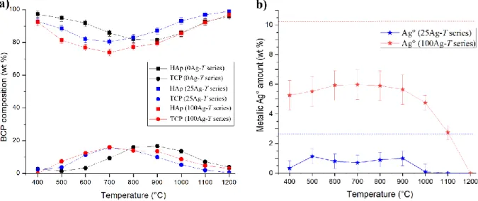
or an Ag°/HAp nanocomposite material. The difference is – of course – fundamental, since in the first
61
case the single-phase material contains Ag+ cations that substitute Ca2+ from the HAp crystal
62
structure, while in the second case metallic silver particles are intimately mixed in a multi-phase
63
composite. Although these two alternatives are listed and described in the literature [42] there is a
64
lack of understanding the mechanisms that lead to one or the other.
65
The purpose of this paper is to fully characterize the silver-HAp doping mechanism in the case
66
of sol-gel synthesis followed by gradual thermal treatment leading to silver auto-reduction. The
67
study is a continuation of our previous work on the BCP (biphasic calcium phosphate) doping
68
mechanism with cations from the first-row transition metals [48]. The case of zinc doping, with an
69
interstitial mode of incorporation into the HAp crystal structure, was first fully characterized [49-51],
70
then investigated for iron [52]. In the case of copper, it transpired that control of the doping
71
mechanism allowed an insight into behaviour in a biological environment [53]. For a complete
72
understanding of the present paper it is very important to note the difference between the notations
73
used: Ag+:HAp and Ag°/HAp. Ag+:HAp corresponds to the incorporation of Ag+ cations within the
74
crystal structure of hydroxyapatite (which is expected in the case of conventional cationic doping),
75
whereas Ag°/HAp means the formation of a composite containing two distinct phases, the
76
hydroxyapatite trapping nanoparticles of Ag° metallic silver.
77
2-. Materials and methods
78
2.1. Sol-gel elaboration of silver containing BCP samples
79
The sol-gel route previously proposed by the authors was used to synthesize both undoped and
80
Ag-doped series of BCP samples [51]. Briefly, to produce 2 g of undoped BCP powder, 4.7 g of
81
Ca(NO3)2.4H2O (Sigma-Aldrich) and 0.84 g of P2O5 (Sigma-Aldrich) were dissolved in ethanol under
82
stirring and refluxed at 85°C for 24 hours. The solution was then maintained at 55°C for 24 hours to
83
obtain a white consistent gel, and further heated at 80°C for 10 hours to obtain a white powder.
84
Finally, the powder was heat-treated for 15 hours. This thermal treatment was performed between
85
100°C and 1200°C. Samples from the undoped series were named the 0Ag-T series with T indicating
86
the temperature (from 400°C to 1200°C) of the following thermal treatment. Required amounts of
87
AgNO3 Aldrich) were added to the solution simultaneously with Ca(NO3)2.4H2O
(Sigma-88
Aldrich) in order to synthesize the Ag-doped series. Nominal compositions were calculated,
89
assuming Ag+ calcium substitution (i.e. assuming a (Ca+Ag)/P = 1.67 constant ratio) with two doping
90
levels: a 2.5% calcium substitution (Ag/(Ca+Ag) = 0.025 corresponding to the nominal composition
91
Ca9.75Ag0.25(PO4)6(OH)1.75; named the 25Ag-T series) and a 10% calcium substitution (Ag/(Ca+Ag) = 0.1
corresponding to the nominal composition Ca9Ag1(PO4)6(OH); named the 100Ag-T series).
As-93
synthesized Ag-doped powders were pale yellow; heat treatments turn the colour to light grey, and
94
then to white above 1000°C.
95
2.2. Powder X-Ray Diffraction (PXRD) and Rietveld analyses
96
PXRD patterns were recorded on a Philips X’Pert Pro PANalytical diffractometer (Almelo,
97
Netherlands), with θ-θ geometry, reflection mode, equipped with a solid-state X-Celerator detector
98
and using Cu K radiation ( = 1.54184 Å). PXRD patterns were recorded at room temperature in the
99
interval 3° < 2θ < 120°, with a step size of 2θ = 0.0167° and a counting time of 200s for each data
100
value.
101
Rietveld refinements were systematically performed for each measurement (13 temperatures for
102
the 3 series) using the FullProf.2k program [54]. The Rietveld strategy was detailed in previous
103
related works [48-53]. A new additional phosphate compound was considered here: AgCa10(PO4)7,
104
which crystallizes in the trigonal R3c space group with a = 10.4372 Å and c = 37.3379 Å [55]. It is
105
isotopic with members of the MCa10(PO4)7 (M = Li, Na, K and Cs) series, and is closely related to the
106
structure of β-TCP (β-Ca3(PO4)2, R3c symmetry with a = 10.4352 Å and c = 37.4029 Å [56]). The
half-107
occupied Ca4 site of β-TCP [56], becomes fully occupied by silver cations in AgCa10(PO4)7 [55];
108
explaining the charge compensation between Ca2+ and Ag+. The metal Ag° phase was also considered
109
in several sample; it is a compact cubic structure
Fm3
m
with a = 4.085 Å [57]. An example of a110
Rietveld plot is shown in Figure SEI1.
111
2.3. Scanning Electron Microscopy (SEM)
112
Electronic microscopy observations used a ZEISS SUPRA 55VP with GEMINI Field Emission –
113
Scanning Electron Microscope and were carried out on pressed pellets after gold metallization. No
114
polishing was carried out to avoid the leaching of the silver cations and to avoid the deformation of
115
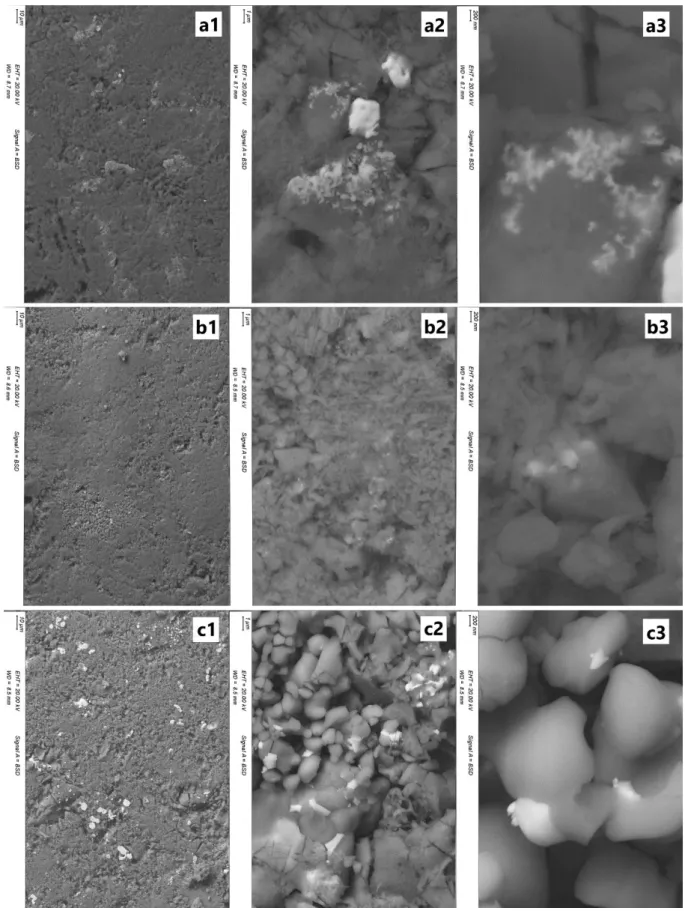
the ductile metallic silver particles. The main object of this analysis is not the quantification, but the
116
localization, of the silver element. Quantitative analyses were performed using an EDS (OXFORD
117
XMAX 80 N+ 80 mm2 Si-detector combined with OXFORD AZtec Advanced V3.3 software) on Ca, P
118
and Ag elements. Measurements were made using an acceleration voltage of 20 kV and 30 seconds
119
of acquisition on three wide areas (magnification x100) before averaging. Some specific isolated
120
measurements were performed using x5000 magnification. A 4QBSD detector was used to acquire
121
chemical contrast images to highlight silver particles.
122
The following nine samples were characterized by SEM: 0Ag-400, 0Ag-700, 0Ag-1200, 25Ag-400,
123
25Ag-700, 25Ag-1200, 100Ag-400, 100Ag-700 and 100Ag-1200.
124
3-. Results: Materials characterization
125
3.1. Quantitative phase analysis
126
To correctly interpret the behaviour of our samples, their phase compositions were extracted
127
from Rietveld analyses (PXRD patterns from the 25Ag-T series are shown in Figure 1). Figure 1
128
evidences the gradual increase in crystallinity of HAp with respect to temperature by the decrease in
129
the diffraction peak width. Phase compositions of the undoped BCP series and of the two Ag-doped
130
BCP series (x = 25 and 100) are presented in Table 1, and Figure 2a represents the thermal composition
131
variation for the two main phases: HAp and -TCP. Supplementary minor phases were observed.
-132
CDP (diCalcium DiPhosphate with composition Ca2P2O7) was observed up to 700°C, with a
133
maximum amount of 4 wt % at 500-600°C. CaO and CaCO3 (calcite) were present in the samples.
134
They are indicated in CaO equivalent content in Table 1, as calcite decarbonation leads to CaO
135
formation at about 800°C. The amount of CaO equivalent becomes negligible at high temperatures,
136
where the BCP were mainly composed of HAp (corresponding to the nominal composition). All these
137
observations – on main and minor phases – were similar to those of our previous studies on first-row
138
transition metal doping [48-53]. The -TCP phase is stabilized for intermediate temperatures, with a
maximum amount close to 20 wt % around 900 °C for the undoped series and around 700 °C for the
140
two silver-doped series. The main difference compared to our previous works concerns the formation
141
of metallic silver, Ag°, already at 400°C in the Ag-doped series (contrary to metal oxide formation for
142
the first-row transition metals, including ZnO and CuO). The amount of metallic silver reached 1 wt
143
% for the 25Ag-T series and 6 wt % for the 100Ag-T series (Table 1 and Figure 2b). These values are
144
to be compared to the quantities introduced during the syntheses: 2.65 wt % for the nominal
145
Ca9.75Ag0.25(PO4)6(OH)1.75 composition and 10.22 wt % for the nominal Ca9Ag1(PO4)6(OH) composition
146
(respectively the 25Ag-T and the 100Ag-T series). Figure 2b highlights the fact that the entirety of the
147
dopant introduced was not found exclusively in metallic form, but rather of the order of half of this
148
quantity. The reduction of the incorporated Ag+ silver cation to Ag° silver metal was autogenous, and
149
monitored by the thermal treatment only, without any reducing agent.
150
The formation of Ag°/HAp nanocomposite by heating Ag+-incorporated hydroxyapatite has
151
already been mentioned in the literature, either using a reducing agent (NaBH4 [34,58], hydrazine
152
[41]) or not [17,22,26,38,42]. However, the nanocomposite formation mechanism, the main subject of
153
this paper, has never been studied and detailed. It may first be pointed out that a thermal study on
154
silver nitrate (AgNO3; not shown here) showed decomposition accompanied by reduction at 300 °C,
155
which leads to the production of metallic silver (Ag°). To complete this information, the colour
156
changes of our samples are also interesting. The undoped series remained constantly white, while
157
the doped series were yellow at 400 °C (pale yellow for the 25Ag-T series and yellow-brown for the
158
100Ag-T series), then turned grey up to 1000 °C before bleaching above 1100 °C. The grey colour can
159
be attributed to the silver nanoparticles present in the Ag°/HAp composite. It is therefore normal to
160
lose the grey colour above 1000 °C, since silver has a melting point of 962 °C. Thus it is difficult to
161
design Ag°/HAp composites which could be obtained following heat treatments at more than 1000
162
°C [8,36,39,59]. Our white 25Ag-1100, 25Ag-1200 and 100Ag-1200 samples were then exempt of silver
163
particles, and certainly just composed of undoped HAp counterbalanced by a few wt % of β-TCP due
164
to the introduced Ca/P ratio (1.625 for the 25Ag-T series and 1.5 for the 100Ag-T series instead of the
165
1.67 value for HAp). By wetting calcium phosphate particles, metallic silver was still present in the
166
25Ag-1000 sample (in very little amount) and in 100Ag-1100 sample (although the Ag° amount
167
decrease is very clear at above 900 °C).
168
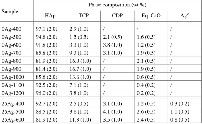
Table 1. Quantitative phase analysis (wt %) of the 27 samples; standard deviations are indicated in
169
brackets (CDP: Ca2P2O7 and ‘Eq. CaO’ considers both the CaO and CaCO3 amounts).
170
Sample
Phase composition (wt %)
HAp
TCP
CDP
Eq. CaO
Ag°
0Ag-400
97.1 (2.0)
2.9 (1.0)
/
/
/
0Ag-500
94.8 (2.0)
1.5 (0.5)
2.1 (0.5)
1.6 (0.5)
/
0Ag-600
91.8 (2.0)
3.3 (1.0)
3.8 (1.0)
1.2 (0.5)
/
0Ag-700
85.8 (2.0)
9.3 (1.0)
3.1 (1.0)
1.9 (0.5)
/
0Ag-800
81.9 (2.0)
16.0 (1.0)
/
2.1 (0.5)
/
0Ag-900
81.4 (2.0)
16.7 (1.0)
/
1.9 (0.5)
/
0Ag-1000
85.8 (2.0)
13.6 (1.0)
/
0.6 (0.5)
/
0Ag-1100
92.5 (2.0)
7.1 (1.0)
/
0.4 (0.2)
/
0Ag-1200
96.0 (2.0)
3.8 (1.0)
/
0.2 (0.2)
/
25Ag-400
92.7 (2.0)
2.5 (0.5)
3.1 (1.0)
1.2 (0.5)
0.3 (0.2)
25Ag-500
88.5 (2.0)
3.6 (1.0)
4.1 (1.0)
2.6 (0.5)
1.1 (0.5)
25Ag-600
81.9 (2.0)
11.3 (1.0)
3.5 (1.0)
2.4 (0.5)
0.8 (0.5)
25Ag-700
80.4 (2.0)
15.8 (1.0)
1.1 (0.5)
2.2 (0.5)
0.7 (0.5)
25Ag-800
82.9 (2.0)
14.0 (1.0)
/
2.2 (0.5)
0.9 (0.5)
25Ag-900
87.1 (2.0)
19.9 (1.0)
/
2.0 (0.5)
1.0 (0.5)
25Ag-1000
93.1 (2.0)
5.3 (1.0)
/
1.5 (0.5)
0.1 (0.2)
25Ag-1100
96.8 (2.0)
2.0 (0.5)
/
1.2 (0.5)
/
25Ag-1200
99.0 (2.0)
0.5 (0.2)
/
0.5 (0.2)
/
100Ag-400
92.3 (2.0)
/
/
2.4 (0.5)
5.3 (1.0)
100Ag-500
81.4 (2.0)
7.4 (1.0)
2.3 (0.5)
3.4 (1.0)
5.5 (1.0)
100Ag-600
76.8 (2.0)
12.5 (1.0)
1.8 (0.5)
3.0 (1.0)
5.9 (1.0)
100Ag-700
73.8 (2.0)
16.1 (1.0)
1.0 (0.5)
3.2 (1.0)
6.0 (1.0)
100Ag-800
77.3 (2.0)
13.9 (1.0)
/
2.9 (0.5)
5.9 (1.0)
100Ag-900
79.4 (2.0)
13.5 (1.0)
/
1.4 (0.5)
5.6 (1.0)
100Ag-1000 85.5 (2.0)
8.7 (1.0)
/
1.1 (0.5)
4.8 (1.0)
100Ag-1100 92.2 (2.0)
4.7 (1.0)
/
0.4 (0.2)
2.8 (0.5)
100Ag-1200
96.8 (2.0)
3.0 (1.0)
/
0.1 (0.2)
/
171
172
173
Figure 1. PXRD patterns from the 25Ag-T series. Scattering signals are mainly those from the HAp
174
phase, and other phases are identified thanks to their intense diffraction peaks (*: Ag°, #: CaO, ¤:
175
CaCO3 and $: β-TCP).
176
177
Figure 2. Thermal variation in a) the two main phase contents (squares: HAp; circles: TCP) from the
178
BCP samples and b) the metallic silver amount (stars) for the undoped 0Ag-T series (black symbols),
179
the 25Ag-T series (blue symbols) and 100Ag-T series (red symbols). Dotted lines indicate the nominal
180
silver amounts, and lines are only guides for the eyes.
181
3.2. Thermal variation in the HAp structural parameters
182
Thermal variations in the HAp lattice parameters are shown in Figure 3 and collated in Table
183
SEI1. Figure 3 clearly shows that the parameters of the HAp network evolved for the lowest
184
temperatures and then showed no more variations. The undoped series presents a relatively weak
185
contraction between 400 °C and 500 °C for both a and c lattice parameters (and consequently for the
186
unit cell volume). This contraction is also observed between 400 °C and 500 °C on the basal a lattice
187
parameter for the two silver-doped series, but much more markedly. Concerning the hexagonal c
188
lattice parameter, the difference between the undoped and the two doped series extends up to 700
189
°C, which of course also affects the unit cell volume difference up to 700 °C. For the lowest
190
temperatures, an enlargement of the HAp network is in favour of a calcium-to-silver substitution,
191
explained by the larger cationic radius of the Ag+ cation: 1.28 Å for Ag+ against 1.12 Å for Ca2+ in
192
eightfold coordination [60]. This indication of a silver substitution mechanism to form Ag+:HAp with
193
larger lattice parameters is consistent with previous literature results [32,33,38], but more marked in
194
our study. Indeed, the large values found here (a = 9.488 Å and c = 6.901 Å for the 100Ag-400 sample)
195
have never been evinced before. Despite the electronic contrast between Ag+ and Ca2+ cations, the last
196
Rietveld refinement cycles failed to clearly locate the silver atoms in the hydroxyapatite structure.
197
Nevertheless, the indications obtained support the notion of a substitution mechanism at the Ca1 and
198
Ca2 sites: the results indicate substitution rates which are too weak, but which are present (silver
199
occupancies about 3 % and 5 % – instead of the expected 10 % – for the Ca1 and Ca2 sites respectively),
200
and show the absence of chemical elements at the interstitial site within the hexagonal channel (i.e.
201
the 2b Wyckoff site). This leads us to privilege the Ca10-xAgx(PO4)6(OH)2-x stoichiometry for the doped
202
Ag+:HAp phase, and not that proposed by Geng et al., Ca10-xAg2x(PO4)6(OH)2 [32], which implies a
203
double mechanism by substitution and insertion. Our work on the structural parameters of
204
hydroxyapatite enables us to conclude that the HAp silver-doped phase is present in our samples
205
from 400 °C to 600 °C. This is correlated with the refined isotropic thermal parameters (see Biso in
206
Table SEI1) of hydroxyl anions, which are exaggerated in these samples: 400, 500,
25Ag-207
600, 100Ag-400, 100Ag-500 and 100Ag-600. We will consider below for these temperatures that the
208
Ag+:HAp phase presents the nominal stoichiometry, that is to say Ca9.75Ag0.25(PO4)6(OH)1.75 for the
209
25Ag-T series and Ca9Ag1(PO4)6(OH) for the 100Ag-T series, although there is no definitive
210
experimental proof for these compositions.
212
Figure 3. Thermal variation in the HAp lattice parameters for the three series of samples. Left: a
213
(squares) and c (circles) hexagonal lattice parameters, right: unit cell volume (stars). Lines are guides
214
for the eyes only.
215
3.3. Thermal variation in the β-TCP structural parameters
216
Thermal variations in the TCP lattice parameters, shown in Figure 4 and collated in Table SEI2,
217
are very different from those presented above for HAp. The basal a lattice parameter remained
218
constant whatever the synthesis series and heat treatment temperature. On the other hand, variations
219
in the axial c lattice parameter provide interesting information. The c lattice parameter – and
220
consequently the unit cell volume – for the two silver-doped series is below the value refined for the
221
undoped series, except for the temperature of TCP appearance (400 °C for the 25Ag-T series and 500
222
°C for the 100Ag-T series) and for the higher temperatures (1100 °C and 1200 °C). It corresponds to
223
the formation of the ordered calcium-to-silver substitution in β-TCP that leads to the AgCa10(PO4)7
224
compound. This compound is isotypic to β-TCP (trigonal R3c space group, 18 independent atomic
225
positions) with its half-occupied Ca4 site (at the special 6a Wyckoff site [56]) fully filled by Ag+ cations
226
[55]. The content of the unit cell is then Ag6Ca60(PO4)42; i.e. AgCa10(PO4)7 corresponding to Ca
3-227
xAg2x(PO4)2 with x = 0.143. This ordered substitution mechanism, leading to a definite compound, is
228
accompanied by a decrease in the axial c lattice parameter despite the larger size of Ag+: 37.403 Å for
229
β-TCP [56] against 37.338 Å for AgCa10(PO4)7 [55]. Thus, the value of the c axial lattice parameter
230
allows us to determine whether the present TCP phase corresponds to the undoped β-TCP phase or
231
the silver-containing AgCa10(PO4)7 phase. Table SEI2 indicates the TCP phase attribution for each
232
sample; either β-TCP (implied undoped Ca3(PO4)2) or AgCa10(PO4)7 (called Ag+:TCP here).
234
Figure 4. Thermal variation in TCP lattice parameters for the three series of samples. Left: a (squares)
235
and c (circles) trigonal lattice parameters; right: unit cell volume (stars). Lines are guides for the eyes
236
only.
237
3.4. Ag location studied by SEM analyses
238
SEM analyses were used to evaluate the silver amount and location in selected samples from the
239
doped series. Results from elemental quantifications made with EDS spectroscopy are summarized
240
in Table 2. The calculated Ca/P ratio indicates that results should be considered as semi-quantitative
241
only because of large deviations from the expected values as well as large variations within a single
242
series. This must be connected to the unpolished surface state of the pellets and the use of internal
243
calibration lines in the software. However, interesting additional information is provided by these
244
analyses.
245
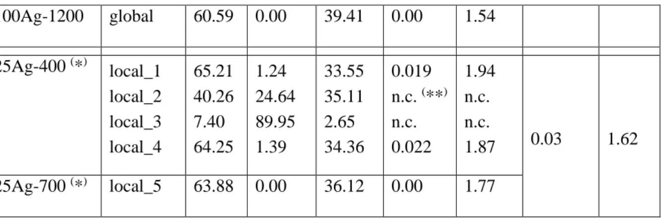
Table 2. Quantitative elemental analyses performed by EDS/SEM.
246
Sample
Analysis Atomic composition (%)
Calculated ratio
Expected ratio
Ca
Ag
P
Ag/Ca
Ca/P
Ag/Ca
Ca/P
0Ag-400
global
65.63
0.00
34.37
0.00
1.91
0.00
1.67
0Ag-700
global
59.80
0.00
40.20
0.00
1.49
0Ag-1200
global
68.02
0.00
31.98
0.00
2.13
25Ag-400
global
66.47
2.17
31.37
0.03
2.12
0.03
1.62
25Ag-700
global
60.14
1.43
38.43
0.02
1.56
25Ag-1200
global
65.48
0.00
34.52
0.00
1.90
100Ag-400
global
62.39
3.94
33.68
0.06
1.85
0.11
1.50
100Ag-700
global
62.35
3.03
34.61
0.05
1.80
100Ag-1200
global
60.59
0.00
39.41
0.00
1.54
25Ag-400
(*
)local_1
local_2
local_3
local_4
65.21
40.26
7.40
64.25
1.24
24.64
89.95
1.39
33.55
35.11
2.65
34.36
0.019
n.c.
(**
)n.c.
0.022
1.94
n.c.
n.c.
1.87
0.03
1.62
25Ag-700
(*
)local_5
63.88
0.00
36.12
0.00
1.77
(*
)corresponding images are shown in Figure 6.
247
(
**
)non-calculated ratio because the analysed area did not correspond to a calcium phosphate.
248
249
250
First, results from Table 2 confirm the complete disappearance of silver in doped series after heat
251
treatment at 1200°C. We can conclude that above the melting point of silver, not only did the metallic
252
Ag° particles leave the sample, but the calcium phosphate phases were also completely free of Ag+
253
cations. The chemical contrast imaging performed with the backscattered electron detector makes it
254
possible to highlight the notion of Ag° particles (Figure 5). The use of low magnifications shows the
255
presence of large, micrometric, particles (already present in the 25Ag-400 sample, and in large
256
quantities in the 100Ag-700 sample). Then, thanks to higher magnifications, it is possible to visualize
257
nanometric particles of small size. Figure 5a3 evidences the presence of silver particles of less than
258
100 nm diameter in 25Ag-400. These particles merged on heating to reach diameters of 200-400 nm in
259
the 25Ag-700 sample (Figure 5b3) and 500-1000 nm in the 100Ag-700 sample (Figure 5c3). Images
260
from Figure 5 clarify the composite feature of the samples.
261
Finally, local elemental analyses were performed to conclude the SEM characterization (see
262
selected zones in Figure 6). Results from selected areas (EDS shown in Figure 7) are presented in
263
Table 2 for the 25Ag-T series. These results confirm that nanoparticles were exclusively composed of
264
silver: metallic Ag° as shown in area 3 (and also area 2) in the 25Ag-400 sample. Indeed, the results
265
from the local zone ‘3’ are in favor of particle containing only the silver element (the minor Ca and P
266
contents come from the not really punctual electron beam), and therefore of a metallic nature (i.e. Ag°
267
particles). These results also confirm the presence of silver in the phosphate calcium phases for the
268
lowest heat treatment temperatures. Areas 1 and 4 indicate the presence of small amounts of silver,
269
in combination with large amounts of calcium and phosphor at 400°C, corresponding to an
270
Ag+:doped HAp phase. This is no longer true at 700 °C, where the calcium and phosphorus elements
271
are completely separated from the silver element: area 5 in sample 25Ag-700 is only composed of Ca
272
and P corresponding to undoped calcium phosphate compound (Figure 7 and Table 2).
274
Figure 5. Electron Backscattered SEM images showing the metallic silver particles in the samples. a)
275
25Ag-400; b) 25Ag-700 and c) 100Ag-700 samples using 1: x500, 2: x5k and 3: x20k magnifications.
276
277
Figure 6. Electron Backscattered SEM photographs used for local elemental analyses (presented in
278
Table 2) of the 25Ag-400 and 25Ag-700 samples.
279
280
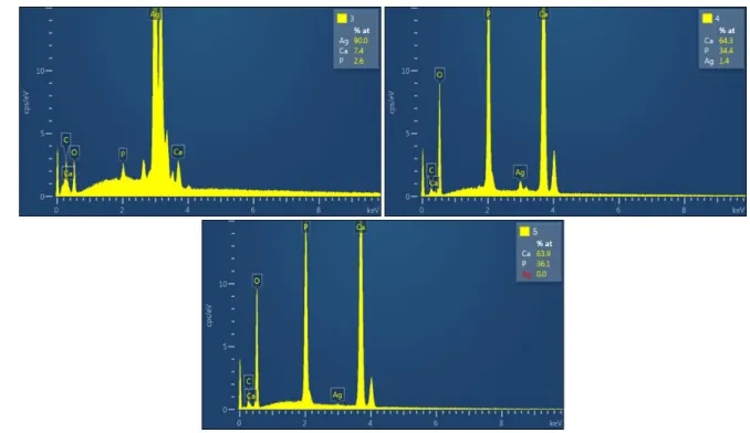
281
Figure 7. Selected local EDS analyses (presented in Table 2) from zones ‘3’ and ‘4’ of the 25Ag-400
282
sample and from zone ‘5’ of the 25Ag-700 sample (shown in Figure 6).
283
284
4-. Discussion
285
The results presented above evidence three distinct locations for silver atoms in the synthesized
286
BCP samples, including two distinct chemical states (metallic and ionic). A part of the introduced
287
silver cations is found in the metallic state, in the form of nanoparticles, already present at 400 °C.
288
The remainder is in the cationic form in substitution for calcium cations, either in the HAp phase (at
289
low temperatures: from 400 °C to 600 °C) or in the TCP phase (at the following temperatures and up
290
to 1000 °C). From 1000 °C silver leaves the samples, especially because of the melting point of Ag°
291
(962 °C). The cumulative amounts of located silver atoms are shown in Figure 8 for both the 25Ag-T
292
and 100Ag-T doped series. It appears that calculations for the lowest (400 °C) temperature are in good
293
agreement with the nominal introduced amounts (i.e. 2.67 wt % for the 25Ag-T series and 10.22 wt %
294
for the 100Ag-T series). We then observe an overestimation of the silver content up to 600°C, before
295
seeing it start to plummet at 700 °C. These fluctuations around the expected values indicate that the
296
amount of silver in the HAp phase is overestimated above 500 °C, and conversely the amount of
297
silver in substitution in β-TCP is certainly underestimated from 800 °C upwards.
299
Figure 8. Cumulative silver amount taken from metallic Ag° (black), substituted HAp (red) and
300
AgCa10(PO4)7 (or substituted TCP, blue) for the 25Ag-T series (left) and the 100Ag-T series (right).
301
Dotted lines indicate the nominal silver amounts, and error bars correspond to cumulative standard
302
deviation considering the tree phases.
303
Nanoparticles of metallic silver therefore formed very quickly in our samples by the autogenous
304
reduction of silver nitrate. The set of samples can thus be considered as a Ag°/BCP composite.
305
However, at low temperatures, we also observe the formation of a silver-doped Ag+:HAp phase
306
whose composition is close to the nominal composition (i.e. Ca9.75Ag0.25(PO4)6(OH)1.75 for the 25Ag-T
307
series and Ca9Ag1(PO4)6(OH) for the 100Ag-T series). Beyond 400 °C, this doped Ag+:HAp phase
308
persists while gradually lowering its doping element content. The drop in the doping rate was
309
notably predictable by observing the variations in the HAp lattice parameters between 400 °C and
310
700°C (Figure 3). Both a and c lattice parameters of the Ag+:HAp phase are vastly superior to those
311
from the undoped series at 400 °C. This difference fades at 500 °C and disappears at 700°C. SEM
312
analyses confirmed the presence of dopant in the calcium phosphate phase at 400 °C, followed by its
313
exclusion at 700°C. Concomitantly, cationic Ag+ substitutes calcium cations into the β-TCP phase,
314
which stabilizes around 700 °C. First, the AgCa10(PO4)7 definite compound is formed; it is
315
distinguishable from the undoped β-TCP phase thanks to the smaller c lattice parameter (Figure 4).
316
The underestimation of silver content from 800 °C upwards suggests that the Ag+ amount in the TCP
317
phase continues to increase; however, no experimental proof has been brought to support this.
318
The characterization of the doped Ag+:HAp phase with increased lattice parameters, by
319
comparison with an undoped HAp lattice, has already been reported in the literature. This is notably
320
the case in the study of Geng et al. [32], where the Ca10-xAg2x(PO4)6(OH)2 doped composition was
321
considered. This nominal composition suggests two kinds of doping mechanism: only half of the
322
silver cations (x value) can substitute calcium cations into the Ca1 or Ca2 crystallographic sites of the
323
HAp crystal structure. The second half of silver cations (to reach the 2x doping level) must be located
324
at interstitial sites. Our crystallographic study, based on Rietveld refinement, did not enable us to
325
highlight the presence of silver cations at interstitial crystallographic sites, not to mention that this
326
situation is not preponderant for large cations like Ag+. For these reasons we preferred to note the
327
chemical composition of the doped Ag+:HAp phase as follows: Ca10-xAgx(PO4)6(OH)2-x, in which only
328
the substitution mechanism was considered in agreement with the paper by Badrour et al. [61].
329
However, we must admit that our Rietveld refinements did not make it possible to clearly quantify
330
the silver substitution rates at both the Ca1 and Ca2 crystallographic sites in the HAp structure.
331
5-. Conclusion
332
During the past decade, several studies have been devoted to silver incorporation in HAp (or
333
BCP) samples due to their high potential for biomaterial applications, namely because of the
well-334
known bactericide properties of silver. Unanimously, the results have shown a very interesting
335
bactericidal effect following the doping of calcium phosphates with silver. However, the material
aspect of the samples studied in the literature showed some disparities, in particular due to a lack of
337
understanding of the doping mechanism. Some works mention metallic silver nanoparticles; others
338
focus on cationic substitution in phosphate phases. The results presented in the present paper are
339
especially devoted to the description of the mechanism by which silver is incorporated into BCP
340
samples. This work follows the experience previously acquired on the BCP doping mechanism of the
341
first-row transition elements. It appears that both electronic states are simultaneously present in the
342
silver-doped samples: metallic Ag° and cationic Ag+. Synthesized samples are composites comprising
343
closely-mixed Ag° nanoparticles and Ag+:doped calcium phosphate phases. At the lowest
344
temperature (400 °C), it is the HAp phase which presents a silver-to-calcium substitution leading to
345
the doped Ca10-xAgx(PO4)6(OH)2-x compounds. Then, by increasing the temperature (above 600 °C) the
346
Ag+ cations migrate to the β-TCP phase to form the definite Ca10Ag(PO4)7 compound corresponding
347
to Ca3-xAg2x(PO4)2 with x = 0.143 (i.e. Ca2.857Ag0.286(PO4)2). Finally, the composite aspect of the prepared
348
samples disappears above 1000 °C because the relatively low melting point of silver (962 °C). In the
349
light of these results, it appears important for the study of biological properties to distinguish the
350
behaviour in biological conditions of metallic silver nanoparticles from that of substituted cationic
351
Ag+, particularly in terms of bactericidal power and release rate/kinetics in the human body.
352
Acknowledgments: The authors warmly thank Anne-Marie Gelineau for the electron microscopy performed at
353
2MATech - Clermont-Ferrand, a few days before her retirement. Reviewers and guest editor, Francesco Capitelli,
354
are also grateful for their questions and comments that have improved the content and understanding if this
355
paper.
356
References
357
[1] Dorozhkin, S.V. Biocomposites and hybrid biomaterials based on calcium orthophosphates.
358
Biomaterials. 2011, 1, 3–56.
359
[2] Dahl, S.G.; Allain, P.; Marie, P.J.; Mauras, Y.; Boivin, G.; Ammann, P.; Tsouderos, Y.; Delmas, P.D.;
360
Christiansen, C. Incorporation and distribution of strontium in bone. Bone 2001, 8, 446–453.
361
[3] Lagier, R.; Baud, C.A. Magnesium whitlockite, a calcium phosphate crystal of special interest in
362
pathology. Pathol. Res. Pract. 2003, 199, 329–335.
363
[4] Lee, R.S.; Kayser, M.V.; Ali, S.Y. Calcium phosphate microcrystal deposition in the human
364
intervertebral disc. J. Anat. 2006, 208, 13–19.
365
[5] Rey, C.; Combes, C; Drouet, C.; Glimcher, M.J. Bone mineral: update on chemical composition and
366
structure. Osteoporos Int. 2009, 20, 1013-1021.
367
[6] Cazalbou, S.; Combes, C.; Eichert, D.; Rey, C. Adaptative physico-chemistry of bio-related calcium
368
phosphates. J. Mater. Chem. 2004, 14, 2148-2153.
369
[7] Elliot, J.C. Structure and chemistry of the apatite and other calcium orthophosphates, Amsterdam:
370
Elsevier, 1994.
371
[8] Turkoz, M.; Atilla, A.O.; Evis, Z. Silver and fluoride doped hydroxyapatite: Investigation by
372
microstructure, mechanical and antibacterial properties, Ceram. Int. 2013, 39 8925-8931.
373
[9] Diaz, M.; Zia, R.; Sameemi, F.; Ikram, H.; Bashir, F. In vitro antimicrobial activity of ZnO based
374
glass-ceramics against pathogenic bacteria. J. Mater. Sci. Mater. Med. 2015, 26:268.
375
[10] Khan, M.S.; ur Rehman, S.; Ali, M.A.; Sultan, B.; Sultan, S. Infection in orthopedic implant
376
surgery, its risk factors and outcome. J. Ayub Med. Coll. Abbottabad 2008, 20, 23-25.
377
[11] Cremet, L.; Corvec, S.; Bemer, P.; Bret, L; Lebrun, C.; Lesimple, B.; Miegeville, A.F.; Reynaud, A.;
378
Lepelletier, D.; Caroff, N. Orthopaedic-implant infection by Escherichia coli: molecular and
379
phenotypic analysis of the causative strains. J. Infect. 2012, 64, 169-175.
380
[12] Salwiczek, M.; Qu, Y.; Gardiner, J.; Strugnell, R.A.; Lithgow, T.; McLean, K.M.; Thissen, H.
381
Emerging rules for effective antimicrobial coatings. Trends Biotechnol. 2014, 32, 82-90.
382
[13] Nasser, S. Prevention and treatment of sepsis in total hip replacement surgery. Orthop. Clin. North
383
Am. 1992, 23, 265-277.
384
[14] Li, P.L.; Zamora, J.; Bentley, G. The results at ten years of the Insall-Burstein II total knee
385
replacement: clinical, radiological and survivorship studies. J. Bone Joint Surg. Br. 1999, 81, 647-653.
[15] Hendriks, J.G.E.; van Horn, J.R.; van der Mei, H.C.; Busscher, H.J. Background of
antibiotic-387
loaded bone cement and prosthesis-related infection. Biomaterials 2004, 25, 545-556.
388
[16] Fan, J.C.H.; Hung, H.H.; Fung, K.Y. Infection in primary total knee replacement. Hong Kong Med.
389
J. 2008, 14, 40-45.
390
[17] Sygnatowicz, M.; Keyshar, K.; Tiwari, A. Antimicrobial properties of silver-doped
391
hydroxyapatite nano-powders and thin films. Bio. Biomed. Mater. 2010, 62, 65-70.
392
[18] Rauschmann, M.A.; Wichelhaus, T.A.; Stirnal, V.; Dingeldein, E.; Zichner, L.; Schnettler, R.; Alt,
393
V. Nanocrystalline hydroxyapatite and calcium sulphate as biodegradable composite carrier material
394
for local delivery of antibiotic in bone infections. Biomaterials 2005, 26, 2677-2684.
395
[19] Baradari, H.; Damia, C.; Dutreih-Colas, M.; Laborde, E.; Pecout, N.; Champion, E.; Chulia, D.;
396
Viana, M. Calcium phosphate porous pellets as drug delivery systems: effect of drug carrier
397
composition on drug loading and in vitro release. J. Eur. Ceram. Soc. 2012, 32, 2679-2690.
398
[20] Clement, J.L.; Jarrett, P.S. Antibacterial silver. Metal-Based Drugs 1994, 1, 467-482.
399
[21] Marambio-Jones, C.; Hoek, E.M.V. A review of the antibacterial effects of silver nanomaterials
400
and potential implications for human health and the environment. J. Nano. Res. 2010, 12, 1531-1551.
401
[22] Iqbal, N.; Kadir, A.R.M.; Malek, N.N.A.N.; Mahmood, H.N.; Murali, R.M.; Kamarul, T. Rapid
402
microwave assisted synthesis and characterization of nanosized silver-doped hydroxyapatite with
403
antibacterial properties. Mater. Lett. 2012, 89, 118-122.
404
[23] Ning, C.; Wang, X.; Li, L.; Zhu, Y.; Li, M.; Yu, P.; Zhou, L.; Zhou, Z.; Chen, J.; Tan, G.; Zhang, Y.;
405
Wang, Y.; Mao, C. Concentration ranges of antibacterial cations for showing the highest antibacterial
406
efficacy but the least cytotoxicity against mammalian cells: implications for a new antibacterial
407
mechanism. Chem. Res. Toxicol. 2015, 28, 1815-1822.
408
[24] Kim, T.N.; Feng, Q.L.; Kim, J.O.; Wu, J.; Wang, H.; Chen, G.C.; Cui, F.Z. Antimicrobial effects of
409
metal ions (Ag+, Cu2+, Zn2+) in hydroxyapatite. J. Mater. Sci. Mater. Med. 1998, 8, 129-134.
410
[25] Feng, Q.L.; Wu, J.; Chen, G.Q.; Cui, F.Z.; Kim, T.N. A mechanistic study of the antibacterial effect
411
of silver ions on Escherichia coli and staphylococcus aureus. J. Biomed. Mater. Res. 2000, 52, 662-668.
412
[26] Rameshbabu, N.; Sampath Kumar, T.S.; Prabhakar, T.G.; Sastry, V.S.; Murty, K.V.G.K.; Prasad
413
Rao, K. Antibacterial nanosized silver substituted hydroxyapatite: synthesis and characterization. J.
414
Biomed. Mater. Res. A 2007, 80, 581-591.
415
[27] Klasen, H.J. Historical review of the use of silver in the treatment of burns: I. Early uses. Burns
416
2000, 26, 117-130.
417
[28] Gosheger, G.; Hardes, J.; Ahrens, H.; Streitburger, A.; Buerger, H.; Erren, M.; Gunsel, A.; Kemper,
418
F.H.; Winkelmann, W.; von Eiff, C. Silver-coated megaendoprostheses in a rabbit model – an analysis
419
of the infection rate and toxicological side effects. Biomaterials 2004, 25, 5547-5556.
420
[29] Ciobanu, C.S.; Iconaru, S.L.; Pasuk, I.; Vasile, B.S.; Lupu, A.R.; Hermenean, A.; Dinischiotu, A.;
421
Predoi, D. Structural properties of silver doped hydroxyapatite and their biocompatibility. Mater. Sci.
422
Eng. C 2013, 33, 1395-1402.
423
[30] Jadalannagari, S.; Deshmukh, K.; Ramanan, S.R.; Kowshik, M. Antimicrobial activity of
424
hemocompatible silver doped hydroxyapatite nanoparticles synthesized by modified sol-gel
425
technique. Appl. Nanosci. 2014, 4, 133-141.
426
[31] Fu, C.; Zhang, X.; Savino, K.; Gabrys, P.; Gao, Y.; Chaimayo, W.; Miller, B.L.; Yates, M.Z.
427
Antimicrobial silver-hydroxyapatite composite coating through two-stage electrochemical synthesis.
428
Surf. Coat. Tech. 2016, 301, 13-19.
429
[32] Geng, Z.; Cui, Z.; Li, Z.; Zhu, S.; Liang, Y.; Liu, Y.; He, X.; Yu, X.; Wang, R.; Yang, W. Strontium
430
incorporation to optimize the antibacterial and biological characteristics of silver-substituted
431
hydroxyapatite coating. Mater. Sci. Eng. C 2016, 58, 467-477.
432
[33] Gokcekaya, O.; Webster, T.J.; Ueda, K.; Narushima, T.; Ergun, C. In vitro performance of
Ag-433
incorporated hydroxyapatite and its adhesive porous coating deposited by electrostatic spaying.
434
Mater. Sci. Eng. C 2017, 77, 556-564.
435
[34] Wang, J.; Gong, X.; Hai, J.; Li, T. Synthesis of silver-hydroxyapatite composite with improved
436
antibacterial properties. Vacuum 2018, 152, 132-137.
[35] Riaz, M.; Zia, R.; Ijaz, A.; Hussain, T.; Mohsin, M.; Malik, A. Synthesis of monophasic Ag doped
438
hydroxyapatite and evaluation of antibacterial activity. Mater. Sci. Eng. C 2018, 90, 308-313.
439
[36] Dubnika, A.; Loca, D.; Rudovica, V.; Parekh, M.B.; Berzina-Cimdina, L. Functionalized
silver-440
doped hydroxyapatite scaffolds for controlled simultaneous silver ion and drug delivery. Ceram. Int.
441
2017, 43, 3698-3705.
442
[37] Zhang, X.; Chaimayo, W.; Yang, C.; Yao, J.; Miller, B.L.; Yates, M.Z. Silver-hydroxyapatite
443
composite coatings with enhances antimicrobial activities through heat treatment. Surf. Coat. Tech.
444
2017, 325, 39-45.
445
[38] Gokcekaya, O.; Ueda, K.; Narushima, T.; Ergun, C. Synthesis and characterization of
Ag-446
containing calcium phosphates with various Ca/P ratios. Mater. Sci. Eng. C 2015, 53, 111-119.
447
[39] Iqbal, N.; Kadir, M.R.A.; Mahmood, N.H.; Salim, N.; Froemming, G.R.A.; Balaji, H.R.; Kamarul,
448
T. Characterization, antibacterial and in-vitro compatibility of zinc-silver doped hydroxyapatite
449
nanoparticles prepared through microwave synthesis. Ceram. Int. 2014, 40, 4507-4513.
450
[40] Kaygili, O.; Keser, S.; Dorozhkin, S.V.; Yakuphanoglu, F.; al-Ghamdi, A.A.; Kirbag, S.; Sertkaya,
451
D.; Ates, T.; Gursoy, N.C. Structural and dielectrical properties of Ag- and Ba-substituted
452
hydroxyapatites. J. Inorg. Organomet. Polym. 2014, 24, 1001-1008.
453
[41] Liu, X.; Mou, Y.; Wu, S.; Man, H.C. Synthesis of silver-incorporated hydroxyapatite
454
nanocomposites for antimicrobial implant coatings. Appl. Surf. Sci. 2013, 273, 748-757.
455
[42] Vukomanovic, M.; Bracko, I.; Poljansek, I.; Uskokovic, D.; Skapin, S.D.; Suvorov, D. The growth
456
of silver nanoparticles and their combination with hydroxyapatite to form composites via a
457
sonochemical approach. Cryst. Growth Des. 2011, 11, 3802-3812.
458
[43] Chen, Y.; Zheng, X.; Xie, Y.; Ji, H.; Ding, C.; Li, H.; Dai, K. Silver release from silver-containing
459
hydroxyapatite coatings. Surf. Coat. Tech. 2010, 205, 1892-1896.
460
[44] Chen, W.; Oh, S.; Ong, A.P.; Oh, N.; Liu, Y.; Courtney, H.S.; Appleford, M.; Ong, J.L. Antibacterial
461
and osteogenic properties of silver-containing hydroxyapatite coatings produced using sol-gel
462
process. J. Biomed. Mater. Res. A 2007, 82, 899-906.
463
[45] Chen, W.; Liu, Y.; Courtney, H.S.; Bettenga, M.; Agrawal, C.M.; Bumgardner, J.D.; Ong, J.L. In
464
vitro anti-bacterial and biological properties of magnetron co-sputtered silver-containing
465
hydroxyapatite coating. Biomaterials 2006, 27, 5512-5517.
466
[46] Feng, Q.L.; Kim, T.N.; Wu, J.; Park, E.S.; Kim, J.O.; Lim, D.Y.; Cui, F.Z. Antibacterial effects of
467
Ag-HAp thin films on alumina substrates. Thin Sol. Films 1998, 335, 214-219.
468
[47] Shirkhanzadeh, M.; Azadegan, M.; Liu, G.Q. Bioactive delivery systems for the slow release of
469
antibiotics: incorporation of Ag+ ions into micro-porous hydroxyapatite coatings. Mater. Lett. 1995,
470
24, 7-12.
471
[48] Renaudin, G.; Gomes, S.; Nedelec, J.-M. First-row transition metal doping in calcium phosphate
472
bioceramics: a detailed crystallographic study. Materials 2017, 10, 92-113.
473
[49] Gomes, S.; Nedelec, J.-M.; Renaudin, G. On the effect of temperature on the insertion of zinc into
474
hydroxyapatite. Acta Biomater. 2012, 8, 1180-1189.
475
[50] Gomes, S.; Kaur, A.; Nedelec, J.-M.; Renaudin, G. X-ray Absorption Spectroscopy shining
476
(synchrotron) light onto the insertion of Zn2+ in calcium phosphate ceramics and its influence on their
477
behaviour in biological conditions. J. Mater. Chem. B 2014, 2, 536-545.
478
[51] Gomes, S.; Nedelec, J.-M.; Jallot, E.; Sheptyakov D.; Renaudin, G. Unexpected mechanism of Zn2+
479
insertion in calcium phosphate bioceramics. Chem. Mat. 2011, 23, 3072-3085.
480
[52] Gomes, S.; Kaur, A.; Grenèche, J.-M.; Nedelec, J.-M.; Renaudin, G. Atomic scale modeling of
iron-481
doped biphasic calcium phosphate bioceramics. Acta Biomater. 2017, 50, 78-88.
482
[53] Gomes, S.; Vichery, C.; Descamps, S.; Martinez, H.; Kaur, A.; Jacobs, A.; Nedelec, J.-M.; Renaudin,
483
G. Cu-doping of calcium phosphate bioceramics: from mechanism to the control of cytotoxicity. Acta
484
Biomater. 2018, 65, 462-474.
485
[54] Rodriguez-Carvajal, J. PROGRAM FullProf.2k – version 3.20; Laboratoire Léon Brillouin
(CEA-486
CNRS): Saclay, France, 2005; FullProf.2k manual available on
http://www-487
llb.cea.fr/fullweb/fp2k/fp2k_divers.htm. See also Rodriguez-Carvajal, J.; Roisnel, T. EPDIC-8; May
23-488
26, 2002; Trans. Tech. Publication: Uppsala, Sweden; Mater. Sci. Forum 2004; 123:443.
[55] Strutynska, N.Y.; Zatovsky, I.V.; Ogorodnyk, I.V.; Slobodyanik, N.S. Rietveld refinement of
490
AgCa10(PO4)7 from X-ray powder data. Acta Cryst. E 2013, 69, i23.
491
[56] Yashima, M.; Sakai, A.; Kamiyama, T.; Hoshikawa, A. Crystal structure analysis of
beta-492
tricalcium phosphate Ca3(PO4)2 by neutron powder diffraction. J. Sol. State Chem. 2003, 175, 272–277.
493
[57] Owen, E.A.; Yates, E.L. Precision measurement of crystal parameters. Phil. Mag. 1933, 15,
472-494
488.
495
[58] Mocanu, A.; Furtos, G.; Rapuntean, S.; Horovitz, O.; Flore, C.; Garbo, C.; Danisteanu, A.;
496
Rapuntean, G.; Prejmerean, C.; Tomoaia-Cotisel, M. Synthesis; characterization and antimicrobial
497
effects of composites based on multi-substituted hydroxyapatite and silver nanoparticles. Appl. Surf.
498
Sci. 2014, 298, 225-235.
499
[59] Singh, B.; Dubey, A.K.; Kumar, S.; Saha, N.; Basu, B.; Gupta, R. In vitro biocompatibility and
500
antimicrobial activity of wet chemically prepared Ca10-xAgx(PO4)6(OH)2 (0.0 ≤ x ≤ 0.5) hydroxyapatites.
501
Mater. Sci. Eng. C 2011, 31, 1320-1329.
502
[60] Shannon, R.D. Revised effective ionic radii and systematic studies of interatomic distances in
503
halides and chalcogenides, Acta Cryst. A 1976, 32, 751-767.
504
[61] Badrour, L.; Sadel, A.; Zahir, M.; Kimakh, L.; el Hajbi, A. Synthesis and physical and chemical
505
characterization of Ca10-xAgx(PO4)6(OH)2-x□x apatites. Ann. Chim. Sci. Mat. 1998, 23, 61-64.