HAL Id: inserm-01877891
https://www.hal.inserm.fr/inserm-01877891
Submitted on 20 Sep 2018
HAL is a multi-disciplinary open access
archive for the deposit and dissemination of
sci-entific research documents, whether they are
pub-lished or not. The documents may come from
teaching and research institutions in France or
abroad, or from public or private research centers.
L’archive ouverte pluridisciplinaire HAL, est
destinée au dépôt et à la diffusion de documents
scientifiques de niveau recherche, publiés ou non,
émanant des établissements d’enseignement et de
recherche français ou étrangers, des laboratoires
publics ou privés.
marrow transplantation.
Pierre Aucouturier, Anne Barra, Liliane Intrator, Catherine Cordonnier,
Dominique Schulz, Francoise Duarte, Jean-Paul Vernant, Jean-Louis
Preud’Homme
To cite this version:
Pierre Aucouturier, Anne Barra, Liliane Intrator, Catherine Cordonnier, Dominique Schulz, et al..
Long lasting IgG subclass and antibacterial polysaccharide antibody deficiency after allogeneic bone
marrow transplantation.. Blood, American Society of Hematology, 1987, 70, pp.779-785.
�inserm-01877891�
Blood, Vol 70, No 3 (September). 1987: pp 779-785 779
Long
Lasting
IgG
Subclass
and
Antibacterial
Polysaccharide
Antibody
Deficiency
After
Allogeneic
Bone
Marrow
Transplantation
By Pierre Aucouturier, Anne Barra, Liliane Intrator, Catherine Cordonnier, Dominique Schulz, Francoise Duarte, Jean-Paul Vernant, and Jean-Louis Preud’homme
Serum lgG subclasses were measured by a competitive
indirect immunoassay with monoclonal antibodies in 31
leukemic patients before and after bone marrow
transplan-tation. Antibodies to Hemophilus influenzae type b (Hib)
capsular polysaccharide were determined in 28 cases.
Abnormally low or borderline subclass (mostly lgG2 and
lgG4) levels were found late after transplant in 23 infected
and noninfected patients. These levels persisted for as long
as 25 months. in association with low or borderline IgA
levels in 78% of the cases. IgG2. lgG4, and IgA often
showed a parallel evolution. whereas lgGl . lgG3. and 1gM
often varied together in the opposite way. Class but not
subclass deficiencies were more frequent in patients with
A
COMBINED cellular and humoral immunodeficiency affects patients after allogeneic bone marrow trans-plantation (BMT). Reconstitution is slow, and certain long-term survivors suffer severe and/or recurrent infections. Serum immunoglobulin (Ig) classes may be depressed for - Iyear for IgG and 1gM and even longer for IgA, with a correlation between infections, Ig class deficiency, and chronic graft-v-host disease (GVHD).’’#{176} IgG subclass defi-ciency is a common feature of primary immunodeficiency states. It predominantly affects IgG2 and IgG4 and may be observed as a “selective” deficiency or in various immunode-ficiency syndromes,” especially IgA deficiency occurring as an apparently isolated defect’2’7 or in ataxia
telangiecta-sia.’82’ A frequent pathogen in such patients is Hemophilus
influenzae type b (Hib),”22’23 whose major antigen is the capsular polysaccharide polyribosylribitolphosphate
(PRP).24 This is not surprising, since IgG2 appears to be the predominant subclass of antibacterial carbohydrate
anti-bodies (Ab).227 The observation of a relatively high mci-dence of Hib pneumonia in our transplanted patients28 led us to study serum IgG subclass and anti-PRP Ab levels in transplanted patients affected with Hib pneumonia. Sera from patients with other infections or without infections were studied as controls.
graft-v-host disease (GVHD). Subclass abnormalities
pro-dominated in infected patients. with mean levels
correlat-ing with the severity of infections; however, the
abnormali-ties are not clearly predictive of infections in individual
cases. Most patients with Hib pneumonia showed virtually
no lgG antibody response to Hib. and one-half of the
patients had a moderate 1gM and IgA response. In the
whole series. many sera collected >1 year after graft
contained very low or undetectable antibodies. Correlation
between anti-Hib antibody and lgG2 levels was significant
but weak because of discrepancies that were only partially
explained by the subclass distribution of the antibodies.
a 1987 by Grune & Stratton. Inc.
of relapses. The conditioning regimen uniformly consisted of
cyclo-phosphamide (60 mg/kg on days - S and - 4) and total body
irradiation (10 gray, with lung shielding above 8 gray). To prevent GVHD, methotrexate was given until day 102 to all patients receiving allogeneic BMT according to the Seattle regimen.’ Fur-thermore, patient 16 received I-depleted bone marrow. Patient 29, the only patient who had received a syngeneic transplant, was not submitted to GVHD prophylaxis. GVHD was diagnosed and graded according to usual criteria.’#{176} No noninfected patient suffered GVHD. In contrast, most patients with repeated infections were affected with severe chronic GVHD.
Measurement of immunoglobulin class and subclass
1ev-els. Ninety-six sera were collected before transplant and after BMI (follow-up 4 to 25 months, mean I 3.2 months). Ig class levels were measured by laser nephelemetry. IgG subclass levels were
determined in coded samples by a competitive indirect immunoassay
(ELISA) with monoclonal Ab, as previously described.3’32 Normal values’5’31’32 were established in a study of 129 to 186 sera from
normal blood donors aged 20 to 50 years. Serum of the donors of
bone marrow grafts was not examined. Values below or at the lower limit of the 95% percentile range of normal sera (ie, 4.0 mg/mL, 0.6 mg/mL, and 0.18 mg/mL for IgGI, IgG2, and IgG3; 6.1, 0.8, and
0.5 mg/mL for IgG, IgA, and 1gM, respectively) are described
below as subclass or class deficiencies. For IgG4, whose level differs according to sex,3’ a 95% percentile range limit can be defined in men only (0.03 mg/mL). IgG2 and IgG4 display very heterogeneous distributions in healthy subjects, and low normal values are likely to PATIENTS AND METHODS
Patients. Nine patients who received allogeneic BMT and expe-rienced Hib pneumonia were retrospectively selected for the study. In view of the results, 22 other transplanted patients who survived >90 days and in whom serial posttransplant sera were available also were chosen as control cases. None of the patients received any IgG therapy during the study. None of the control patients had experi-enced Hib pneumonia, and we purposely selected patients with or without GVHD, viral, or other bacterial infections. Consequently, patients were listed according to the nature of their infections (Table
1): patients infected with extracellular bacteria, mostly Hib
(com-patible with impaired humoral immunity) (7 cases); patients with viral and/or intracellular bacterial infections (suggestive of cellular immune defect)
(
10 cases; two further patients suffered pneumonia of unknown origin); patients with both types of infections (5 cases),including Hib pneumonia in 2 cases; and patients free of infections
(7 cases). Chemotherapy regimens preceding the graft varied
according to the type and staging of the leukemias and to the number
From the Laboratory of Immunology and Immunopathology (CNRS UA 1 172), Poitiers University Hospital, Laboratory of Hematology and Immunology and Unit of Bone Marrow Trans-plantation, CHU Henri Mondor, Cr#{232}teiland Institut M#{234}rieux, Marcy L ‘Etoile, France.
Submitted February 23. 1987; accepted May 13, 1987.
Supported in part by Minist#{232}rede l’Education Nationale (Direc-tion de Ia Recherche) and Fondation pour la Recherche M#{234}dicale.
Address reprint requests to Professor Jean-Louis Preud’homme, CNRS UA I 1 72, CHUR La Mil#{233}trie,BP 577, F 86021 Poitiers Cedex, France.
The publication costs ofthis article were defrayed in part by page
charge payment. This article must therefore be hereby marked “advertisement” in accordance with 18 U.S.C. §1734 solely to
indicate this fact.
© I 987 by Grune & Stratton, Inc. 0006-4971/87/7003-0027$3.00/0
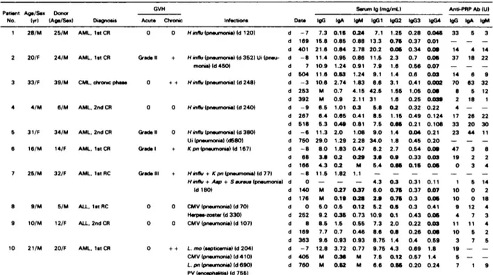
Table 1. Diagnosis. Infection Patterns. Serum 1g. and Anti-PRP Ab Levels Patient No. Ag./Sx (yrl Donor (Ag/Sex) Diagnosis GVH
Acute Chronic InfCtIOnS Date
Serum olma/mtl Anti-PRP Ab (UI
IgG gA gM IgGi IgG2 IgG3 IgG4 tgG gA gM
1 25/M 25/M AML. 1st CR 0 0 Hw,flu (pneumonia) (d 120) d
2 20/F 24/M AML. 1st R
3 33/F 39/M cMi. c’oc pha.
4 4/M 6/M AMI. 2nd CR 5 31/F 34/M AML. 2nd CR 6 16/PA 14/F AML. 1st CR 7 25/M 32/F AMI. lit RC -7 7.3 0.15 0.24 7.1 1.25 d 169 15.8 0.85 0.58 13.3 0.75 d 401 21.6 0.84 2.75 20.2 0.05
ade II + Hinflu (pneumonial (d 352) Ui (pn.u- d -8 1 1 .4 0.95 0.86 1 1 .5 2.3
monia)(d450) d 7 10.9 1.24 0.91 7.9 i.6 d 504 11.6 0.53 1.24 9.1 1.4 0 ++ Hinflu(pn.umonsal(d248) d -3 10.6 2.74 1.83 6.6 3.1 d 253 M 0.7 4.15 42.5 1.55 d 392 M 0.9 2.11 31 1.6 0 0 Hinflu(pn.umonEa((d240) d -9 6.5 1.01 0.3 5.5 0.2 d 267 6.4 0.65 0.41 5.5 1.15 d 51 5.3 0.49 0.81 7.5 0.65 &adeII 0 Hinflu(pneumoni.((d380I d -6 11.3 2.0 1.08 9.0 1.4 Ui (pn.umonel(d5801 d 750 29.0 1.29 2.28 34.0 1.5 adeI + Kpn(pneumonial(d167) d -8 8.0 1.83 0.47 6.2 2.7 d 65 3.5 0.2 0.29 3.6 0.9 d 166 4.3 0.2 M 5.4 0.65 adeIIl + HW,PPJ +Kpn(pn.wnoni.((d77( d -.5 11.5 1.82 1.1 -
-Hw,flu + Asp + Sar.us (pneumonial d 0 - - - 4.3 0.3
(d 180) d 140 M 0.27 0.37 6.0 0.75 d 176 N 0.19 0.25 2.9 0.75 d 0 5.0 0.5 0.12 5.2 0.5 d 252 9.2 0.35 0.73 10.9 0.1 d 8 8.5 1.5 0.55 7.3 2.0 d 169 7.7 0.7 0.46 8.6 0.5 d 363 9.6 0.93 0.93 8.75 1.4 d -7 12.5 3.72 0.77 9.75 4.3 d 405 N 0.36 M 7.5 0.12 0.28 0.045 33 5 3 0.37 0.01 - - -0.34 0.09 14 4 14 0.7 0.05 37 16 22 0.56 0.07 - - -0.6 0.03 14 6 9 0.41 0.002 70 63 32 1.05 0.06 5 5 12 0.25 0.039 2 15 1 0.32 0.22 4 - -0.49 0.124 17 26 22 0.21 0.108 33 20 30 0.04 0.21 23 44 11 0.45 0.20 - - -0.54 0.09 47 3 a 0.33 0.03 19 2 2 0.15 0.05 0 3 4 0.31 0.11 1 5 14 0.37 0.07 10 0 2 0.3 0.05 10 0 15 0.3 0.41 9 12 4 0.43 0.05 4 7 3 0.22 0.03 11 11 4 0.26 0.06 10 5 2 0.4 0.59 3 7 5 0.69 1.8 19 - -0.57 1.4 5 - -7 1 9
a 9/M 5/M ALL. lit RC 0 0 CMV (pneumcnial (d 701
H.rp..-zost Id 3301
9 10/M 12/F ALL. 2nd CR 0 0 CMV (pneumoni.I (d 107)
10 2 1/M 20/F AML. lit CR 0 ++ L.mo (septicemi.I Id 2041
CMV (pneumoni& (d 410)
L.pn(pn.umonial(d 6901
Pv (.ncephaktis) (d 755)
d 760 M 0.52 M 6.6 0.56 0.20 0.24
780 AUCOUTURIER ET AL
include deficiencies.32 Borderline levels (0.95 mg/mL for IgG2 and <0. 1 mg/mL in both sexes for IgG4) are therefore considered in the analysis.
Determination ofanti-PRP antibody levels. Anti-PRP Ab were measured in 79 sera from 28 patients (mean time between BMT and collection of the last serum studied was 14.3 months). PRP was prepared from culture supernatants by precipitation with a cationic detergent and further purification by saline and phenol extractions33 and was coupled to tyramine according to the method of Anthony et al.’ Microtitration plates were coated with PRP-tyramine (5 g/ ml PRP in phosphate-buffered saline [PBSI) and saturated with 1%
bovine serum albumin (BSA) in PBS containing 0.05% Tween-20. One-half of every plate was treated identically except that PRP was replaced by PBS (blanks). Sera diluted ‘/o in 0.5% BSA containing PBS-Tween were incubated for 90 minutes at 37#{176}Cin triplicates in the PRP-coated and control wells. The plates were washed six times in PBS-Tween and incubated with peroxidase-tagged goat antisera specific for human -y, a, and Ig chains (Institut Pasteur Produc-tion, Paris). After washings, bound conjugate was revealed with o-phenylenediamine and H202, and optical densities (OD) were recorded on a microplate reader.3’ Each ELISA plate contained standard positive and negative sera, and, as a reproducibility test, three further known positive sera (interassay coefficients of variation were 9.8% for IgG Ab, 7.8% for IgA Ab, and I 1 .3% for 1gM Ab). For every sample, the experiment was repeated on the same plate with serum previously incubated for 45 minutes with high-concen-tration (500 g/mL) PRP. Only specific OD (inhibited by PRP) were taken into account. Results were expressed in arbitrary units calculated from specific OD in PRP-coated wells after results on uncoated wells were deducted by comparing the same data with the
positive and negative standard sera.
Due to lack of material, the subclass distribution of lgG anti-PRP Ab could be studied in four patients only. These experiments were performed by a procedure similar to that previously described except that bound anti-PRP Ab were revealed by subclass-specific
mono-clonal Ab followed by peroxidase-coupled rabbit IgG anti-mouse IgG extensively absorbed with human IgG, as described in detail
previously.”
Statistical analysis. Ig and anti-PRP Ab levels in patients and controls were compared by the Student’s ttest when possible, or by the Mann and Whitney’s test when the number of subjects was too small. Percentages were compared using the Chi-square test.
RESULTS
Detailed results of measurement of Ig class and subclass and anti-PRP antibody levels are shown in Table I. Before transplant, several patients had low (patients 4, 7, 8, and 26) or borderline (patients 18 and 19) IgG2 and/or IgG4 (patients I
,
2, 3, 6, 24, 28, and 3 1)
levels. IgA and IgG3 were rarely depressed (patients 1, 5, and 28, respectively), whereas abnormally low 1gM levels were noticed in patients I, 4, 8, 12, and 18. None of these patients had any past history of abnormal infections. The evolution of the levels of the various Ig isotypes after BMT varied greatly from patient to patient. However, comparison of the curves drawn for each patient (not shown), revealed a strong tendency for certain isotypes to vary together: IgG1 and IgG3 showed a roughly parallel evolution in 68% of cases, IgGI and 1gM in 48% of cases, and IgG3 and 1gM in 45% of cases. On the other hand, roughly parallel curves were observed for IgG2 and IgG4 (61% ofcases), IgG2 and IgA (52% ofcases), and IgG4 and IgA (48% ofcases). Any other association between Ig isotypes was noted in <19% of cases. In contrast, IgGl and IgG2 varied in the opposite way in 61% of cases. Consequently, IgG2 levels were lower in late (>350 days after transplant) than in early samples in 1 1 cases. Subclass deficiencies or borderline levels at the end of follow-up1 1 26/M 30/M ALl.. let CR 0 + CMV (pneumonial (d 90)
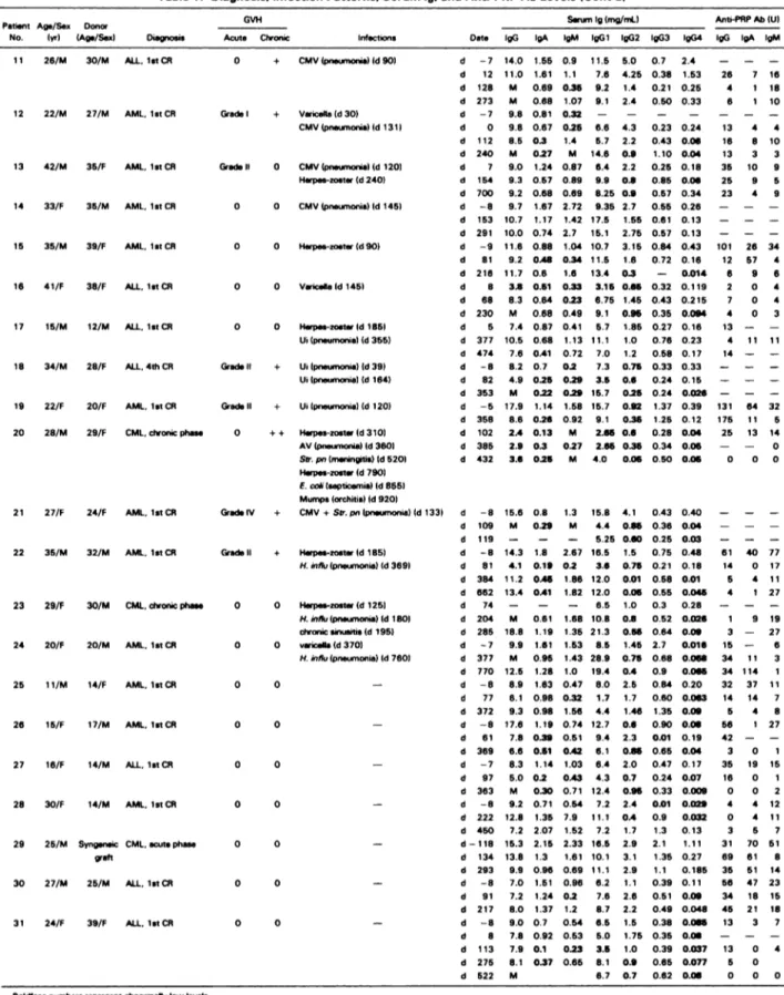
Table 1. Diagnosis. Infection Patterns. Serum 1g. and Anti-PRP Ab Levels (Cont’d)
. Patient No. Age/Sex )yr) Donor (Age/Sex) Diagnosis GVH
Acute Chronic Infections Date
Serum Ig(mg/mU Anti-PRP Ab (U)
lgG gA gM )gGl IgG2 lgG3 IgG4 lgG gA gM 0 0 Herpes-zoster Id 90) 0 0 Varicella(d145) d -7 14.0 1.55 0.9 11.5 5.0 0.7 2.4 d 12 11.0 1.61 1.1 7.6 4.25 0.38 1.53 d 128 M 0.69 0.35 9.2 1.4 0.21 0.25 d 273 M 0.68 1.07 9.1 2.4 0.50 0.33 d -7 9.8 0.81 0.32 - - - -d 0 9.8 0.67 0.25 6.6 4.3 0.23 0.24 d 112 8.5 0.3 1.4 5.7 2.2 0.43 0.06 d 240 M 0.27 M 14.6 0.9 1.10 0.04 d 7 9.0 1.24 0.87 6.4 2.2 0.25 0.18 d 154 9.3 0.57 0.89 9.9 0.6 0.85 0.06 d 700 9.2 0.68 0.69 8.25 0.9 0.57 0.34 d -8 9.7 1.67 2.72 9.35 2.7 0.55 0.26 d 153 10.7 1.17 1.42 17.5 1.55 0.61 0.13 d 291 10.0 0.74 2.7 15.1 2.75 0.57 0.13 d -9 11.6 0.88 1.04 10.7 3.15 0.84 0.43 d 81 9.2 0.48 0.34 11.5 1.6 0.72 0.16 d 216 11.7 0.6 1.6 13.4 0.3 - 0.014 d 8 3.5 0.51 0.33 3.15 0.65 0.32 0.119 d 68 8.3 0.64 0.23 6.75 1.45 0.43 0.215 d 230 M 0.68 0.49 9.1 0.95 0.35 0.094 d 5 7.4 0.87 0.41 5.7 1.85 0.27 0.16 d 377 10.5 0.68 1.13 11.1 1.0 0.76 0.23 d 474 7.6 0.41 0.72 7.0 1.2 0.58 0.17 d -8 8.2 0.7 0.2 7.3 0.75 0.33 0.33 d 82 4.9 0.25 0.29 3.5 0.6 0.24 0.15 d 353 M 0.22 0.29 15.7 0.25 0.24 0.026 d -5 17.9 1.14 1.58 15.7 0.92 1.37 0.39 d 358 8.6 0.26 0.92 9.1 0.35 1.25 0.12 d 102 2.4 0.13 M 2.65 0.6 0.28 0.04 d 385 2.9 0.3 0.27 2.65 0.35 0.34 0.06 d 432 3.6 0.25 M 4.0 0.06 0.50 0.06 12 22/M 27/M AML. let CR 13 42/M 35/F AMI. lstCR 14 33/F 35/M AML. let CR 15 35/M 39/F AML. 1st CR 16 41/F 38/F ALL. lstcR 17 15/M 12/M ALL. lstCR I8 34/M 28/F ALL. 4th CR
19 22/F 20/F AML. let CR Grade I) +
20 28/M 29/F CML. chronic phase 0 ++ Herpes-zoster (d310)
AV (pneumonia) Id360) S.,.. pn )menintis) (d 520) Herpes-zost Id 790)
F. cch)septicemi.) (d 855) Mumps (orchitis) Id 920)
21 27/F 24/F AML. let CR Grads IV + CMV + Str. pn (pneumonia) Id 133)
22 35/M 32/M AMI. let CR 23 29/F 30/M CML. ctwonic phase 24 20/F 20/M AML. let CR 25 1 1/M 14/F AML. let CR 26 15/F 17/M AML. 1st CR 27 16/F 14/M ALL. lstCR 28 30/F 14/M AMI. lstCR
29 25/M Syngeneic CML. acute phase 1 30 27/M 25/M ALL,letCR 31 24/F 39/F ALL. lstCR 0 0 0 0 0 0 0 0 0 0
Abbreviations: AML acute myeloidleukemia; CR. complete remission; CML. chronic myeloid letemia; ALL. acute lymphoblastic leMmi. H. influ, HMusmnzas; K. pn. KMbaiepnetanonise;
Asp.. Aspwlus llen,g.t&e; S. aoreus; St,1i#{216}QcOcCUS aor.m; CMV, cytomegelovirue; L.ion. Listens inonocyrogeoes; L. pn. LacmeIa pn.c.vnofiIa; PV. povavirue; AV. adeno*ue; SU. Pt’.
Streptococcus pneinonrne; C. co& EschchS, co8; Ui. unidentified pathogen.
M, not measured because o the presence o monoclonal tg
Grads I + Varicella Id 30) CMV (pneumonia) Id 131) &aI, 0 CMV(pneumonia)(d 120) Herpes-zoeter (d 240) 0 0 CMV (pneumonia) Id 145) 0 0 Ilerpes-zoeterld 155) Ui(pneumonia) (d 355) Grade II + Ui (pneumonia) Id 391 Ui
(pneumonia)
Id164) Ui(pneumonia) (d120) &adell + Herpes-zoster(d185) H.influ (pneumonia) Id 369) 0 0 Hsrpes-zost (d 1251 H. w,flu (pneumonia) Id 1801 chronicsinusitis(d 1951 0 0 varicsll.(d370( H.influ(pneumonia)
Id 760) 0 0 0 0 d -8 15.6 0.8 d 109 M 0.29 d 119 - -d -8 14.3 1.8 d 81 4.1 0.19 d 384 11.2 0.46 d 662 13.4 0.41 d 74 - -d 204 N 0.61 d 285 18.8 1.19 d -7 9.9 1.61 d 377 M 0.95 d 770 12.5 1.28 d -8 8.9 1.63 d 77 6.1 0.98 d 372 9.3 0.98 d -8 17.6 1.19 d 61 7.8 0.39 d 369 6.6 0.51 d -7 8.3 1.14 d 97 5.0 0.2 d 363 M 0.30 d -8 9.2 0.71 d 222 12.8 1.35 d 450 7.2 2.07 d-118 15.3 2.15 d 134 13.8 1.3 d 293 9.9 0.96 d -8 7.0 1.51 d 91 7.2 1.24 d 217 8.0 1.37 d -8 9.0 0.7 d 8 7.8 0.92 d 113 7.9 0.1 d 275 8.1 0.37 d 522 M 1.3 15.8 4.1 0.43 0.40 M 4.4 0.55 0.36 0.04 - 5.25 0.60 0.25 0.03 2.67 16.5 1.5 0.75 0.48 0.2 3.6 0.75 0.21 0.18 1.86 12.0 0.01 0.58 0.01 1.82 12.0 0.06 0.55 0.045 - 8.5 1.0 0.3 0.28 1.68 10.8 0.5 0.52 0.026 1.35 21.3 0.55 0.64 0.09 1.53 8.5 1.45 2.7 0.016 1.43 28.9 0.75 0.68 0.066 1.0 19.4 0.4 0.9 0.065 0.47 8.0 2.5 0.84 0.20 0.32 1.7 1.7 0.60 0.063 1.56 4.4 1.46 1.35 0.09 0.74 12.7 0.6 0.90 0.06 0.51 9.4 2.3 0.01 0.19 0.42 6.1 0.86 0.65 0.04 1.03 6.4 2.0 0.47 0.17 0.43 4.3 0.7 0.24 0.07 0.71 12.4 0.95 0.33 0.009 0.54 7.2 2.4 0.01 0.029 7.9 11.1 0.4 0.9 0.032 1.52 7.2 1.7 1.3 0.13 2.33 16.5 2.9 2.1 1.11 1.61 10.1 3.1 1.35 0.27 0.69 11.1 2.9 1.1 0.185 0.96 6.2 1.1 0.39 0.11 0.2 7.6 2.6 0.51 0.09 1.2 8.7 2.2 0.49 0.048 0.54 6.5 1.5 0.38 0.065 0.53 5.0 1.75 0.35 0.06 0.23 3.5 1.0 0.39 0.037 0.65 8. 1 0.9 0.65 0.077 6.7 0.7 0.62 0.06 26 7 16 4 1 18 6 1 10 13 4 4 16 8 10 13 3 3 35 10 9 25 9 5 23 4 9 101 26 34 12 57 4 6 96 2 04 7 04 4 03 13 - -4 11 11 14 - -131 64 32 175 11 5 25 13 14 - - 0 0 00 61 40 77 14 0 17 5 4 11 4 1 27 1 9 19 3 - 27 15 - 6 34 11 3 34 114 1 32 37 11 14 14 7 5 4 8 56 1 27 42 - -3 0 1 35 19 15 16 0 1 0 0 2 4 4 12 0 4 11 3 5 7 31 70 51 69 61 8 35 51 14 56 47 23 34 18 15 45 21 18 13 3 7 13 0 4 5 0 0 00 Boldface numbers r.prsssnt abnormally Pow levels.782 AUCOUTURIER ET AL predominantly affected IgG2 (20 patients) and IgG4 (19
patients), together with low or borderline IgA levels in 78% of cases.
The analysis of subclass levels in the last studied samples showed that long-term subclass deficiencies were observed predominantly in infected patients (only one noninfected patient had IgG4 deficiency). Every patient with an infection pattern compatible with both cellular and humoral ID was subclass-deficient, with a maximum follow-up of 25 months. Borderline levels were observed in all patients’ groups, including noninfected subjects, in whom the incidence of complete recovery was the highest. Differences according to the infection patterns are reflected in the mean IgG subclass values in the last studied samples (Table 2). On the whole, IgG 1 and IgG3 were higher and IgG2 and IgG4 lower than
in normal subjects (differences between patients and controls
were significant except for IgG3 levels in infected patients); the lowest values for the two latter isotypes were in patients with infections of both types (mean 0.33 and 0.08 mg/mL, respectively) and the highest in noninfected patients. Because of the small size of the group of noninfected patients, differences between infected and noninfected patients were significant for IgG2 levels only (P < .05). No
significant correlation was observed between the ages of the donors and recipients of the graft and the incidence of subclass deficiency.
Serum collected at periods close to infectious episodes was available in certain patients only. Comparison with subclass levels in the sera from noninfected patients showed an incidence of subclass deficiency possibly higher in infected patients (the difference is not significant in such a small series). Indeed, for IgG2, low levels were observed during 8 of 21 infectious episodes (4 with extracellular bacteria and 4 with intracellular bacteria or viruses) and borderline levels during 6 other episodes, whereas of 14 samples collected from noninfected subjects at similar periods after graft, one contained low and 5 contained borderline IgG2 levels. Despite this, the finding of normal subclass levels during one-third of the infectious episodes and the rare but possible occurrence of low levels in noninfected patients make the finding of subclass deficiency suggestive but not predictive of occurrence of infection in individual cases.
Comparison of serum Ig levels in patients with and without GVHD clearly shows that class deficiencies occur-ring late after BMT were observed predominantly in chronic GVHD: Only patients with chronic GVHD had total IgG and 1gM deficiencies, and IgA deficiency was more frequent
in GVHD (75% v 32% of cases, P < .02). As for IgG subclasses, the incidence of deficiency did not differ accord-ing to the presence or absence of acute or chronic GVHD. However, the patient who received a syngeneic graft (patient 29) is one of the only two patients in whom every isotype was at normal or high levels throughout the study.
Six of nine patients affected with Hib pneumonia had abnormally low or borderline serum IgG2 levels, an mci-dence that is not different from that observed in patients in whom Hib pneumonia could not be demonstrated. This led us to determine serum levels of anti-PRP Ab. Before transplant, patients 4 and 7, who had low IgG2, also had low Ab levels (Table 1). Mean Ab levels before graft, calculated excluding patients with IgG2 deficiency, were similar in the different patient groups and comparable to those in normal adults. We had enough material to study anti-PRP Ab at the moment of Hib pneumonia in only two patients (patients 3 and 7); their levels were low although IgG2 was in the normal range in case 3. After Hib infection, IgG anti-PRP Ab remained low or continued to decrease except in patients 4 and 24, in whom they reached levels comparable to those in normal subjects without known Hib infection. Patient 7, who showed a poor response after I-Iib pneumonia, experienced a second bout of Hib infection 3 months later. Mean IgG anti-PRP Ab level was not higher in patients infected with Hib than in the other patients under study (Table 3). Patients 4 and 24 had low IgG2 levels. Due to lack of material, we could not study the subclass distribution of these IgG Ab. This experiment was performed in patient I at day 401; the patient’s serum contained subthreshold levels of IgG2 and low but detectable IgG anti-PRP Ab. Two-thirds of these Ab belonged to the IgGl subclass, and one-third belonged to IgG2.
A moderate increase in 1gM and IgA Ab was observed just after the pneumonia in four and three cases, respectively. A strong IgA response was manifested by one case. Conse-quently, mean 1gM and IgA Ab levels after Hib infection were similar to normal subject levels (Table 3).
Most patients without known Hib infection had low anti-PRP Ab of the three classes, the latest samples studied often showing the lowest values. A number of sera collected after 1 year contained no detectable Ab or levels similar to those in young children (Tables I and 3). However, there were exceptions, such as patients 19, 29, and 30. Patient 19 had high Ab levels before transplant and I year later, despite a very low IgG2 level. This striking contrast led us to study the subclass ofthe Ab in the sample collected at day 358. In fact, we found that anti-PRP Ab in this serum was restricted to
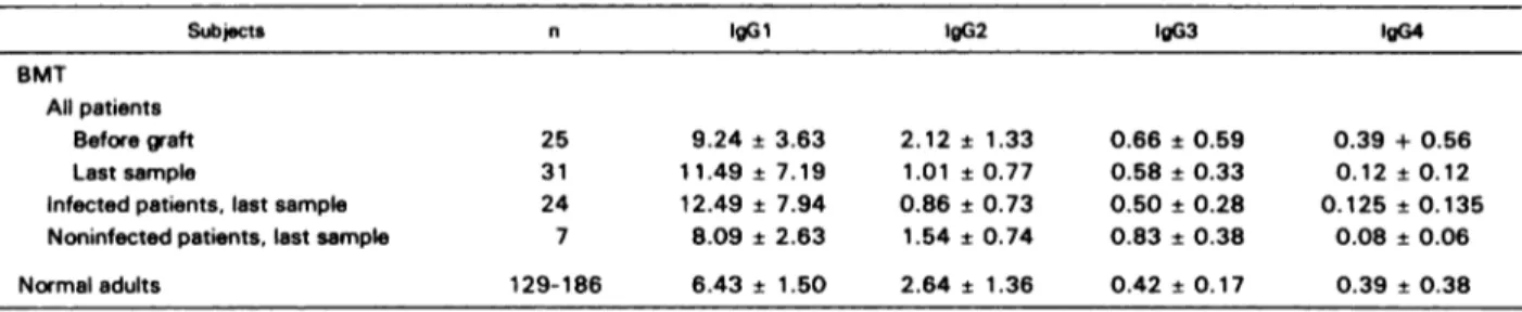
Table 2. Serum lgG Su bclass Levels in BM T Patients and Controls
Subjects n lgG 1 lgG2 lgG3 lgG4
BMT
All patients
Beforeaft
Last sample
Infected patients, last sample Noninfected patients, last sample
25 31 24 7 9.24 ± 3.63 1 1.49 ± 7.19 12.49 ± 7.94 8.09 ± 2.63 2.12 ± 1.33 1.01 ± 0.77 0.86 ± 0.73 1.54 ± 0.74 0.66 ± 0.59 0.58 ± 0.33 0.50 ± 0.28 0.83 ± 0.38 0.39 + 0.56 0.12 ± 0.12 0.125 ± 0.135 0.08 ± 0.06 Normal adults 129-186 6.43 ± 1.50 2.64 ± 1.36 0.42 ± 0.17 0.39 ± 0.38
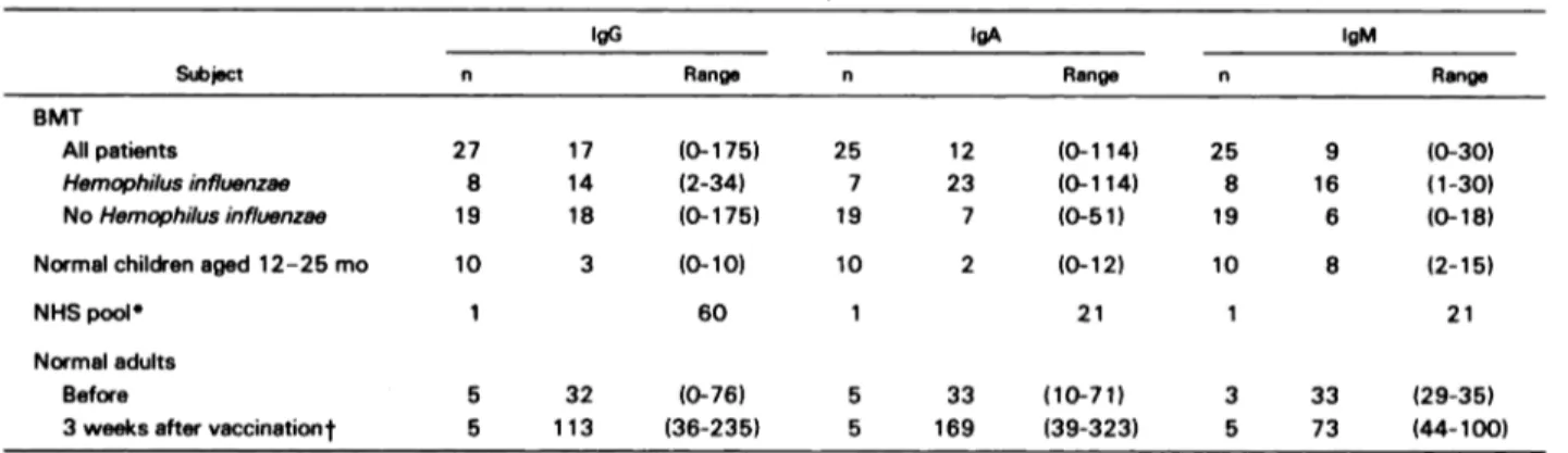
Table 3. Anti-P RP Ab Lev els in the L ast Studied S amples Fr om BMT P atients and in Control Sera
Subject
IgG lgA lgM
n Range n Range n Range
BMT All patients Hemophilusinfluenzae NoHemophilusinfluenzae 27 8 19 17 14 18 (0-175) (2-34) (0-175) 25 7 19 12 23 7 (0-1 14) (0-114) (0-51) 25 8 19 9 16 6 (0-30) (1-30) (0-18)
Normal children aged 12-25 mo 10 3 (0-10) 10 2 (0-12) 10 8 (2-15)
NHSpool 1 60 1 21 1 21 Normal adults Before 3weeksaftervaCCinationt 5 32 113 (0-76) (36-235) 5 5 33 169 (10-71) (39-323) 3 5 33 73 (29-35) (44-100)
Values are units, means, and ranges. Ab levels in patients were significantly lower than in normal (nonvaccinated) subjects. Due to the small size of the
groups, differences between Hib-infected and non-Hib-infected patients are not significant.
aNormal human sera from 60 adult blood donors.
tone injection of PRP (25 ag).
IgG2. Two other sera with fair Ab and low IgG2 levels were studied for subclass distribution (patient 20 at day 102 and patient 26 at day - 8), and the Ab were equally distributed in the IgGi and IgG2 isotypes. The correlation between IgG2 and anti-PRP Ab in the whole study, although significant
(P < .05), was weak (r - .23). DISCUSSION
The present study shows that IgG subclass deficiencies occur with a very high frequency in long-term survivors after BMT. They predominantly affect IgG2 and IgG4, as in a variety of primary and acquired immunodeficiency syn-dromes.’2”5 Similar findings were recently reported in abstract form.36 A striking observation in our study is that IgG2-IgG4 deficiencies, often associated with low IgA levels, frequently developed late after transplant, whereas 1gM, IgGl, and IgG3 and total IgG had reached normal or high levels. Indeed, IgG2, IgG4, and IgA often showed a parallel evolution, whereas IgGi, IgG3, and 1gM tended to vary together in the opposite way. This is reminiscent of several observations: in childhood, the increase of serum IgG I and IgG3 levels is rapid and follows total IgG, whereas IgG2 and IgG4 reach adult levels only around puberty (as IgA does)37
39; in common variable immunodeficiency subclass imbal-ance is very common, with predominant deficiencies of either IgG2 and IgG4 or IgG 1 and IgG3’5; patients with “selective” IgA deficiency often have IgG2-IgG4 deficiency, with fre-quent IgGl-IgG3 increase in infected patients.’2’7 These subclasses also differ with respect to Ab specificity, with an IgG1-IgG3 predominance of Ab to viral proteins#{176}43 and an IgG2 predominance of antibacterial polysaccharide Ab.227 Finally, studies of in vitro B cell maturation are compatible with different regulations of IgG2-IgG4 and IgGl-IgG3 expression.”
The asynchronous evolution of the subclasses and the well-known existence of profound T cell disturbances after BMT’’47#{176} suggest that subclass deficiency might result from an impaired T cell regulation rather than from an intrinsic B cell defect. Experimental work in the mouse51’52 favors the hypothesis that the expression of the isotypes
whose genes are located downstream in the CH locus (as IgG2 and IgG4 are in humans) requires more T cell help than does control of upstream isotypes. The finding of IgG2-IgG4 deficiency in human syndromes featuring impaired T-B cell cooperation, such as the Di George’s syndrome, the severe combined immunodeficiency syndrome with abnormal expression of HLA class II antigens, ataxia telangiectasia, and AIDS’5”9’21’53 is in keeping with this hypothesis. Although bacterial polysaccharides are T-inde-pendent antigens, antipolysaccharide Ab production is highly dependent on T cell regulation.TM Therefore, both subclass and anti-PRP Ab deficiencies may result from abnormal T cell function. Cells from five of the present patients were studied for in vitro suppressor activity?#{176} There
is no correlation between excessive suppressor activity and
the present findings, suggesting that impaired helper
func-tion might play a major role.
Previous work’’#{176} showed that GVHD is a significant factor in the occurrence of infections and of Ig class deficien-cy; this is confirmed in the present study. In contrast, we observed an incidence of subclass deficiency similar in
patients with and without GVHD. However, the finding of
fully normal Ig levels throughout the study was exceptional and concerned virtually only the patient who received a syngeneic transplant. Therefore, that clinically undetectable GVHD plays a role in the pathogenesis of subclass deficien-cies is a possibility. Both the mean subclass levels and incidence of subclass deficiencies differed according to the infection patterns. Subclass deficiencies were observed in patients infected with extracellular bacteria as well as in patients with viral or intracellular bacterial infections in whom they are unlikely to play a direct role. They were constant in patients with both types of infections and may reflect the general level of immunodeficiency in these patients. The subclass deficiencies may contribute to the occurrence of extracellular bacterial infections, but certain patients free of infections had low or borderline subclass levels, whereas some infected patients had normal class and subclass levels at the period of infectious episodes. The explanation of the occurrence of infections is therefore more
784 AUCOUTURIER ET AL
10. Brenner MK, Wimperis iZ, Reittie iE, Patterson i, Asherson complex than a mere subclass deficiency, even in patients with Hib pneumonia. Anti-PRP Ab defect might play some role, since we observed low levels just before pneumonia in a patient with normal IgG2 level and since there was a very poor IgG Ab response after pneumonia (protection against Hib is predominantly mediated by IgG Ab55). One patient with low IgG response experienced a second bout of Hib pneumonia 3 months later. Several patients who had no Hib pneumonia developed anti-PRP Ab (patients 19, 24, 29, and 30), but we do not know whether the other patients without pneumonia have been infected by Hib. Humoral immunode-ficiency thus probably plays a role in the genesis of infections observed in BMT recipients. Thus, substitutive IgG therapy of infectious episodes may be indicated even in patients without class and/or subclass deficiency. Whether a pro-longed systematic IgG treatment is indicated for prophylaxis of infections remains to be determined. On the other hand, the high incidence of Hib pneumonia in our series28 and the defective anti-PRP Ab response raises the questions of the indication and timing ofvaccination in transplanted patients. In such cases, a protein conjugate PRP vaccine should be used, since it is effective in IgG2-deficient patients who do not respond to PRP alone.56
REFERENCES
I. Storb R, Thomas ED: Allogeneic bone marrow transplanta-tion. Immunol Rev 71:77, 1983
2. Witherspoon RP, Lum LG, Storb R: Immunologic reconstitu-tion after human marrow grafting. Semin Hematol 2 1:2, 1984
3. Elfenbein GJ, Anderson PN, Humphrey RL, Mullins GM, Sensenbrenner LL, Wards JR, Santos GW: Immune system recon-stitution following allogeneic bone marrow transplantation in man: A multiparameter analysis. Transplant Proc 8:641, 1976
4. Noel DR, Witherspoon RP, Storb R, Atkinson K, Doney K, Mickelson EM, Ochs HD, Warren RP, Weiden PL, Thomas ED:
Does graft-versus-host disease influence the tempo of immunologic recovery after allogeneic human marrow transplantation? An obser-vation on 56 long-term survivors. Blood 5 1: I 087, 1978
5. Sullivan KM. Shulman HM, Storb R, Weiden PL,
Wither-spoon RP, McDonald GVB, Schulbert MM, Atkinson K, Thomas
ED: Chronic graft-versus-host disease in 52 patients: Adverse natu-ral course and successful treatment with combination
immunosup-pression. Blood 57:267, 1981
6. Witherspoon RP, Kopecky K, Storb RF, Flournoy N, Sullivan KM. Sosa R, Deeg Hi, Ochs HD, Cheever MA, Fefer A, Thomas ED: Immunological recovery in 48 patients following syngeneic marrow transplantation for hematological malignancy.
Iransplanta-tion 33:143, 1982
7. Atkinson K, Farewell V. Storb R, Isoi MS, Sullivan KM. Witherspoon RP, Fefer A, Clift R, Goodell B, Thomas ED: Analysis of late infections after human bone marrow transplantation: Role of genotype nonidentity between marrow donor and recipient and of nonspecific suppressor cells in patients with chronic graft-versus-host disease. Blood 60:714, 1982
8. Winston Di, 1-b WG, Champlin RE, Gale RP: Infectious complications of bone marrow transplantation. Exp Hematol I 2:205,
1984
9. Perreault C, Giasson M, Gyger M, Belanger R, David M,
Bonny Y, Boileau i, Bercelo R, Moquin iP: Serum immunoglobulin
levels following allogeneic bone marrow transplantation. Blut
51:137, 1985
GL, Hoffbrand AV, Prentice HG: Recovery of immunoglobulin
isotypes following I-cell depleted allogeneic bone marrow
transplan-tation. Br J Haematol 64:125, 1986
1 1. Oxelius VA: IgG subclasses and human disease. Am J Med
76:7, 1984
12. Oxelius VA, Laurell AB, Lindquist B, Golebiowska H, Axels-son U, Bjjorkander i, Hanson LA: IgG subclass in selective IgA deficiency. N Engl i Med 304:1476, 1981
13. Ugazio AG, Out TA, Plebani A, Duse M, Monafo V. Nespoli L, Burgio GR: Recurrent infections in children with “selective” IgA deficiency: Association with IgG2 and IgG4 deficiency, in
Wedg-wood Ri, Rosen FS, Paul NW (eds): Primary Immunodeficiency Disease, Birth Defects. Original Article Series 19. New York, Liss,
1983, p 169
14. Cunningham-Rundles C, Oxelius VA, Good RA: IgG2 and IgG3 subclass deficiencies in selective IgA deficiency in the United States, in Wedgwood Ri, Rosen FS, Paul NW (eds): Primary Immunodeficiency Diseases, Birth Defects. Original Article Series 19. New York, Liss, 1983, p 173
I 5. Aucouturier P. Bremard-Oury C, Clauvel JP, Debre M, Griscelli C, Seligmann M, Preud’homme JL: Serum IgG subclass levels in primary and acquired immunodeficiency, in Hanson LA, S#{246}derstr#{246}mI, Oxelius VA (eds): Immunoglobulin Subclass Defi-ciencies. Monographs in Allergy 20, Basel, Karger, 1986, p 62
16. Skvaril F, Scherz R: lgG subclasses in IgG deficient patients with anti-IgG antibodies, in Hanson LA, S&Ierstr#{246}mI, Oxelius VA (eds): Immunoglobulin Subclass Deficiencies. Monographs in Allergy 20, Basel, Karger, 1986, p 164
17. Plebani A, Monafo V, Avanzini AA, Ugazio G, Burgio R: Relationship between IgA and IgG subclass deficiencies: A reap-praisal, in Hanson LA, SOderstr#{246}mI, Oxelius VA (eds): Immuno-globulin Subclass Deficiencies. Monographs in Allergy 20, Basel, Karger, 1986, p 171
18. Rivat-Peran L, Buriot D, Salier iP, Rivat C, Dumitresco SM, Griscelli C: Immunoglobulins in ataxia-telangiectasia: Evidence for IgG4 and IgA2 subclass deficiencies. Clin Immunol Immunopathol 20:99, 1981
19. Oxelius VA, Berkel Al, Hanson LA: IgG2 deficiency in ataxia-telangiectasia. N EngI i Med 306:515, 1982
20. Berkel Al: Studies of IgG subclasses in ataxia-telangiectasia patients, in Hanson LA, S#{246}derstr#{246}mI, Oxelius VA (eds): Immuno-globulin Subclass Deficiencies. Monographs in Allergy 20, Basel, Karger, 1986, p 100
21. Aucouturier P. Br#{233}mard-Oury C, Griscelli C, Berthier M, Preud’homme iL: Serum IgG subclass deficiency in ataxia-telangiectasia. Clin Exp Immunol 68:392, 1987
22. Br#{233}mard-Oury C, Aucouturier P. Debr#{233}M, Preud’homme JL, Griscelli C: Immunoglobulin G subclasses in patients with immunodeficiencies, in Hanson LA, S#{246}derstr#{246}mT, Oxelius VA (eds): Immunoglobulin Subclass Deficiencies. Monographs in Allergy 20, Basel, Karger, 1986, p 75
23. Shackelford P0, Polmar SH, Mayus JL, iohnson WL, Corry iM, Nahm MH: Spectrum of IgG2 subclass deficiency in children with recurrent infections: Prospective study. i Pediatr 108:647, 1986
24. Lagergard T, Nylen 0, Sandberg I, Trollfors B: Antibody responses to capsular polysaccharide, lipopolysaccharide, and outer membrane in adults infected with Haemophilus influenzae type b. J Clin Microbiol 20:1154, 1984
25. Yount Wi, Dorner NM, Kunkel HG, Kabat EA: Studies on human antibodies. IV Selective variations in subgroup composition and genetic markers. i Exp Med 127:633, 1968
26. Siber GR, Schur PH, Aisenberg AC, Weitzman SA, Schiff-man G: Correlation between serum IgG2 concentration and the
antibody response to bacterial polysaccharide antigens. N Engl i Med 303:178, 1980
27. Bird P, Lowe i, Stokes RP, Bird AG, Ling NR, iefferis R: The separation of human serum IgG into subclass fractions by immunoaffinity chromatography and assessment of specific
anti-body activity. J Immunol Methods 71:91, 1984
28. Cordonnier C, Bernaudin JF, Bierling P, Huet Y, Vernant JP: Pulmonary complications occurring after allogeneic bone mar-row transplantation. A study of 130 consecutive transplanted patients. Cancer 58:1047, 1986
29. Thomas ED, Storb R, Clift RA, Fefer A, iohnson FL, Neiman PE, Lerner KG, Jlucksberg H, Buckner CD: Bone marrow transplantation (first of two parts). N Engl J Med 292:832, 1975
30. Schulman HM, Sullivan KM. Weiden PL, McDonald GB, Striker GE, Sale GE, Hackman R, Tsoi MS. Storb R, Thomas ED: Chronic graft-versus-host syndrome in man. A long-term clinico-pathologic study of 20 Seattle patients. Am J Med 69:204, 1980
31. Aucouturier P. Danon F, Daveau M, Guillou B, Sabbah A,
Besson i, Preud’homme JL: Measurement of serum IgG4 levels by a
competitive immunoenzymatic assay with monoclonal antibodies. J Immunol Methods 74:151, 1984
32. Aucouturier P. Mounir 5, Preud’homme iL: Distribution of IgG subclass levels in normal adult sera as determined by a competitive enzyme immunoassay using monoclonal antibodies. Diag Immunol 3:191, 1985
33. Anderson P. Smith DH: Isolation of the capsular polysac-charide from culture supernatant of Haemophilus infiuenzae type b. Infect Immunol 15:472, 1977
34. Anthony BF, Concepcion NF, McGeary SA, Ward ii, Heiner DC, Shapshak P. Insel RA: Immunospecificity and quantita-tion of an enzyme-linked immunosorbent assay for group B
strepto-coccal antibody. J Clin Microbiol 16:350, 1982
35. Barra A, Aucouturier P. Preud’homme JL: Isotypic distribu-tion of human anti-thyroglobulin IgG antibodies: Methodological difficulties. Diag Immunol 4:228, 1986
36. Riches PU, Walker SA, Rogers IR, Hobbs JR: Relative deficiency of serum IgA, IgG2 and IgG4 during reconstitution following BMT: Relationship to infection. Bone Marrow Transplant
1:53, 1986 (suppl 1)
37, Oxelius VA: lgG subclass levels in infancy and childhood. Acta Paediatr Scand 68:23, 1979
38. Van der Giessen M, Rossouw E, Algra-Van Veen I, Van Loghem E, Zegers BJM, Sander PC: Quantification of IgG sub-classes in sera of normal adults and healthy children between 4 and
12 years ofage. Clin Exp Immunol 21:501, 1975
39. Zegers BJM, Van der Giessen M, Reerink-Brongers EE, Stoop JW: The serum IgG subclass levels in healthy infants of 13-62 weeks ofage. Clin Chim Acta 101:265, 1980
40. Morell A, Roth-Wicky B, Skvaril F: Immunoglobulin G subclass restriction ofantibodies against hepatitis B surface antigen. Infect Immun 39:565, 1983
41. Linde GA, Hammarstrom L, Persson MAA, Smith CIE, Sundqvist VA, Wahren B: Virus-specific antibody activity of dif-ferent subclasses of immunoglobulins G and A in cytomegalovirus infections. Infect Immunol 42:237, 1983
42. Skvaril F, Schilt U: Characterization of the subclasses and light chain types of IgG antibodies to rubella. Clin Exp Immunol
55:671, 1984
43. Sundqvist VA, Linde A, Wahren B: Virus-specific immuno-globulin G subclasses in herpes simplex and varicella-zoster virus infections. J Clin Microbiol 20:94, 1984
44. Mayumi M, Kuritani T, Kubagawa H, Cooper MD: IgG subclass expression by human lymphocytes and plasma cells: B lymphocyte precommitted to IgG subclass can be preferentially induced by polyclonal mitogens with I cell help. i Immunol I 30:671,
1983
45. Walker L, iohnson GD, MacLennan 1CM: The IgG subclass response of human lymphocytes to B-cell activators. Immunology 50:269, 1983
46. Le Ihi Bich-Ihuy, Revillard iP: Modulation of polyclonally activated human peripheral B cells by aggregated IgG and IgG-binding factors: Differential effect on IgG subclass synthesis. J Immunol 133:544, 1984
47. Atkinson K, Hansen iA, Storb R, Goehle 5, Goldstein G, Thomas ED: I-cell subpopulations identified by monoclonal
anti-bodies after human marrow transplantation. I. Helper-inducer and
cytotoxic-suppressor subsets. Blood 59:1292, 1982
48. Linch DC, Knott i, Thomas RM, Harper P. Goldstone AH, Davis EG, Levinski RJ: I cell regeneration after allogeneic and autologous bone marrow transplantation. Br i Haematol 53:451,
1983
49. Rozans MK, Smith Bi, Burakoff SJ, Miller RA: Long-lasting deficit of functional I cell precursors in human bone marrow transplant recipients revealed by limiting dilution methods. J Immunol 136:4040, 1986
50. Leroy E, Calvo CF. Divine M, Gourdin MF, Baujean F, Ben Ariba MH, Mishal Z, Vernant iP, Farcet iP, Senik A: Persistence of I8/HNK-1 + suppressor lymphocytes in the blood of long-term
surviving patients after allogeneic bone marrow transplantation. i Immunol 137:2180, 1986
51. Martinez-Alonso C, Couthino A, Andrei AA: Immunoglobu-lin C-gene expression. 1. The commitment of lgG subclass of secretory cells is determined by the quality of the nonspecific stimuli. Eur i Immunol 10:698, 1980
52. Mongini PKA, Paul WE, Metcalf ES: I cell regulation of immunoglobulin class expression in the antibody response to trinitro-phenyl-Ficoll. Evidence for I cell enhancement of the immunoglobu-lin class switch. J Exp Med 155:884, 1982
53, Aucouturier P. Couderc Li, Gouet D, Danon F, Gombert i, Matheron 5, Saimot AG, Clauvel iP, Preud’homme iL: Serum immunoglobulin G subclass dysbalances in the lymphadenopathy syndrome and acquired immune deficiency syndrome. Clin Exp Immunol 63:234, 1986
54, Khater M, Macai I, Genyea C, Kaplan i: Natural killer cell regulation of age-related and type-specific variations in antibody responses to pneumococcal polysaccharides. J Exp Med 164:1505,
I986
55. iohnston RB, Anderson P. Rosen FS, Smith DH:
Character-ization of human antibody to polyribophosphate, the capsular
anti-gen of Hemophilus influenzae type B. Clin Immunol Immunopathol
1:234, 1973
56. Insel RA, Anderson PW: Response to oligosaccharide-protein conjugate vaccine against Hemophilus influenzae in two patients with IgG2 deficiency unresponsive to capsular polysaccharide vac-cine. N EngI J Med 315:499, 1986