Contents lists available atScienceDirect
Industrial Crops & Products
journal homepage:www.elsevier.com/locate/indcropResidues from the Brazilian pepper tree (Schinus terebinthifolia Raddi)
processing industry: Chemical profile and antimicrobial activity of extracts
against hospital bacteria
Rodrigo Borges de Araujo Gomes
a,b,⁎, Erica Santana de Souza
c, Nataly Senna Gerhardt Barraqui
d,
Cristina Luz Tosta
a, Ana Paula Ferreira Nunes
c, Ricardo Pinto Schuenck
c, Fabiana Gomes Ruas
e,
José Aires Ventura
e, Paulo Roberto Filgueiras
a, Ricardo Machado Kuster
a,⁎aLaboratório de Cromatografia, Departamento de Química, Universidade Federal do Espírito Santo, Vitória, ES, Brazil bInstituto Federal do Espírito Santo – IFES, Aracruz, ES, Brazil
cPrograma de Pós-Graduação em Doenças Infecciosas, Universidade Federal do Espírito Santo, Vitória, ES, Brazil dPrograma de Pós-Graduação em Biologia Vegetal, Universidade Federal do Espírito Santo, Vitória, ES, Brazil eInstituto Capixaba de Pesquisa, Assistência Técnica e Extensão Rural – Incaper, Vitória, ES, Brazil
A R T I C L E I N F O
Keywords: Schinus terebinthifolia
Brazilian pepper tree Residues
Antimicrobial activity Phenolic compounds
A B S T R A C T
Schinus terebinthifolia Raddi is a plant used in folk medicine in the treatment of various diseases and has several biological potentials. Its fruit is used as condiment and has high demand in the spice market. In the present study extracts of different polarities prepared from residues from the Brazilian pepper tree processing industry were characterized chemically by gas chromatography/mass spectrometry (GC–MS) and negative-ion mode electro-spray ionization Fourier transform ion cyclotron resonance mass spectrometry (ESI(-)FT-ICR MS). The anti-bacterial activity of the extracts was evaluated against multidrug-resistant strains of hospital origin (Staphylococcus aureus, Enterococcus faecium, Enterococcus faecalis, Pseudomonas aeruginosa and Acinetobacter baumannii) and standard strains (ATCC). The apolar fractions (dichloromethane and hexane) presented tri-terpenes as main components and the polar extracts (methanol and hydroethanolic extracts) were characterized by high contents of phenolic compounds, especially gallotannins, gallic acid and flavonoids. The methanolic fraction and the hydroethanolic extract of the residues were the most active mainly against S. aureus (MIC 0.60–0.90 mg/mL), E. faecium and E. faecalis (MIC 1.20–2.10 mg/mL). These results demonstrate the richness of bioactive compounds present in the residues and indicate a possible application of this material for the devel-opment of biotechnological products with potential against multidrug-resistant bacteria.
1. Introduction
Schinus terebinthifolia Raddi (Syn.: Schinus terebinthifolius Raddi) (Anacardiaceae), is known as Brazilian pepper tree or aroeira. It is a species widely distributed along the Brazilian coast (Morton, 1978; Carvalho et al., 2013). Its fruits are used as condiment and have high demand in the international market. In Brazil a large part of the fruit production originates from the extractive exploitation, mainly in Espírito Santo and Rio de Janeiro states. Currently, some producers in the state of Espírito Santo have been growing aroeira (rose pepper) for the international market due to high prices reached in this agribusiness (Neves et al., 2016). Therefore, Espírito Santo has become one of the most important Brazilian state for that agricultural production. In
addition to being used in food industry, S. terebinthifolia is also widely used in folk medicine due to its different biological activities, such as anti-allergic (Cavalher-Machado et al., 2008), anti-inflammatory (Rosas et al., 2015;da Silva et al., 2017b) and antimicrobial (Gomes et al., 2013;Muhs et al., 2017;da Silva et al., 2017a). Phytochemical studies have described the presence of gallic acid, methyl gallate, ethyl gallate, flavonoids myricitrin, myricetin and quercitrin (Ceruks et al., 2007; Santana et al., 2012), anthocyanins, bioflavonoids, hydrolyzable tan-nins such as galloylglucose and galloyl shikimic acids (Feuereisen et al., 2014,2017). This species is present in the Phytotherapeutic Form of the Brazilian Pharmacopoeia (2011) (Anvisa, 2011) and in the list of phy-totherapics of the National Relation of Essential Medicines (Brazilian Ministry of Health, 2017).
https://doi.org/10.1016/j.indcrop.2019.05.079
Received 7 January 2019; Received in revised form 24 May 2019; Accepted 27 May 2019
⁎Corresponding authors at: Laboratório de Cromatografia, Departamento de Química, Universidade Federal do Espírito Santo, Vitória, ES, Brazil. E-mail addresses:rodrigo.gomes@ifes.edu.br,rodrigobagomes@hotmail.com(R.B.d.A. Gomes),kusterrm@gmail.com(R.M. Kuster).
Available online 05 June 2019
0926-6690/ © 2019 Elsevier B.V. All rights reserved.
Bacterial resistance is considered a global public health problem. Infections caused by bacteria resistant to antibiotics are associated with excess mortality, prolonged hospitalization and increased hospital costs (Cosgrove, 2006;de Kraker et al., 2011). Therefore, control initiatives have been proposed in the attempt to combat infections caused by sistant microorganisms. In 2017 the World Health Organization re-leased a list of priority pathogens for research and development of new antibiotics. Among them are methicillin-resistant Staphylococcus aureus (MRSA), vancomycin-resistant Enterococcus faecium (VRE), Acineto-bacter baumannii and Pseudomonas aeruginosa resistant to carbapenem (WHO, 2017). This information demonstrates the importance and the concern with the development of research on active agents against the strains of these microorganisms. Considering the potential of S. ter-ebinthifolia Raddi as an antimicrobial plant of popular use, the high availability of raw material (residues) and the need for research in-volving these pathogens, this work aimed to characterize the residues from the Brazilian pepper tree processing industry and to evaluate its activity against multidrug-resistant bacteria.
2. Material and methods
2.1. Plant material
The residues were collected in an industry located in the munici-pality of Boa Esperança - ES, which receives fruits produced in the south and in the north of Espírito Santo under cultivation guidance by the Capixaba Institute of Research, Technical Assistance and Rural Extension (Incaper). This material is composed mostly of leaves and minority of unusable fruits. Another material composed exclusively of the fruit peels discarded in the process was also collected for the pre-paration of extracts.
2.2. Preparation of the extracts
2.2.1. Extraction of Brazilian pepper tree residues (leaves and fruits) The extracts were prepared by subjecting the sample (50 g) to the organic solvents hexane (HF) (3 × 130 mL), dichloromethane (DF) (3 × 130 mL) and methanol (MF) (3 × 130 mL) successively. At each extraction, in the maceration mode, the solvent remained in contact with the sample for 24 h at room temperature and then it was perco-lated through the plant. Similarly, a new amount of sample (50 g) was subjected to extraction but using a 54% (v/v) hydroethanolic solution (HEE) as the extracting solvent. After being prepared, all extracts were dried in the rotary evaporator.
2.2.2. Extraction of Brazilian pepper tree fruit peels
The fruit peels that are discarded in the beneficiation process were weighed (50 g) and subjected to percolation with a 54% (v/v) hydro-ethanolic solution (FPE) (3 × 130 mL) at room temperature. After preparation, the extract was also dried in rotary evaporator.
2.3. Esterification reaction - dichloromethane fraction
The dichloromethane extract (0.8 g) was refluxed with 10 mL of 0.5 mol L−1KOH solution for 10 min. Then the reactional medium was cooled and 5 mL of a methanolic solution of BF3were added to it and a new refluxing procedure was carried out for another 10 min. After cooling, the contents were transferred to a separatory funnel and par-titioned between hexane and a saturated NaCl solution. The organic phase was collected, filtered and the solvent evaporated to give the esterified dichloromethane fraction.
2.4. Gas chromatography/mass spectrometry (GC/MS) analysis
The hexane, dichloromethane and esterified dichloromethane frac-tions were analyzed by gas chromatography coupled to mass
spectrometry (GC–MS) on an Agilent 7890B (Agilent, California, USA) equipped with an HP-5MS column (30 m × 0.25 mm, film thickness 0.25 μm) and an Agilent 5977A MSD mass detector. The initial column temperature was set at 40 °C, remaining for 1 min. Thereafter, a heating rate of 5 °C/min was used up to 240 °C, increased to 310 °C at 30 °C/min and maintained for 20 min. The total analysis time was 63 min. The carrier gas was helium at a flow rate of 1 mL/min. Injector and detector temperatures were set at 290 °C and 310 °C, respectively. The MS was operated in electron impact ionization mode (70 eV, 50–500 Da). The samples were diluted in hexane (5.8 mg/mL) and dichloromethane (10 mg/mL) and the injected volume was 1.0 μL with the injector in split mode 1:10. A series of n-alkanes was analyzed under the same chromatographic conditions for the calculation of the retention index of the substances. The main chemical constituents of the samples were proposed by comparing the mass spectra with the NIST library data base, afterwards by comparing the retention indices to the literature (Adams, 2009; The Pherobase-Database, 2018; Chemistry WebBook, 2018).
2.5. ESI(-)FT-ICR MS and ESI(-)FT-ICR MS/MS analyses
The methanolic, dichloromethane and hydroethanolic extracts were analyzed by Fourier Transform Ion Cyclotron Mass Spectrometry (FT-ICR MS) to determine the chemical profile. The samples were solubi-lized (1 mg/mL) in a solution of acetonitrile:water (1:1) which was infused at a rate of 5 μL/min in the negative mode operated electro-spray (ESI) source. The mass spectrometer (model 9.4 T Solarix, Bruker Daltonics, Bremen, Germany) was set to operate over a range of m/z 150–1500. Among the ESI source conditions used in the analyzes are the nebulizer gas pressure of 1.0 bar, the capillary voltage of 3.8 kV and the capillary transfer temperature of 200 °C. In addition, the accumu-lation time of the ions was 0.010 s and each spectrum was acquired by accumulating 32 scans of time-domain transient signals in 16 mega-point time-domain data sets. The tandem mass spectrometry (MS2) experiments were performed on a quadrupole analyzer coupled to the FT-ICR mass spectrometer. The MS2spectra were acquired using the ion accumulation time of 1 s, isolation window of 1.0 (m/z units) and 25–45% of the collision energy. The mass spectra were acquired and processed using Data Analysis software (Bruker Daltonics, Bremen, Germany).
2.6. Antimicrobial activity 2.6.1. Microorganism strains
The antimicrobial activity was evaluated against 15 clinical bac-terial strains isolated from different hospitals of Espírito Santo, Brazil: 5 strains of MRSA, 5 strains of VRE (2 strains of E. faecium and 3 strains of E. faecalis), 5 strains of carbapenemase-producers non-fermenting Gram-negative bacilli (2 strains of A. baumannii and 3 strains of P. aeruginosa). The following reference strains from American Type Culture Collection (ATCC) were also evaluated: S. aureus (ATCC 29213), E. faecalis (ATCC 29212), A. baumannii (ATCC 19606) and P. aeruginosa (ATCC 27853). TheTables 1 and 2 of the Supporting In-formation showed the antimicrobial susceptibility profile and genes of antimicrobial resistance of clinical strains.
2.6.2. Determination of the minimum inhibitory concentration (MIC) and minimum bactericidal concentration (MBC)
The MIC and MBC were determined by the microdilution method according to the methodology proposed by Clinical and Laboratory Standards Institute (CLSI, 2017) using Mueller-Hinton broth (MHb) as culture medium and dimethylsulfoxide (DMSO) as emulsifier. The bacterial strains from cultures grown in non-selective solid medium for 20–24 h of incubation at 37 °C were suspended in 0.85% saline and adjusted to the standard of 0.5 of the McFarland scale. In the microplate with 96 wells, each well was filled with 180 μL of solution of each
extract at concentrations raging from between 0.30 and 2.70 mg/mL and 20 μL of bacterial suspension. The results were based on the visual growth of the bacterial after 24 h of incubation at 37 °C, and confirmed with the addition of 20 μL of resazurin solution (0.015% w/v) after more 3 h of incubation at 37 °C. MIC was defined as the minimum ex-tract concentration capable of inhibiting the visible growth of cells. Five microliters of solution from each well where no growth was observed were applied to plates containing nutrient agar. The plates were then incubated in a bacteriological oven for 24 h. MBC was considered the lowest concentration where bacterial growth was not observed. The experiments were performed in triplicate.
3. Results and discussion
3.1. Extraction yields
Yields for HEE, MF, DF and HF were 23.62% 22.94%, 2.55% and 4.39%, respectively. A higher extraction efficiency was observed when polar solvents were used. These results agree with the findings ofScheid et al. (2018)that reported higher yield of the polar extract (methanol) of the leaves when compared with hexane and dichloromethane frac-tions.Braga et al. (2007)andBarbieri et al. (2014)obtained yields of 26.67% and 24.31%, respectively, for the methanol extracts of the leaves of S. terebinthifolia. The FPE was the one that presented the highest yield (32.4%) and is close to the results obtained byMuhs et al. (2017)who obtained 28.46% for the methanol extract of fruits. 3.2. Gas chromatography/mass spectrometry (GC/MS) analysis 3.2.1. Hexane fraction
The chromatographic profile of the hexane fraction is shown in Fig. 1.
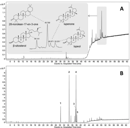
It was possible to identify 39 substances (Table 1). The pentacyclic triterpenes lupeol (16.32%), lupenone (12.75%) and 28-norolean-17-en-3-one (10.69%) as well as their precursor squalene (9.69%) are among the major compounds accounting for 49.45% of the total area of the chromatogram. Lupeol is a triterpene found in various fruits and vegetables such as olives, mango and fig, and in many medicinal herbs (Saleem et al., 2004). This substance was also identified in a hexane extract of Schinus molle L. together with lupenone, which was the main component (Batista et al., 2016). The literature describes the presence of triterpene 28-norolean-17-en-3-one in plants of the genus Pistacia, which belongs to the same family as S. terebinthifolia (Anacardiaceae) (Stern et al., 2003;Vuorinen et al. (2015)), being one of the main tri-terpene found in samples of Pistacia lentiscus (Assimopoulou and Papageorgiou, 2005).
As the residues are fragmented and exposed to the environment, smaller peaks related to monoterpenes (7.98%) and sesquiterpenes (16.06%) were expected due to the volatilization of these compounds. However, it was possible to identify some terpenes produced by this species such as the monoterpenes p-cymene (3.06%), limonene (1.92%), 3-carene (1.65%), α-pinene (0.96%) and the sesquiterpenes spatulenol (2.22%), β-caryophyllene (2.08%), caryophyllene oxide (1.66%) and α-eudesmol (1.36%). These substances were described as components of the essential oil of S. terebinthifolia (Lloyd et al., 1977; Bendaoud et al., 2010;Carvalho et al., 2013;Ennigrou et al., 2017). It has been reported that essential oils obtained from leaves of S. ter-ebinthifolia, collected at different sites, have monoterpenes in their composition, mainly α-pinene, limonene and p-cymene (Barbosa et al., 2007). Regarding the volatile compounds of the green and mature fruits of this plantSchimitberger et al. (2018)identified α-pinene, δ-3-carene and limonene as the predominant substances and proposed them as chemical markers of the species.
3.2.2. Dichloromethane and dichloromethane esterified fractions
The GC–MS chromatogram of the dichloromethane fraction shows the predominance of peaks in the triterpene region between 45 and 54 min (Fig. 2A). The presence of the steroid β-sitosterol (11.29%) to-gether with 28-norolean-17-en-3-one (29.18%), lupenone (8.04%) and lupeol (9.08%) were observed in the dichloromethane fraction. Re-garding the esterified dichloromethane fraction (Fig. 2B), a significant difference between the both chromatographic profiles could be verified. In the esterified sample, intense peaks related to fatty acid methyl esters appear in the region between 30 and 38 min, with a decrease of the peaks of the triterpenes. The presence of these signals demonstrates the high concentration of fatty acids in comparison with triterpenes and indicates a satisfactory esterification process was, allowing the identi-fication of these acids in the form of their methyl esters (Table 2). Table 1
Chemical composition of the hexane fraction of residues.
No. Retention
time (min) Compound Area (%) RI
a Monoterpenes 1 9.359 α-pinene 0.96 928 2 11.621 3-carene 1.65 1004 3 12.047 p-cymene 3.06 1018 4 12.182 limonene 1.92 1023 5 16.976 α-terpineol 0.19 1185 6 20.022 thymol 0.20 1295 Sesquiterpenes 7 21.039 δ-elemene 0.22 1333 8 22.076 α-copaene 0.34 1373 9 22.466 β-elemene 0.95 1388 10 23.218 β-caryophyllene 2.08 1417 11 23.503 γ-elemene 0.24 1429 12 23.706 aromadendrene 0.32 1437 13 24.064 humulene 0.14 1452 14 24.593 γ-muurolene 0.64 1473 15 24.743 germacrene D 0.23 1479 16 24.878 β-selinene 0.85 1484 17 25.086 α-selinene 1.07 1493 18 25.164 α-muurolene 0.23 1496 19 25.517 γ-cadinene 0.39 1511 20 25.709 δ-cadinene 0.88 1519 21 26.310 elemol 0.23 1545 22 27.032 spathulenol 2.22 1576 23 27.187 caryophyllene oxide 1.66 1582 24 27.743 rosifoliol 0.50 1606 25 28.256 γ-eudesmol 0.32 1629 26 28.438 α-muurolol 0.42 1638 27 28.687 β-eudesmol 0.75 1649 28 28.754 α-eudesmol 1.36 1652 Triterpenes/Steroids/Others 29 37.736 phytol 0.54 2102 30 44.803 squalene 9.69 2816 31 45.145 octacosane 8.20 2874 32 46.889 α-tocoferol 4.93 3136 33 48.814 β-sitosterol 3.63 3346 34 49.078 olean-12-en-3-one 1.82 3373 35 49.416 28-norolean-17-en-3-one 10.69 3405 36 49.592 lupenone 12.75 3417 37 49.867 lupeol 16.32 3437 38 51.097 lupeol acetate 4.10 3524 39 52.041 ursonic aldehyde 3-acetate 2.19 3592
a Retention index obtained with reference to a standard of n-alkanes using an HP-5MS column.
Table 2
Main compounds of the esterified dichloromethane fraction.
No. Retention time (min) Area (%) Compound RIa
1 30.352 8.74 methyl tetradecanoate 1725 2 34.472 26.51 methyl hexadecanoate 1928 3 37.658 7.69 methyl linoleate 2098 4 37.783 31.21 methyl linolenate 2104 5 38.218 4.60 methyl stearate 2129
a Retention index obtained with reference to a standard of n-alkanes using an HP-5MS column.
Fig. 1. Chromatogram of the hexane fraction of residues.
It was possible to identify esters of myristic, palmitic, linoleic, li-nolenic and stearic acids, with esters of lili-nolenic and palmitic acids accounting for 57.72% of the total area. Recently, the composition of fatty acids in S. terebinthifolia Raddi leaves and twigs was determined by gas chromatography. Thirteen fatty acids were identified, and α-lino-lenic, palmitic and linoleic acids were the main components (Ennigrou et al., 2018).
3.3. ESI(-)FT-ICR MS and ESI(-)FT-ICR MS/MS analyses 3.3.1. Dichloromethane fraction of residues
In the mass spectra obtained (Fig. 3), the regions with the most intense signals are related to the main classes of compounds identified in the dichloromethane fraction, acid triterpenes (m/z 453 to 503) and fatty acids (m/z 227 to 339). Among the acid triterpenes, two of the main ones found in S. terebinthifolia were identified: masticadienoic acid (m/z 453) and schinol (m/z 455) (Jain et al., 1995;Morais et al., 2014). In addition, nine other ions had molecular formulas associated with acid triterpenes being one of them in glycoside form. These results indicate the presence of substances not yet isolated from this plant (Table 3). The analysis allowed the identification of other fatty acids beyond the five identified by GC–MS.
These results are in agreement withda Silva et al. (2017a) who identified by ESI(-)-TOF MS the fatty acids (myristic, palmitic, stearic, among others) and acid triterpenes when different fractions of ethanolic extracts of fruits and leaves of S. terebinthifolia were analyzed. When studying antifungal compounds of the hydroethanolic extract of the leaves of this plant,Johann et al. (2010)isolated the schinol from the hexane fraction and identified it by ESI-MS and NMR. Years laterVieira et al. (2015)isolated the 3β-masticadienolic (schinol) and masticadie-nonic acids when performing the fractionation of a dichloromethane extract of the fruits.
3.3.2. Polar extracts
After the FT-ICR MS analysis, mass spectra of the MF, HEE and FPE were obtained (Fig. 4). The most intense signals were m/z 183 (methyl gallate), m/z 353 (quinic acid hexoside) and m/z 325 (galloylshikimic acid), for MF, HEE and FPE, respectively. The chemical composition of polar extracts analyzed by ESI(-)FT-ICR MS is presented in Table 4, where the substances or classes were proposed based on the generated ions, molecular formula, double bound equivalent (DBE) and literature data regarding the identification of the substances in the species, gender or family.
The literature describes the identification of gallic acid and its de-rivatives in the leaves and fruits of S. terebinthifolia (Ceruks et al., 2007; Santana et al., 2012;Feuereisen et al., 2014;Camaroti et al., 2018). The ion of the deprotonated molecule at m/z 169, its chloride adduct (m/z 205) and its dimer [2M-H]−at m/z 339 confirm the presence of this substance. In relation to their derivatives, the ions at m/z 183 and 197 suggest the presence of the methyl and ethyl gallates, respectively, in addition to their adducts with chloride (m/z 219 and 233). Beyond gallic acid, shikimic acid (m/z 173) was also identified in the depro-tonated form. It is one of the precursors of the phenolic compound biosynthesis (Dewick, 2009).
Some sugars were identified in the samples as disaccharides (m/z 341), monosaccharide (m/z 179) and their adducts with chloride (m/z 215 and 377), mainly in the fruit peels. The disaccharide was also de-tected clustering with water (m/z 359).
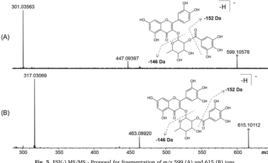
The presence of flavonoids was detected at m/z 301, 317, 433, 447, 449, 463, 477, 479, and 493. Some of them have been found in S. terebinthifolia, such as quercetin (m/z 301), quercitrin (m/z 447), myr-icetin (m/z 317) and myricitrin (m/z 463) (Ceruks et al., 2007; Carvalho et al., 2013). Myricitrin was found to form a cluster with the chloride anion (m/z 499) and in a more complex structure at m/z 615. Likewise, quercitrin was also detected as part of another structure, at m/z 599. ESI(-) MS/MS experiments were performed to aid in the identification of these compounds formed by these flavonoids (Fig. 5). The ions of the deprotonated molecules at m/z 599 and 615 were identified as quercitrin O-gallate and myricitrin O-gallate. The two structures lose the galloyl fraction (−152 Da) to give the fragments m/z 447 (quercitrin) and 463 (myricitrin), which, with subsequent loss of the sugar portion (−146 Da), do originate m/z 301 and 317 signals Fig. 3. ESI(-)FT-ICR mass spectra of dichloromethane fraction of the residues.
Table 3
Substances proposed from ESI(-)FT-ICR MS data for the dichloromethane fraction of the residues.
[M-H]−(m/z) MS/MS
fragments Molecularformula [M-H]− Error (ppm) DBE a Proposed substance 227.20176 – C14H27O2 −0.46 1 myristic acid 241.21746 – C15H29O2 −0.63 1 pentadecanoic acid 255.23300 – C16H31O2 −0.18 1 palmitic acid 269.24865 – C17H33O2 −0.18 1 heptadecanoic acid 277.21739 – C18H29O2 −0.32 4 linolenic acid 279.23302 – C18H31O2 −0.22 3 linoleic acid 281.24870 – C18H33O2 −0.34 2 oleic acid 283.26434 – C18H35O2 −0.31 1 stearic acid 311.29568 – C20H39O2 −0.41 1 eicosanoic acid 339.32691 – C22H43O2 −0.15 1 behenic acid 453.33722 407 C30H45O3 0.45 8 masticadienoic acid 455.35305 173 C30H47O3 0.04 7 schinol 467.31677 423, 407 C30H43O4 −0.18 9 triterpene acid 469.33235 439, 425, 409, 369 C30H45O4 −0.03 8 triterpene acid 471.34808 – C30H47O4 −0.20 7 triterpene acid 483.31170 – C30H43O5 −0.20 9 triterpene acid 485.32732 455, 423, 387 C30H45O5 −0.14 8 triterpene acid 487.34299 – C30H47O5 −0.19 7 triterpene acid 501.32231 – C30H45O6 −0.30 8 triterpene acid 503.33799 – C30H47O6 −0.35 7 triterpene acid 633.40121 453, 179 C36H57O9 −0.63 8 triterpene glycoside a Double bound equivalente (DBE).
characteristic of the aglycones quercetin and myricetin, respectively. Similar compounds were identified in fruits of Rhus coriaria L. (Anacardiaceae) (Abu-Reidah et al., 2015). As far as we know, the quercitrin derivative is reported here for the first time in S. ter-ebinthifolia.
Signals at m/z 537 and 541 related to biflavonoids have been identified. They are agathisflavone or its amentoflavone isomer for m/z 537 and tetrahydroamentoflavone or its isomer (tetrahydrorobusta-flavone) at m/z 541. The literature describes the presence of these substances in the fruits of S. terebinthifolia (Feuereisen et al., 2017; Muhs et al., 2017).
The polar extracts were abundant in gallotannins and these sub-stances were responsible for some of the most intense signals of the mass spectra. The ion at m/z 331 was identified as galloylglucose. The major fragments formed from MS/MS (Table 4) were related to two losses of two formaldehyde moieties [M-H-60]− and [M-H-60-60]− from glucose to give the ions m/z 271 and 211, respectively (Tan et al., 2011). Loss of glucose from m/z 331 [M-H-162]−to originate the ion at m/z 169. The ion m/z 483 (digalloylglucose) suffered decarboxylation [M-H-44]−(m/z 439) and showed loss of a galloyl fraction (−152 Da) and of gallic acid (−170 Da) to give the signals m/z 331 and 313 re-spectively. The ion at m/z 635 lost galloyl [M-H-152]−(m/z 483) and gallic acid [M-H-170]− (m/z 465) and has been identified as tri-galloylglucose. The ion at m/z 939 also loses galloyl (−152 Da) and gallic acid (−170 Da) to give the ions m/z 787 and 769, respectively. A further loss of these structures can also be verified with the signals m/z 635 and 617. The fragments shown correspond to the penta-galloylglucose molecule (Fig. 1, Supporting Information).
Collision induced dissociation (CID) of the ion m/z 325 showed the presence of a fragment at m/z 169 for the deprotonated gallic acid as a result of the loss of a shikimate residue (−156 Da). Based on this in-formation, galloylshikimic acid was proposed for the compound. For the ion m/z 477, digalloylshikimic acid was attributed to it as it pro-vided the fragment at m/z 325 originated from the loss of galloyl (−152 Da) and the m/z 169 fragment. The m/z 629 ion showed the signals 477 and 325 generated by the consecutive loss of galloyl units. This ion was attributed to trigalloylshikimic acid (Fig. 2, Supporting Information). Gallotannins, galloylglucose and galloylshikimic acids compounds are described as metabolites identified in S. terebinthifolia (Cavalher-Machado et al., 2008;Feuereisen et al., 2014).
The deprotonated molecules in m/z 335 and 349 showed loss of a
galloyl fraction (−152 Da) to give the m/z 183 and 197 fragments, respectively. The compounds were characterized as methyl digallate and ethyl digallate. The ions of m/z 343 and 495 were identified as mono- and digalloylquinic acids due to sequential loss of galloyl moi-eties (−152 Da) and the formation of ions m/z 191 (deprotonated quinic acid) and m/z 169 (deprotonated gallic acid). In the fragmen-tation of the m/z 353 ion signals were formed in m/z 179 for the loss of a fraction of the quinic acid (−174 Da) and in m/z 173 for the for-mation of that acid with its subsequent dehydration. These fragments suggest that this substance is the quinic acid hexoside.
The presence of methyl gallate (m/z 183) in the methanolic fraction and of ethyl gallate (m/z 197) in the hydroethanolic extracts was ob-served. It was suspected that the extractive solvent had reacted with some substance present in the sample to furnish the gallates in question. Thus, extracts of the residues were prepared with different solvents (methanol, ethanol, water and acetone) and analyzed by the same technique (ESI(-)FT-ICR MS) to verify the appearance of the corre-sponding gallate signals (Fig. 6).
ESI(-)FT-ICR MS showed the presence of methyl galate (m/z 183) in the methanolic extract, whereas the ethyl gallate (m/z 197) was found only in the ethanolic extract. It was observed the absence of these ions in the other extracts indicating the interference of the extracting solvent in the production of these gallates. Studies in which methanol was used as the extracting solvent described the identification of methyl gallate (Cavalher-Machado et al., 2008; Rosas et al., 2015; da Silva et al., 2017b). In contrast, those using ethanol as the extracting solvent de-scribed ethyl gallate (Bulla et al., 2015;da Silva et al., 2017a). There are also papers that report the two substances (Ceruks et al., 2007; Santana et al., 2012), but the plant extract had contact with both the solvents, one in the extraction process and the other in the purification step. The literature has described the methanolysis reaction, when gallotannins with a higher degree of galloylation are treated with me-thanol to give products such as methyl gallate, methyl digallate and pentagalloylglucose. This reaction also assists in the quantification by HPLC of hydrolysable tannins present in foods (Chen and Hagerman, 2004;Newsome et al., 2016). A further indication of the solvent effect was the presence of methyl digallate (m/z 335) only in the methanolic fraction and ethyl digallate (m/z 349) in the hydroethanolic extract (Table 4). Therefore, it is possible to suppose that the formation of these substances occurs during the preparation of extracts since hydrolysable tannins under heating (for example, during extract concentration) can Fig. 4. ESI(-)FT-ICR mass spectra: (A) MF, (B) HEE, (C) FPE.
suffer solvolysis. TheFig. 7shows the reaction between the solvent and the hydrolysable tannins present in the extract. When performing the nucleophilic attack (1) the solvent will induce the production of methyl or ethyl digallate. Another possibility would be the reaction (2) pro-ducing methyl or ethyl gallate.
3.4. Antimicrobial activity
The results of the antimicrobial activity are shown inTable 5. The extracts MF and HEE were the most active, mainly against Gram-posi-tive bacteria. The higher sensitivity of these strains may be related to the difference in the constitution and arrangement of the cell
membrane of these microorganisms in relation to the Gram-negative (Kołodziejczyk et al., 2013). The most susceptible strains were those of S. aureus with the two extracts presenting MIC 0.6–0.9 mg/mL and MBC from 1.8 mg/mL onwards. For strains 10A and ATCC 29213 the MBC was not reached at the concentrations tested. These extracts showed activity against strains of E. faecium and E. faecalis with MIC values 1.20–1.50 mg/mL and MBC 1.80–2.70 mg/mL for MF and MIC 1.20–2.10 mg/mL for HEE, in addition to inhibiting the growth of one A. baumannii strain (30B) and three multidrug-resistant P. aeruginosa strains. Although FPE, DF and HF had no bactericidal effect at the concentrations tested, FPE showed inhibitory activity against S. aureus (MIC 1.20–1.80 mg/mL), E. faecium (101E), E. faecalis (ATCC 29212) Table 4
Substances proposed from ESI(-)FT-ICR MS data for polar extracts.
[M-H]−(m/z) MS/MS fragments Molecular formula
[M-H]− Error (ppm) DBE
a Proposed substance MFb HEEc FPEd
169.01428 – C7H5O5 −0.20 5 gallic acid + + +
173.04559 – C7H9O5 −0.26 3 shikimic acid + + +
179.05609 – C6H11O6 0.09 1 monosaccharide – – +
183.02993 168 C8H7O5 −0.19 5 methyl gallate + – –
197.04559 169, 168 C9H9O5 −0.21 5 ethyl gallate – + +
204.99097 169 C7H6ClO5 −0.20 5 gallic acid chloride cluster + – +
215.03284 179 C6H12ClO6 −0.23 1 monosaccharide chloride cluster + + +
219.00666 183 C8H8ClO5 −0.08 5 methyl gallate chloride cluster + – –
233.02229 197 C9H10ClO5 −0.28 5 ethyl gallate chloride cluster – + +
301.03530 – C15H9O7 0.25 11 quercetin + + – 317.03055 – C15H9O8 −0.81 11 myricetin + – – 321.02537 169 C14H9O9 −0.51 10 digallic acid + + + 325.05661 169 C14H13O9 −0.33 8 galloylshikimic acid + + + 331.06723 271, 241, 211, 169 C13H15O10 −0.47 6 galloylglucose + + + 335.04098 183 C15H11O9 −0.38 10 methyl digallate + – –
339.03596 169 C14H11O10 −0.57 9 gallic acid cluster + + +
341.10920 – C12H21O11 −0.77 2 disaccharide – + –
343.06722 191, 173, 169 C14H15O10 −0.44 7 galloylquinic acid – + +
349.05666 197 C16H13O9 −0.43 10 ethyl digallate – + +
353.10913 179, 173 C13H21O11 −0.55 3 quinic acid hexoside + + +
359.11972 179, 161 C12H23O12 −0.61 1 disaccharide / H2O – + +
367.04392 331 C13H16ClO10 −0.47 5 galloylglucose chloride cluster + – +
377.08584 341, 215, 179 C12H22ClO11 −0.60 1 disaccharide chloride cluster + + +
433.07793 – C20H17O11 −0.67 12 flavonoid + – – 447.09364 301, 273, 271, 179 C21H19O11 −0.79 12 quercitrin + – – 449.07280 – C20H17O12 −0.56 12 flavonoid + – – 453.33762 407 C30H45O3 −0.45 8 masticadienoic acid – + + 455.35321 173 C30H47O3 −0.30 7 schinol – + + 463.08855 317, 179 C21H19O12 −0.76 12 myricitrin + + + 469.33231 439, 425, 409, 369 C30H45O4 0.05 8 triterpene acid – + + 471.34861 – C30H47O4 −1.33 7 triterpene acid – + – 477.06775 325, 313, 289, 263, 169 C21H17O13 −0.59 13 digalloylshikimic acid – + + 477.10413 – C22H21O12 −0.59 12 flavonoid + + + 479.08363 – C21H19O13 −1.07 12 flavonoid + + – 483.07835 439, 331, 313, 271, 169 C20H19O14 −0.67 11 digalloylglucose + + + 485.32768 455, 423, 387 C30H45O5 −0.89 8 triterpene acid – + – 493.06275 – C21H17O14 −0.76 13 flavonoid + + – 495.07832 343, 191, 169 C21H19O14 −0.59 12 digalloylquinic acid + + + 499.06536 463 C21H20ClO12 −0.96 11 myricitrin chloride cluster + + – 501.32251 – C30H45O6 −0.69 8 triterpene acid – + – 537.08305 443, 375 C30H17O10 −0.61 22 agathisflavone/ amentoflavone – + + 541.11446 415, 389 C30H21O10 −0.81 20 tetrahydroamentoflavone/ tetrahydrorobustaflavone – + + 599.10484 447, 301 C28H23O15 −1.00 17 quercitrin O-gallate + + – 615.09980 463, 317 C28H23O16 −1.04 17 myricitrin O-gallate + + – 629,07902 477, 325 C28H21O17 −0,96 18 trigalloylshikimic acid – – + 633.40135 453, 179 C36H57O9 −0.86 8 triterpene glycoside – + + 635.08929 483, 465 C27H23O18 −0.48 16 trigalloylglucose + + + 635.41745 455, 179 C36H59O9 −1.57 7 triterpene glycoside – – + 649.39632 469, 179 C36H57O10 −0.92 8 triterpene glycoside – – + 939.11240 787, 769, 635, 617 C41H31O26 −1.59 26 pentagalloylglucose + + – (+) - Substance present. (-) - Substance absent.
a Double bound equivalent (DBE). b Methanolic fraction (MF). c Hydroethanolic extract (HEE). dFruit peels extract (FPE).
and A. baumannii (101B). The activity against S. aureus is in agreement withMuhs et al. (2017)who verified that one of the fractions of the methanolic extract of the fruits presented inhibitory activity against MRSA and demonstrated a significant reduction in the dermonecrosis caused by this microorganism. The DF was active against two strains of S. aureus and was the only extract that inhibited all strains of A. bau-mannii (MIC 2.40 mg/mL). This activity may be related to the acid
triterpenes identified in the sample. When studying natural Medi-terranean plants, Karygianni et al. (2014)verified that extracts con-taining acid triterpenes were active against oral bacteria, especially Gram-negative bacteria. The HF also showed no bactericidal activity but inhibited the growth of all S. aureus strains (MIC 2.40–2.70 mg/ mL). In general, the polar extracts were more active, and this higher activity may be related to the presence of phenolic compounds, because Fig. 5. ESI(-) MS/MS - Proposal for fragmentation of m/z 599 (A) and 615 (B) ions.
Fig. 6. Mass spectra of S. terebinthifolia extracts prepared with methanol, ethanol, water and acetone.
substances belonging to this class such as gallic acid, methyl gallate, galloylglucose compounds and flavonoids have shown antimicrobial activity (Kang et al., 2008;Cushnie and Lamb, 2011;Engels et al., 2012; Farhadi et al., 2018). Phenolic compounds are also known to have strong antioxidant activity (Cai et al., 2004). Phenolic substances identified in extracts of S. terebinthifolia were considered responsible for the antioxidant activity observed (El-Massry et al., 2009; Bernardes et al., 2014;Uliana et al., 2016;da Silva et al., 2017b;Tlili et al., 2018). Our results agree with the literature that describes the antimicrobial potential of S. terebinthifolia. Assays performed with alcoholic extracts of leaves against S. aureus obtained MIC values close to those found in the present study, 0.75 mg/mL (El-Massry et al., 2009) and 0.50 mg/mL (Uliana et al., 2016). The differences in the results presented in these works may be related to the variation of the chemical composition of each of the extracts. In addition to activity against S. aureus, the polar extracts were also active against E. coli, P. aeruginosa, C. albicans (Guerra et al., 2000;El-Massry et al., 2009;Uliana et al., 2016) and E. faecalis (de Costa et al., 2012). In another study the antimicrobial po-tential of fruit extract against S. aureus and Bacillus cereus was related to the phenolic compounds present in the sample (Degáspari et al., 2005). In addition to the extracts, the essential oils are also described with antimicrobial activity (El-Massry et al., 2009; Cole et al., 2014; Ennigrou et al., 2018). Even a study was carried out aiming at the application of the essential oil as a biopreservative food (da Silva Dannenberg et al., 2016).
4. Conclusion
It was possible to identify different classes of substances present in the residues from the Brazilian pepper tree processing industry, mainly phenolic compounds such as gallic acid, gallotannins and flavonoids in the polar extracts and triterpenes in the apolar extracts. This work describes for the first time the occurrence of quercitrin O-gallate in the species. Extracts rich in phenolic substances showed significant activity against multidrug-resistant bacteria, mainly against MRSA and VRE. Due to the richness of these compounds, residues from the Brazilian pepper tree processing industry can be used as source of substances
with antimicrobial potential, antioxidant activity, besides the applica-tion as food preservative, being necessary more studies on the use of this material. These findings provide opportunities to explore the use of industrial by-products to reduce residues streams and recover bioactive compounds.
Conflicts of interest
The authors declare no conflict of interest related to this work.
Acknowledgments
This study was financed in part by the Coordenação de Aperfeiçoamento de Pessoal de Nível Superior Brasil (CAPES) -Finance Code 001", in part by CNPq (process number 443063/2014-1) and PQ (process number 308631/2016-1). The authors extend the thanks to Foundation for Support to Research and Innovation of Espírito Santo (FAPES-83784233).
Appendix A. Supplementary data
Supplementary material related to this article can be found, in the online version, at doi:https://doi.org/10.1016/j.indcrop.2019.05.079.
References
Abu-Reidah, I.M., Ali-Shtayeh, M.S., Jamous, R.M., Arráez-Román, D., Segura-Carretero, A., 2015. HPLC-DAD-ESI-MS/MS screening of bioactive components from Rhus
cor-iaria L. (Sumac) fruits. Food Chem. 166, 179–191.https://doi.org/10.1016/j. foodchem.2014.06.011.
Adams, R.P., 2009. Identification of Essential Oil Components by Gas Chromatography/ Mass Spectrometry, 4 ed. Carol Stream: Allured Publishing Corporation, USA. ANVISA, 2011. Formulário de Fitoterápicos Farmacopeia Brasileira, 1.ed. . Available
from:http://www.anvisa.gov.br/hotsite/farmacopeiabrasileira/conteudo/ Formulario_de_Fitoterapicos_da_Farmacopeia_Brasileira.pdf(accessed 04.05.2017. Assimopoulou, A.N., Papageorgiou, V.P., 2005. GC-MS analysis of penta- and tetra-cyclic
triterpenes from resins of Pistacia species. Part I. Pistacia lentiscus varChia. Biomed. Chromatogr 19, 285–311.https://doi.org/10.1002/bmc.454.
Barbieri, D.S.V., Tonial, F., Lopez, P.V.A., Sales Maia, B.H.L.N., Santos, G.D., Ribas, M.O., Glienke, C., Vicente, V.A., 2014. Antiadherent activity of Schinus terebinthifolius and
Table 5
Minimum inhibitory concentration (MIC) and minimum bactericidal concentration (MBC) of residues extracts from the Brazilian pepper tree processing industry.
MFb HEEc FPEd DFe HFf
Species Strains MICa MBCa MICa MBCa MICa MBCa MICa MBCa MICa MBCa
S. aureus 10A 0.90 > 2.70 0.90 > 2.70 1.50 > 2.70 2.40 > 2.70 2.40 > 2.70 23A 0.90 1.80 0.60 1.80 1.20 > 2.70 > 2.70 > 2.70 2.70 > 2.70 35A 0.60 1.80 0.60 1.80 1.50 > 2.70 > 2.70 > 2.70 2.40 > 2.70 67A 0.60 2.10 0.90 1.80 1.50 > 2.70 > 2.70 > 2.70 2.40 > 2.70 114A 0.60 2.70 0.60 1.80 1.20 > 2.70 > 2.70 > 2.70 2.40 > 2.70 ATCC 29213 0.60 > 2.70 0.90 > 2.70 1.80 > 2.70 1.80 > 2.70 2.40 > 2.70 E. faecium 70E 1.50 2.10 2.10 > 2.70 > 2.70 > 2.70 > 2.70 > 2.70 > 2.70 > 2.70 101E 1.50 2.70 1.80 > 2.70 2.10 > 2.70 > 2.70 > 2.70 > 2.70 > 2.70 E. faecalis 1277 1.50 2.70 1.80 > 2.70 > 2.70 > 2.70 > 2.70 > 2.70 > 2.70 > 2.70 6885 1.20 2.70 2.10 > 2.70 > 2.70 > 2.70 > 2.70 > 2.70 > 2.70 > 2.70 168557 1.20 2.70 1.80 > 2.70 > 2.70 > 2.70 > 2.70 > 2.70 > 2.70 > 2.70 ATCC 29212 1.20 1.80 1.20 2.70 1.50 > 2.70 2.40 > 2.70 > 2.70 > 2.70 A. baumannii 30B > 2.70 > 2.70 > 2.70 > 2.70 > 2.70 > 2.70 2.40 > 2.70 > 2.70 > 2.70 101B 1.20 > 2.70 1.80 > 2.70 2.70 > 2.70 2.40 > 2.70 > 2.70 > 2.70 ATCC 19606 > 2.70 > 2.70 > 2.70 > 2.70 > 2.70 > 2.70 2.40 > 2.70 > 2.70 > 2.70 P. aeruginosa 34B 2.40 > 2.70 2.70 > 2.70 > 2.70 > 2.70 > 2.70 > 2.70 > 2.70 > 2.70 39B 2.40 > 2.70 2.70 > 2.70 > 2.70 > 2.70 > 2.70 > 2.70 > 2.70 > 2.70 121B 2.40 > 2.70 2.40 > 2.70 > 2.70 > 2.70 > 2.70 > 2.70 > 2.70 > 2.70 ATCC 27853 > 2.70 > 2.70 > 2.70 > 2.70 > 2.70 > 2.70 > 2.70 > 2.70 > 2.70 > 2.70 a Values expressed in mg/mL. b Methanolic fraction (MF). c Hydroethanolic extract (HEE). dFruit peels extract (FPE). e Dichloromethane fraction (DF). f Hexane fraction (HF).
Croton urucurana extracts on in vitro biofilm formation of Candida albicans and Streptococcus mutans. Arch. Oral Biol. 59, 887–896.https://doi.org/10.1016/j. archoralbio.2014.05.006.
Barbosa, L.C.A., Demuner, A.J., Clemente, A.D., De Paula, V.F., Ismail, F.M.D., 2007. Seasonal variation in the composition of volatile oils from Schinus terebinthifolius Raddi. Quim. Nova 30, 1959–1965.
https://doi.org/10.1590/S0100-40422007000800030.
Batista, L.C.S.O., Cid, Y.P., De Almeida, A.P., Prudêncio, E.R., Riger, C.J., De Souza, M.A.A., Coumendouros, K., Chaves, D.S.A., 2016. In vitro efficacy of essential oils and extracts of Schinus molle L. Against Ctenocephalides felis felis. Parasitology 143, 627–638.https://doi.org/10.1017/S0031182016000081.
Bendaoud, H., Romdhane, M., Souchard, J.P., Cazaux, S., Bouajila, J., 2010. Chemical composition and anticancer and antioxidant activities of Schinus molle L. And Schinus
terebinthifolius Raddi berries essential oils. J. Food Sci. 75, 466–472.https://doi.org/ 10.1111/j.1750-3841.2010.01711.x.
Bernardes, N.R., Heggdorne-Araújo, M., Borges, I.F.J.C., Almeida, F.M., Amaral, E.P., Lasunskaia, E.B., Muzitano, M.F., Oliveira, D.B., 2014. Nitric oxide production, in-hibitory, antioxidant and antimycobacterial activities of the fruits extract and fla-vonoid content of Schinus terebinthifolius. Braz. J. Pharmacogn. 24, 644–650.
https://doi.org/10.1016/j.bjp.2014.10.012.
Braga, F.G., Bouzada, M.L.M., Fabri, R.L., de, M., Moreira, F.O., Scio, E., Coimbra, E.S., 2007. Antileishmanial and antifungal activity of plants used in traditional medicine in Brazil. J. Ethnopharmacol. 111, 396–402.https://doi.org/10.1016/j.jep.2006.12. 006.
Brazilian Ministry of Health, 2017. Brasília. Relação Nacional de Medicamentos Essenciais: RENAME 2017. pp. 210.
Bulla, M.K., Hernandes, L., Baesso, M.L., Nogueira, A.C., Bento, A.C., Bortoluzzi, B.B., Serra, L.Z., Cortez, D.A.G., 2015. Evaluation of photoprotective potential and per-cutaneous penetration by photoacoustic spectroscopy of the Schinus terebinthifolius Raddi extract. Photochem. Photobiol. 91, 558–566.https://doi.org/10.1111/php. 12419.
Cai, Y., Luo, Q., Sun, M., Corke, H., 2004. Antioxidant activity and phenolic compounds of 112 traditional Chinese medicinal plants associated with anticancer. Life Sci. 74, 2157–2184.https://doi.org/10.1016/j.lfs.2003.09.047.
Camaroti, J.R.S.L., de Almeida, W.A., do Rego Belmonte, B., de Oliveira, A.P.S., de Albuquerque Lima, T., Ferreira, M.R.A., Paiva, P.M.G., Soares, L.A.L., Pontual, E.V., Napoleão, T.H., 2018. Sitophilus zeamais adults have survival and nutrition affected by Schinus terebinthifolius leaf extract and its lectin (SteLL). Ind. Crops Prod. 116, 81–89.https://doi.org/10.1016/j.indcrop.2018.02.065.
Carvalho, M.G., Melo, A.G.N., Aragão, C.F.S., Raffin, F.N., Moura, T.F.A.L., 2013. Schinus
terebinthifolius Raddi: chemical composition, biological properties and toxicity. Rev.
Bras. Pl. Med. 15, 158–169.https://doi.org/10.1590/S1516-05722013000100022. Cavalher-Machado, S.C., Rosas, E.C., de Brito, F.A., Heringe, A.P., de Oliveira, R.R.,
Kaplan, M.A.C., Figueiredo, M.R., Henriques, M.G.M.O., 2008. The anti-allergic ac-tivity of the acetate fraction of Schinus terebinthifolius leaves in IgE induced mice paw edema and pleurisy. Int. Immunopharmacol. 8, 1552–1560.https://doi.org/10. 1016/j.intimp.2008.06.012.
Ceruks, M., Romoff, P., Fávero, O.A., Lago, J.H.G., 2007. Constituíntes fenólicos polares de Schinus terebinthifolius Raddi (Anacardiaceae). Quim. Nova 30, 597–599.https:// doi.org/10.1590/S0100-40422007000300018.
Chemistry WebBookhttps://webbook.nist.gov/chemistry/.
Chen, Y., Hagerman, A.E., 2004. Characterization of soluble non-covalent complexes between bovine serum albumin and β-1, 2, 3, 4, 6-penta-O-galloyl-D-glucopyranose by MALDI-TOF MS. J. Agric. Food Chem. 52, 4008–4011.
CLSI, 2017. Performance Standards for Antimicrobial Susceptibility Testing, 27th edition. CLSI M100. Clinical and Laboratory Standards Institute, 950 West Valley Road, Suite 2500, Wayne, Pennsylvania 19087 USA.
Cole, E.R., dos Santos, R.B., Lacerda Júnior, V., Martins, J.D.L., Greco, S.J., Cunha Neto, A., 2014. Chemical composition of essential oil from ripe fruit of Schinus
ter-ebinthifolius Raddi and evaluation of its activity against wild strains of hospital origin.
Braz. J. Microbiol. 45, 821–828. https://doi.org/10.1590/S1517-83822014000300009.
Cosgrove, S.E., 2006. The relationship between antimicrobial resistance and patient outcomes: mortality, length of hospital stay, and health care costs. Clin. Infect. Dis. (Suppl 2), 82–89.
de Costa, E.M.M.B., de Evangelista, A.P.A., de Medeiros, A.C.D., Dametto, F.R., de Carvalho, R.A., 2012. In vitro evaluation of the root canal cleaning ability of plant extracts and their antimicrobial action. Braz. Oral Res. 26, 215–221.https://doi.org/ 10.1590/S1806-83242012000300006.
Cushnie, T.P.T., Lamb, A.J., 2011. Recent advances in understanding the antibacterial properties of flavonoids. Int. J. Antimicrob. Agents 38, 99–107.https://doi.org/10. 1016/j.ijantimicag.2011.02.014.
da Silva Dannenberg, G., Funck, G.D., Mattei, F.J., da Silva, W.P., Fiorentini, Â.M., 2016. Antimicrobial and antioxidant activity of essential oil from pink pepper tree (Schinus
terebinthifolius Raddi) in vitro and in cheese experimentally contaminated with Listeria monocytogenes. Innov. Food Sci. Emerg. Technol. 36, 120–127.https://doi.org/10. 1016/j.ifset.2016.06.009.
da Silva, J.H.S., Simas, N.K., Alviano, C.S., Alviano, D.S., Ventura, J.A., de Lima, E.J., Seabra, S.H., Kuster, R.M., 2017a. Anti-Escherichia coli activity of extracts from
Schinus terebinthifolius fruits and leaves. Nat. Prod. Res. 32, 1365–1368.https://doi. org/10.1080/14786419.2017.1344657.
da Silva, M.M., Iriguchi, E.K.K., Kassuya, C.A.L., do Vieira, M.C., Foglio, M.A., de Carvalho, J.E., Ruiz, A.L.T.G., de Souza, K.P., Formagio, A.S.N., 2017b. Schinus
ter-ebinthifolius: Phenolic constituents and in vitro antioxidant, antiproliferative and in vivo anti-inflammatory activities. Braz. J. Pharmacogn 27, 445–452.https://doi.org/ 10.1016/j.bjp.2016.12.007.
de Kraker, M.E.A., Davey, P.G., Grundmann, H., on behalf of the BURDEN study group, 2011. Mortality and hospital stay associated with resistant Staphylococcus aureus and
Escherichia coli bacteremia: estimating the burden of antibiotic resistance in Europe.
PLoS Med. 8 (10), 1–8.
Degáspari, C.H., Wasczynskyj, N., Prado, M.R.M., 2005. Atividade antimicrobiana de
Schinus terebenthifolius Raddi. Ciênc. Agrotec. 29, 617–622.
Dewick, P.M., 2009. Medicinal Natural Products: a Biosynthetic Approach. John Wiley & Sons, New York.
El-Massry, K.F., El-Ghorab, A.H., Shaaban, H.A., Shibamoto, T., 2009. Chemical compo-sitions and antioxidant/antimicrobial activities of various samples prepared from
Schinus terebinthifolius leaves cultivated in Egypt. J. Agric. Food Chem. 57,
5265–5270.https://doi.org/10.1021/jf900638c.
Engels, C., Gänzle, M.G., Schieber, A., 2012. Fast LC–MS analysis of gallotannins from mango (Mangifera indica L.) kernels and effects of methanolysis on their antibacterial activity and iron binding capacity. Food Res. Int. 45, 422–426.https://doi.org/10. 1016/j.foodres.2011.11.008.
Ennigrou, A., Casabianca, H., Laarif, A., Hanchi, B., Hosni, K., 2017. Maturation-related changes in phytochemicals and biological activities of the Brazilian pepper tree (Schinus terebinthifolius Raddi) fruits. S. Afr. J. Bot. 108, 407–415.https://doi.org/10. 1016/j.sajb.2016.09.005.
Ennigrou, A., Casabianca, H., Vulliet, E., Hanchi, B., Hosni, K., 2018. Assessing the fatty acid, essential oil composition, their radical scavenging and antibacterial activities of
Schinus terebinthifolius Raddi leaves and twigs. J. Food Sci. Technol.https://doi.org/ 10.1007/s13197-018-3049-6.
Farhadi, F., Khameneh, B., Iranshahi, M., Iranshahy, M., 2018. Antibacterial activity of flavonoids and their structure–activity relationship: an update review. Phytother. Res. 1–28.https://doi.org/10.1002/ptr.6208.
Feuereisen, M.M., Hoppe, J., Zimmermann, B.F., Weber, F., Schulze-Kaysers, N., Schieber, A., 2014. Characterization of phenolic compounds in brazilian pepper (Schinus
ter-ebinthifolius Raddi) exocarp. J. Agric. Food Chem. 62, 6219–6226.https://doi.org/ 10.1021/jf500977d.
Feuereisen, M.M., Zimmermann, B.F., Schulze-Kaysers, N., Schieber, A., 2017. Differentiation of Brazilian peppertree (Schinus terebinthifolius Raddi) and Peruvian peppertree (Schinus molle L.) fruits by UHPLC-UV-MS analysis of their anthocyanin and biflavonoid profiles. J. Agric. Food Chem. 65, 5330–5338.https://doi.org/10. 1021/acs.jafc.7b00480.
Gomes, F.S., Procópio, T.F., Napoleão, T.H., Coelho, L.C.B.B., Paiva, P.M.G., 2013. Antimicrobial lectin from Schinus terebinthifolius leaf. J. Appl. Microbiol. 114, 672–679.https://doi.org/10.1111/jam.12086.
Guerra, M.J.M., Barreiro, M.L., Rodriguez, Z.M., Rubalcaba, Y., 2000. Actividad anti-microbiana de un extracto fluido al 80% de Schinus terebinthifolius Raddi (copal). Rev. Cuba. Plantas Med. 5, 23–25.
Jain, M.K., Yu, Bao-Zhu, Rogers, J.M., Smith, A.E., Boger, E.T.A., Ostrander, R.L., Rheingold, A.L., 1995. Specific competitive inhibitor of secreted phospholipase A2 from berries of Schinus terebinthifolius. Phytochemistry 39, 537–547.https://doi. org/10.1016/0031-9422(94)00960-2.
Johann, S., Sá, N.P., Lima, L.A.R.S., Cisalpino, P.S., Cota, B.B., Alves, T.M.A., Siqueira, E.P., Zani, C.L., 2010. Antifungal activity of schinol and a new biphenyl compound isolated from Schinus terebinthifolius against the pathogenic fungus Paracoccidioides
brasiliensis. Ann. Clin. Microbiol. Antimicrob. 9, 30. https://doi.org/10.1186/1476-0711-9-30.
Kang, M.-S., Oh, J.-S., Kang, I.-C., Hong, S.-J., Choi, C.-H., 2008. Inhibitory effect of methyl gallate and gallic acid on oral bacteria. J. Microbiol. 46, 744–750.https://doi. org/10.1007/s12275-008-0235-7.
Karygianni, L., Cecere, M., Skaltsounis, A.L., Argyropoulou, A., Hellwig, E., Aligiannis, N., Wittmer, A., Al-Ahmad, A., 2014. High-level antimicrobial efficacy of representative Mediterranean natural plant extracts against oral microorganisms. Biomed Res. Int. 2014.https://doi.org/10.1155/2014/839019.
Kołodziejczyk, K., Sójka, M., Abadias, M., Viñas, I., Guyot, S., Baron, A., 2013. Polyphenol composition, antioxidant capacity, and antimicrobial activity of the extracts obtained from industrial sour cherry pomace. Ind. Crops Prod. 51, 279–288.https://doi.org/ 10.1016/j.indcrop.2013.09.030.
Lloyd, H.A., Jaouni, T.M., Evans, S.L., Morton, J.F., 1977. Terpenes of Schinus ter-ebinthifolius. Phytochemistry 16, 1301–1302. https://doi.org/10.1016/S0031-9422(00)94384-X.
Morais, T.R., Da Costa-Silva, T.A., Tempone, A.G., Borborema, S.E.T., Scotti, M.T., De Sousa, R.M.F., Araujo, A.C.C., De Oliveira, A., De Morais, S.A.L., Sartorelli, P., Lago, J.H.G., 2014. Antiparasitic activity of natural and semi-synthetic tirucallane tri-terpenoids from Schinus terebinthifolius (Anacardiaceae): Structure/activity relation-ships. Molecules 19, 5761–5776.https://doi.org/10.3390/molecules19055761. Morton, J.F., 1978. Brazilian pepper-Its impact on people, animals and the environment.
Econ. Bot. 32, 353–359.https://doi.org/10.1007/BF02907927.
Muhs, A., Lyles, J.T., Parlet, C.P., Nelson, K., Kavanaugh, J.S., Horswill, A.R., Quave, C.L., 2017. Virulence inhibitors from Brazilian peppertree block quorum sensing and abate dermonecrosis in skin infection models. Sci. Rep. 7, 1–15.https://doi.org/10.1038/ srep42275.
Neves, E.J.M., Santos, A.M., Gomes, J.B.V., Ruas, F.G., Ventura, J.A., 2016. Cultivo da aroeira-vermelha (Schinus terebinthifolius Raddi) para produção de pimenta-rosa. Colombo: Embrapa Florestas 10 Documentos, 294.
Newsome, A.G., Li, Y., Van Breemen, R.B., 2016. Improved quantification of free and ester-bound gallic acid in foods and beverages by UHPLC-MS/MS. J. Agric. Food Chem. 64, 1326–1334.https://doi.org/10.1021/acs.jafc.5b04966.
Rosas, E.C., Correa, L.B., Pádua, T.D.A., Costa, T.E.M.M., Luiz Mazzei, J., Heringer, A.P., Bizarro, C.A., Kaplan, M.A.C., Figueiredo, M.R., Henriques, M.G., 2015. Anti-in-flammatory effect of Schinus terebinthifolius Raddi hydroalcoholic extract on neu-trophil migration in zymosan-induced arthritis. J. Ethnopharmacol. 175, 490–498.
https://doi.org/10.1016/j.jep.2015.10.014.
Saleem, M., Afaq, F., Adhami, V.M., Mukhtar, H., 2004. Lupeol modulates NF-κB and PI3K/Akt pathways and inhibits skin cancer in CD-1 mice. Oncogene 23, 5203–5214.
https://doi.org/10.1038/sj.onc.1207641.
Santana, J.S., Sartorelli, P., Lago, J.H.G., Matsuo, A.L., 2012. Isolamento e avaliação do potencial citotóxico de derivados fenólicos de Schinus terebinthifolius Raddi (Anacardiaceae). Quim. Nova 35, 2245–2248. https://doi.org/10.1590/S0100-40422012001100029.
Scheid, T., Moraes, M.S., Henriques, T.P., Paula, A., Riffel, K., Belló-klein, A., Von Poser, G.L., Ethur, E.M., Partata, W.A., 2018. Effects of methanol fraction from leaves of
Schinus terebinthifolius Raddi on nociception and spinal-cord oxidative biomarkers in
rats with neuropathic pain. Evid. Based Complement. Altern. Med.https://doi.org/ 10.1155/2018/5783412.
Schimitberger, V.M.B., Pratti, D.L., de, A., Cavalcanti, L.C., Ramalho, V.F., da Costa, A.P.F., Scherer, R., Kuster, R.M., Ramos, A.C., da Silva, A.G., 2018. Volatile com-pounds profile changes from unripe to ripe fruits of Brazilian pepper (Schinus
ter-ebinthifolia Raddi). Ind. Crops Prod. 119, 125–131.https://doi.org/10.1016/j. indcrop.2018.04.011.
Stern, B., Heron, C., Corr, L., Serpico, M., Bourriau, J., 2003. Compositional variations in aged and heated Pistacia resin found in late bronze age canaanite amphorae and bowls from Amarna, Egypt. Archaeometry 45, 457–469.https://doi.org/10.1111/ 1475-4754.00121.
Tan, H.P., Ling, S.K., Chuah, C.H., 2011. Characterisation of galloylated cyanogenic glucosides and hydrolysable tannins from leaves of Phyllagathis rotundifolia by LC-ESI-MS/MS. Phytochem. Anal. 22, 516–525.https://doi.org/10.1002/pca.1312.
The Pherobase-Database of pheromones and semiochemicalshttp://www.pherobase. com/.
Tlili, N., Yahia, Y., Feriani, A., Labidi, A., Ghazouani, L., Nasri, N., Saadaoui, E., Khaldi, A., 2018. Schinus terebinthifolius vs Schinus molle: a comparative study of the effect of species and location on the phytochemical content of fruits. Ind. Crops Prod. 122, 559–565.https://doi.org/10.1016/j.indcrop.2018.05.080.
Uliana, M.P., Fronza, M., da Silva, A.G., Vargas, T.S., de Andrade, T.U., Scherer, R., 2016. Composition and biological activity of Brazilian rose pepper (Schinus terebinthifolius Raddi) leaves. Ind. Crops Prod. 83, 235–240.https://doi.org/10.1016/j.indcrop. 2015.11.077.
Vieira, M.N., das Costa, F.N., Leitão, G.G., Garrard, I., Hewitson, P., Ignatova, S., Winterhalter, P., Jerz, G., 2015. Schinus terebinthifolius scale-up countercurrent chromatography (Part I): HIGH performance countercurrent chromatography frac-tionation of triterpene acids with off-line detection using atmospheric pressure che-mical ionization mass spectrometry. J. Chromatogr. A 1389, 39–48.https://doi.org/ 10.1016/j.chroma.2015.02.005.
Vuorinen, A., Seibert, J., Papageorgiou, V.P., Rollinger, J.M., Odermatt, A., Schuster, D., Assimopoulou, A.N., 2015. Pistacia lentiscus oleoresin: virtual screening and identi-fication of masticadienonic and isomasticadienonic acids as inhibitors of 11 β-hy-droxysteroid dehydrogenase 1. Planta Med. 81, 525–532. https://doi.org/10.1055/s-0035-1545720.
WHO - World Health Organization, 2017. Global Priority List of Antibiotic-resistant Bacteria to Guide Research, Discovery, and Development of New Antibiotics. Available from: (accessed 13.06.2017). http://www.who.int/medicines/ publications/WHO-PPL-Short_Summary_25Feb-ET_NM_WHO.pdf.