HAL Id: hal-00824737
https://hal.archives-ouvertes.fr/hal-00824737
Submitted on 22 May 2013
HAL is a multi-disciplinary open access
archive for the deposit and dissemination of
sci-entific research documents, whether they are
pub-lished or not. The documents may come from
teaching and research institutions in France or
abroad, or from public or private research centers.
L’archive ouverte pluridisciplinaire HAL, est
destinée au dépôt et à la diffusion de documents
scientifiques de niveau recherche, publiés ou non,
émanant des établissements d’enseignement et de
recherche français ou étrangers, des laboratoires
publics ou privés.
Thermal Behavior of Fluorinated Double-Walled Carbon
Nanotubes
Lyubov Gennadievna Bulusheva, Pavel Nikolaevich Gevko, Alexander
Vladimirovich Okotrub, Yuliya Vladimirovna Lavskaya, Nikolay Fedorovich
Yudanov, Lyudmila Ivanovna Yudanova, Oleg Gennadievich Abrosimov, Egor
Mikhailovich Pazhetnov, Andrey Ivanovich Boronin, Emmanuel Flahaut
To cite this version:
Lyubov Gennadievna Bulusheva, Pavel Nikolaevich Gevko, Alexander Vladimirovich Okotrub, Yuliya
Vladimirovna Lavskaya, Nikolay Fedorovich Yudanov, et al.. Thermal Behavior of Fluorinated
Double-Walled Carbon Nanotubes. Chemistry of Materials, American Chemical Society, 2006, vol. 18, pp.
4967-4971. �10.1021/cm0613915�. �hal-00824737�
O
pen
A
rchive
T
oulouse
A
rchive
O
uverte (
OATAO
)
OATAO is an open access repository that collects the work of Toulouse researchers and
makes it freely available over the web where possible.
This is an author-deposited version published in:
http://oatao.univ-toulouse.fr/
Eprints ID : 2674
To link to this article
:
URL :
http://dx.doi.org/10.1021/cm0613915
To cite this version
:
Bulusheva, L. G. and Gevko, P. N. and Okotrub, A. V. and
Lavskaya, Yu. V. and Yudanov, N. F. and Yudanova, L. I. and Abrosimov, O. G.
and Pazhetnov, E. M. and Boronin , A. I. and Flahaut, Emmanuel ( 2006)
Thermal
Behavior of Fluorinated Double-Walled Carbon Nanotubes.
Chemistry of
Materials, vol. 18 (n° 20). pp. 4967-4971. ISSN 0897-4756
Any correspondence concerning this service should be sent to the repository
administrator:
staff-oatao@inp-toulouse.fr
Thermal Behavior of Fluorinated Double-Walled Carbon Nanotubes
L. G. Bulusheva,*
,†P. N. Gevko,
†A. V. Okotrub,
†Yu. V. Lavskaya,
†N. F. Yudanov,
†L. I. Yudanova,
†O. G. Abrosimov,
‡E. M. Pazhetnov,
‡A.I. Boronin,
‡and E. Flahaut
§NikolaeV Institute of Inorganic Chemistry SB RAS, pr. Ak. LaVrentieVa 3, NoVosibirsk 630090, Russian Federation, BoreskoV Institute of Catalysis SB RAS, pr. Ak. LaVrentieVa 5, NoVosibirsk 630090, Russian Federation, and CIRIMAT, UMR CNRS 5085/ LCMIE, Centre InteruniVersitaire de Recherche et
d’Ingenierie des Materiaux, UniVersite´ Paul-Sabatie´r, 31062 Toulouse cedex 9, France
Double-walled carbon nanotubes (DWNTs), produced by a catalytic chemical vapor deposition method, have been fluorinated using a volatile mixture of BrF3and Br2. Optical absorption spectroscopic study
on the product detected nonfluorinated nanotubes, which could correspond to the inner walls of DWNTs. The fluorinated DWNTs have been annealed in vacuum at fixed temperatures, and X-ray photoelectron spectroscopy showed almost no fluorine in the sample heated to 300 °C. Comparison between X-ray fluorescent C KR spectra of the pristine DWNT sample and the annealed fluorinated sample revealed change of the atomic structure of graphitic shells in the process of thermal defluorination.
Introduction
Fluorinated carbon nanotubes can be freed of fluorine atoms with hydrazine treatment and annealing in an inert atmosphere.1The latter method is more attractive owing to
its promise for controlled removal of fluorine with variation of temperature. At present, the process of thermal defluori-nation has been examined in more detail for single-walled carbon nanotubes (SWNTs).2Systematic investigation of the
spectroscopic characteristics of fluorinated SWNTs under different annealing temperatures indicated that the fluorine loss usually takes place between 100 and 400°C.3Raman
spectroscopy showed that the thermal recovery of the fluorinated SWNTs to their original state is not complete and the higher relative intensity of the disorder mode for defluorinated sample compared to that for the pristine one could be due to creation of defects and amorphous carbon.4
IR spectroscopic study of the products of thermal decom-position of fluorinated SWNTs has showed that the fluorine is driven off the tubes in the form of various fluorocarbon species.5 Extraction of carbon atoms from the fluorinated
SWNT walls could be a reason for tube cutting.6
Double-walled carbon nanotubes (DWNTs) consisting of two concentric shells could possess a specific reactivity, as compared to SWNTs. Fluorination of DWNTs has been carried out with fluorine gas at 200 °C and has yielded a product with CF0.3composition.7A Raman spectrum of the
product indicated the disappearance of radial breathing modes below 250 cm-1originating from the outer shells of DWNTs;
the inner tube shells remained intact.
In the present work, fluorination of DWNTs was per-formed at room temperature using gaseous BrF3as the
fluo-rinating agent. The change in the structure and electronic state of the fluorinated DWNTs with an annealing was exam-ined by means of optical absorption, X-ray photoelectron, and X-ray fluorescent spectroscopy methods.
Experimental Section
DWNTs were produced by catalytic chemical vapor deposition (CCVD) decomposition of CH4 over Mg1-xCoxO solid solution
containing a small addition of molybdenum.8After the CCVD, the
catalyst and byproducts were removed by treatment of the sample with concentrated aqueous HCl solution. High-resolution transmis-sion electron microscopy showed that a typical sample consists of
∼80% DWNTs, 20% SWNTs, and a few triple-walled nanotubes.
The diameter distribution of the DWNTs ranged from 0.5 to 2.5 nm for the inner tubes and from 1.2 to 3.2 nm for the outer tubes. Fluorination of DWNTs was carried out following a procedure previously applied to arc-produced multiwall carbon nanotubes.9
A sample placed in a teflon flask was held in a vapor over a solution of BrF3in Br2for 7 days. Thereafter, the flask content was dried
by a flow of N2up to the termination of Br2evolution. The structure
of the product was examined by a JEM-2010 transmission electron microscope (TEM) and a Solver Pro (NT-MDT) atomic force microscope (AFM).
Optical absorption spectra of DWNT samples were recorded using a Shimadzu UV 3101 PC instrument in a wavelength range of 190-3200 nm with a 3-nm resolution. The pristine and
fluori-†Nikolaev Institute of Inorganic Chemistry. ‡Boreskov Institute of Catalysis.
§Universite´ Paul-Sabatie´r.
(1) Khabashesku, V. N.; Billups, W. E.; Margrave J. L. Acc. Chem. Res.
2002, 35, 1087.
(2) Bettinger, H. F. ChemPhysChem 2003, 4, 1283.
(3) Zhao, W.; Song, Ch.; Zheng, B.; Liu, J.; Viswanathan T. J. Phys.
Chem. B 2002, 106, 293.
(4) Pehrsson, P. E.; Zhao, W.; Baldwin, J. W.; Song, Ch.; Liu, J.; Kooi, S.; Zheng, B. J. Phys. Chem. B 2003, 107, 5690.
(5) Bettinger, H. F.; Peng, H. J. Phys. Chem. B 2005, 109, 23218. (6) Gu, Z.; Peng, H.; Hauge, R. H.; Smalley, R. E.; Margrave, J. L. Nano
Lett. 2002, 2, 1009.
(7) Muramatsu, H.; Kim, Y. A.; Hayashi, T.; Endo, M.; Yonemoto, A.; Arikai, H.; Okino, F.; Touhara, H. Chem. Commun. 2005, 2002. (8) Flahaut, E.; Bacsa, R.; Peigney, A.; Laurent, C. Chem. Commun. 2003,
1442.
(9) Yudanov, N. F.; Okotrub, A. V.; Shubin, Yu. V.; Yudanova, L. I.; Bulusheva, L. G.; Chuvilin, A. L.; Bonard, J.-M. Chem. Mater. 2002,
14, 1472.
nated nanotube-containing powders have been suspended in ethanol and heptane, respectively, followed by their sonication in a bath for ∼1 h. Thin films were deposited on sapphire substrates using an airbrush. Evaporation of the solvent was aided by heating the substrate during airbrushing at about 70°C. The homogeneity of the obtained film was controlled visually.
X-ray photoelectron spectra (XPS) of DWNT samples were collected on a ‘‘VG Escalab HP” spectrometer using the Al KR radiation (hν ) 1486.6 eV). The spectrometer energy scales were calibrated against the Au4f7/2and Cu2p3/2lines with binding energy
of 84.0 and 932.7 eV, respectively. A sample was suspended in freon and then spread on a tantalum holder, which prior to sample application was cleaned by argon bombardment. The holder coated with fluorinated DWNT sample was resistively heated in the preparation chamber of the spectrometer at 190, 270, 300, and 500°C for a half hour. The temperature was measured with a Pt-Pt/Rh thermocouple spot-welded to the back of the holder. After heating, the sample was cooled to room temperature, and its XPS spectrum was recorded. The base pressure in the spectrometer chamber during the experiments was 10-9 mbar; the main gases
were CO, CO2, and H2O. The curve-fitting of the XPS spectra was
carried out using a Gaussian and Lorentzian function convolution. X-ray fluorescent C KR spectra of polycrystalline nontextured graphite and pristine and fluorinated DWNTs were recorded with a laboratory spectrometer. The samples were coated onto a copper support. To study the effect of thermal annealing on the electronic structure of fluorinated DWNTs, the sample was annealed in the vacuum chamber of an X-ray tube at the fixed temperatures during 10 min and then cooled down to the temperature of liquid nitrogen. The operating conditions of the X-ray tube with the copper cathode were U ) 6 kV, I ) 0.5 A. A single crystal of ammonium biphthalate (NH4AP) was used as an analyzing crystal. How this
crystal is used to obtain the C KR spectrum was described elsewhere.10Determination of the X-ray band energy was accurate
to (0.15 eV with a spectral resolution of ∼0.5 eV. The spectra were normalized with reference to the maximum intensity.
Results and Discussion
A TEM image of the fluorinated sample is presented in Figure 1. As can be seen from the image, the applied fluorination procedure is not destructive to nanotubes. The DWNTs are in the individual form or combined into thin bundles. A thermogravimetric (TG) curve for the decomposi-tion of the fluorinated sample in a helium atmosphere is shown in Figure 2. The heating rate was 10 °C/min. One can see that the sample lost weight starting from ∼50°C, and during the process, the overall weight loss was ∼34%.
The differential TG (DTG) curve exhibits two minimums located around ∼100 and at ∼480°C. The first weight-loss step could be attributed to water desorption; the second one is indicative of exothermic decomposition of the fluorinated carbon species. Taking into account the TG results, the temperature of the fluorinated sample annealing in a vacuum was no more than 500°C.
Figure 3 compares the overall spectra for the tantalum holder and the fluorinated sample of DWNTs. The holder surface was not completely covered with substance, and thus, tantalum lines are present in the XPS spectrum of the inves-tigated sample. This spectrum exhibits three main peaks corresponding to the C 1s, F 1s, and O 1s lines; the low-energy features are indicative of a small amount of bromine in the sample. Notice that the fluorinated DWNT sample contains a significant level of oxygen. It could be caused by high surface area of the sample and presence of the electro-negative fluorine atoms, increasing the chemisorption of the oxygen-containing molecule.11Furthermore, reaction of the
fluorinated DWNTs with atmospheric moisture cannot be excluded.5
The C 1s spectra of the fluorinated DWNT sample and that annealed at the different temperatures are compared in Figure 4. The spectrum of fluorinated DWNTs has two clearly defined maximums, A and C, with energy of 284.6 and 288.5 eV; a shoulder, B, around 285.9 eV; and a high-energy wide feature, D, around 293.7 eV. The maximum A has the same energy as the principal C 1s peak of the pristine
(10) Okotrub, A. V.; Bulusheva, L. G. Fullerene Sci. Technol. 1998, 6,
405. (11) Wang, Y.-Q.; Sherwood, P. M. A. Chem. Mater. 2004, 16, 5427.
Figure 1. TEM image of the fluorinated DWNT sample.
Figure 2. TG and DTG curves for the fluorinated DWNT sample decomposed in He atmosphere with heating rate of 10°C/min.
Figure 3. XPS overall spectra of the tantalum holder surface (1) and the
DWNTs and corresponds to the nonfluorinated carbon spe-cies. The maximum C is attributed to carbon atoms bounded to fluorine atoms, and the shoulder B is due to carbon atoms directly connected to the CF groups. Furthermore, the oxygen-grafted carbon atoms have binding energy close to the shoulder B position,12although the portion of
oxygen-containing groups estimated from XPS data is considerably lower than that of the CF groups. The wide feature D is assigned to the carbon atoms comprising the CF2and CF3
groups.13
Results of deconvolution of the C 1s spectra of the fluori-nated DWNT sample and the sample annealed at 190 °C and 270 °C are presented in Figure 5a. The low-intensity feature D was not fitted, and the ratio of the carbon atoms corresponding to it was determined by the intensity integra-tion after the background subtracintegra-tion. The integral intensity of the maximum A in the spectrum of nonheated fluorinated sample is equal to ∼47%. Variation in the spectral compo-nents ratio depending on the annealing temperature is shown in Figure 5b. The thermal treatment of the fluorinated DWNT
sample results in rather rapid lowering of the intensity of components B, C, and D in the temperature range of 20-270 °C, and the feature D is virtually absent in the C 1s spectrum of the sample annealed at 270°C (insert in Figure 5a). Components B and C are slowly changed with additional heating beyond 300 °C, and they still exist in the final spectrum. Similar behavior of components B and C is indica-tive of their assignment to the same specimens with ∼CF0.35
composition. The separation of the B and C features co-incides with the value characteristic of graphite fluoride, C2F,14 produced using the same fluorination method, that
supports the suggestion about the covalent character of the C-F bond in the fluorinated DWNTs. A slight change in the B/C ratio with heating is due to the contribution from the oxygen-grafted carbon with binding energy of ∼286.0 eV. Figure 6 compares the C 1s spectra of the pristine DWNT sample and the fluorinated sample heated at 500°C. Sample annealing results in a broadening of the C 1s spectrum and an increase in intensity in the region of 282-283 eV. The observed spectral variation indicates a difference between the electronic state of carbon atoms in the pristine sample and in the fluorine- and heat-treated sample.
The structural modifications of the fluorinated DWNT sample with annealing were studied by optical absorption spectroscopy. The spectra of the pristine DWNT sample, the fluorinated one and the fluorinated sample annealed in vacuum at temperatures ranging from 100 to 400 °C are compared in Figure 7. The features in the energy region of 0.6-1.3 eV and 1.3-2.0 eV correspond, respectively, to the first and second interband electronic transitions of the semi-conducting nanotubes. The absorption lines between 2.0 and 3.0 eV are mainly assigned to the first interband transitions of the metallic carbon nanotubes.15Fluorination results in a
considerable decrease in the sample absorption; however, the features become more clearly resolved as compared to those in the spectrum of pristine DWNTs. A similar effect is usually observed when using a surfactant for carbon
nano-(12) Marcoux, P. R.; Schreiber, J.; Batail, P.; Lefrant, S.; Renouard, J.; Jacob, G.; Albertini, D.; Mevellec, J.-Y. Phys. Chem. Chem. Phys.
2002, 4, 2278.
(13) Lee, Y. S.; Cho, T. H.; Lee, B. K.; Rho, J. S.; An, K. H.; Lee, Y. H.
J. Fluorine Chem. 2003, 120, 99.
(14) Asanov, I. P.; Paasonen, V. M.; Mazalov, L. N.; Nazarov, A. S. Russ.
J. Struct. Chem. 1998, 39, 928.
(15) Hamon, M. A.; Itkis, M. E.; Niyogi, S.; Alvaraez, T.; Kuper, C.; Menon, M.; Haddon, R. C. J. Am. Chem. Soc. 2001, 123, 11292.
Figure 4. The C 1s spectra of the fluorinated DWNT sample heated at
different temperatures.
Figure 5. (a) Decomposition of C 1s spectra of the fluorinated DWNT
sample heated at different temperatures; insert shows a 20× magnification of component D. (b) Variation of the spectral features ratio, depending on the annealing temperature.
Figure 6. C 1s spectra of pristine DWNT sample (1) and fluorinated DWNT
tube isolation,16and in our case, it could be associated with
an increase in intertube distance as a result of fluorination. The optical absorption spectrum of the fluorinated DWNTs (Figure 7, trace 2) is virtually identical to the spectrum measured for the nonfluorinated DWNT suspended in sodium cholate.17Existence of peculiarities in the optical spectrum
of the fluorinated sample indicates that part of the DWNTs remained intact or attached a small portion of fluorine atoms having no considerable effect on the density of electronic states around the Fermi level. Assignment of the spectral features for the fluorinated DWNTs has been made in ref 18. Since the core tubes of DWNTs mainly absorb in the spectral region examined, we conclude some of them are nonfluorinated.
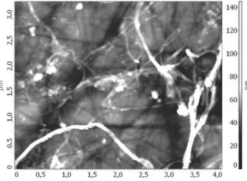
Annealing of the fluorinated DWNTs causes a change in the optical absorption spectrum; in particular, the features are broadened and their intensities are lowered. Possibly, it is indicative that some low-fluorinated nanotubes are dis-rupted with sample heating. The spectrum of the sample, annealed at 400°C and almost free of fluorine, still has more fine structure than the spectrum of pristine nonfluorinated DWNTs. The better resolution of optical features is achieved with debundling of nanotubes, and hence, the fluorinated samples consist of smaller-diameter ropes. Retention of tubular species in the heat-treated sample was supported by the AFM examination (Figure 8).
The electronic structure of the DWNT samples was probed using X-ray fluorescent spectroscopy (Figure 9). A C KR spectrum arises as a result of electron transitions from occu-pied valence states to a previously created core hole. Owing to the dipole selection rules and localization of the core orbitals, the spectrum contains data on the density of the occupied C 2p states. C KR spectra of the fluorinated DWNT sample and the sample annealed at temperatures from 150 to 450°C are compared in Figure 9a. The spectra exhibit three main features, lettered A, B, and C, and the intensity and structure are not changed monotonically with the annealing temperature. Despite the fact that the C KR spectra
of the fluorinated sample before and after annealing are simi-lar in appearance, the origin of their spectral features is different. In the spectrum of the sample heated at 450°C (contained trace of fluorine), the maximum A corresponds to the π-system of the graphitic-like particles, the intense maximum C is attributed to the σ-system, and the shoulder B is formed as a result of X-ray transitions of both types of electrons.19Since fluorination of DWNTs leaves part of the
(16) Hagen, A.; Moos, G.; Talalaev, V.; Hertel, T. Appl. Phys. A 2004,
78, 1137.
(17) Hertel, T.; Hagen, A.; Talalaev, V.; Arnold, K.; Hennrich, F.; Kappes, M.; Rosenthal, S.; McBride, J.; Ulbricht, H.; Flahaut, E. Nano Lett.
2005, 5, 511.
(18) Gevko, P. N.; Bulusheva, L. G.; Okotrub, A. V.; Yudanov, N. F.; Yushina, I. V.; Grachev, K. A.; Pugachev, A. M.; Surovtsev, N. V.; Flahaut, E. Fullerenes, Nanotubes, Carbon Nanostruct. 2006, 14, 233. (19) Okotrub, A. V.; Gusel’nikov, A. V.; Bulusheva, L. G. In
Nanoengi-neered Nanofibrous Materials; NATO Science Series; Guceri, S.I.,
Gogotsi, Y., Kuznetsov, V., Eds.; Kluver Academic Book Publishers: Dordrecht, Netherlands, 2004; Volume NATO-ASI (PST 979397); p 347.
Figure 7. Background-corrected optical absorption spectra of the pristine
DWNT sample (1), fluorinated DWNT sample (2), and fluorinated sample
annealed at temperatures 100 (3), 200 (4), 300 (5), and 400°C (6). Figure 8. AFM image of the fluorinated DWNT sample annealed at 400°C.
Figure 9. (a) C KR spectra of fluorinated DWNT samples annealed at
different temperatures; (b) C KR spectra measured for the pristine DWNT sample (1), fluorinated sample annealed at 450°C (2), and graphite (3).
inner walls unfluorinated, the C KR spectrum of the fluori-nated DWNT sample can be considered a composition of the spectra of graphitic and fluorographitic particles. It follows from the interpretation of the C KR spectrum of graphite fluoride C2F20that the maximums A and C of the
fluorine-containing samples arise from the participation of the electrons of the CF groups. Therefore, the spectra of fluori-nated samples heated at mild temperatures and at tempera-tures above 300°C cannot be directly compared. The spectral changes at the early stages of sample annealing are related to the defluorination process. The fluorinated samples annealed at the highest temperatures, as the XPS data showed, contain only traces of fluorine, and changes ob-served in the density of C 2p states are more likely to result from a rearrangement of the graphitic network.
Figure 9b compares the C KR spectra of pristine DWNTs, a fluorinated sample annealed at 450°C, and graphite. The spectrum of the pristine DWNTs has the highest intensity of the short-wave maximum A that is indicative of a large portion of defects in the tube walls. The similar enhancement of the density of weak bonding states has been detected in the C KR spectrum of HiPCO SWNTs subjected to a multistage purification. Comparison of this spectrum with the theoretical C KR spectra plotted as the result of quantum-chemical calculations on carbon tube models revealed the occurrence of a substantial quantity of 2-fold coordinated carbon atoms, which could constitute zigzag-like boundaries of large holes in the graphitic shells.21The C KR spectrum
of the annealed fluorinated sample exhibits a lowering of the maximum A intensity, a short-wave shift of the main maximum C, and an increase in the shoulder B intensity. These changes suggest a distinction between the electronic state of carbon in the treated DWNT sample and that in the pristine one. As was mentioned above, thermal defluorination of carbon nanotubes results in the extraction of carbon atoms from the graphitic network. Increasing the temperature could aid in healing of the developed vacancies with topological defect (nonhexagonal rings) formation. Actually, the theo-retical X-ray spectrum plotted for the carbon atoms making
the topological defects has shown a decrease in the density of high-energy states and a short-wave shift of the main maximum C22related to the theoretical spectrum of carbon
atoms located at the vacancy boundary.23 The intensity of
the maximum A in the spectrum of topological defects was close to that in the spectrum of the perfect graphitic network. The C KR spectrum of the defluorinated sample exhibits an enhanced intensity in the maximum A, as compared to that of the graphite spectrum (Figure 9b). Hence, we conclude the fluorinated DWNT sample annealed at 450°C still may contain a portion of vacancies, which remained unrecon-structed due to an insufficiently high temperature.
Conclusion
Interaction of DWNTs with a gaseous BrF3and Br2
mix-ture at room temperamix-ture results mainly in covalent attach-ment of fluorine atoms to the outer tube shells. Fluorination of DWNTs resolves the features of the optical absorption spectrum, allowing the observation of the configuration of the inner shells. TG analysis and XPS examination of the fluorinated DWNT sample revealed the fluorine removal begins at 100 °C, and decomposition of the fluorocarbon particles is ended at ∼500°C. The thermal defluorination of the DWNT sample has virtually no effect on the structure of the inner tube shells and considerably changes the electronic state of carbon from the outer shells. During annealing, fluorine atoms leave the sample, together with carbon atoms of the graphitic network, creating vacancies which, under the enhanced temperature conditions, are likely to be partially reconstructed by the formation of topological defects.
Acknowledgment. This work was supported by the Russian
Foundation for Basic Research (Project 03-03-32286) and a Grant of the President of Russian Federation for the leading scientific schools (No. 4419.2006.3).
CM0613915
(20) Bulusheva, L. G.; Okotrub, A. V.; Yudanov, N. F. Phys. Low-Dimen.
Struct. 2002, 7/8, 1.
(21) Bulusheva, L. G.; Okotrub, A. V.; Dettlaff-Weglikowska, U.; Roth, S.; Heggie, M. I. Carbon 2004, 42, 1095.
(22) Bulusheva, L. G.; Okotrub, A. V.; Asanov, I. P.; Fonseca, A.; Nagy, J. B. J. Phys. Chem. B 2001, 105, 4853.
(23) Bulusheva, L. G.; Okotrub, A. V.; Gusel’nikov, A. V.; Dettlaff-Weglikowska, U.; Roth S. In Molecular Nanostructures: XVII
International Winterschool/Euroconference on Electronic Properties of NoVel Materials; Kuzmany, H., Fink, J., Mehring, M., Roth, S.,