Conjugated Polymers That Respond
to Oxidation with Increased Emission
The MIT Faculty has made this article openly available. Please sharehow this access benefits you. Your story matters.
Citation Dane, Eric L., Sarah B. King, and Timothy M. Swager. “Conjugated Polymers That Respond to Oxidation with Increased Emission.” Journal of the American Chemical Society 132.22 (2010): 7758–7768. As Published http://dx.doi.org/ 10.1021/ja1019063
Publisher American Chemical Society (ACS)
Version Author's final manuscript
Citable link http://hdl.handle.net/1721.1/74229
Terms of Use Article is made available in accordance with the publisher's policy and may be subject to US copyright law. Please refer to the publisher's site for terms of use.
Conjugated Polymers that Respond to Oxidation with Increased Emission
Journal: Journal of the American Chemical Society Manuscript ID: ja-2010-019063.R1
Manuscript Type: Article Date Submitted by the
Author:
Complete List of Authors: Dane, Eric; Massachusetts Institute of Technology, Chemistry King, Sarah; Massachusetts Institute of Technology, Chemistry Swager, Timothy; Mass. Inst. of Tech., Chemistry
1
Conjugated Polymers that Respond to Oxidation with
Increased Emission
Eric L. Dane, Sarah B. King, and Timothy M. Swager*
Department of Chemistry, Massachusetts Institute of Technology,
77 Massachusetts Avenue, Cambridge, Massachusetts 02139
EMAIL: *tswager@mit.edu
RECEIVED DATE (to be inserted):
TITLE RUNNING HEAD. CPs that Respond to Oxidation with Increased Emission
ABSTRACT. Thioether-containing poly(paraphenylene-ethynylene) (PPE) copolymers show a strong fluorescence turn-on response when exposed to oxidants in solution as a result of the selective conversion of thioether substituents into sulfoxides and sulfones. We propose that the increase in fluorescence quantum yield (ΦF) upon oxidation is the result of both an increase in the rate of
fluorescence (kF) as a result of greater spatial overlap of the frontier molecular orbitals in the oxidized
materials and an increase in the fluorescence lifetime (τF) due to a decrease in the rate of non-radiative
decay. Contrary to established literature, the reported sulfoxides do not always act as fluorescence quenchers. The oxidation is accompanied by spectral changes in the absorption and emission of the polymers, which are dramatic when oxidation causes the copolymer to acquire a donor-acceptor interaction. The oxidized polymers have high fluorescence quantum yields in the solid state, with some having increased photostability. A turn-on fluorescence response to hydrogen peroxide in organic
ACS Paragon Plus Environment
1 2 3 4 5 6 7 8 9 10 11 12 13 14 15 16 17 18 19 20 21 22 23 24 25 26 27 28 29 30 31 32 33 34 35 36 37 38 39 40 41 42 43 44 45 46 47 48 49 50 51 52 53 54 55 56 57 58 59 60
2 solvents in the presence of an oxidation catalyst indicates the potential of thioether-containing materials for oxidant sensing. The reported polymers show promise as new materials in applications where photostability is important, where tunability of emission across the visible spectrum is desired, and where efficient emission is an advantage.
Introduction
Living in an oxidizing environment is essential for most life, but it has generally been an impediment to the use of fluorescent conjugated polymers (CPs) in practical applications. Fluorescent CPs are an important class of functional materials that have found use in applications ranging from organic light emitting diodes (OLEDs)1 to the detection of TNT vapor at ppb-levels via amplified fluorescence quenching.2 Because most conjugated polymers become less emissive when oxidized, photobleaching attributed to reactions involving oxygen has limited their use.3 In order to better understand how to synthesize materials that are robust in our oxygen-rich atmosphere, we became interested in CPs that upon oxidation showed productive changes in emission, such as a large increase in emission, a significant shift in emission wavelength, or greater photostability.
Materials that show an increase in emission when oxidized are also candidates for the turn-on fluorescence sensing of peroxide oxidants. Being able to detect organic peroxides, such as triacetone triperoxide (TATP), is important because they can be used to make dangerous explosive devices.4 On the other hand, hydrogen peroxide plays important biological roles, including being a disease marker and acting as a signaling molecule.5 Therefore, compounds that show an increase in fluorescence when oxidized by peroxides are important to the development of new sensing materials.
Previous studies on the photobleaching of fluorescent CPs have focused on understanding the mechanism. For example, fluorenone defects in polyfluorene act as low-energy trap sites that cause decreased emission intensity.6 In poly (paraphenylene-vinylene) (PPV) polymers, it has been suggested that the main degradation pathway involves the reaction of singlet oxygen with the alkenyl-group,
ACS Paragon Plus Environment
1 2 3 4 5 6 7 8 9 10 11 12 13 14 15 16 17 18 19 20 21 22 23 24 25 26 27 28 29 30 31 32 33 34 35 36 37 38 39 40 41 42 43 44 45 46 47 48 49 50 51 52 53 54 55 56 57 58 59 60
3 resulting in the formation of carbonyl defects that eventually result in chain scission.3 In poly(paraphenylene-ethynylene) (PPE)7 polymers, the mechanism of photobleaching has not been elucidated, but studies suggest that in model systems alkynes are less prone to reaction with singlet oxygen than alkenes.8 However, it is not clear that the observed photobleaching results solely from covalent bond formation between the CPs and oxygen species, because in some cases it has been shown to be reversible.9 Although the specific mechanism is not known, CPs show greater photostability when irradiated under anaerobic conditions indicating that the presence of oxygen is important to photobleaching.10 Previous work from our group has found that antioxidants and triplet-quenchers, such as cyclooctatetraene derivatives, can be added to thin-films of CPs to attenuate photobleaching under irradiation.11 Additionally, we have shown that polymers with high ionization potentials, due to the presence of electron-withdrawing perfluoroalkyl substituents, showed resistance to photobleaching.12 Building on these previous investigations, this report focuses on designing CPs that respond positively to oxidation.
Previous examples of conjugated polymers where it was shown that oxidation did not cause a decrease in emission include polyfluorenes that contain small amounts of either a phosphafluorene or phosphafluorene-oxide comonomer.13 Compared to the phosphine polymer’s emission, the phosphine oxide polymer’s emission was red-shifted in the solid state, but not in solution. However, the oxidized polymers had roughly the same emission efficiency as the unoxidized polymers, and in this example, the oxidation was carried out on the monomer before polymerization. We chose to append sulfur atoms, in the form of thioethers, directly to the polymer backbone because thioethers can be selectively oxidized in the presence of alkynes. In organic compounds, sulfur can have a formal oxidation state ranging from -2 to +6.14 In this investigation, we were interested in what effect changing the formal oxidation state from a thioether (-2) to that of a sulfoxide (0) or a sulfone (+2) would have on the photophysical properties of a PPE. Additionally, we hypothesized that the electron-withdrawing nature of the oxidized thioethers would lead to changes in absorption and emission wavelengths due to donor-accepter interactions.
ACS Paragon Plus Environment
1 2 3 4 5 6 7 8 9 10 11 12 13 14 15 16 17 18 19 20 21 22 23 24 25 26 27 28 29 30 31 32 33 34 35 36 37 38 39 40 41 42 43 44 45 46 47 48 49 50 51 52 53 54 55 56 57 58 59 60
4 Conjugated polymers that contain sulfur atoms are ubiquitous in materials chemistry, but almost exclusively the sulfur atom is contained in a thiophene ring.15 Surprisingly, we can find relatively few examples of the incorporation of thioethers into CPs, though a review by Gingras16 summarizes how chemists have exploited the properties of persulfurated aromatic compounds. Lehn17 synthesized a series of diacetylene-linked poly(phenylthio)-substituted benzenes that showed the ability to be multiply reduced, taking advantage of sulfur’s ability to stabilize negative charge. Additionally, several examples of thioether-containing PPV CPs have been reported.18 In general, aromatic sulfones, and especially aromatic sulfoxides, have not been extensively utilized in materials chemistry.
We designed a monomer that contains four thioethers divided between two dithiolane rings. Because the sulfur atoms of 1 (Scheme 1) are not members of an aromatic ring system, it was anticipated that they would behave differently than a sulfur atom in a thiophene ring system. The five-member rings have the advantage of tying back the sulfur atoms (θCCS = 123-124˚, see Scheme 1, X-ray
structure of 1) and minimizing their steric interference with the reactive positions used in polymerization. Additionally, they allow for the facile introduction of solubilizing groups from readily available symmetrical and asymmetrical ketones via acid-catalyzed condensation. It was anticipated that the branching of the aliphatic solubilizing groups would aid in solubility and prevent aggregation in the solid-state. Monomers and polymers with a similar geometry with oxygen in place of sulfur have been recently described.19
Results
Synthesis
Monomer 1 is available in three steps from inexpensive starting materials without the need for chromatography. Compound 2 was synthesized via complete nucleophilic aromatic substitution of 1,2,4,5-tetrachlorobenzene with sodium 2-methylpropane thiolate in refluxing dimethylformamide, followed by recrystallization from refluxing methanol.20 The t-butyl groups were deprotected in situ and
ACS Paragon Plus Environment
1 2 3 4 5 6 7 8 9 10 11 12 13 14 15 16 17 18 19 20 21 22 23 24 25 26 27 28 29 30 31 32 33 34 35 36 37 38 39 40 41 42 43 44 45 46 47 48 49 50 51 52 53 54 55 56 57 58 59 60
5 the resulting thiols were condensed with 5-nonanone in the presence of HBF4•Et2O in refluxing toluene.
Compound 3 could not be successfully brominated or iodinated via electrophilic substitution, presumably due to nucleophilic attack by the thioethers on the electrophilic reagents and subsequent side-reactions. Rather than pursue a method of sequential lithiations, we used an excess of lithium tetramethylpiperidine and chlorotrimethylsilane to drive the double deprotonation of 3 to completion in one flask. The resulting di-trimethylsilyl product was carried through to be iodinated with ICl in CH2Cl2.21 Monomer 3 was purified by recrystallization from hexane and obtained in an overall yield of
38% as an air-stable yellow solid.
Model compounds (MC1 and MC2) and polymers (P1 and P2) were synthesized via Sonagashira cross-coupling in good yield. P1 was isolated as a high molecular weight polymer (Mn = 236,000 g/mol) according to gel permeation chromatography (GPC) and was soluble in common organic solvents such as CH2Cl2 and THF. Under the same conditions, P2 formed high molecular weight
polymers that became insoluble, either because with increased chain length the polymers become insoluble or because of the presence of cross-linking. In order to isolate soluble polymers, the temperature of the polymerization was lowered from 65 ˚C to 45 ˚C. At the lower reaction temperature, the molecular weight of P2 (Mn = 18,900 g/mol) decreased and the isolated polymer was soluble. In addition to GPC, the polymers were characterized by 1H- and 13C-NMR and FT-IR.
Oxidation of Model Compounds
Model compounds MC1 and MC2 were oxidized with one and two equivalents of m-chloroperbenzoic acid and the mono- and disulfoxides of each (MC1_1ox, MC1_2ox, MC2_1ox, and
MC2_2ox) were isolated via chromatography and recrystallization (Scheme 2). The mono- and disulfoxides were characterized with 1H-and 13C-NMR, FT-IR, and HRMS. For MC2_1ox and
MC1_2ox_cis, X-ray crystallography was used to ensure the identity of the compound (see Supporting Information for ORTEP-diagrams). For each of the disulfoxides, four diastereomers were obtained (Scheme 2). The cis-isomers, in which the polar sulfoxide bonds point in the same direction and
ACS Paragon Plus Environment
1 2 3 4 5 6 7 8 9 10 11 12 13 14 15 16 17 18 19 20 21 22 23 24 25 26 27 28 29 30 31 32 33 34 35 36 37 38 39 40 41 42 43 44 45 46 47 48 49 50 51 52 53 54 55 56 57 58 59 60
6 increase the overall dipole of the molecule, had a significantly longer retention time on silica gel as compared to the trans-isomers, in which the dipoles point in opposite directions. The two isomers were easily separated via chromatography. As expected, the directing influence of the thioethers favored a meta-orientation for the sulfoxides over a para- or an ortho-relationship, although a small amount of para-compound was observed in each case. The meta- and para-isomers show diagnostic 1H- and 13 C-NMR, with the para-isomer having fewer unique 1H- and 13C-signals because of its higher symmetry. For MC1_2ox, the cis- and trans-products contained less than 10% of the para-isomer according to integration of the proton NMR. For MC2_2ox, the isolated cis- and trans-products contained the meta- and para-isomer in a ratio of roughly 3:1. The selectivity for the second oxidation to occur at the meta-position as opposed to the para-meta-position results from the first sulfoxide withdrawing electron-density from the sulfur atoms in an ortho- and para-orientation more effectively than from the sulfur in a meta-orientation. The ortho-compound was not observed. The oxidation of the second sulfur in dithioacetals is often much slower as compared to the oxidation of the first, with the oxidation of the sulfoxide to sulfone as a competing process.22
Optical and Photophysical Properties of the Model Compounds in Solution.
Both MC1 and MC2 have similar absorbance and emission properties (see Table 1, Figure 1) in solution (CH2Cl2). MC1 and MC2 have strong absorbance peaks between 300 and 400 nm, with a
weaker absorbance between 400 and 500 nm. The absorbance maxima for MC2 (375 nm) is red-shifted as compared to MC1 (355 nm), as a result of the electron-donating methoxy groups at the 1- and 4-positions of the outer aryl rings. However, the same effect is not seen in the less intense band in the visible region, where the absorbance maximum of MC1 (434 nm) is nearly the same as that of MC2 (435 nm). MC1 and MC2 are relatively non-emissive, with quantum yields of less than 0.01, and they show broad, featureless emission centered near 490 nm.
Interestingly, when thoroughly degassed with bubbling nitrogen, both MC1 and MC2 show an additional sharp, red-shifted emission peak (MC1, 560 nm; MC2, 580 nm) and a vibronic shoulder
ACS Paragon Plus Environment
1 2 3 4 5 6 7 8 9 10 11 12 13 14 15 16 17 18 19 20 21 22 23 24 25 26 27 28 29 30 31 32 33 34 35 36 37 38 39 40 41 42 43 44 45 46 47 48 49 50 51 52 53 54 55 56 57 58 59 60
7 above 600 nm (see Figure S1 in Supporting Information). Exposure of the samples to air causes the peak to completely disappear. Quenching of the emission by oxygen indicates that the emissive process involves an excited triplet state rather than a singlet state. Room temperature phosphorescence is unusual for organic compounds of this type, and it indicates that the four thioether substituents on the central aromatic ring promote intersystem crossing from the singlet excited state to triplet excited state.23
For both MC1 and MC2, oxidation to the mono-sulfoxide causes an increase in fluorescence quantum yield relative to the parent compound, with the effect being more dramatic for MC2. This increase in emissivity is accompanied by a blue-shift in emission, which is more pronounced for MC1. The emission maxima of MC1 shifts from 489 nm for the unoxidized version to 458 nm for the monosulfoxide (MC1_1ox). The emission maxima of MC2 shifts from 493 nm for the unoxidized version to 467 nm for the monosulfoxide (MC2_1ox). Further oxidation of MC1_1ox to MC1_2ox does not cause a significant change in emissivity; however it is accompanied by a shift in emission from 458 nm to 403 nm and a change in the structure of the emission. Conversely, further oxidation of
MC2_1ox to MC2_2ox results in a 10-fold increase in fluorescence quantum yield, and is accompanied by a small red-shift of the emission maxima from 467 nm to 474 nm. As discussed in the previous section, cis- and trans-isomers of MC1_2ox and MC2_2ox were isolated and characterized separately. There appears to be no significant difference in the photophysical properties of the cis- and trans-isomers. However, it should be noted that in all cases we isolated the cis- and trans-isomers as a mixture of regioisomers. Because the compounds were not separated, we cannot comment on how the meta- versus para-orientation of the sulfoxides affects the photophysical properties.
To investigate the effect of further oxidation, we observed the absorbance and emission changes caused by adding increasing amounts of m-CPBA to dilute solutions of MC1 and MC2 (see Figure S2 in Supporting Information). Increasing oxidation of MC1 is accompanied by blue-shifted absorbance and emission, and a general increase in emission. Comparison of the spectrum with isolated compounds
ACS Paragon Plus Environment
1 2 3 4 5 6 7 8 9 10 11 12 13 14 15 16 17 18 19 20 21 22 23 24 25 26 27 28 29 30 31 32 33 34 35 36 37 38 39 40 41 42 43 44 45 46 47 48 49 50 51 52 53 54 55 56 57 58 59 60
8 indicates that after the initial formation of MC1_1ox, a significant amount of MC1_2ox is formed. Additionally, a peak emerges at 422 nm that we attribute to sulfone-containing compounds with significantly higher quantum yields than the sulfoxides.24 When MC2 is titrated with oxidant, there is an initial blue shift in absorbance and emission, also accompanied by a substantial increase in emission. However, as the compound is further oxidized the donor-acceptor interaction that develops between the electron-rich terminal aromatic rings and the increasingly electron-poor center ring causes a red-shift in absorbance and emission. The red-shift in emission is accompanied by a decrease in emission efficiency, however the resulting fluorophores are still substantially more emissive than the parent compound.
Oxidation of Polymers
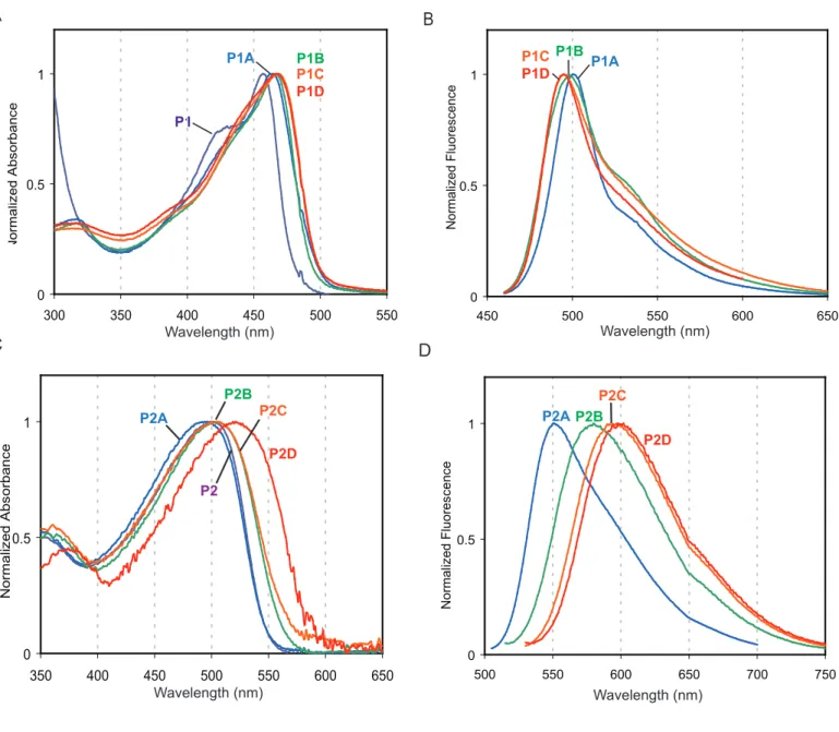
Polymers P1 and P2 were oxidized with 2.10, 3.15, 4.20, 6.30, and 8.40 equivalents of m-CPBA overnight at room temperature in a CH2Cl2 solution (Figure 2, P1A-D and P2A-D). The oxidation
products of P1 did not show substantial changes in color; however the reaction mixtures became noticeably green fluorescent under ambient light. Conversely, the color of P2 changed from yellow to red with increasing amounts of oxidation. The effect of the oxidation on molecular weight was monitored with GPC and is summarized in Table 2. The Mn of the polymers decreased with greater amounts of oxidation. The addition of 8.4 equiv of oxidant caused the molecular weight of P1 to decrease by half. This suggests that oxidation at the alkyne linkages becomes more competitive with sulfur oxidation with higher amounts of oxidant. It has been reported that diphenylacetylene slowly reacts with m-CPBA in chloroform at room temperature to give a variety of oxidation products resulting from the initial formation of an oxirene.25 Some of the oxidation products reported result in cleavage of the diphenylacetylene into two molecules, while others contain linkages that are susceptible to cleavage.25
The presence and relative abundance of sulfoxides and sulfones in the oxidized samples of P1 and P2 were evaluated with infrared spectroscopy and compared to similar spectra for the model
ACS Paragon Plus Environment
1 2 3 4 5 6 7 8 9 10 11 12 13 14 15 16 17 18 19 20 21 22 23 24 25 26 27 28 29 30 31 32 33 34 35 36 37 38 39 40 41 42 43 44 45 46 47 48 49 50 51 52 53 54 55 56 57 58 59 60
9 compounds (see Figure S3 and Figure S4 in Supporting Information). Oxidation causes significant changes in the spectra of both polymers between 1600 and 850 cm-1 (Figure 3). Initially, P1 has a relatively featureless spectrum. Oxidation with 2.1 equiv. of m-CPBA causes a strong absorption band at 1077 cm-1 to appear, indicating sulfoxide formation. This band continues to grow and broaden with further oxidation. Beginning with P1C and continuing in P1D, two peaks at 1335 and 1160 cm-1 belonging to sulfones appear. The FT-IR spectrum of P2 is more challenging to interpret, because signals from the aryl ether groups on the comonomer of P2 between 1150 and 1000 cm-1 interfere with the observation of the sulfoxide band in the oxidized polymers. P2A shows a strong sulfoxide band at 1081 cm-1, but does not show sulfone bands. Sulfone bands appear in P2B and are accompanied by a decrease in the initial signal belonging to sulfoxides, which suggests that some of the sulfoxides have been oxidized to sulfones. Beginning with P2C and continuing in P2D, the sulfone peaks at 1335 and 1161 cm-1 continue to grow as the sulfoxide signal weakens.
In summary, FT-IR shows that both P1 and P2 are first oxidized to sulfoxides and then oxidized to sulfones. P2 appears to be more easily oxidized to sulfones relative to P1, because of its more electron-rich comonomer. It is difficult to comment quantitatively on the level of oxidation using FT-IR, however the spectra support some important qualitative observations. The P1A and P2A spectra suggest that most of the thioether-containing aromatic rings have been oxidized to the disulfoxide, which is in accord with the reactivity seen in the model compounds. The weak sulfone bands seen in
P1D indicate minimal sulfone formation with 8.40 equiv of oxidant, which suggests that most of the thioether repeating units have four or less oxygen atoms. This is in contrast with the strong sulfone bands observed in P2D, which suggest that each thioether repeating unit contains at least four oxygen atoms. In both P1 and P2, the oxidized polymers have complex structures. Although this extra-dimension of polydispersity makes characterization challenging, it likely aids in solubility and reduces crystallinity.
Optical and Photophysical Properties of the Polymers in Solution.
ACS Paragon Plus Environment
1 2 3 4 5 6 7 8 9 10 11 12 13 14 15 16 17 18 19 20 21 22 23 24 25 26 27 28 29 30 31 32 33 34 35 36 37 38 39 40 41 42 43 44 45 46 47 48 49 50 51 52 53 54 55 56 57 58 59 60
10 Our primary method of evaluating the effect of oxidation on the polymers is absorption and fluorescence spectroscopy. As can be seen in Figure 2 and Table 2, the parent polymer is relatively nonemissive, with a quantum yield less than 0.01. The addition of two equivalents of oxidant, which leads to the formation of mostly sulfoxides, causes P1’s absorbance maximum to red shift from 444 nm to 457 nm. P1A’s emission is blue-shifted from 512 nm to 492 nm relative to the parent polymer, and its quantum yield increases dramatically (35-fold). When P1 is treated with 4.2 equivalents of oxidant (P1B), the absorbance red-shifts and the emission blue-shifts. The spectral changes for P1B are accompanied by an increase in quantum yield to 0.53, but the effect of further oxidation is minor.
Similar to P1, P2 is relatively non-emissive in solution, with a quantum yield less than 0.01. The quantum yield of P2A is 0.49, which represents a more than 49-fold increase in emission when the polymer is treated with 2.1 equivalents of oxidant. Additional oxidation causes continued red-shifting in absorbance and emission, as can be seen in P2B, P2C, and P2D. However, greater oxidation is accompanied by a decrease in quantum yield, which appears to be at least partially a result of a decrease in the rate of fluorescence (kF).
Response to Hydrogen Peroxide in Solution
Surprisingly, the model compounds and polymers did not react with hydrogen peroxide in solution at room temperature. To facilitate a reaction, the oxidation catalyst methylrheniumtrioxide (MTO)26 was added as described by Malashikin and Finney.4a The absorbance and emission of dilute solutions of MC2 and P2 in quartz cuvettes were recorded after the addition of catalyst ([MTO] = 0.5 mM), but before the addition of oxidant.27 Following the addition of hydrogen peroxide, the stirred solutions were monitored at 15 minutes, 3 hours, and 20 hours. Initially, three solvents were screened (THF, EtOH, DCM), and the extent of reaction was greatest in DCM. Ethanol showed the same response at a slower rate, but no reaction was observed in THF (unstabilized).
ACS Paragon Plus Environment
1 2 3 4 5 6 7 8 9 10 11 12 13 14 15 16 17 18 19 20 21 22 23 24 25 26 27 28 29 30 31 32 33 34 35 36 37 38 39 40 41 42 43 44 45 46 47 48 49 50 51 52 53 54 55 56 57 58 59 60
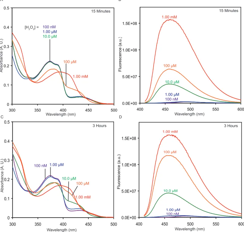
11 The response at 3 hrs of a 5.00 µM solution of P2 to a range of H2O2 concentrations is shown in
Figure 4. At 3 hrs, fluorescence increases in the 1.00 mM, 100 µM, 10.0 µM, 1.00 µM, and 100 nM samples, but not in the 10.0 nM and 1.0 nM H2O2 solutions. The response continues to develop over
time, and it can be seen by eye under UV-illumination after 20 hours (Figure 4, inset picture) for all concentrations of H2O2 except 1.00 nM (see Figure S5 in Supporting Information).
To better understand and characterize the response of P2 to hydrogen peroxide, a 10.0 µM solution of MC2 was subjected to similar conditions (see Figure S6 in Supporting Information). At 15 minutes, a fluorescence increase is observed in the samples with a H2O2 concentration of 1.00 mM, 100
µM, and 10.0 µM. Based on the absorbance spectrum, it appears that after 15 minutes MC2 has been transformed to MC2_2ox in the 1.00 mM H2O2 solution. After three hours there is no further reaction,
indicating that the disulfoxide is a stopping point under these conditions. At 15 minutes, the 100 µM H2O2 solution, which represents 10 equiv of H2O2 per molecule of MC2, has been oxidized to the
monosulfoxide based on the absorbance spectrum. After 3 hours, the sample shows further oxidation, though it has not yet reached the same point as the 1.00 mM H2O2 solution. The 10.0 µM H2O2 solution
shows a significant increase in fluorescence at 15 minutes, but its absorbance spectrum is relatively unchanged. After 3 hours, the absorbance spectrum of the 10.0 µM H2O2 appears more like that of the
monosulfoxide, and the fluorescence has continued to increase. In the 1.00 µM H2O2 sample, there is a
significant increase in fluorescence, presumably from the formation of some amount of the monosulfoxide, but the absorbance spectrum changes only slightly. The 100 nM H2O2 solution shows
no response.
Thin-film photophysics of polymers
Thin-films of polymers P1, P1A-D, P2, and P2A-D were prepared by spin-casting a chloroform solution (1.0 mg/ml) onto silanized glass slides. The films were annealed at 120 ˚C for 10 minutes. The films show behavior similar to the dilute solutions, with no signs of aggregation (Table 2, Figure S7 in Supporting Information). For P1, P1A, and P1B, the thin-film absorption and emission maxima are
red-ACS Paragon Plus Environment
1 2 3 4 5 6 7 8 9 10 11 12 13 14 15 16 17 18 19 20 21 22 23 24 25 26 27 28 29 30 31 32 33 34 35 36 37 38 39 40 41 42 43 44 45 46 47 48 49 50 51 52 53 54 55 56 57 58 59 60
12 shifted compared to the solution values. For P1C and P1D, the absorption maxima are relatively unchanged going from solution to thin-film, however the emission maxima are red-shifted in the solid-state. For P2 and P2A, the thin-film absorption and emission maxima are red-shifted compared to the solution values. P2B displays no change in absorption maxima in moving from solution to the solid-state, however its emission does red-shift. P2C and P2D show a slight blue-shift in absorption when moving from solution to the solid-state, however in both cases the emission is red-shifted.
The films made from the oxidized forms of P1, specifically P1B and P1C, were highly emissive, with quantum yields above 0.50. We compared films of P1C to films of a previously reported non-aggregating PPE (P3) with a fluorescent quantum yield of 0.33 in the solid state.2a In Figure 5, the emission of P1C is significantly greater than that of a film of P3 when both are excited with monochromatic light of 420 nm, a wavelength at which the two films have nearly identical optical densities. Films of P2A are also highly fluorescent with a quantum yield of 0.56. Comparatively, films of the more highly oxidized polymers (P2B-D) are less emissive, however the fluorescence can be clearly seen by eye under UV irradiation as shown in Figure 6.
Thin-film photobleaching studies
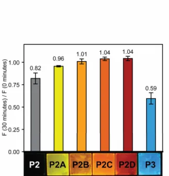
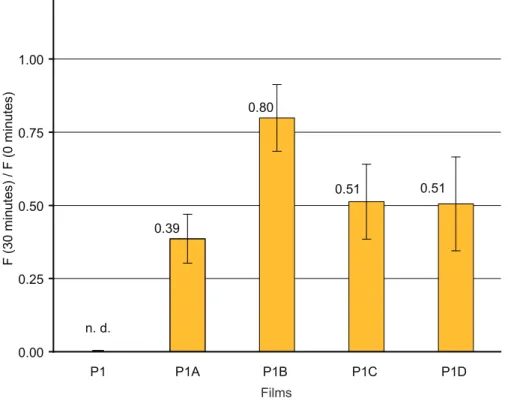
Films of P1, P1A-D, P2, and P2A-D were tested for photostability by comparing the emission before and after the films were irradiated at their absorbance maxima for 30 minutes, using monochromatic light generated from a 450 W steady-state Xe lamp as the irradiation source under aerobic conditions.11 The approximate area of irradiation was 0.25 cm2 and the average power density of irradiation was estimated to be 6 mW/cm2. Films of P1 and P1A-D had an optical density (OD) of 0.10 ± 0.01. Films of P2, P2A-D, and P3 had an OD of 0.05 ± 0.01. The films were not moved during the process to ensure that the measurements reflect the effect of irradiation on one area of the film. All measurements were done in triplicate and the error is reported as the standard deviation. Films of P1 and P1A-D did not show improved photostability as compared to P3 under identical conditions11 (see Figure S8 in Supporting Information). However, films of P2A-D proved resistant to photobleaching
ACS Paragon Plus Environment
1 2 3 4 5 6 7 8 9 10 11 12 13 14 15 16 17 18 19 20 21 22 23 24 25 26 27 28 29 30 31 32 33 34 35 36 37 38 39 40 41 42 43 44 45 46 47 48 49 50 51 52 53 54 55 56 57 58 59 60
13 (Figure 6). P2A-D retain more than 95% percent of their original fluorescence after 30 minutes of irradiation, with P2B-P2D even showing a slight increase in emission. They are more photostable than
P3, which retains only 60% of its initial emission when irradiated at its absorbance maximum (440 nm) at the same OD, in accordance with previously reported results.11
Discussion
Understanding the Photophysical Behavior of the Model Compounds
Because of its relatively weak intensity, the absorbance band of MC1 and MC2 centered at approximately 435 nm (Figure 1) suggests that the thioethers on the center ring impart intramolecular charge transfer (ICT) character to the HOMO-LUMO optical transition. The ICT character of the transition results from poor spatial overlap of the frontier molecular orbitals (FMOs), which we examined with quantum mechanical calculations (Figure 7a, Supporting Information Figure S9).19 Qualitatively, for S1 (a simplified version of MC1 for ease of computation) the HOMO is primarily located on the center ring with most of the orbital density on the sulfur atoms, while the LUMO is delocalized through out the molecule’s π-system with minimal density on the sulfur atoms. The presence of ICT character has been observed in simple thioether containing aromatic compounds, such as phenyl-n-propyl sulfide.28 Additionally, ICT character is observed in compound 719 (Chart 1), which is an oxygen analogue of the thioethers studied here.
Upon oxidation of MC1 to MC1_1ox, the ICT band blue shifts, and with oxidation to the disulfoxide, the ICT band disappears. The computed HOMO and LUMO for S2 are in agreement with this observation, as it appears that the spatial overlap of the HOMO and LUMO is significantly greater for S2 as compared to S1 (Figure 7). The situation is more complex in the case of MC2 and its mono- and disulfoxides because the oxidation of the thioethers introduces a donor-acceptor interaction between the electron-rich outer rings and the electron-poor middle ring. However, comparing the absorption spectrum of MC2 to that of MC2_1ox reveals that oxidation to the monosulfoxide causes a blue-shift
ACS Paragon Plus Environment
1 2 3 4 5 6 7 8 9 10 11 12 13 14 15 16 17 18 19 20 21 22 23 24 25 26 27 28 29 30 31 32 33 34 35 36 37 38 39 40 41 42 43 44 45 46 47 48 49 50 51 52 53 54 55 56 57 58 59 60
14 similar to that seen in MC1_1ox. The origin of the weak red-shifted absorbance present in both isomers of MC2_2ox may be elucidated by future computational studies.
For MC1 and MC2, the fluorescence quantum yield is low in solution (ΦF = 0.003, Table 1), for
two reasons. First, MC1 and MC2 have short fluorescence lifetimes of approximately 100 ps, which indicates that another relaxation process is competing with fluorescence. Based on previous studies of thioethers we suggest that intersystem crossing (ISC) from the singlet excited state to the triplet excited state accounts for the shortened lifetime. In compounds as simple as phenyl-n-propyl sulfide, ISC is believed to be the predominant deactivation pathway of the excited state.28 An example of an aromatic chromophore containing more than one thioether is 2,3,6,7,10,11-hexakis(n-hexylsulfanyl)triphenylene (Chart 1, Compound 8), which has a reduced fluorescence quantum yield compared to the parent triphenylene (0.015 vs. 0.066), but displays significant phosphorescence at 77K.29 Compound 8 has a significantly shortened lifetime (0.7 ns) as compared to the parent triphenylene (36.6 ns) or the same compound with the sulfur atoms replaced by oxygen atoms (8.0 ns). For MC1 and MC2, the observation of room temperature phosphorescence (Figure S1, Supporting Information) in degassed samples further supports the argument that ISC is a main deactivation pathway for the singlet excited state.
The second reason that the unoxidized model compounds have low emission is that they have lower rates of fluorescence (kF) as compared to the oxidized versions. Oxidation of MC1 to MC1_1ox
and both isomers of MC1_2ox is accompanied by an increase in kF. The increase in kF in the
disulfoxide as compared to the unoxidized compound appears to be a consequence of better spatial overlap between the HOMO and LUMO (see Figure 7). 23,30 The oxidation of MC2 to MC2_1ox causes the fluorescence lifetime to increase substantially from 0.099 ns to 2.7 ns, but does not increase kF.
Further oxidation to MC2_2ox causes an order of magnitude increase in kF from 0.020 ns-1 to ~ 0.20 ns-1
and a minor increase in the lifetime.
ACS Paragon Plus Environment
1 2 3 4 5 6 7 8 9 10 11 12 13 14 15 16 17 18 19 20 21 22 23 24 25 26 27 28 29 30 31 32 33 34 35 36 37 38 39 40 41 42 43 44 45 46 47 48 49 50 51 52 53 54 55 56 57 58 59 60
15
MC2 shows a more than 150-fold increase in ΦF when oxidized to the disulfoxide, but the
increase is more modest (7- to 13-fold based on the isomer) when MC1 is oxidized to the disulfoxide. We contend that the sulfoxides in MC1_2ox are behaving differently than the sulfoxides in MC2_2ox. It has been reported that the fluoresence of aromatic sulfoxides is low because of nonradiative decay pathways involving the sulfoxide. For example, pyrene has a quantum yield of 0.32 and phenyl 1-pyrenyl sulfide has a quantum yield of 0.34 in CH2Cl2. Oxidation of the sulfur in phenyl 1-pyrenyl
sulfide to a sulfoxide causes the quantum yield to decrease to 0.02, whereas further oxidation causes the quantum yield to increase to 0.76.4a A similar trend, in which the presence of a sulfoxide attached directly to an aromatic core quenches a fluorophore that is relatively unaffected by the presence of a thioether or a sulfone in the same position, is observed for naphthalene, biphenyl, and anthracene sulfides.31 However, in the compounds we have examined sulfoxides do not appear to act as fluorescence quenchers except in MC1_2ox (and perhaps in MC1_1ox), in which case the emission is blue-shifted towards the near-UV. This suggests that when irradiated at higher energies (shorter wavelengths) sulfoxides may quench fluorescence via a mechanism that is not operative at lower energies (longer wavelengths). The behavior of our model compounds and polymers is not unprecedented. Within a previously reported series of 21 tetracene derivatives (Chart 1), 15 of which are sulfoxides (9) and 6 of which are sulfones (10), the tetracene derivatives with sulfoxides are more emissive than the tetracene sulfones in nearly every case.32
Understanding the Photophysical Behavior of the Polymers
In contrast to the model compounds, the HOMO-LUMO optical transitions of P1 and P2 appear to be allowed, thus suggesting that the HOMO is partially delocalized along the polymer’s π-backbone and has better spatial overlap with the LUMO. In contrast, when the oxygen-containing monomer 7 is incorporated into PPEs the HOMO remains localized.19 P1 and P2, which like MC1 and MC2 are not very emissive, show large increases in ΦF when oxidizied due to an increase in both kF and τF (Table 2).
For example, when P1 is oxidzied to P1A with 2.1 equivalents of oxidant, kF increases from 0.074 ns-1
ACS Paragon Plus Environment
1 2 3 4 5 6 7 8 9 10 11 12 13 14 15 16 17 18 19 20 21 22 23 24 25 26 27 28 29 30 31 32 33 34 35 36 37 38 39 40 41 42 43 44 45 46 47 48 49 50 51 52 53 54 55 56 57 58 59 60
16 to 0.52 ns-1 and τF increases from 0.13 ns to 0.67 ns. When P2 is oxidized to P2A, kF increases from
0.050 ns-1 to 0.36 ns-1 and the lifetime increases from 0.17 ns to 1.35 ns. Based on the trends observed for the oxidized model compounds and the relavent literature compounds, it appears that the increase in kF is due to greater spatial overlap of the FMOs due to delocalization of the HOMO along the
π-backbone and that the increase in τF is due primarily to a decrease in the rate of ISC.
Understanding the behavior of the polymers in thin films
Because oxidation in solution causes P1 and P2 to become emissive, thin-films of the thioether-containing polymers were expected to show a fluorescence turn-on response when irradiated in air. However, this phenomenon was not observed. Instead, the already weak emission further decreased with prolonged irradiation (Figure 6, P2), even though thioethers are known to react with singlet oxygen.33 There are two possible explanations for this behavior. The first is that the thioethers are not being oxidized to sulfoxides or sulfones under the conditions. The second is that oxidation is so minimal that any oxidized moieties are effectively quenched by the remaining unoxidized segments.
Blue-shifted versus red-shifted fluorescence turn-on response to H2O2
As discussed in the results section, both MC2 and P2 show a fluorescence turn-on response to oxidation by hydrogen peroxide in the presence of a catalyst (MTO). However, they differ in that
MC2’s emission blue-shifts and P2’s emission red-shifts under these oxidation conditions. As a conjugated polymer, P2 offers the opportunity for exciton migration along the polymer backbone to the most highly oxidized, and therefore red-shifted, emissive sites.2,34 In contrast, each molecule of MC2 in dilute solution behaves independently. However, MC2 offers the opportunity for a dark field fluorescence turn-on response.35 A dark field turn-on is when the emissive species is blue-shifted relative to the initial species, and therefore avoids overlapping emission from the initial species’ vibronic manifold of transitions so that there is no initial background signal to subtract. In this situation, the fluorescence turn-on is limited more by the inherent noise in the detector rather than the emissivity
ACS Paragon Plus Environment
1 2 3 4 5 6 7 8 9 10 11 12 13 14 15 16 17 18 19 20 21 22 23 24 25 26 27 28 29 30 31 32 33 34 35 36 37 38 39 40 41 42 43 44 45 46 47 48 49 50 51 52 53 54 55 56 57 58 59 60
17 of the fluorophore. For example, our instrumentation shows an average turn-on of 900-fold between 430-440 nm for MC2 in 1.00 mM [H2O2] after 15 minutes (see Supplementary Information, Figure S6),
even though the λmax is above 450 nm. In contrast, when P2 is oxidized there is still substantial
emission from the parent polymer to subtract, even though P2 does show a slight blue-shift in emission upon initial oxidation. Therefore, MC2 offers an advantage in situations where the increase in fluorescence intensity is most important, and P2 offers an advantage in situations where a change in emission color is desired.
Conclusions
In summary, we have efficiently synthesized model compounds and polymers containing thioethers that show large increases in fluorescence quantum yield when oxidized. We demonstrate that oxidation is accompanied by a red-shift in emission when the compounds contain electron-rich aromatic rings that act as donor groups. Oxidation with hydrogen peroxide in the presence of an organometallic catalyst points to the potential application of these systems in peroxide sensing. Thin films of the oxidized polymers are shown to be very emissive, and in some cases, to have improved photostability. We conclude that oxidation of the sulfur atoms causes an increase in ΦF by both increasing the rate of
fluorescence (kF) and decreasing the rate of non-radiative decay, thereby increasing the fluorescence
lifetime (τF). Based on photophysical studies and computation, we propose that the increase in kF is
caused by greater spatial overlap of the frontier molecular orbitals in the oxidized compounds as compared to the unoxidized compounds.
Acknowledgement. We thank Dr. Peter Müller for collecting and solving X-ray crystal structures, Phil Reusswig for help obtaining solid-state quantum yields, and Dr. Jing Zhao of Prof. Moungi G. Bawendi’s research group and Casandra Cox of Prof. Daniel G. Nocera’s research group for help obtaining fluorescence lifetimes.
ACS Paragon Plus Environment
1 2 3 4 5 6 7 8 9 10 11 12 13 14 15 16 17 18 19 20 21 22 23 24 25 26 27 28 29 30 31 32 33 34 35 36 37 38 39 40 41 42 43 44 45 46 47 48 49 50 51 52 53 54 55 56 57 58 59 60
18
Supporting Information Available: Methods, materials, and experimental details. X-ray crystallographic details of compounds 1, MC2_1ox, and MC1_2ox_cis. Additional UV/Vis and fluorescence spectra, including results of H2O2 oxidation of MC2 and P2, and spectra of polymer
thin-films. FT-IR spectra for MC1, MC2, and their oxidation products. P1 photostability results. Additional materials relating to Figure 7, including a data table and chemical structures for 9 and 10.
1
H- and 13C-NMR spectra for all previously unreported compounds and polymers. This material is available free of charge via the Internet at http://pubs.acs.org.
FIGURE 1.
ACS Paragon Plus Environment
1 2 3 4 5 6 7 8 9 10 11 12 13 14 15 16 17 18 19 20 21 22 23 24 25 26 27 28 29 30 31 32 33 34 35 36 37 38 39 40 41 42 43 44 45 46 47 48 49 50 51 52 53 54 55 56 57 58 59 60
19
Figure 1. Model compound oxidation. (a and c) The absorption spectra of MC1, MC2, and oxidation products. (b and d) The fluorescence spectra of MC1, MC2, and oxidation products. Within each plot, the fluorescence intensity is scaled to reflect the relative emissivity of each fluorophore as determined by its absolute quantum yield (see Table 1. for values). λex: 420 nm (MC1, MC2), 390 nm (MC1_1ox, MC2_1ox, MC2_2ox), 370 nm (MC1_2ox). All spectra were taken in CH2Cl2 and all fluorescence
spectra are corrected for PMT response.
FIGURE 2.
ACS Paragon Plus Environment
1 2 3 4 5 6 7 8 9 10 11 12 13 14 15 16 17 18 19 20 21 22 23 24 25 26 27 28 29 30 31 32 33 34 35 36 37 38 39 40 41 42 43 44 45 46 47 48 49 50 51 52 53 54 55 56 57 58 59 60
20
Figure 2. Polymer oxidation. (a and b) The oxidation of P1 and P2 with increasing amounts of oxidant is shown. Photographs show solutions of the polymers in CDCl3 (5 mg/mL) under ambient light (left)
and irradiated at 365 nm with a handheld lamp (right). (c and d) The absorbance spectra of dilute solutions of P1, P1A-D, P2, and P2A-D in CH2Cl2 are shown. (e and f) The fluorescence spectra of
dilute solutions of P1, P1A-D, P2, and P2A-D in CH2Cl2 are shown and are corrected for PMT
response. λex: 440-450 nm (P1, P1A-D), λmax minus 10 nm (P2, P2A-D).
FIGURE 3.
ACS Paragon Plus Environment
1 2 3 4 5 6 7 8 9 10 11 12 13 14 15 16 17 18 19 20 21 22 23 24 25 26 27 28 29 30 31 32 33 34 35 36 37 38 39 40 41 42 43 44 45 46 47 48 49 50 51 52 53 54 55 56 57 58 59 60
21
Figure 3. Polymer infrared spectra. A portion of the FT-IR spectra of P1, P1A-D, P2, and P2A-D is shown with blue lines indicating sulfone bands and red lines indicating sulfoxide bands. Samples were drop-cast on KBr plates from concentrated CH2Cl2 solutions.
FIGURE 4.
ACS Paragon Plus Environment
1 2 3 4 5 6 7 8 9 10 11 12 13 14 15 16 17 18 19 20 21 22 23 24 25 26 27 28 29 30 31 32 33 34 35 36 37 38 39 40 41 42 43 44 45 46 47 48 49 50 51 52 53 54 55 56 57 58 59 60
22
Figure 4. Fluorescence response of P2 to hydrogen peroxide. The response of P2 to H2O2 after 3 hours
at room temperature is plotted for a series of hydrogen peroxide concentrations. The inset shows the same solutions under UV irradiation (365 nm) at 20 hours. For all solutions: λex = 460 nm, [P2] = 5.00
µM, [MTO] = 0.5 mM, [urea] = [H2O2] in CH2Cl2 with < 5% ethanol.
ACS Paragon Plus Environment
1 2 3 4 5 6 7 8 9 10 11 12 13 14 15 16 17 18 19 20 21 22 23 24 25 26 27 28 29 30 31 32 33 34 35 36 37 38 39 40 41 42 43 44 45 46 47 48 49 50 51 52 53 54 55 56 57 58 59 60
23
FIGURE 5.
Figure 5. Solid-state emission of P1C versus P3. The fluorescence emission at an angle of 22.5˚ to the face of the film is shown for P3 and P1C. The two samples had nearly identical optical densities at the excitation wavelength (420 nm). Representative samples of P3 and P1C under UV irradiation (365 nm) are shown in the inset.
ACS Paragon Plus Environment
1 2 3 4 5 6 7 8 9 10 11 12 13 14 15 16 17 18 19 20 21 22 23 24 25 26 27 28 29 30 31 32 33 34 35 36 37 38 39 40 41 42 43 44 45 46 47 48 49 50 51 52 53 54 55 56 57 58 59 60
24
FIGURE 6.
Figure 6. Photostability of P2 thin-films. Thin-films of P2, P2A-D, and P3 of the same optical density (OD = 0.05 ± 0.01) were irradiated for 30 minutes at their absorbance maxima with monochromatic light and the ratio of fluorescence at 30 minutes to the initial fluorescence was measured.
Representative samples under UV irradiation (365 nm) are shown at the bottom of the plot.
ACS Paragon Plus Environment
1 2 3 4 5 6 7 8 9 10 11 12 13 14 15 16 17 18 19 20 21 22 23 24 25 26 27 28 29 30 31 32 33 34 35 36 37 38 39 40 41 42 43 44 45 46 47 48 49 50 51 52 53 54 55 56 57 58 59 60
25
FIGURE 7.
Figure 7. HOMO-LUMO Computations. (a and b) Frontier orbital topologies (B3LYP 6-311+G*, Gaussian03) for structures S1 and S2. Structures S1 and S2 represent MC1 and MC1_2ox_trans, respectively, with the butyl-groups being replaced with methyl-groups for ease of computation.
ACS Paragon Plus Environment
1 2 3 4 5 6 7 8 9 10 11 12 13 14 15 16 17 18 19 20 21 22 23 24 25 26 27 28 29 30 31 32 33 34 35 36 37 38 39 40 41 42 43 44 45 46 47 48 49 50 51 52 53 54 55 56 57 58 59 60
26
Scheme 1. Preparation of 1, MC1, MC2, P1, and P2.
ACS Paragon Plus Environment
1 2 3 4 5 6 7 8 9 10 11 12 13 14 15 16 17 18 19 20 21 22 23 24 25 26 27 28 29 30 31 32 33 34 35 36 37 38 39 40 41 42 43 44 45 46 47 48 49 50 51 52 53 54 55 56 57 58 59 60
27
Scheme 2. Oxidation of MC1 and MC2.
ACS Paragon Plus Environment
1 2 3 4 5 6 7 8 9 10 11 12 13 14 15 16 17 18 19 20 21 22 23 24 25 26 27 28 29 30 31 32 33 34 35 36 37 38 39 40 41 42 43 44 45 46 47 48 49 50 51 52 53 54 55 56 57 58 59 60
28
Table 1. Model compound photophysics in CH2Cl2.
Compound λmax(nm) / ε(M-1•cm-1) λem (nm) ΦFa τF (ns) kFe (ns-1) MC1 355 (28,000), 372 (24,000), 434 (4,600) 489 0.0030 0.094b 0.032 MC1_1ox 304 (35,000), 353 (26,000), 422 (7,600) 458 0.029 <0.4b,c >0.07c MC1_2ox_cis 308 (38,000), 358 (22,000) 403 0.038 <0.5b,c >0.08c MC1_2ox_trans 309 (38,000), 360 (21,000) 403 0.022 <0.4b,c >0.06c MC2 375 (22,000), 435 (4,500) 493 0.0030 0.099b 0.030 MC2_1ox 308 (23,000), 381 (20,000) 467 0.055 2.7d 0.020 MC2_2ox_cis 310 (25,000), 387 (18,000) 473 0.62 2.9d 0.21 MC2_2ox_trans 310 (26,000), 387 (17,000) 473 0.52 2.9d 0.18
aAbsolute quantum yields were determined by comparision with 9,10-diphenylanthracene in cyclohexane (ΦF = 0.90). bFluorescence lifetimes were measured using a pulsed-femtosecond
laser (350 nm) and were fit to monoexponential decays with R2>0.99. cObtained lifetimes could be fit to monoexponential decays, but the emission profile suggested that photodegradation via deoxygenation to the sulfide occurred during excitation. Therefore, only an upper bound is placed on the lifetime, and a minimum kF is calculated. dFluorescence lifetimes were measured
using a pulsed-picosecond laser (415 nm) and were fit to monoexponential decays with R2>0.99. e
ΦF/τF.
ACS Paragon Plus Environment
1 2 3 4 5 6 7 8 9 10 11 12 13 14 15 16 17 18 19 20 21 22 23 24 25 26 27 28 29 30 31 32 33 34 35 36 37 38 39 40 41 42 43 44 45 46 47 48 49 50 51 52 53 54 55 56 57 58 59 60
29
Table 2. Polymer photophysics.
a
Molar absorptivity based on molecular weight of repeat unit. bAbsolute quantum yields were determined for P1, P1A-D, P2, and P2A-B by comparision with coumarin-6 in ethanol (ΦF = 0.76). P2C and P2D were compared to rhodamine B in ethanol (ΦF = 0.49). cThin film quantum yields of P1B, P1C, and P2A were determined using an integrating sphere and monochromatic light of several wavelengths near the λmax. Thin-film quantum yields of P1, P2, P2B, P2C, P2D were determined by
comparision with thin-films of perylene in PMMA (ΦF = 0.78). dFluorescence lifetimes were measured
using a pulsed-femtosecond laser (350 nm, P1, P2) or pulsed-picosecond (415 nm, P1A-D, P2A-D) laser and were fit to monoexponential decays with R2>0.99. eΦF/τF. fDetermined with GPC(THF)
against polystyrene standards. gMw/Mn. hDegree of polymerization (DP) based on polymer repeat unit is
260 for P1 and 21 for P2..
λmax (nm)/ ε(M-1•cm-1)a λem (nm) ΦF τFd (ns) kFe (ns-1) Mnf Mwf PDIg Polymer
CH2Cl2 film CH2Cl2 film CH2Cl2b Filmc CH2Cl2 CH2Cl2 (kDa) (kDa)
P1 444 (44,000) 457 512 517 0.0096 <0.01 0.13 0.074 236h 1,990 8.40 P1A 459 (38,000) 464 492 500 0.35 n.d. 0.67 0.52 265 2,710 10.2 P1A’ 463 (50,000) n.d. 480 n.d. 0.61 n.d. 0.68 0.90 n.d. n.d. n.d. P1B 463 (54,000) 467 479 498 0.53 0.57 0.73 0.72 215 2010 9.35 P1C 468 (69,000) 469 483 495 0.59 0.70 0.71 0.83 112 587 5.24 P1D 468 (48,000) 468 483 495 0.48 n.d. 0.80 0.60 102 384 3.76 P2 457 (43,000) 489 526 503 0.0085 <0.01 0.17 0.050 18.9 h 31.1 1.65 P2A 494 (29,000) 500 532 551 0.49 0.56 1.35 0.36 18.8 32.3 1.72 P2A’ 497 (30,000) n.d. 540 n.d. 0.42 n.d. 1.34 0.31 n.d. n.d. n.d. P2B 503 (25,000) 503 556 580 0.28 <0.10 1.51 0.19 15.9 23.6 1.48 P2C 513 (29,000) 512 568 594 0.25 <0.10 1.66 0.15 13.0 20.3 1.56 P2D 524 (23,000) 520 579 599 0.19 <0.10 1.72 0.11 14.3 22.4 1.57
ACS Paragon Plus Environment
1 2 3 4 5 6 7 8 9 10 11 12 13 14 15 16 17 18 19 20 21 22 23 24 25 26 27 28 29 30 31 32 33 34 35 36 37 38 39 40 41 42 43 44 45 46 47 48 49 50 51 52 53 54 55 56 57 58 59 60
30
Chart 1. Literature Compounds.
aR
1, R2, R3 = H, OMe, t-Bu, Cl, Br.
REFERENCES
1Grimsdale, A. C.; Chan, K. L.; Martin, R. E.; Jokisz, P. G.; Holmes, A. B. Chem. Rev. 2009, 109,
897-1091.
2(a)Yang, Y-S.; Swager, T. M. J. Am. Chem. Soc. 1998, 120, 5321-5322, 11864-11873. (b) Thomas
III, S. W.; Joly, G. D.; Swager, T. M. Chem. Rev. 2007, 107, 1339-1386. (c) McQuade, D. T.; Pullen, A. E.; Swager, T. M. Chem. Rev. 2000, 100, 2537-2574.
3
(a) Scurlock, R. D.; Wang, B. J.; Ogilby, P. R.; Sheats, J. R.; Clough, R. L. J. Am. Chem. Soc. 1995, 117, 10194–10202. (b) Dam, N.; Scurlock, R. D.; Wang, B.; Ma, L.; Sundahl, M.; Ogilby, P. R. Chem. Mater. 1999, 11, 1302-1305.
4
(a) Malashikhin, S.; Finney, N. S. J. Am. Chem. Soc. 2008, 130, 12846-12847. (b) Schulte-Labeck, R.; Vogel, M.; Karst, U. Anal. Bioanal. Chem. 2006, 386, 559-565.
5 Molecular Biology of Free Radicals in Human Diseases; Aruoma, O. I., Halliwell, B., Eds.; OICA
International: Micoud, St. Lucia, 1998.
6(a)Romaner, L.; Pogantsch, A.; de Freitas, P. S.; Scherf, U.; Gaal, M.; Zojer, E.; List, E. J. W. Adv.
Funct. Mater. 2003, 13, 597–601. (b) Cho, S. Y.; Grimsdale, A. C.; Jones, D. J.; Watkins, S. E.; Holmes, A. B. J. Am. Chem. Soc. 2007, 129, 11910–11911.
7Bunz, U. H. F. Chem. Rev. 2000, 100, 1605-1644.
ACS Paragon Plus Environment
1 2 3 4 5 6 7 8 9 10 11 12 13 14 15 16 17 18 19 20 21 22 23 24 25 26 27 28 29 30 31 32 33 34 35 36 37 38 39 40 41 42 43 44 45 46 47 48 49 50 51 52 53 54 55 56 57 58 59 60
31
8
McIlroy, S. P.; Cló, E.; Nikolajsen, L.; Frederiksen, P. K.; Nielsen, C. B.; Mikkelsen, K. V.; Gothelf, K. V.; Ogilby, P. R. J. Org. Chem. 2005, 70, 1134–1146.
9Park, S.-J.; Gesquiere, A. J.; Yu, J.; Barbara, P. F. J. Am. Chem. Soc. 2004, 126, 4116–4117. 10
(a) Yan, M.; Rothberg, L. J.; Papadimitrakopoulos, F.; Galvin, M. E.; Miller, T. M. Phys. Rev. Lett.
1994, 73, 744-747. (b) Kocher, C.; Montali, A.; Smith, P.; Weder, C. Adv. Funct. Mater. 2001, 11, 31– 35.
11 Andrew, T. L.; Swager, T. M. Macromolecules 2008, 41, 8306. 12
(a) Kim, Y.-M.; Swager, T. M. Chem. Comm. 2005, 372-374. (b) Kim, Y.; Whitten, J. E.; and Swager T. M. J. Am. Chem. Soc. 2005, 127, 12122-1213.
13Chen, R.; Zhu, R.; Fan, Q.; Huang, W. Org. Lett. 2008, 10, 2913-2916.
14 McNaught, A. D.; Wilkinson, A. IUPAC Compendium of Chemical Terminology, 2nd Edition;
Blackwell Science, 1997.
15
Roncali, J. Chem. Rev. 1992, 92, 711-738.
16
Gingras, M.; Raimundo, J.; Chabre, Y. M. Angew. Chem. Int. Ed. 2006, 45, 1686-1712.
17
Mayor, M.; Lehn, J.; Fromm, K. M.; Fenske, D. Angew. Chem. Int. Ed. Engl. 1997, 36, 2370-2372.
18
(a) Yoon, C.-B.; Kang, I.-N.; Shim, H.-K. J. Polym. Sci., Part A: Polym. Chem. 1997, 35, 2253– 2258. (b) Shim, H. K.; Yoon, C. B.; Ahn, T.; Hwang, D. H.; Zyung, T. Synth. Met. 1999, 101, 134–135. (c) Reisch, H. A.; Scherf, U. Macromol. Chem. Phys. 1999, 200, 552–561. (d) Hou, J.; Fan, B.; Huo, L.; He, C.; Yang, C.; Li, Y. J. Polym. Sci., Part A: Polym. Chem. 2006, 44, 1279–1290. (e) Gutierrez, J. J.; Luong, N.; Zepeda, D.; Ferraris, J. P. Polym. Prepr. (Am. Chem. Soc., Div. Polym. Chem.) 2004, 45, 172–173.
19 Dutta, T.; Woody, K. B.; Parkin, S. R.; Watson, M. D.; Gierschner, J. J. Am. Chem.
Soc., 2009, 131, 17321–17327.
20Reddy, T. J.; Iwama, T.; Halpern, H. J.; Rawal, V. H. J. Org. Chem. 2002, 67, 4635-4639. 21Krizan, T. D.; Martin, J. C. J. Am. Chem. Soc. 1983, 105, 6155-6157.
22 Aggarwal, V. K.; Davies, I. W.; Franklin, R.; Maddock, J.; Mahon, M. F.; Molloy, K. C. J.
Chem. Soc. Perkin Trans. 1 1994, 2363-2368.
23Turro, N. J. Modern Molecular Photochemistry; University Science Books: Sausalito, CA, 1991. 24
To understand the effect of sulfone formation, MC1 was treated with 8.0 equiv of m-CPBA in refluxing CH2Cl2 overnight. The least polar fractions were separated by chromatography. The mixture
obtained showed strong sulfone signals in FT-IR, an absorption maximum at 392 nm, and a broad emission with a maximum near 414 nm. The sulfone-containing compounds are also much more emissive than the sulfoxides, with an average quantum yield of 0.40 measured at several excitation wavelengths between 350 nm and 380 nm.
25 Stille, J. K.; Whitehurst, D. D. J. Am. Chem. Soc. 1964, 86, 4871-4876. 26
(a) Herrmann, W. A.; Kühn, F. E. Acc. Chem. Res. 1997, 30, 169-180. (b) Vassell, K. A.; Espenson, J. H. Inorg. Chem. 1994, 33, 5491-5498. (c) Yamazaki, S. Bull. Chem. Soc. Jpn. 1996, 69, 2955-2959.
27
MC1 and P1 were not investigated because it was anticipated they would respond more slowly to hydrogen peroxide than the more electron-rich MC2 and P2.
28
Becker, R. S.; Jordan, A. D.; Kolc, J. J. Chem. Phys. 1973, 59, 4024-4028.
29
(a) Baunsgaard, D.; Larsen, M.; Harrit, N.; Frederiksen, J.; Wilbrandt, R.; Stapelfeldt, H. J. Chem. Soc., Faraday Trans. 1997, 93, 1893-1901. (b) Marguet, S.; Markovitsi, D.; Millie, P.; Sigal, H. J. Phys. Chem. B. 1998, 102, 4697-4710.
30 Zucchero, A. J.; Wilson, J. N.; Bunz, U. H. F. J. Am. Chem. Soc. 2006, 128, 11872-11881.
31 (a) Lee, W.; Jenks, W. J. Org. Chem. 2001, 66, 474-480. (b) Jenks, W. S.; Matsunaga, N.;
Gordon, M. J. Org. Chem. 1996, 61, 1275-1283.
32
Lin, Y.; Lin, C. Org. Lett. 2007, 9, 2075-2078.
33 Clennan, E. L. Acc. Chem. Res. 2001, 34, 875-884.
ACS Paragon Plus Environment
1 2 3 4 5 6 7 8 9 10 11 12 13 14 15 16 17 18 19 20 21 22 23 24 25 26 27 28 29 30 31 32 33 34 35 36 37 38 39 40 41 42 43 44 45 46 47 48 49 50 51 52 53 54 55 56 57 58 59 60
32
34
Kim, T.-H.; Swager, T. M. Angew. Chem. Int. Ed. 2003, 42, 4803-4806 35
Thomas, S. W.; Venkatesan, K.; Müller, P.; Swager, T. M. J. Am. Chem. Soc. 2006, 128, 16641-16648.
ACS Paragon Plus Environment
1 2 3 4 5 6 7 8 9 10 11 12 13 14 15 16 17 18 19 20 21 22 23 24 25 26 27 28 29 30 31 32 33 34 35 36 37 38 39 40 41 42 43 44 45 46 47 48 49 50 51 52 53 54 55 56 57 58 59 60
33 TOC Graphic
ACS Paragon Plus Environment
1 2 3 4 5 6 7 8 9 10 11 12 13 14 15 16 17 18 19 20 21 22 23 24 25 26 27 28 29 30 31 32 33 34 35 36 37 38 39 40 41 42 43 44 45 46 47 48 49 50 51 52 53 54 55 56 57 58 59 60
S1
Supporting Information for “Conjugated Polymers that Respond
to Oxidation with Increased Emission”
Eric L. Dane, Sarah B. King, and Timothy M. Swager* Department of Chemistry, Massachusetts Institute of Technology,
77 Massachusetts Avenue, Cambridge, Massachusetts 02139 *tswager@mit.edu
Contents
General Methods and Instrumentation. 2
Experimental Details. 2
Figure S1. Effect of Oxygen on Emission. 9
Figure S2. Titration of Model Compounds with m-CPBA. 10
Figure S3. MC1 FT-IR. 11
Figure S4. MC2 FT-IR. 12
Figure S5. Oxidation of P2 with H2O2. 13
Figure S6. Oxidation of MC2 with H2O2. 14
Figure S7. Thin-film Spectra of Polymers. 15
Figure S8. Photostability of P1 thin-films. 16
Figure S9. Frontier Molecular Orbitals of S1 and S2. 17
Compound 1 NMR Spectra 18
Compound 3 NMR Spectra 19
Model Compound 1 NMR Spectra 20
MC1_1ox NMR Spectra 21
MC1_2ox_cis NMR Spectra 22
MC1_2ox_trans NMR Spectra 23
Model Compound 2 NMR Spectra 24
MC2_1ox NMR Spectra 25
MC2_2ox_cis NMR Spectra 26
MC2_2ox_trans NMR Spectra 27
Polymer 1 NMR Spectra 28
Polymer 2 NMR Spectra 29
S2 Synthetic manipulations were carried out under an argon atmosphere using standard Schlenk techniques. 1H-NMR and 13C-1H-NMR spectra were recorded on a Varian 500 MHz spectrometer. Chemical shifts of each signal were reported in units of δ (ppm) and referenced to the residual proton signal of the solvent (chloroform-d: 7.27) or the solvent’s carbon-13 signal (77.23). Coupling constants (J) are reported in Hz. Samples for FTIR were pre-pared by drop casting a solution of the sample in dichloromethane on a KBr plate, and recorded on a Perkin-Elmer Model 2000 FTIR. Polymer molecular weights were determined by gel permeation chromatography (GPC) versus polystyrene standards (Agilent Technologies, Inc.) using THF as the eluent at a flow rate of 1.0 mL/min in a Hewlett-Packard series 1100 GPC system equipped with three PLgel 5 μm 105, 104, 103 (300 X 7.5 mm) columns in series and a diode array detector at 450 nm. The UV-vis absorption and fluorescence spectra were measured in a 1-cm quartz cuvette. Fluorescence and phosphorescence spectra were measured at a right-angle
geometry with a SPEX Fluorolog-τ2 fluorometer (model FL112, 450 W xenon lamp). Compounds 41, 52, and
63 were prepared according to literature procedures. Unless otherwise mentioned, all chemicals were purchased
from Aldrich Chemical Co. or Alfa-Aesar, and used as received.
Experimental Details.
2,2,6,6-tetra-n-butyl-4,8-diiodo benzo[1,2-d;4,5-d’]bis[1,3]dithiole (1). To an oven-dried 500-mL Schlenk
flask containing a magnetic stir bar were added 0.030 g (0.22 mmol, 0.10 equiv) of triethylammoniumchloride, 220 mL of anhydrous tetrahydrofuran, 2.62 mL (15.4 mmol, 7.0 equiv) of tetramethylpiperidine, and 8.53 mL (13.64mmol, 6.2equiv) of n-butyl lithium (1.5 M in hexanes) and the reaction mixture was cooled to -78 ºC. Af-ter 30 minutes of stirring, 1.00 g (2.20 mmol, 1.00 equiv) of compound 3 was added as a solid. AfAf-ter 30 minutes at -78 ºC, 3.35 mL (26.4 mmol, 12.00 equiv) of chlorotrimehthylsilane was added. The reaction was held at -78 ºC for two hours, warmed to 0ºC and held for six hours, and then stirred at 20 ºC overnight. The reaction was quenched with aqueous ammonium chloride and extracted into hexanes, washing three times with 0.1M HCl, once with brine, and drying over sodium sulfate. Removal of the solvent yielded 1.53 g of an off-white solid that was carried through unpurified to the next step. The crude material was dissolved in 140 mL of anhydrous dichloromethane and cooled to 0 ºC. To the reaction mixture, 1.071 g (6.6mmol, 3 equiv) of Iodinemonochloride dissolved in 6 mL of anhydrous dichloromethane was added dropwise over 15 minutes. The reaction mixture was stirred at 0 ºC for 2 hours and then warmed to room temperature and stirred for 0.5 hours. The reaction was
S3 with 0.10 M sodium hydroxide, once with brine, and then dried over sodium sulfate. The crude material was
recrystallized from refluxing hexane to yield 1.24 g (1.77 mmol) of a greenish-yellow product in 80 % yield. 1H
NMR (500 MHz, CDCl3): δ 2.04 (8H, psuedo-t, J = 7.5 Hz), 1.46 (8H, quint, J = 7.5 Hz), 1.36 (8H, sext, J = 7.0
Hz), 0.93(12H, t, J = 7.0 Hz); 13C NMR (125 MHz, CDCl
3): δ 138.5, 80.5, 68.1, 42.0, 28.1, 22.9, 14.2; HRMS
(ESI): 705.9772 [calc’d for M+: 705.9784]; FTIRνmax KBr/cm-1: 2953m, 2920m, 2848m, 1460m, 1431m, 1374m,
1315s, 1235s, 1069m, 734m, 676m. MP: 149-151ºC (hexane).
2,2,6,6-Tetra-n-butylbenzo[1,2-d;4,5-d’]bis[1,3]dithiole (3). To an oven-dried 100-mL Schlenk flask
con-taining a magnetic stir bar, 3.00 g (6.90 mmol, 1.00 equiv) of compound 2 and 30 mL of dry toluene were added. To this mixture, 3.56 mL (20.7 mmol, 3.00 equiv) of 5-nonane and 1.87 mL (13.8 mmol, 2.00 equiv) of tetra-fluoroboric acid diethyl ether complex were added and the reaction was refluxed overnight. The reaction was quenched by adding aqueous sodium bicarbonate and then extracted into diethylether and dried over sodium sul-fate. The crude material was recrystalized from refluxing methanol to yield 2.51 g (5.52 mmol) of a white powder
in 80% yield. 1H NMR (500 Mz, CDCl
3): δ 6.94 (2H, s), 2.05 (8H, pseudo-t, J = 8.0 Hz), 1.43 (8H, quint, J = 8.0
Hz), 1.34 (8H, sext, J = 7.5 Hz), 0.91 (12H, t, J = 7.0 Hz); 13C NMR (125 Mz, CDCl
3): δ 135.3, 116.3, 74.9, 41.0,
28.4, 23.0, 14.2; HRMS (ESI): 454.1858 [calc’d for M+: 454.1851]; FTIR νmax KBr/cm-1: 2956s, 2928s, 2856m,
1457m, 1428s, 1329m, 1263m, 1103s, 870s, 739m, 649m; MP: 123-125 ºC (hexane).
Model Compound 1 (MC1). In a 100-mL Schlenk flask, 0.100 g of diiodo 1 (0.141 mmol), 0.008 g of Pd(0)
[P(Ph)3]4 (5 mol%), and 0.003 g CuI (10 mol%) were dissolved in 30 ml of a degassed 2:1 mixture of toluene
and diisopropylamine. The reaction mixture was degassed by vigorously bubbling argon from a syringe for five minutes, and then 0.047 mL of phenylacetylene (0.424 mmol, 3.0 equiv) was added. The flask was sealed and heated to 60 ºC overnight. After cooling, the reaction mixture was extracted into diethylether and washed with saturated ammonium chloride solution, dilute acid, and brine. After drying over sodium sulfate and removal of solvent, the crude mixture was dry loaded onto a silica column and chromatographed, beginning with hexane and finally eluting with a 4:1 mixture of hexane/DCM. The isolated product was further purified by recrystallization
from refluxing hexane to yield 0.067 g (73% yield) of a yellow crystal. 1H NMR (500 Mz, CDCl
3): δ 7.57 (4H,
dd, J = 7.0, 3.0 Hz), 7.36 (6H, m), 2.09 (8H, pseudo-t, J=8.0 Hz), 1.53 (8H, quint, J=7.5 Hz), 1.36 (8H, sext, J=7.5
Hz), 0.94 (12H, t, J=7.0 Hz); 13C NMR (125 Mz, CDCl


