HAL Id: hal-02644988
https://hal.inrae.fr/hal-02644988
Submitted on 28 May 2020
HAL is a multi-disciplinary open access
archive for the deposit and dissemination of
sci-entific research documents, whether they are
pub-lished or not. The documents may come from
teaching and research institutions in France or
abroad, or from public or private research centers.
L’archive ouverte pluridisciplinaire HAL, est
destinée au dépôt et à la diffusion de documents
scientifiques de niveau recherche, publiés ou non,
émanant des établissements d’enseignement et de
recherche français ou étrangers, des laboratoires
publics ou privés.
signaling and direct defenses
Tapan Kumar Mohanta, Andrea Occhipinti, Simon Atsbaha Zebelo, Maria
Foti, Judith Fliegmann, Simone Bossi, Massimo E. Maffei, Cinzia M. Bertea
To cite this version:
Tapan Kumar Mohanta, Andrea Occhipinti, Simon Atsbaha Zebelo, Maria Foti, Judith Fliegmann,
et al.. Ginkgo biloba responds to herbivory by activating early signaling and direct defenses. PLoS
ONE, Public Library of Science, 2012, 7 (3), �10.1371/journal.pone.0032822�. �hal-02644988�
Methodology/Principal Findings:Early and late responses in mechanically wounded leaves and in leaves damaged by S. littoralis included plasma transmembrane potential (Vm) variations, time-course changes in both cytosolic calcium concentration ([Ca2+]cyt) and H2O2production, the regulation of genes correlated to terpenoid and flavonoid biosynthesis,
the induction of direct defense compounds, and the release of volatile organic compounds (VOCs). The results show that G. biloba responded to hebivory with a significant Vm depolarization which was associated to significant increases in both [Ca2+]cytand H2O2. Several defense genes were regulated by herbivory, including those coding for ROS scavenging enzymes
and the synthesis of terpenoids and flavonoids. Metabolomic analyses revealed the herbivore-induced production of several flavonoids and VOCs. Surprisingly, no significant induction by herbivory was found for two of the most characteristic G. biloba classes of bioactive compounds; ginkgolides and bilobalides.
Conclusions/Significance:By studying early and late responses of G. biloba to herbivory, we provided the first evidence that this ‘‘living fossil’’ plant responds to herbivory with the same defense mechanisms adopted by the most recent angiosperms.
Citation: Mohanta TK, Occhipinti A, Atsbaha Zebelo S, Foti M, Fliegmann J, et al. (2012) Ginkgo biloba Responds to Herbivory by Activating Early Signaling and Direct Defenses. PLoS ONE 7(3): e32822. doi:10.1371/journal.pone.0032822
Editor: Gustavo Bonaventure, Max Planck Institute for Chemical Ecology, Germany Received January 9, 2012; Accepted February 6, 2012; Published March 20, 2012
Copyright: ß 2012 Mohanta et al. This is an open-access article distributed under the terms of the Creative Commons Attribution License, which permits unrestricted use, distribution, and reproduction in any medium, provided the original author and source are credited.
Funding: This work was partly supported by the Centre of Excellence CEBIOVEM of the University of Turin. The funders had no role in study design, data collection and analysis, decision to publish, or preparation of the manuscript. There are no current external funding sources for this study.
Competing Interests: The authors have declared that no competing interests exist. * E-mail: massimo.maffei@unito.it
.These authors contributed equally to this work.
¤ Current address: Department of Biological Science, College of Basic Science and Humanities, G.B. Pant University of Agriculture and Technology, Pant Nagar, Udham Singh Nagar, Uttarakhand, India
Introduction
Dating back more than 200 million years (Myr), Ginkgo biloba, the only species remaining from the family Ginkgoaceae, is one of the oldest seed plants often referred to as a ‘‘living fossil’’ because it is known to have existed early in the Jurassic period [1]. Evolutionary studies on fossil leaves and reproductive organs revealed that the morphology of G. biloba has little changed during the last 100 Myr [2,3], and molecular analysis of the G. biloba genome (incomplete) suggests a much closer relationship to cycads than to conifers [4,5]. Paleoecological inferences based on both morphology and sedimentary environments support the idea that G. biloba was displaced in riparian habitats by angiosperms with better adaptations to frequent disturbance [3]. G. biloba cDNA libraries have been constructed [6,7] and, recently, a total of 64,057 ESTs were generated using the 454 GS FLX sequencing platform and integrated with the Ginkgo ESTs in GenBank [8].
G. biloba has a broad spectrum of resistance or tolerance to many pathogens and herbivores and because of its hardiness the trees are frequently planted in large cities [1]. G. biloba anatomy, structure and growth of the shoot apex, heterophylly, patterns of venation and internal secretory structures have been described since the beginning of the last century [9,10].
Upon herbivore attack, chemical defense mechanisms are usually divided into constitutive and induced, both of them acting either directly or indirectly. Inducibility, or the ability to increase defensive traits after herbivore attack, is viewed as a way for plants to cope with high resource demands and the unpredictability of herbivore attack [11]. All induced defenses require a cascade of events starting from the recognition of the initial herbivore attack to the production of specific defense molecules, upon gene expression and metabolic activation [12–14]. With regards direct defenses, some plants that store monoterpenes, like Mentha aquatica,
respond to herbivory by increasing terpenoid production and up-regulating the expression of genes involved in terpenoid biosyn-thesis [15]. Species of milkweed (Asclepias spp.) use cardenolides to fight both above and belowground herbivores [16], whereas cotton (Gossypium spp.) produces gossypol and a variety of other gossypol-like terpenoids that exhibit toxicity to a wide range of herbivores [17]. Important constituents present in G. biloba leaves are terpene trilactones (e.g., ginkgolides A, B and C), many flavonol glycosides, biflavones, proanthocyanidins, alkylphenols, simple phenolic acids, 6-hydroxykynurenic acid, 4-O-methylpyridoxine, polyprenols, bilobalide, and ginkgotoxin [18,19]. Ginkgolide biosynthesis is initiated by the cyclization of the diterpene levopimaradiene and the isolation and characterization of a cDNA encoding G. biloba levopimaradiene synthase has been described [20]. The antioxi-dant, antiischemic, cardioprotective, neurosensory, cerebral, pharmacokinetics, and antiaging activity has been established on standardized G. biloba extract, EGb 761 [21–25]. EGb 761 has also been demonstrated to be a potent scavenger of free radicals [26,27]. Thus, EGb might have a potential for scavenging reactive oxygen species (ROS) [28]. The antioxidant properties of G. biloba flavonoids can also result from their ability to complex metal ions such as Cu2+, Fe2+, Zn2+and Mg2+[29].
The pharmacological properties of G. biloba correlate with its strong repellent effect on herbivores. In fact, the fecundity of spider mites was almost zero, because they did not survive the intake of toxic G. biloba leaf constituents, making impossible rearing spider mites on G. biloba, while the rearing of spider mites on other plants was successful [30,31]. The potential of G. biloba and its synthetic metabolites for preventing apple feeding and infestation by neonate lame of the codling moth, Cydia pomonella, has been demonstrated [32].
In order to react promptly to herbivore attacks, plants must be able to detect their predators and to react quickly with early signaling. Early events include calcium signaling and the production of ROS, leading to unbalances in the ion distribution across the plasma membrane that eventually alter the plasma transmembrane (Vm) potential, as recently reviewed [14]. These early events and the effect of herbivore-associated elicitors [33] are followed by activation of protein kinase cascades [34], eventually leading to gene expression and production of direct and indirect defenses [35,36]. Plant tissues that are attacked by herbivores also emit volatile organic compounds (VOCs), that may both induce defense on the same or different plants and attract predators of the attacking herbivore [11,37,38].
To better understand the role of direct and indirect defenses in G. biloba, we evaluated early and late responses in leaves either mechanically wounded or damaged by the generalist herbivore Spodoptera littoralis. Here we show that S. littoralis feeding on G. biloba induces the typical signaling pathways found in angiosperms. These responses include Vm variations, time-course changes in both cytosolic calcium concentration ([Ca2+]
cyt) and H2O2
production, the regulation of gene expression, the induction of direct defense compounds and the release of VOCs.
Results
Herbivory induces early response defense signaling in G. biloba: Vm, Ca2+and H2O2variations
G. biloba is characterized by different leaf types, depending on the age and shape of the leaf: bilobed, multi-dissected and fan-shaped. Before herbivore wounding (HW), we mapped the distribution of Vm values in healthy leaves belonging to the three leaf types. We found that bilobed and fan-shaped leaves had almost the same average values (P.0.05), whereas multi-dissected
leaves showed statistically lower values (P,0.05). A more careful analysis of fan-shaped leaves showed that epidermal cells had statistically lower Vm values (P,0.05) when compared to palisade and spongy parenchyma cells (Fig. 1).
When Vm was evaluated after mechanical damage (MD) a small and not significant depolarization was observed, no matter the time lapsing after the MD event. On the other hand, a significant Vm depolarization was found up to 6 h after HW (Fig. 1) in all three leaf types (bilobed: 11.0160.98 mV, P,0.05; multi-dissected: 7.5560.71 mV, P,0.01; fan-shaped: 6.5860.53 mV, P,0.01).
G. biloba has a particular venation pattern. Leaf blades show unconnected veins, veins which are anastomosed marginally but unconnected basally, and veins which end a considerable distance from the margin. It was speculated that the anastomoses found in G. biloba are of a simple, archaic type and are apparently analogous to the anastomoses in the leaves of certain ferns and in the leaflets of various cycads [39]. When Vm was measured below and aside the wounding zone no significant differences were found, indicating that the depolarizing signals is transmitted independent of the anastomotic pattern (Fig. 1). Based on the above results, we chose to run all following analyses on fan-shaped leaves.
In order to evaluate whether G. biloba uses the same signaling pathway demonstrated in angiosperms (e.g., Lima bean [40]), MD and HW fan-shaped leaves were pre-incubated with the dyes calcium orange (for the quantitative determination of cytosolic calcium concentration, [Ca2+]cyt) and Amplex Red (for the
quantitative determination of H2O2production) [41,42].
Figure 2 shows time-course variations of [Ca2+]cytfollowing MD
(Fig. 2A) and HW (Fig. 2B), with respect to intact leaves. No significant differences were observed between MD and intact leaves (data not shown). After 30 min, a significant increase in [Ca2+]cytwas only found following HW (Fig. 2B). However, after
4 h of HW, [Ca2+]cyt drastically decreased (Fig. 2B). DPI
(diphenyleneiodonium) is a suicide inhibitor of the phagocytic NADPH oxidase and an inhibitor of NADH-dependent H2O2
production by peroxidase [43]. DPI prompted a strong inhibition of the increase of [Ca2+]cytin HW at 30 min; however, values were
significantly higher with respect to MD (compare Figs. 2A and 2B). The calcium ion chelating agent, EGTA has been used to demonstrate the specificity of the effect of Ca2+[44]. When EGTA was used after 30 min of feeding, the chelating agent was found to inhibit the increase of [Ca2+]cyt(Fig. 2B). Even in this case, HW
showed significantly (P,0.05) higher [Ca2+]cytvalues than MD in
response to EGTA (Fig. 2B). Verapamil is a voltage-gated Ca2+ channel antagonist which has a significant effect on herbivore-induced Ca+2 release [45,46]. Verapamil significantly reduced HW [Ca2+]cytafter 30 min, although values were still higher with
respect to MD (Figs. 2A and 2B). In general, the pharmacological agents all inhibited early HW-dependent [Ca2+]
cyt increases and
had no effects on late HW-induced [Ca2+]cytvariations.
One of the first reactions to biotic attack is the production of ROS [47]. Hydrogen peroxide (H2O2) is generated upon
herbivore attack in several angiosperms [45]. G. biloba fan-shaped leaves showed a significantly higher H2O2production 30 min after
HW, when compared to MD leaves; however, after 4 h from feeding, HW values dropped to MD levels (Fig. 3). The use of DPI inhibited HW-dependent H2O2production that remained at MD
levels, and the same was found after 30 min of HW by using EGTA. Verapamil had no effect on MD-dependent H2O2
production (Fig. 3A) and significantly increased H2O2 in HW,
especially after 30 min of herbivory (Fig. 3B).
The subcellular localization of [Ca2+]cytwas found mainly at the
cytoplasmic level and was evidenced by the calcium orange dye as patches not associated with specific organelles (Fig. 4A); on the
other hand, H2O2 localization by Amplex Red showed a clear
association with microbodies (probably peroxisomes) and/or mitochondria (Fig. 4B).
Heterologous gene expression analysis of G. biloba on Arabidopsis microarray reveals the presence of several conserved defense genes
Analysis of the G. biloba transcriptome after herbivory by heterologous microarray hybridization on Arabidopsis thaliana genome microarrays revealed the presence of several conserved up- and down-regulated defense genes (see Table S1).
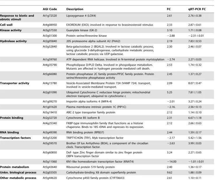
Bioinfor-matic approaches aimed to find orthologous sequences in G. biloba ESTs using the oligonucleotide sequences present on the Arabidopsis microarray showed generally a low percentage of sequence identity, with the exception of a protein kinase (At3g01300) (Table S2). Among 146 significantly (fold change $2, P#0.05) modulated genes on the Arabidopsis microarray, we chose 24 genes (17 up and 7 down-regulated) for real-time PCR (qPCR) validation on G. biloba cDNA. qPCR confirmed the differential expression for most of these genes, with the exception of some down-regulated genes in the microarray data that were found up-regulated by qPCR, after 4 h of larvae feeding on leaves
Figure 1.G. bilobais characterized by different leaf types, depending on the age and shape of the leaf. A, bilobed; B, multi-dissected and;C, fan-shaped. Vm values are reported along with standard errors (in brackets) as mV (n<50). Herbivore wounding is shown in leaf segments and Vm values are indicated below and aside the wounding zone. The leaf section of C shows Vm values of the different mesophyll and epidermal cells of a fan-shaped leaf.D, Spodoptera littoralis feeding on G. biloba leaves.
(Table 1). Most of these genes were associated with biotic and abiotic stress responses. Some were transcription factor regulators: these included a Dof-type zinc finger protein, the phosphate-responsive protein EXO, a MYB transcription factor, and a F-box family protein transcription factor. Other genes, encoding b-galactosidase, guanylate kinase, lipoxygenase, ABC transporter protein, and phospholipase D, are usually involved in plant stress responses. A strong up-regulation was found for a gene (similar to VAMP 724) which encodes a protein that plays a key role in vesicle trafficking to vacuoles and delivery of molecules to their destination. High fold-change expression values were also found for 20S proteasome alpha subunit PAA2 and a putative
cytochrome b5. Ubiquinol cytochrome c reductase, belonging to the family of reductases specifically acting on diphenols, was up-regulated. Down-regulation was confirmed for a protein kinase similar to Arabidopsis APK1A (Table 1).
Considering the indication that ROS levels are modulated upon feeding, as shown by confocal laser scanning microscopy (CLSM), we extended our gene expression study to four genes coding for ROS-scavenging enzymes: superoxide dismutase (SOD), peroxi-dase (POX), ascorbate peroxiperoxi-dase (APX) and catalase (CAT), following MD and HW treatment (Fig. 5). With respect to MD (dotted line), SOD and CAT were up-regulated at both time points, whereas POX and APX showed opposing trends: POX was
up-Figure 2. Calcium variations inG. bilobaupon mechanical damage and herbivore wounding. A. Mechanically wounded G. biloba leaves, values (n = 5) are expressed as mM Ca2+calculated from a calibration curve. The same letter indicates not significant (P.0.05) variation. B. Herbivore wounded G. biloba leaves, values (n = 5) are expressed as mM Ca2+. Different letters indicate significant (P,0.05) differences, the asterisks indicate significant (P,0.05) differences with respect to mechanical damage. In both panels, calcium orange indicates the absence of pharmacological inhibitors.
doi:10.1371/journal.pone.0032822.g002
Figure 3. H2O2variations inG. bilobaupon mechanical damage and herbivore wounding. A. Mechanically-wounded G. biloba leaves, values (n = 5) are expressed as mM H2O2calculated from a calibration curve. The same letter indicates not significant (P.0.05) variation. B. Herbivore-wounded G. biloba leaves, values (n = 5) are expressed as mM H2O2. Different letters indicate significant (P,0.05) differences, the asterisk indicate significant (P,0.05) differences with respect to mechanical damage. In both panels, amplex indicates the absence of pharmacological agents. doi:10.1371/journal.pone.0032822.g003
regulated at 30 min and down-regulated at 4 h, whereas APX was down-regulated at 30 min and up-regulated at 4 h (Fig. 5).
Herbivory induces the regulation of G. biloba direct defenses: flavonoid biosynthesis and gene expression
G. biloba leaves are characterized by the presence of several secondary metabolites, including the terpenoids ginkgolide A, B and C, and bilobalide, and several glycosylated flavonoids (Table 2). Analysis of MD and HW G. biloba leaves revealed that the main flavonoid backbones present were quercetin, kaempferol, myricetin and isorhamnetin, which were glycosylated in position 3 by b-D-glucose
and a-L-rhamnose (Fig. 6). Chemical analyses were performed 4 h
after both MD or HW, which was considered a time long enough to identify trends in metabolic adaptations to insect feeding.
With respect to MD, HW prompted an almost two fold increase in several glycosylated flavonoids, particularly 3-O-(b-D
-glucosyl)k-aempferol (2.74-fold, P,0.01), 3-O-[6-O-(a-L-rhamnosyl)-b-D -glucosyl]isorhamnetin (2.6-fold, P,0.05), 3-O-[6-O-(a-L
-rhamno-syl)-b-D-glucosyl]kaempferol (2.50-fold, P,0.05) and 3-O-[2-O-(b-D-glucosyl)-a-L-rhamnosyl]quercetin (2.46-fold, P,0.05). HW induced the synthesis of two new compounds: glycosyl myricetin and 3-O-[2-O-(b-D-glucosyl)-a-L-rhamnosyl]kaempferol.
Surpris-ingly, no significant differences were found between HW and MD for one of the most bioactive compounds of G. biloba, bilobalide, whereas ginkgolides A, B and C were significantly reduced by HW treatment with respect to MD (21.33-fold, P,0.05; 21.44-fold, P,0.05 and; 21.47-fold, P,0.05, respectively) (Table 2 and Fig. 6). Control analyses performed on intact leaves showed no significant differences with respect to MD (data not shown).
We then measured the expression levels of some genes related to phenylpropanoid and terpenoid biosynthesis, respectively, since these compounds are modulated by G. biloba responses to HW. Chalcone synthase (CHS), which catalyzes the first committed step in flavonoid biosynthesis, was induced comparably at 30 min and 4 h, whereas phenylalanine ammonia lyase (PAL), flavanone 3-hydroxylase (F3H), and anthocyanidin reductase (ANR) were significantly up-regulated by HW only after 4 h. In contrast, flavonol synthase (FLS) was down-regulated at both time points (Fig. 7). In G. biloba, the universal sesquiterpene precursor farnesyl diphosphate (FPP) is synthesized from geranyl diphosphate by the enzyme FPP synthase (FPPS), whereas the diterpene precursor geranylgeranyl diphosphate (GGPP) is synthesized from
isopente-nyl diphosphate and FPP by the enzyme GGPP synthase (GGPPS) [8]. Ginkgolide biosynthesis is initiated by protonating GGPP to give labdadienyl diphosphate, then the allylic diphosphate ionization is followed by cyclization, 1,4 hydride shift, methyl migration, and deprotonation to yield levopimaradiene. Levopi-maradiene synthase (LPS) catalyzes the initial cyclization step in ginkgolide biosynthesis [48]. A transient up-regulation of FPPS was observed after 30 min of herbivory, which dropped back to control levels at 4 h (Fig. 7). GGPPS was not significantly regulated by herbivory, whereas a significant decrease of LPS expression was observed with time (Fig. 7)
Herbivory induces G. biloba VOCs emission
Although G. biloba reacts to herbivory by inducing potentially toxic defense compounds, the plant also emits VOCs (Table 3). We analyzed the composition and quantity of VOCs by Tenax TA adsorption and GC-MS analysis of the headspace of treated leaves after 4 h and 24 h and found a significantly (P,0.05) higher emission after infestation by S. littoralis in comparison to mechanical damage (Table 3).
After 4 h feeding by S. littoralis, the emission of 1-octanol (3-fold), 2-heptenal (2.4-fold) and the sesquiterpenes a-copaene (6-fold) and b-caryophyllene (4.6-(6-fold) was always significantly (P,0.05) higher in HW with respect to MD. The green leaf volatile (GLV) 2-hexenal did not show significant changes, whereas the emission of 2-octenal was significantly higher in MD. After 24 h, the emission of 2-methyl butane increased significantly in HW plants with respect to MD (Table 3). A significant increase was also observed in HW for the two GLVs, 2-hexenal and 2-heptenal. In HW leaves, the emission of 1-octanol was still significantly enhanced, and a significant increase was found for 2-nonenal and ethyl benzoate. The emission of the two sesquiterpenes a-copaene (2.6-fold) and b-caryophyllene (3-fold) was still higher in HW in comparison to MD, although to a lesser extent with respect to 4 h time point.
Discussion
Plants and insects have coexisted for as long as 350 million years, if the earliest forms of land plants and insects are included, and have developed a series of relationships affecting the organisms at all levels, from basic biochemistry to population
Figure 4. Subcellular localization of [Ca2+]cytand H2O2inG. bilobaleaves upon herbivory. A. False color images from confocal laser scanning microscopy shows that upon herbivory [Ca2+]cytwas found mainly in the cytosol, indicated by the calcium orange dye as green patches not associated with any specific organelle. Metric bar = 10 mm.B. H2O2localization by Amplex Red shows a clear associations with microbodies (probably peroxisomes) and/or mitochondria but not with chloroplasts. Metric bar = 20 mm. In both panels, single arrows indicate the dye, double arrows indicate chloroplasts.
genetics. Although some of the relationships between the two kingdoms, such as pollination, are mutually beneficial, the most common interaction involves insect folivory and plant direct and indirect defenses against herbivorous insects [35,49,50]. On the basis of this long-standing relationship, it is not surprising that the strategies used by plants to resist or evade insect herbivores may be based on a common strategy. Although some species accumulate high levels of toxic compounds which function as direct biochemical defenses, other may not commit resources for this strategy, but seek to minimize herbivore damage through rapid growth and development, dispersion, choice of habitat, or by emitting VOCs able to attract enemy’s enemies [11,36,51]. Despite this diversity, our study on G. biloba shows that there is a general common defensive mechanism for plant response to herbivore wounding.
Our results show that the so called ‘‘living fossil plant’’ G. biloba uses early and late responses which are comparable to those found in angiosperms [13,14,40,52]. This is not surprising, since lower plants
like the fern Pteris vittata have been shown to respond to herbivory by ROS production and the emission of volatile compounds [53]. In G. biloba, Vm variations, although small, were significantly different between HW and MD, indicating that also in this species early detection of herbivory involves an ion imbalance across the plasma membrane [40] and possibly the perception of insect elicitors by plant cell receptors. In angiosperms such as Lima bean, Vm variations are associated to changes in calcium homeostasis [46]. G. biloba leaves reacted to HW with a burst of [Ca2+]cyt, that was
inhibited by the use of the calcium chelator EGTA and the inward calcium channel inhibitor Verapamil, as found in angiosperms [13,41]. Surprisingly, DPI also inhibited HW-induced [Ca2+]cyt,
suggesting an interplay between H2O2 and calcium homeostasis
[45]. In fact, the use of Verapamil induced a significant burst of H2O2, whereas EGTA reduced H2O2production. The subcellular
localization of calcium and H2O2signaling upon HW were in the
cytosol and mitochondria/peroxisomes, respectively, as already observed in angiosperms [45,46,52].
Table 1. Gene expression of G. biloba leaves after 4 h from S. littoralis herbivory.
AGI Code Description FC qRT-PCR FC Response to biotic and
abiotic stimuli
At1g72520 Lipoxygenase 4 (LOX4) 2.61 2.7660.38 Cell wall At4g08950 EXORDIUM (EXO); involved in response to brassinosteroid stimulus 2.33 2.8760.61 Kinase activity At3g57550 Guanylate kinase (GK-2) 3.10 1.7160.08 At3g01300 Protein serine/threonine kinase 22.88 22.3160.01 Hydrolase activity At2g05840 20S proteasome alpha subunit A2 (PAA2) 2.19 7.8360.53
At3g52840 Beta-galactosidase 2 (BGAL2). Involved in lactose catabolic process, using glucoside 3-dehydrogenase, carbohydrate metabolic process, lactose catabolic process via UDP-galactose.
2.30 2.4660.07
At1g59760 ATP dependent RNA helicase. Involved in N-terminal protein myristoylation 22.74 2.2760.03 At4g35790 Phospholipase D/PLD Delta. Involved in phospolipase metabolism.
Mutants are affected in hydrogen peroxide mediated cell death.
2.53 1.7460.32 At5g66080 Protein phosphatase 2C family protein/PP2C family protein. Protein
serine/threonine phosphatase activity
2.45 1.3760.27 Transporter activity At4g15780 Vesicle-Associated Membrane Protein 724 (VAMP 724). transport,
involved in vesicle-mediated transport.
2.09 8.0760.47 At2g01090 Ubiquinol Cytochrome C reductase hinge protein; mitochondrial
electron transport, ubiquinol to cytochrome c
5.25 7.8161.05 At1g09270 Importin alpha isoforms 4 (IMPA-4) 22.01 3.2760.24 At1g01620 Plasma membrane intrinsic protein 1C (PIP1C) 22.16. 2.5660.13 At5g19410 ABC-2 type transporter family protein 2.12 1.1460.10 Protein binding At2g32720 Cytochrome B5 isoform B 2.31 6.6761.18
At4g25340 FKBP-type immunophilin family that functions as a histone chaperone. Binds to 18S rDNA and represses its expression.
2.12 2.6660.63 RNA binding At3g49390 RNA binding protein (RPB37) 2.44 1.5960.17 Transcription factors At5g53200 TRIPTYCHON (TRY), Myb transcription factor 22.17 5.4261.56
At5g59570 Brother Of lux Arrhythmo (BOA), a component of the circadian clock. Transcription factor.
22.63 3.9960.03 At5g60850 Dof- type Zinc finger domain similar to zinc finger protein
OBP4 transcription factor
3.24 2.2760.65 At5g11060 KN1-like homeodomain transcription factor (KNAT4) 214.00 21.0160.01 Protein metabolism At5g43640 Ribosomal protein S19 family protein 2.40 1.3660.17 Unkn. biological process At2g03505 Carbohydrate-binding X8 domain superfamily protein 9.62 1.8860.09 Other metabolic process At5g44620 Cytochrome p450 family protein (CYP706A3) 2.61 1.1060.11 Data are expressed as fold change by considering gene expression in mechanically damaged leaves equal to 1. Microarray data from the heterologous hybridization performed on Arabidopsis microarrays are listed along with qPCR data using G. biloba cDNAs. Genes are grouped by GO annotations. (6SD). Microarray Fold Change (FC).
Upon HW, several genes were differentially expressed with respect to MD. The observed increase in H2O2was in accordance
with the increased transcript levels of SOD and CAT at all time points, as previously found upon herbivory in the model plant Lima bean [45]. On the other hand POX was found to be significantly down regulated by herbivory at later times, whereas APX was down-regulated at early times. The down-regulation of POX has been associated to the effect of insect’s oral secretions [54].
The results obtained with our heterologous microarray experiment showed that most of the modulated genes were associated with biotic and abiotic stress responses. A strong up-regulation was found for a gene encoding a protein with transporter activity, the Vesicle-Associated Membrane Protein 724 (VAMP 724, v-SNARE). This protein forms a complex known as SNARE (soluble N-ethylmaleimide-sensitive-factor attachment protein receptor), that plays a key role in vesicle trafficking to vacuoles and delivery of molecules to their destination. The major role carried out by this protein is to move ROS from endosomes to vacuoles. Suppression of Arabidopsis vesicle VAMP 724 expression inhibits fusion of H2O2 containing vesicles with vacuoles [55].
Another up-regulated gene with transporter activity is ubiquinol cytochrome c reductase (cytochrome bc1 complex or complex III). The activity of the gene product is involved in mitochondrial ROS production, particularly H2O2, which acts not only as a damaging
oxidant but also as a signaling molecule through either direct (oxidation of its target) or indirect (e.g., involving peroxiredoxins) action [56]. Interestingly, the 20S proteasome alpha subunit PAA2 proved to be highly induced by herbivory. Dahan and co-workers [57] hypothesized a complex organization and regulation of the 20S plant proteasome and its possible stress-induced modification into a so-called ‘‘plant defense proteasome’’, which might be involved in the activation of plant defense reactions. The same authors also demonstrated that 20S proteasome alpha subunit is up regulated by elicitins in tobacco cells. Other up-regulated genes involved in transport processes were importin a´ (IMPA-4), one of the two factors of the nuclear pore-targeting complex which was found to interact with virulence (Vir) proteins encoded by the Ti plasmid of Agrobacterium tumefaciens [58]; and an aquaporin (PIP1c), which is
involved in water transport activity, and which has been recently correlated to ROS signaling and/or oxidative stress response [59]. Upon herbivory cytochrome b5 was also up-regulated. Cyto-chrome b5 is a heme-binding protein and functions as an electron transfer component involved in a number of oxidative reactions, such as the anabolic metabolism of lipids and the catabolism of xenobiotics and compounds of endogenous metabolism [60]. The oxidative reactions mediated by cytochrome b5 are also associated with sugar supply and cytochrome b5 plays a regulatory role by physically interacting with sugar transporters [61].
The gene encoding for a protein serine/threonine kinase, similar to protein kinase APK1A, was found to be down regulated upon herbivory. The involvement of protein kinases in plant-herbivore interaction has been recently reviewed [14].
Two transcription factors, TRIPTYCHON (TRY) and a component of the circadian clock (BROTHER OF LUX ARRHYTHMO, BOA), showed a consistent up-regulation. TRY, which encodes a CPC-homologous MYB-related transcription factor, is a negative regulator of trichome development functioning in lateral inhibition and hence most probably in cell-cell signaling [62]. BOA is a GARP family transcription factor and is regulated by circadian rhythms in A. thaliana. Overexpression of BOA exhibits physiological and develop-mental changes, including delayed flowering time and increased vegetative growth under standard growing conditions [63].
Phenolic compounds are apparently important in the defense mechanisms of conifers [64] and the induction of leaf flavonoids is a specific defense response of many plants against insect herbivory [65,66]. Our results on G. biloba flavonoid metabolism and gene expression indicate an involvement of flavonoids in response to herbivory. PAL, CHS, F3H and ANR gene expression were up-regulated after 4 h. This increased gene expression was accom-panied by the increased abundance of several flavonoids like 3-O-(b-D-glucosyl)kaempferol, 3-O-[6-O-(a-L-rhamnosyl)-b-D-gluco-syl]kaempferol, 3-O-[2-O-(b-D-glucosyl)-a-L-rhamnosyl]kaemp-ferol and 3-O-[6-O-(a-L-rhamnosyl)-b-D-glucosyl]isorhamnetin. Kaempferol diglycoside, kaempferol triglycoside, and quercetin glycosides were also found to be significantly increased by beetle damage [67]. In G. biloba,
3-O-[2-O-(b-D-glucosyl)-a-L-rhamno-Figure 5. Time-course quantitative gene expression of some ROS scavenging genes inG. bilobaupon herbivory. Gene expression of superoxide dismutase (SOD) and catalase (CAT) was up-regulated by herbivory at all times. Upon herbivory, peroxidase (POX) was significantly down-regulated after 4 h, whereas ascorbate peroxidase (APX) was down-regualted after 30 min. The dotted lines represent control values (mechanical damage), different letters indicate significant (P,0.05) differences, asterisk indicates significant (P,0.05) differences with respect to control. doi:10.1371/journal.pone.0032822.g005
syl]quercetin was also increased after 4 h herbivory, although the gene expression of a flavonol synthase (FLS), which leads to the synthesis of quercetin from dihydroflavonols, was down-regulated. Although increased contents of bilobalide, ginkgolide A and ginkgolide B have been observed in G. biloba cell cultures induced with biotic elicitors of Candida albicans [68] or in plants exposed to elevated levels of O3[69], the concentration of bilobalide did not
show significant changes in response to herbivory, whereas the ginkgolides A, B and C were all significantly reduced by insect feeding. Ginkgolide B has been shown to confer bioactivity by inhibiting oxidative stress generation, and the dose-response effects of ginkgolide B on ROS generation in human cells has been demonstrated [70]. We may speculate that the increased ROS activity upon HW may exert a negative effect on ginkgolide production. The observed down-regulation of LPS at all times correlated with the reduction of ginkgolide content.
Herbivory also induced G. biloba VOCs emissions. Previous attempts to induce VOC emission from G. biloba with spider mites (Tetranychus urticae) were unsuccessful, because leaves were not accepted as a host plant. However, treatment of G. biloba leaves with 1 mM jasmonic acid (JA) induced VOC emission [31], suggesting the potential of this plant to emit terpenoids. The generalist S. littoralis accepted G. biloba leaves although feeding induced a delay in molting and the death of some insect (unpublished results). Herbivore-induced VOCs included the sesquiterpenes a-copaene and b-caryophyllene, which have been shown to be released by G. biloba upon JA treatment [31]. These two
sesquiterpenes have also been described to be involved in attraction of insect’s predators in several plant-interactions [51]. Finally the increased emission of some HW-induced sesquiterpenes was accom-panied by the up-regulation of FPPS, a key gene in sesquiterpene synthesis [71]. The conversion of FPP to the sesquiterpenes a-copaene [72] and b-caryophyllene [73] has been demonstrated.
In conclusion, we showed that the ‘‘living fossil’’ plant G. biloba responds to herbivory by inducing early responses, such as the variation of the plasma transmembrane potential and the induction of both calcium and ROS signaling. These events preceded the activation of ‘‘second line’’ defense systems including the activation of defense genes and the production of secondary plant metabolites (e.g., many glycosylated flavonoids). Furthermore the emission upon herbivory of specific VOCs indicates the ability of the plant to potentially activate indirect defenses along with the activation of direct defenses, although the ability of emitted VOCs to attract predators of herbivores was not yet demonstrated. Current research in our laboratory is under way to evaluate the possible attraction of predators by emitted VOCs as well as S. littoralis tolerance to G. biloba toxic metabolites and the mechanisms underlying its resistance.
Materials and Methods Plant and animal material
Ginkgo biloba L. seeds were collected from an adult (.100 years old) female G. biloba tree growing in the Botanical Garden of the Table 2. Comparative analysis of flavonoids, bilobalide and ginkolides between mechanically damaged (MD) and Spodoptera littoralis wounded (HW) Ginkgo biloba leaves after 4 h feeding.
Compound Spectra [M-H]2
MD HW
Quinic acid MS:190.8; MS2
[190.8]: 173; 127; 111; 93; 85 3.25 ( 1.81) 3.81 ( 0.48) 3-O-[2-O, 6-O-Bis(a-L-rhamnosyl)-b-D-glucosyl]quercetin MS:755; MS2[755]: 609; 301 1.01 ( 0.69) 1.66 ( 0.17) 3-O-[6-O-(a-L-rhamnosyl)-b-D-glucosyl]myricetin MS:625; MS2[625]: 317 MS3[317]: 288; 271; 179 0.51 ( 0.38) 1.60 ( 0.53)
3-O-(b-D-glucosyl)quercetin MS:463; MS2
[463]: 301 1.78 ( 1.34) 5.66 ( 1.12) 3-O-[6-O-(a-L-rhamnosyl)-b-D-glucosyl]isorhamnetin MS:623; MS2
[623]: 315 18.80 ( 5.35) 49.34 ( 0.78) Glucosyl myricetin MS:625; MS2
[625]: 317 MS3
[317]: 288; 270; 179 nd 1.44 ( 0.21) 3-O-[2-O-(b-D-glucosyl)-a-L-rhamnosyl]isorhamnetin MS:623; MS2
[623]: 315 MS3
[315]: 301; 272; 255 8.64 ( 2.82) 16.01 ( 1.85) 3-O-[6-O-(a-L-rhamnosyl)-b-D-glucosyl]quercetin MS:609.1; MS2
[609]: 301 20.42 ( 2.38) 39.09 (10.19) 3-O-[6-O-(a-L-rhamnosyl)-b-D-glucosyl]-39-methylmyricetin MS:639.1; MS2[639]: 331 MS3[331]:316; 289; 271 3.05 ( 1.31) 5.27 ( 0.44)
3-O-(a-L-rhamnosyl)isorhamnetin MS:463; MS2
[463]: 315 MS3
[315]:301 46.10 (14.89) 93.44 ( 0.13) 3-O-[2-O-(b-D-glucosyl)-a-L-rhamnosyl]quercetin MS:609.1; MS2
[609]: 301 MS3
[301]:271; 24.11 ( 9.37) 59.12 ( 4.96) 3-O-[6-O-(a-L-rhamnosyl)-b-D-glucosyl]kaempferol MS:593; MS2[593]:285 MS3[285]: 257; 229 12.08 ( 2.86) 30.22 ( 6.67)
3-O-[2-O-(b-D-glucosyl)-a-L-rhamnosyl]-39-methylmyricetin MS:639; MS2
[639]: 331 MS3 [331]:316; 287; 271 1.57 ( 0.83) 2.45 ( 0.08) Diglucosyl isorhamnetin MS:623.1; MS2 [623]: 315 MS3 [315]: 300; 271; 255 14.10 ( 1.95) 19.62 ( 0.28) Glucosyl quercetin MS:463; MS2 [463]: 301 51.49 ( 5.59) 56.73 ( 2.44) 3-O-[2-O-(b-D-glucosyl)- a-L-rhamnosyl]kaempferol MS:593; MS2
[593]:285 nd 7.72 ( 1.95) 3-O-(b-D-glucosyl)kaempferol MS:447; MS2 [447]: 285 MS3 [285]: 255, 227, 151 2.34 ( 0.88) 6.42 ( 0.52) 3-O-(a-L-rhamnosyl)quercetin MS:447; MS2[447]: 301 MS3[301]: 179; 151 26.46 ( 2.52) 50.94 ( 9.78) 7-O-(a-L-rhamnosyl)kaempferol MS:431; MS2 [431]: 285; 227 19.01 ( 2.83) 22.90 ( 4.45) Bilobalide MS:325; MS2 [325]: 251; 207; 193; 163 265.28 (15.02) 233.03 (15.77) Ginkgolide A [M+CO2] 2 453; MS2[453]:407; 379; 351 318.47 ( 9.31) 239.87 (17.69) Ginkgolide B MS:423; MS2 [423]: 395; 367 404.22 (24.76) 279.91 ( 8.43) Ginkgolide C MS:439; MS2 [439]: 411; 383; 321 65.74 ( 8.17) 44.60 ( 3.09) nd, not detected.
Values (n = 5–8) are expressed as ng g21
fr. wt. (6SEM). In the same row, boldface HW values indicate significant (P,0.05) differences between HW and mechanically damaged (MD) leaves.
University of Turin. Seeds were germinated in plastic pots with sterilized potting soil at 27uC during the day and 22uC during the night and 60% humidity using daylight fluorescent tubes at approximately 120mmol m22s21 with a photoperiod of 16 hours. Experiments were conducted with three-month old
plants on fully developed leaves which were found to be the most responsive leaves.
Larvae of the generalis herbivore Spodoptera littoralis (Boisd. 1833) (Lepidoptera, Noctuidae) (kindly supplied as egg clutches by Dr. Roland Reist, Syngenta Crop. Protection Mu¨nchwilen AG, Stein,
Figure 6. Structure formulae of the main representativeG. bilobacompounds. doi:10.1371/journal.pone.0032822.g006
Switzerland), were used because to our knowledge there are no reports on herbivores feeding on G. biloba. Larvae were reared in Petri dishes at 22–24uC with a 14–16 h light phase. They were fed on artificial diet as previously described [46].
All experiments, except preliminary Vm tests on bilobed and multi-dissected leaves, were carried out by using G. biloba fan-shaped leaves at the same developmental stage. S. littoralis larvae (third instar) were starved for 24 h before transfer to leaves. The mechanical damage was done by a pattern wheel. Damaged leaves were harvested by cutting the petiole and immediately frozen in liquid nitrogen and stored at 280uC until use.
Membrane potentials
Membrane potentials were determined in leaf segments. G. biloba bilobed, fan-shaped and multi-dissected leaves were analyzed. The transmembrane potential difference (Vm) was determined as previously reported [46]. Vm variations were recorded both on a pen recorder and through a digital port of a PC using a data logger. The results of Vm are shown as the average number of at least 50 Vm measurements.
Determination of intracellular calcium variations using confocal laser scanning microscopy (CLSM) and calcium orange
Calcium orange dye (stock solution in DMSO, Molecular Probes, Leiden, The Netherlands) was diluted in 5 mM MES-Na buffer (pH 6.0) containing 0.5 mM calcium sulfate and 2.5mM, dichlorophenyldimethylurea (DCMU) (Sigma-Aldrich, Milan,
Italy) to a final concentration of 5mM. This solution was applied on G. biloba fan-shaped leaves attached to the plant. The leaf was gently fixed on a glass slide and a drop of 5mM calcium orange solution (about 45ml) was applied and covered with another glass slide. After one hour of incubation with calcium orange, the leaf was mounted on a Nikon Eclipse C1 (Nikon Instruments, Tokyo, Japan) spectral CLSM stage without separating the leaf from the plant in order to assess the basic fluorescence levels as a control. Calcium variations were also monitored following MD and HW for 30 min and 4 h in the presence of either 15mM diphenyle-neiodonium (DPI; Sigma-Aldrich), 100mM Verapamil (Fluka Biochemika, Buchs, Switzerland) or 250mM ethylene glycol-bis(2-aminoethylether)-N,N,N9,N9-tetraacetic acid (EGTA, Sigma-Al-drich).
The microscope operated with a Krypton/Argon laser at 543 nm and 568 nm wavelengths: the first wavelength excited calcium orange, resulting in green fluorescence and the second mainly excited chlorophyll, resulting in red fluorescence. Images generated by the FluoView software were analyzed using the NIH image software as described earlier [41]. Measurements were repeated at least 5 times (biological replicates).
CLSM localization of H2O2and active peroxidases using
10-acetyl-3,7-dihydroxyphenoxazine (Amplex Red)
The amplex red hydrogen peroxide/peroxidase assay (Molec-ular Probes) was used for the detection of H2O2 and active
peroxidases. G. biloba fan-shaped leaves from intact plants in pots were incubated with a 50mM amplex red solution (in 5 mM Mes-Na buffer, pH 6.0, containing 0.5 mM calcium sulfate and 5mM
Figure 7. Time-course quantitative gene expression of some G. biloba genes involved in phenylpropanoid and terpenoid metabolism upon herbivory. Phenylalanine ammonia lyase (PAL) and anthocyanidin reductase (ANR) were significantly up regulated by herbivory only after 4 h, whereas flavonol synthase (FLS) was down-regulated at 30 min and 4 h. Chalcone synthase (CHS) showed a constant up-regulation, whereas flavanone 3-hydroxylase (F3H) showed an increased up regulation after 4 h. Farnesyl diphosphate synthase (FPPS) was significantly upregulated only after 30 min whereas geranylgeranyl diphosphate synthase (GGPP) showed no regulation at all times. Levopimaradiene synthase (PPS) was significantly down-regulated at all times. The dotted lines represent control values (mechanical damage), different letters indicate significant (P,0.05) differences, asterisks indicate significant (P,0.05) differences with respect to control.
DCMU) as reported earlier [45]. Leaves were mounted on a Nikon Eclipse C1 spectral CLSM stage without separating the leaf from the plant in order to determine the background fluorescence. H2O2variations were monitored after MD or HW treatment for
30 min and 4 h, and in addition in the presence of either 15mM DPI, 100mM Verapamil or 250mM EGTA. The microscope was operated with an Ar-Laser (458 nm/5 mW; 476 nm/5 mW; 488 nm/20 mW; 514 nm/20 mW), a HeNe-Laser (543 nm/ 1,2 mW) and a HeNe-Laser (633 nm/10 mW). Measurements were repeated at least 5 times (biological replicates).
Isolation of total RNA and cDNA synthesis
Total RNA was extracted from treated (HW) and control (MD) G. biloba leaves by using the Agilent Plant RNA Isolation Mini Kit (Agilent Technologies), following manufacturer’s instructions. To remove residual genomic DNA, total RNA was treated with RNAse-free DNAse I set (Qiagen, Hilden, Germany) The RNA quality was checked using the Agilent 2100 Bioanalyzer on RNA 6000 Nano LabChips Kit (Agilent Technologies). Quantitative analysis was performed using the NanoDrop ND-1000 micro scale spectrophotometer (Thermo Fisher Scientific, Waltham, MA, US) as previously reported [15].
For cDNA synthesis, High-capacity cDNA Reverse Transcrip-tion Kit (Applied Biosystems) was used according to manufactur-er’s instructions. Briefly, the reactions were prepared by adding 1.5mg total RNA, 2ml of 106 RT buffer, 0.8ml of 256 dNTPs mix (100 mM), 2ml 106 RT random primer, 1ml of Multi-scribeTMreverse transcriptase and nuclease-free sterile water up to 20ml. Then the reaction mixtures were incubated at 25uC for 10 minutes, 37uC for 2 hours, and 85uC for 5 seconds. Samples were stored at 220uC for further analyses.
Heterologous gene microarray hybridization
Five hundred nanograms of total RNA from MD and HW-treated samples were separately reverse-transcribed into double-strand cDNAs by the Moloney murine leukemia virus reverse transcriptase (MMLV-RT) and amplified for 2 h at 40uC using the Agilent Low RNA Input Linear Amplification Kit, two-color (Agilent Technologies, Santa Clara, CA, US). Subsequently, cDNAs were transcribed into antisense cRNA and labeled with either Cy3-CTP or Cy5-CTP fluorescent dyes for 2 h at 40uC following the manufacturer’s protocol. Cyanine-labeled cRNAs were purified using RNeasy Minikit (Qiagen, Hilden, Germany). Purity and dye incorporation were assessed by spectrophotometry and electrophoresis (using the NanoDrop ND-1000 and Agilent
Limonene 1029 18.71( 2.34) 20.25 (2.55) 4.42 (2.90) 12.10 (0.48) 2-Octenal 1056 10.79 (0.78) 6.76 (1.08) 4.07 (0.85) 3.40 (1.36) 1-Octanol 1068 6.38 (1.47) 19.28 (1.16) 2.78 (0.07) 4.33 (0.91) Nonanal 1101 0.28 (0.11) 0.45 (0.27) 0.14 (0.02) 0.10 (0.01) 2-Nonenal 1149 6.74 (2.40) 7.43 (1.52) 3.83 (0.39) 10.41 (1.65) Ethyl benzoate 1173 28.14 (7.48) 29.92 (2.61) 1.29 (0.16) 6.76 (1.83) Decanal 1202 0.73 (0.13) 0.65 (0.52) 0.43 (0.17) 0.13 (0.11) Benzothiazole 1218 2.97 (0.95) 3.60 (1.53) 0.86 (0.69) 0.43 (0.06) 2-Decenal 1249 6.59 (1.68) 8.01 (3.54) 1.58 (0.43) 1.53 (0.24) Undecanal 1307 5.03 (1.81) 4.62 (2.91) 2.38 (0.59) 1.27 (0.93) a-Copaene 1377 1.09 (0.67) 6.58 (0.53) 4.59 (2.77) 11.89 (0.22) 1-Tetradecene 1390 7.43 (1.77) 8.44 (2.69) 11.00 (2.62) 11.39 (2.58) Tetradecane 1400 5.95 (0.38) 8.50 (0.79) 1.91 (0.42) 1.84 (0.39) Dodecanal 1402 8.48 (1.63) 9.19 (2.87) 4.33 (2.87) 2.90 (1.70) (E)-b-caryophyllene 1419 5.25 (1.77) 24.26 (1.52) 2.74 (0.69) 8.30 (1.70) b-Chamigrene 1453 2.67 (0.60) 12.16 (1.82) 6.37 (0.99) 6.25 (1.63) Pentadecane 1478 1.49 (1.14) 1.90 (0.91) 0.58 (0.10) 0.43 (0.09) Total 198.14 (11.84) 250.02 (12.63) 97.91 (18.60) 153.07 (26.64)
Data are expressed as micrograms of VOCs per gram of leaf fresh weight (6SEM). Retention times (RT) and Kova´ts Index (KI) are indicated for each compound. For the same time point, boldface HW values indicate significant (P,0.05) differences between MD and HW. HW, herbivore wounding; MD, mechanical damage.
2100 Bioanalyzer LabChips, respectively). Then, 750 ng of Cy3-labeled RNA of the control condition and 750 ng of Cy5-Cy3-labeled RNA of the experimental condition (HW) were combined and hybridized using the Gene Expression Hybridization Kit (Agilent Technologies) onto 1622 K Arabidopsis (v2) Oligo Microarray (Agilent Technologies).
After a 17 h incubation at 65uC and 10 rpm, the microarray was first washed with gene expression wash buffer 1 for 1 min, then with gene expression wash buffer 2 for 1 min, then with 100% acetonitrile for 30 s, and finally washed in the stabilization and drying solution for 30 s.
The microarray slide was scanned with the Agilent Microarray G2505B Scanner and data were extracted and normalized from the resulting image using Agilent Feature Extraction (FE) software (v.9.5.1). Data were analyzed using the GeneSpring GX 10.1.1 software (Agilent Technologies).
Bioinformatics analyses
About 4300 EST sequences isolated from G. biloba female leaf were downloaded from the National Centre for Biotechnology Informa-tion (NCBI) database (http://www.ncbi.nlm.nih.gov/). BlastX anal-yses were carried out using the NCBI blast tool and The Arabidopsis Information Resource (TAIR) database (http://www.arabidopsis. org/), i) in order to identify G. biloba genes with similarity to those oligonucleotide probes on the microarray which were found to be differentially expressed by cross-hybridization, and ii) in order to predict potential protein functions for these genes. Twenty four genes, out of the almost 100 genes which were significantly modulated in the microarray, were selected and validated by qPCR.
In order to find genes encoding enzymes involved the biosynthesis of phenylpropanoids and in the ROS scavenging system to be employed in expression analyses, an additional search in NCBI (EST database) was carried out.
Quantitative real time PCR (qPCR)
qPCR analyses were carried out using the Stratagene MPX3000 Real Time System (La Jolla, CA, USA). qPCR reactions were run using specific primers designed with Primer3 software (http:// frodo.wi.mit.edu/primer3/) and listed in Supporting Table S3 and G. biloba cDNAs as template. Amplifications were carried out in a 25ml reaction mixture containing 1ml cDNA as template (1:10 dilution of cDNA from 20ml of RT reaction), 12.5ml MaximaTM SYBR Green qPCR master mix (26) (Fermentas, International, Inc, Burlington, ON, Canada) and 100 nM primers (Integrated DNA Technologies, Coralville, IA, US). The applied protocol was the following: initial polymerase activation of 10 min at 95uC; followed by 40 cycles of 30 s at 95uC, 30 s at 57uC, and 30 s at 72uC. Fluorescence was read following each annealing and extension phase. All runs were followed by a melting curve analysis from 55 to 95uC. Three different reference (housekeeping) genes (actin 2, glyceraldehyde-3 phosphate dehydrogenase, 18 S rRNA) were used to calibrate and normalize the results of the qPCR. The best of the three genes was selected using the Normfinder software (www.normfinder.com). The most stable gene was actin 2. PCR conditions were determined by comparing threshold values in dilution series of the RT product, followed by non-template control for each primer pair. Relative expression levels of genes were calculated by using the Pfaffl method [74].
Extraction and analysis of G. biloba compounds induced by MD and HW
One gram of frozen leaves was ground to a fine powder by using liquid nitrogen with the addition of 10 ml methanol (Carlo Erba
Reagents, Arese, Italy). Samples were extracted in a ultrasonic bath at 35uC for 30 min and centrifuged at 5000 g for 10 min at room temperature. The supernatant was transferred and the same extraction procedure was repeated twice. Pooled aliquots were dried under vacuum. Extracts were re-suspended in 500ml methanol and then centrifuged at 5000 g for 10 min at room temperature. Extracts were filtered before injection in LC/MS. Samples were separated by an Agilent 1200 HPLC (Agilent Technologies) equipped with a Luna C18 (3.06150 mm, 3.0mm, Phenomenex, Torrance, CA, USA) reversed-phase column. The binary solvent system was: A) double distilled water with 0.1% v/v formic acid and B), acetonitrile (ACN) with 0.1% v/v formic acid. Separation was performed at 0.2 ml min21 flow rate and 25uC using an ACN gradient. The B mobile phase was held at 25% for 3 min and then increased to 30% at 7 min. Isocratic elution was performed for 8 min. Afterwards, solvent B was increased up to 55% (15 to 22 min), and 95% (23 to 27 min). The column was kept at 95% solvent B for 7 min. The initial mobile phase was re-established for 10 min before the next injection.
Mass spectrometry analyses were performed with a 6330 Series Ion Trap LC-MS System (Agilent Technologies) equipped with an electrospray ionization source (ESI). Qualitative analyses were made by tandem MS3and spectra were acquired in negative mode
with 1.5 kV ion spray voltage, nebulizer curtain gas (N2) at
5 L min21and 325uC, 1.00 V fragmentation amplitude and full scan range 50–1000 m/z. For quantitative analyses, samples were analyzed by LC-ESI-MS2 in MRM mode with the above
acquisition parameters. The monitored mass transitions were m/ z 407R351, 423R367, 439R383 and 325R163 for ginkgolides A, B and C and bilobalide, respectively, and the Y02aglycone ions
for flavonoid-glycosides. Spectral data were processed and analysed by the DataAnalysis for 6330 Series Ion Trap LC/MS 4.0 software (Bruker Daltonik, Bremen, Germany). Identification of spectra was done by manual interpretation and by comparison with literature data [75,76]. External calibration curves were made with standard solutions of rutin, quercetin, kaempferol, ginkgolide A, ginkgolide C and bilobalide (Sigma-Aldrich).
VOC extraction and analysis
Headspace VOCs were collected in 4 L glass desiccators by using four-node cuttings of fan-shaped leaves. Leaves were illuminated with fluorescent light bulbs (70mmol m22s21) with a light phase of 16 h, the temperature inside desiccators was 23uC and the relative humidity about 70%. Glass desiccators were connected to a GC grade air generator (HPZA-3500–220, Parker Balston, Cleveland,OH, USA) through a cork plug with two openings. Air was fluxed into the jars at 200 ml min21flow rate. Clean glass Thermal Desorption Unit (TDU) liners (Gerstel, Mu¨lheim an der Ruhr, Germany) were filled with 20 mg sorbent Tenax TA 60/80 [poly-(2,6-diphenyl)- p-phenylene oxide] (Supelco, Bellefonte, PA, USA). The sorbent was sandwiched by silanized glass wool (Agilent Technologies). Before use, Tenax TA was always preconditioned at 250uC on a Gerstel TDU for 10 min. Undamaged plants as control, MD-leaves and HW-leaves with six third instar S. littoralis larvae were assayed for 4 h and 24 h. All experiments were standardized with 30% of leaf damage. Tenax TA was desorbed in the TDU connected to a Gerstel Cooled Injection System 3 (CIS3 cryofocusing system) which uses liquid CO2as cooling agent. Desorption was carried out in splitless
mode with the following temperature program: 36uC held for 0.5 min, 25uC min21
increase to 260uC. The desorption temper-ature was held for 5 min and the CIS3 was maintained at 240uC. After desorption, CIS3 temperature was raised at a 12uC sec21
and by comparison with the NIST mass spectral search software v2.0 using the libraries NIST 98 library. External calibration curves were made with standard solution of (-)-menthol (99%, Fluka) for quantitative measurements as previously described [15].
Statistical analysis
The overall data sets are expressed as mean values of at least three biological replicates. Three technical replicates were run for each biological replicate. Metric bars indicate standard error. ANOVA and Tukey–Kramer’s HSD test (P,0.05) were used to
Acknowledgments
The authors are grateful to L. Starvaggi and U. Roggero for technical support during Vm analyses.
Author Contributions
Conceived and designed the experiments: MEM CMB. Performed the experiments: TKM AO SAZ MF JF SB CMB. Analyzed the data: MEM CMB JF AO. Contributed reagents/materials/analysis tools: MEM. Wrote the paper: MEM CMB.
References
1. Jacobs BP, Browner WS (2000) Ginkgo biloba: A living fossil. Am J Med 108: 341–342.
2. Zhou ZY, Zheng SL (2003) Palaeobiology: The missing link in Ginkgo evolution - The modern maidenhair tree has barely changed since the days of the dinosaurs. Nature 423: 821–822.
3. Royer DL, Hickey LJ, Wing SL (2003) Ecological conservatism in the ‘‘living fossil’’ Ginkgo. Paleobiology 29: 84–104.
4. Hasebe M (1997) Moleculary phylogeny of Ginkgo biloba: close relationship between Ginkgo biloba and cycads. In: Hori Y, Ridge RW, Tulecke W, Del Tredici P, Tremouillaux-Guiller J, Tobe H, eds. Ginkgo biloba-a Global Treasure. Tokyo: Springer-Verlag. pp 173–181.
5. Zhao YP, Paule J, Fu CX, Koch MA (2010) Out of China: Distribution history of Ginkgo biloba L. Taxon 59: 495–504.
6. Brenner ED, Katari MS, Stevenson DW, Rudd SA, Douglas AW, et al. (2005) EST analysis in Ginkgo biloba: an assessment of conserved developmental regulators and gymnosperm specific genes. Bmc Genomics 6.
7. Wang YQ, Shen JK, Berglund T, Ohlsson AB, Tang XF, et al. (2010) Analysis of expressed sequence tags from Ginkgo mature foliage in China. Tree Genet Genom 6: 357–365.
8. Lin X, Zhang J, Li Y, Luo H, Wu Q, et al. (2011) Functional genomics of a living fossil tree, Ginkgo, based on next-generation sequencing technology. Physiol Plant 143: 207–218.
9. Shaw FJF (1908) A contribution to the anatomy of Ginkgo biloba. New Phytol 7: 85–92.
10. Mundry M, Stu¨tzel T (2006) Morphogenesis of leaves and cones of male short-shoots of Ginkgo biloba L. Flora 199: 437–452.
11. Karban R, Shiojiri K, Ishizaki S (2011) Plant communication - why should plants emit volatile cues? J Plant Interact 6: 81–84.
12. Maffei ME, Mithofer A, Boland W (2007) Insects feeding on plants: Rapid signals and responses preceding the induction of phytochemical release. Phytochemistry 68: 2946–2959.
13. Mitho¨fer A, Boland W, Maffei ME (2009) Chemical ecology of plant–insect interactions. In: Parker J, ed. Molecular aspects of plant disease resistance. Chirchester: Wiley-Blackwell. pp 261–291.
14. Arimura GI, Ozawa R, Maffei ME (2011) Recent advances in plant early signaling in response to herbivory. Int J Mol Sci 12: 3723–3739.
15. Atsbaha Zebelo S, Bertea CM, Bossi S, Occhipinti A, Gnavi G, et al. (2011) Chrysolina herbacea modulates terpenoid biosynthesis of Mentha aquatica L. Plos One 6: e17195.
16. Rasmann S, Agrawal AA (2011) Latitudinal patterns in plant defense: evolution of cardenolides, their toxicity and induction following herbivory. Ecol Lett 14: 476–483.
17. Olson DM, Davis RF, Wackers FL, Rains GC, Potter T (2008) Plant-herbivore-carnivore interactions in cotton, Gossypium hirsutum: Linking belowground and aboveground. J Chem Ecol 34: 1341–1348.
18. van Beek TA (2002) Chemical analysis of Ginkgo biloba leaves and extracts. J Chromatogr A 967: 21–55.
19. Leistner E, Drewke C (2010) Ginkgo biloba and Ginkgotoxin. J Nat Prod 73: 86–92.
20. Schepmann HG, Pang JH, Matsuda SPT (2001) Cloning and characterization of Ginkgo biloba levopimaradiene synthase, which catalyzes the first committed step in ginkgolide biosynthesis. Arch Biochem Biophys 392: 263–269.
21. Bedir E, Tatli II, Khan RA, Zhao JP, Takamatsu S, et al. (2002) Biologically active secondary metabolites from Ginkgo biloba. J Agric Food Chem 50: 3150–3155.
22. Boonkaew T, Camper ND (2005) Biological activities of Ginkgo extracts. Phytomedicine 12: 318–323.
23. Christen Y, Maixent JM (2002) What is Ginkgo biloba extract EGb 761? An overview - From molecular biology to clinical medicine. Cell Mol Biol 48: 601–611.
24. Shi C, Liu J, Wu F, Yew DT (2010) Ginkgo biloba Extract in Alzheimer’s disease: From action mechanisms to medical practice. Int J Mol Sci 11: 107–123. 25. Muller WE, bdel-Kader R, Fehske CJ, Leuner K (2009) Grundlagen der
therapeutischen Anwendung von EGb 761. Pharm unser Zeit 38: 408–416. 26. Maitra I, Marcocci L, Droylefaix MT, Packer L (1995) Peroxyl radical
scavenging activity of Ginkgo biloba Extract Egb 761. Biochem Pharmacol 49: 1649–1655.
27. Shi C, Wu FM, Xu J (2010) H2O2and PAF mediate A beta 1–42-induced Ca 2+
dyshomeostasis that is blocked by EGb761. Neurochem Int 56: 893–905. 28. Altiok N, Ersoz M, Karpuz V, Koyuturk M (2006) Ginkgo biloba extract regulates
differentially the cell death induced by hydrogen peroxide and simvastatin. Neurotoxicology 27: 158–163.
29. Ellnain-Wojtaszek M, Kruczynski Z, Kasprzak J (2002) Variations in the free radical scavenging activity of Ginkgo biloba L. leaves in the period of complete development of green leaves to fall of yellow ones. Food Chem 79: 79–84. 30. Van Den Boom CEM, Dicke M (2003) Differences among plant species in
acceptance by the spider mite Tetranychus urticae Koch. J Appl Ent 127: 177–183. 31. Van Den Boom CEM, van Beek TA, Posthumus MA, De Groot A, Dicke M (2004) Qualitative and quantitative variation among volatile profiles induced by Tetranychus urticae feeding on plants from various families. J Chem Ecol 30: 69–89.
32. Pszczolkowski MA, Durden K, Sellars S, Cowell B, Brown JJ (2011) Effects of Ginkgo biloba constituents on fruit-infesting behavior of codling moth (Cydia pomonella) in apples. J Agric Food Chem 59: 10879–10886.
33. Bonaventure G, VanDoorn A, Baldwin IT (2011) Herbivore-associated elicitors: FAC signaling and metabolism. Trends Plant Sci 16: 294–299.
34. Kanchiswamy CN, Takahashi H, Quadro S, Maffei ME, Bossi S, et al. (2010) Regulation of Arabidopsis defense responses against Spodoptera littoralis by CPK-mediated calcium signaling. Bmc Plant Biol 10.
35. Wu JQ, Baldwin IT (2010) New insights into plant responses to the attack from insect herbivores. Annu Rev Genet 44: 1–24.
36. Wu JQ, Baldwin IT (2009) Herbivory-induced signalling in plants: perception and action. Plant Cell Environ 32: 1161–1174.
37. Kugimiya S, Shimoda T, Takabayashi J (2011) Timing matters: release of plant volatiles that are attractive to parasitoids. J Plant Interact 6: 187–188.
38. Ramadan A, Muroi A, Arimura GI (2011) Herbivore-induced maize volatiles serve as priming cues for resistance against post-attack by the specialist armyworm Mythimna separata. J Plant Interact 6: 155–158.
39. Arnott HJ (1959) Anastomoses in the venation of Ginkgo biloba. Am J Bot 46: 405–411.
40. Maffei ME, Mitho¨fer A, Boland W (2007) Before gene expression: early events in plant-insect interaction. Trends Plant Sci 12: 310–316.
41. Mitho¨fer A, Mazars C, Maffei M (2009) Probing spatio-temporal intracellular calcium variations in plants. In: Pfannschmidt T, ed. Plant signal transduction. Totowa: Humana Press Inc., pp 79–92.
42. Swanson SJ, Choi WG, Chanoca A, Gilroy S (2011) In vivo imaging of Ca2+, pH, and reactive oxygen species using fluorescent probes in plants. Annu Rev Plant Biol 62: 273–297.
43. Frahry G, Schopfer P (1998) Inhibition of O2-reducing activity of horseradish
peroxidase by diphenyleneiodonium. Phytochemistry 48: 223–227.
44. Liu N, Yang YY, Mo SW, Liao JL, Jin JN (2005) Calcium antagonistic effects of Chinese crude drugs: Preliminary investigation and evaluation by45
Ca. Appl Rad Isotop 63: 151–155.
45. Maffei ME, Mitho¨fer A, Arimura GI, Uchtenhagen H, Bossi S, et al. (2006) Effects of feeding Spodoptera littoralis on lima bean leaves. III. Membrane depolarization and involvement of hydrogen peroxide. Plant Physiol 140: 1022–1035.
46. Maffei M, Bossi S, Spiteller D, Mitho¨fer A, Boland W (2004) Effects of feeding Spodoptera littoralis on lima bean leaves. I. Membrane potentials, intracellular calcium variations, oral secretions, and regurgitate components. Plant Physiol 134: 1752–1762.
47. Howe GA, Jander G (2008) Plant immunity to insect herbivores. Annu Rev Plant Biol 59: 41–66.
48. Schwartz M, Arigoni D (1999) Ginkgolide biosynthesis. In: Cane DE, ed. Comprehensive natural product chemistry, isoprenoids including steroids and carotenoids. Tarrytown, NY: Pergamon Press. pp 367–400.
49. Guerrieri E, Digilio MC (2008) Aphid-plant interactions: a review. J Plant Interact 3: 223–232.
50. Sabelis M, Janssen A, Takabayashi J (2011) Can plants evolve stable alliances with the enemies’ enemies? J Plant Interact 6: 71–75.
51. Maffei ME, Gertsch J, Appendino G (2011) Plant volatiles: Production, function and pharmacology. Nat Prod Rep 28: 1359–1380.
52. Arimura G, Maffei ME (2010) Calcium and secondary CPK signaling in plants in response to herbivore attack. Biochem Biophys Res Comm 400: 455–460. 53. Imbiscuso G, Trotta A, Maffei M, Bossi S (2009) Herbivory induces a ROS
burst and the release of volatile organic compounds in the fern Pteris vittata L. J Plant Interact 4: 15–22.
54. Barbehenn R, Dukatz C, Holt C, Reese A, Martiskainen O, et al. (2010) Feeding on poplar leaves by caterpillars potentiates foliar peroxidase action in their guts and increases plant resistance. Oecologia 164: 993–1004.
55. Leshem Y, Melamed-Book N, Cagnac O, Ronen G, Nishri Y, et al. (2006) Suppression of Arabidopsis vesicle-SNARE expression inhibited fusion of H2O2
containing vesicles with tonoplast and increased salt tolerance. Proc Natl Acad Sci USA 103: 18008–18013.
56. Rigoulet M, Yoboue ED, Devin A (2011) Mitochondrial ROS generation and its regulation: Mechanisms involved in H2O2signaling. Antiox Redox Signal 14:
459–468.
57. Dahan J, Etienne P, Petitot AS, Houot V, Blein JP, et al. (2001) Cryptogein affects expression of alpha 3, alpha 6 and beta 1 20S proteasome subunits encoding genes in tobacco. J Exp Bot 52: 1947–1948.
58. Bhattacharjee S, Lee LY, Oltmanns H, Cao HB, Veena, et al. (2008) IMPa-4, an Arabidopsis importin alpha isoform, is preferentially involved in Agrobacter-ium-mediated plant transformation. Plant Cell 20: 2661–2680.
59. Boursiac Y, Boudet J, Postaire O, Luu DT, Tournaire-Roux C, et al. (2008) Stimulus-induced downregulation of root water transport involves reactive oxygen species-activated cell signalling and plasma membrane intrinsic protein internalization. Plant J 56: 207–218.
60. Kumar R, Wallis JG, Skidmore C, Browse J (2006) A mutation in Arabidopsis cytochrome b5 reductase identified by high-throughput screening differentially affects hydroxylation and desaturation. Plant J 48: 920–932.
61. Fan RC, Peng CC, Xu YH, Wang XF, Li Y, et al. (2009) Apple sucrose transporter SUT1 and sorbitol transporter SOT6 interact with cytochrome b5 to regulate their affinity for substrate sugars. Plant Physiol 150: 1880–1901. 62. Schellmann S, Schnittger A, Kirik V, Wada T, Okada K, et al. (2002)
TRIPTYCHON and CAPRICE mediate lateral inhibition during trichome and root hair patterning in Arabidopsis. Embo J 21: 5036–5046.
63. Dai SH, Wei XP, Pei LP, Thompson RL, Liu Y, et al. (2011) Brother of Lux Arrhythmo is a component of the Arabidopsis circadian clock. Plant Cell 23: 961–972.
64. Delvas N, Bauce E, Labbe C, Ollevier T, Belanger R (2011) Phenolic compounds that confer resistance to spruce budworm. Entomol Exper Appl 141: 35–44.
65. Anttila U, Julkunen-Tiitto R, Rousi M, Yang SY, Rantala MJ, et al. (2010) Effects of elevated ultraviolet-B radiation on a plant-herbivore interaction. Oecologia 164: 163–175.
66. Treutter D (2006) Significance of flavonoids in plant resistance: a review. Environ Chem Lett 4: 147–157.
67. O’Neill BF, Zangerl AR, Dermody O, Bilgin DD, Casteel CL, et al. (2010) Impact of elevated levels of atmospheric CO2and herbivory on flavonoids of
soybean (Glycine max L.). J Chem Ecol 36: 35–45.
68. Kang SM, Min JY, Kim YD, Karigar C, Kim SW, et al. (2009) Effect of biotic elicitors on the accumulation of bilobalide and ginkgolides in Ginkgo biloba cell cultures. J Biotechnol 139: 84–88.
69. He X, Huang W, Chen W, Dong T, Liu C, et al. (2009) Changes of main secondary metabolites in leaves of Ginkgo biloba in response to ozone fumigation. J Environ Sci-China 21: 199–203.
70. Chan WH, Hsuuw YD (2007) Dosage effects of ginkgolide B on ethanol-induced cell death in human hepatoma G2 cells. Ann New York Acad Sci 1095: 388–398.
71. Degenhardt J, Koellner TG, Gershenzon J (2009) Monoterpene and sesquiterpene synthases and the origin of terpene skeletal diversity in plants. Phytochemistry 70: 1621–1637.
72. Ducreux LJ, Morris WL, Prosser IM, Morris JA, Beale MH, et al. (2008) Expression profiling of potato germplasm differentiated in quality traits leads to the identification of candidate flavour and texture genes. J Exper Bot 59: 4219–4231.
73. Koellner TG, Held M, Lenk C, Hiltpold I, Turlings TC, et al. (2008) A maize (E)-beta-caryophyllene synthase implicated in indirect defense responses against herbivores is not expressed in most American maize varieties. Plant Cell 20: 482–494.
74. Pfaffl MW (2001) A new mathematical model for relative quantification in real-time RT-PCR. Nucl Acids Res 29.
75. Ding S, Dudley E, Plummer S, Tang J, Newton R, et al. (2008) Fingerprint profile of Ginkgo biloba nutritional supplements by LC/ESI-MS/MS. Phyto-chemistry 69: 1555–1564.
76. Hasler A, Sticher O, Meier B (1992) Identification and determination of the flavonoids from Ginkgo biloba by High-Performance Liquid-Chromatography. J Chromatogr 605: 41–48.